Foundation 2 · 2C · Cell division and specialization

, , and growth control

Sources ↗

Cell division coordinates , , and with nutrient, growth, and damage signals.

Track amount and number

supports growth and preparation, replicates , and prepares for division and checks . M phase includes and usually . is a nondividing state entered from the cycle; some cells re-enter when stimulated, whereas others remain specialized for long periods. is distinct from , a more durable arrest associated with stress or replicative history.

A cell progresses from 2n,2C in to 2n,4C after . number is counted by , so doubles without doubling that count. When sister separate in , each chromatid becomes a ; each completed daughter receives 2n,2C.

Source: 10.2 The Cell Cycle · 10.3 Control of the Cell Cycle

Spindle attachments direct

In condense and the spindle develops. During the breaks down and spindle attach to , assemblies at . At , duplicated align with sister attached to opposite poles. begins after cleavage permits sister separation; shortening and spindle elongation distribute the .

In decondense and envelopes re-form. Animal-cell uses an – ; plant cells assemble a cell plate. A is a region, a is its spindle- complex, and a is a -organizing structure; the similar names describe different objects.

Source: 10.2 The Cell Cycle · 10.3 Control of the Cell Cycle

and coordinate progression

require regulatory and additional activating or inhibitory modifications. Oscillating abundance changes activity through the cycle. control integrates growth signals, resources, and integrity; control restrains division when or repair is incomplete. The spindle-assembly delays when attachments are defective.

At onset, -mediated degradation removes and helps activate , allowing cleavage; degradation also supports mitotic exit. damage can activate and to reduce activity. Loss of multiple allows damaged genomes to proliferate, but a single dividing cell is not automatically malignant.

Source: 10.2 The Cell Cycle · 10.3 Control of the Cell Cycle

Balance , survival, and programmed death

Growth-factor relay signals through pathways that regulate , , and cell-cycle entry. normally support controlled growth; activating changes can create with excessive signaling. restrain , preserve integrity, or facilitate elimination of damaged cells. Contact, anchorage, and nutrient signals also shape whether division proceeds.

uses regulated activation to dismantle a cell while packaging material for clearance. Intrinsic stress can trigger release, while death can initiate extrinsic pathways. Survival signals inhibit these processes. evolution can favor both increased division and reduced , so growth reflects birth and death rates together.

Source: 10.2 The Cell Cycle · 10.3 Control of the Cell Cycle

Practice: apply the mechanism

Question. A cell has replicated its but one remains unattached. Which should delay progression, and what is the cell’s and content before sister separation?

Show reasoning

The spindle-assembly should delay by restraining the machinery that activates . Before sister separation, a cell is 2n,4C: are duplicated but homolog-set number is unchanged. If the fails, missegregation can yield daughters.

Source: 10.2 The Cell Cycle · 10.3 Control of the Cell Cycle

10.2 The — figures and tables

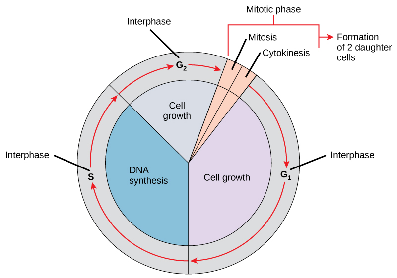
Like a clock, the cell cycles from interphase to the mitotic phase and back to interphase. Most of the cell cycle is spent in interphase, which is subdivided into G subscript 1 baseline, S, and G subscript 2 baseline phases. Cell growth occurs during G subscript 1 baseline, D N A synthesis occurs during S, and more growth occurs during G subscript 2 baseline. The mitotic phase consists of mitosis, in which the nuclear chromatin is divided, and cytokinesis, in which the cytoplasm is divided, resulting in two daughter cells.
Figure 10.5 — 10.2 The

Figure 10.5 The cell cycle in multicellular organisms consists of interphase and the mitotic phase. During interphase, the cell grows and the nuclear DNA is duplicated. Interphase is followed by the mitotic phase. During the mitotic phase, the duplicated chromosomes are segregated and distributed into daughter nuclei. Following mitosis, the cytoplasm is usually divided as well by cytokinesis, resulting in two genetically identical daughter cells.

Mary Ann Clark, Matthew Douglas, and Jung Choi, Biology 2E, OpenStax. · CC BY-NC-SA 4.0 · Original image ↗ · Access for free ↗
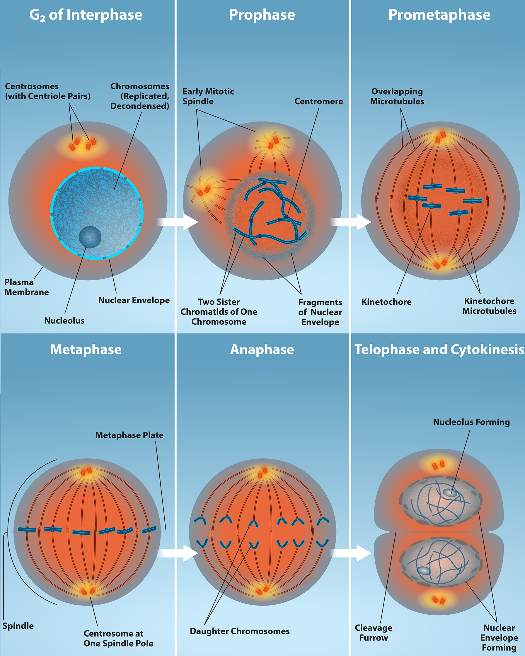
This diagram shows the five phases of mitosis and cytokinesis. During prophase, the chromosomes condense and become visible, spindle fibers emerge from the centrosomes, the nuclear envelope breaks down, and the nucleolus disappears. During prometaphase, the chromosomes continue to condense and kinetochores appear at the centromeres. Mitotic spindle microtubules attach to the kinetochores, and centrosomes move toward opposite poles. During metaphase, the mitotic spindle is fully developed, and centrosomes are at opposite poles of the cell. Chromosomes line up at the metaphase plate and each sister chromatid is attached to a spindle fiber originating from the opposite pole. During anaphase, the cohesin proteins that were binding the sister chromatids together break down. The sister chromatids, which are now called chromosomes, move toward opposite poles of the cell. Non-kinetochore spindle fibers lengthen, elongating the cell. During telophase, chromosomes arrive at the opposite poles and begin to decondense. The nuclear envelope reforms. During cytokinesis in animals, a cleavage furrow separates the two daughter cells. In plants, a cell plate separates the two cells.
Figure 10.6 — 10.2 The

Figure 10.6 G2 of Interphase – The last stage of interphase is the second gap period, G2. During this stage, cells grow, replenish energy and synthesize needed macromolecules, such as proteins and lipids. Mitosis - When G2 is complete, the cell will enter mitosis. Although there are 5 phases in mitosis, with the exception of the metaphase to anaphase transition, these phases are not discrete and happen as a continuous process. Prophase is the first stage in mitosis. The nuclear envelope begins to break down and chromosomes condense and are now visible. Spindle fibers start to appear and centrosomes begin to move towards opposite poles. Prometaphase - Chromosomes continue to condense and are more visible. Kinetochores appear at the centromere and kinetochore microtubules attach. Centrosomes continue to move towards opposite poles. Metaphase – The mitotic spindle is fully developed and centrosomes are at opposite poles. Chromosomes are aligned at the “equatorial plate”, and each sister chromatid rests on one side of the plate, with spindle fibers attached to them. Anaphase - Sister chromatids are pulled apart by spindle fibers and are separated from each other. Each chromatid is now a chromosome. Telophase and Cytokinesis - Chromosomes arrive at opposite poles and start to decondense and become less visible. The nuclear envelope reassembles and begins to surround each new set of chromosomes. The mitotic spindle assembly breaks down and the division of the cytoplasm begins via cytokinesis. This physical separation into two cells are remarkably different processes in plant and animal cells. Credit: Rao, A., Hawkins, A.and Fletcher, S. Department of Biology, Texas A&M University.

Mary Ann Clark, Matthew Douglas, and Jung Choi, Biology 2E, OpenStax. · CC BY-NC-SA 4.0 · Original image ↗ · Access for free ↗
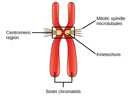
This illustration shows two sister chromatids. Each has a kinetochore at the centromere, and mitotic spindle microtubules radiate from the kinetochore.
Figure 10.7 — 10.2 The

Figure 10.7 During prometaphase, mitotic spindle microtubules from opposite poles attach to each sister chromatid at the kinetochore. In anaphase, the connection between the sister chromatids breaks down, and the microtubules pull the chromosomes toward opposite poles.

Mary Ann Clark, Matthew Douglas, and Jung Choi, Biology 2E, OpenStax. · CC BY-NC-SA 4.0 · Original image ↗ · Access for free ↗
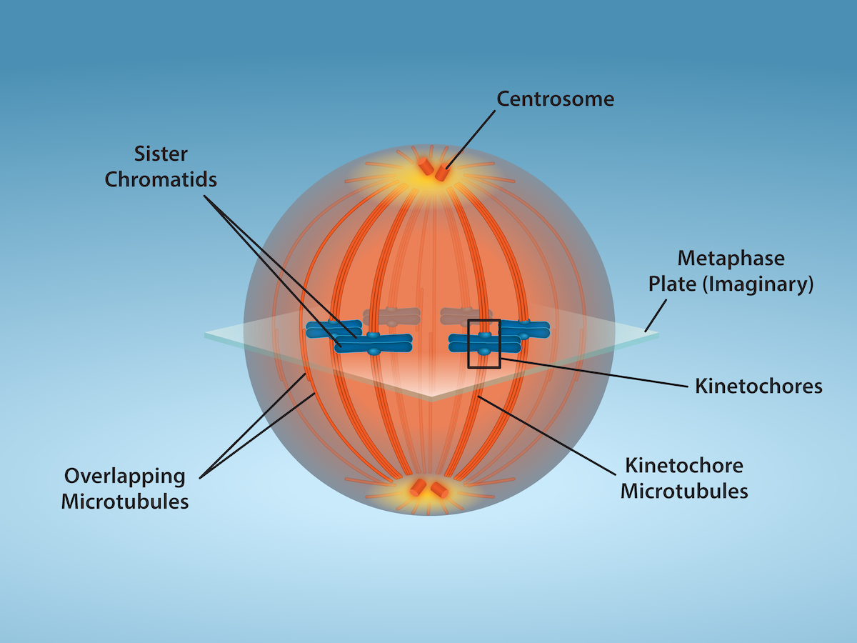
A cell shows overlapping microtubules extending throughout the cell.  At the center of the cell, sister chomratids line up. At their center are kinetochores. And at the very ends of the cell, away from the center, are centrosomes.
Figure 10.8 — 10.2 The

Figure 10.8 Mitotic Cell at Metaphase. The microtubule spindle has completed the alignment of chromosomes at the metaphase plate in preparation for the separation of sister chromatids during anaphase. Credit: Fletcher, S., Ryan, K. and Rao, A., Department of Biology, Texas A&M University.

Mary Ann Clark, Matthew Douglas, and Jung Choi, Biology 2E, OpenStax. · CC BY-NC-SA 4.0 · Original image ↗ · Access for free ↗
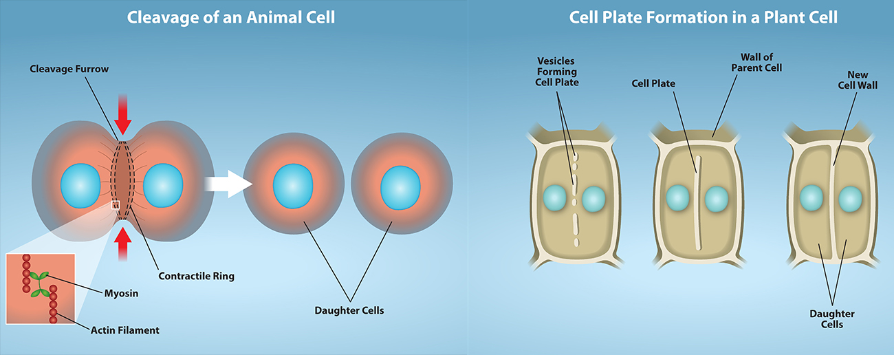
Part a: This illustration shows cytokinesis in a typical animal cell. Part b: Cytokinesis is shown in a typical plant cell. In an animal cell, a contractile ring of actin filaments forms a cleavage furrow that divides the cell in two. In a plant cell, Golgi vesicles coalesce at the metaphase plate. A cell plate grows from the center outward, and the vesicles form a plasma membrane that divides the cytoplasm.
Figure 10.9 — 10.2 The

Figure 10.9 Animal cell cleavage - Contraction of actin filaments by myosin pull the “equator” of the cell causing an invagination of the cell (a fissure called a cleavage furrow). The cleavage furrow gets continuously deeper until it eventually divides the cell into two new, independent daughter cells. Plant cell wall formation - A mixture of enzymes, proteins, and glucose molecules are transported via vesicles to the center of the cell. These vesicles continuously build upon each other until a completely new cell wall has emerged and two new cells are formed, independent of one another. Credit: Rao, A., Ryan, K., Hawkins, A.and Fletcher, S. Department of Biology, Texas A&M University.

Mary Ann Clark, Matthew Douglas, and Jung Choi, Biology 2E, OpenStax. · CC BY-NC-SA 4.0 · Original image ↗ · Access for free ↗
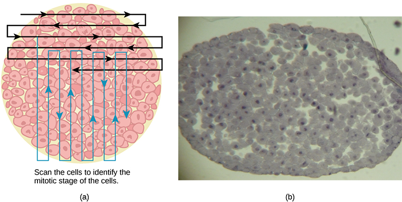
Left: This figure shows an illustration of whitefish blastula cells with a scanning pattern from right to left, and from top to bottom. Right: A micrograph of whitefish blastula cells in various phases of the cell cycle is shown.
Figure 10.10 — 10.2 The

Figure 10.10 Slowly scan whitefish blastula cells with the high-power objective as illustrated in image (a) to identify their mitotic stage. (b) A microscopic image of the scanned cells is shown. (credit “micrograph”: modification of work by Linda Flora; scale-bar data from Matt Russell)

Mary Ann Clark, Matthew Douglas, and Jung Choi, Biology 2E, OpenStax. (credit “micrograph”: modification of work by Linda Flora; scale-bar data from Matt Russell) · CC BY-NC-SA 4.0 · Original image ↗ · Access for free ↗
Phase or StageIndividual TotalsGroup TotalsPercent
Totals100100100 percent

Table 10.1. Mary Ann Clark, Matthew Douglas, and Jung Choi, Biology 2E, OpenStax.

Phase or StagePercentTime in Hours

Table 10.2. Mary Ann Clark, Matthew Douglas, and Jung Choi, Biology 2E, OpenStax.

Source: 10.2 The Cell Cycle

10.3 Control of the — figures and tables

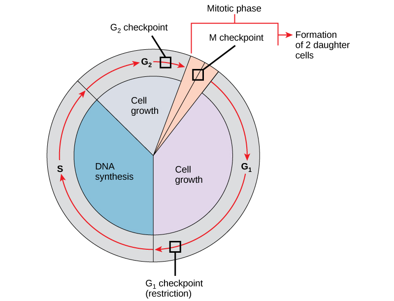
This illustration shows the three major checkpoints of the cell cycle: G subscript 1 baseline checkpoint restriction, G subscript 2 baseline checkpoint, and M checkpoint in the mitotic phase.
Figure 10.11 — 10.3 Control of the

Figure 10.11 The cell cycle is controlled at three checkpoints. The integrity of the DNA is assessed at the G 1 checkpoint. Proper chromosome duplication is assessed at the G 2 checkpoint. Attachment of each kinetochore to a spindle fiber is assessed at the M checkpoint.

Mary Ann Clark, Matthew Douglas, and Jung Choi, Biology 2E, OpenStax. · CC BY-NC-SA 4.0 · Original image ↗ · Access for free ↗
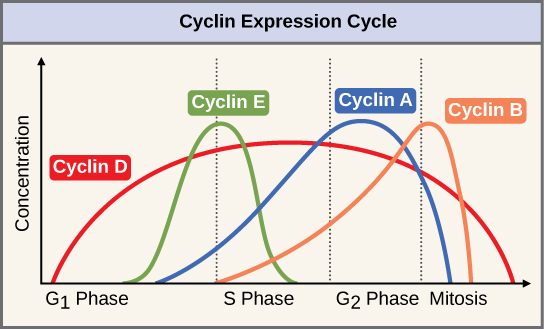
This graph shows the concentrations of different cyclin proteins during various phases of the cell cycle. Cyclin D concentrations increase in G subscript 1 baseline phase, and decrease at the end of mitosis. Cyclin E levels rise during G subscript 1 baseline phase and fall during S phase. Cyclin A levels rise during S phase and fall during mitosis. Cyclin B levels rise in S phase and fall during mitosis.
Figure 10.12 — 10.3 Control of the

Figure 10.12 The concentrations of cyclin proteins change throughout the cell cycle. There is a direct correlation between cyclin accumulation and the three major cell-cycle checkpoints. Also note the sharp decline of cyclin levels following each checkpoint (the transition between phases of the cell cycle), as cyclin is degraded by cytoplasmic enzymes. (credit: modification of work by "WikiMiMa"/Wikimedia Commons)

Mary Ann Clark, Matthew Douglas, and Jung Choi, Biology 2E, OpenStax. (credit: modification of work by "WikiMiMa"/Wikimedia Commons) · CC BY-NC-SA 4.0 · Original image ↗ · Access for free ↗
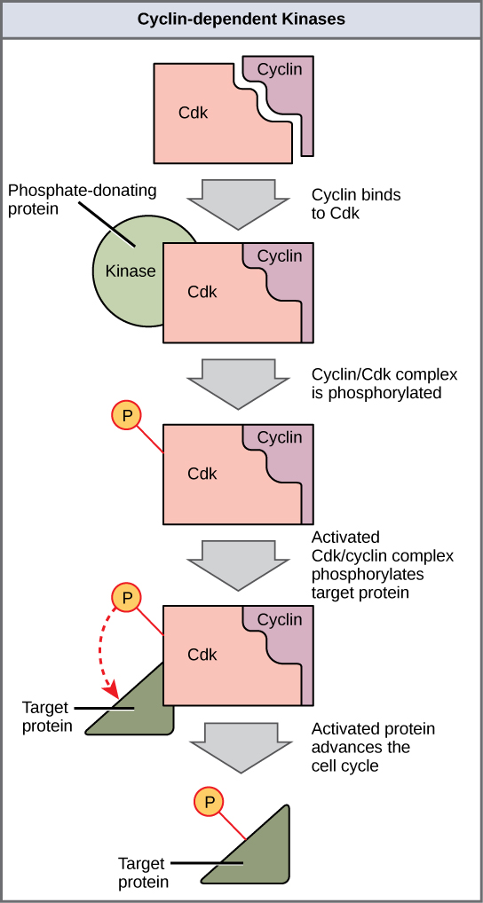
This illustration shows a cyclin protein binding to a upper case C lower case d lower case k. The cyclin slash upper C lower d lower k complex is activated when a kinase phosphorylates it. The cyclin slash upper C lower d lower k complex, in turn, phosphorylates other proteins, thus advancing the cell cycle.
Figure 10.13 — 10.3 Control of the

Figure 10.13 Cyclin-dependent kinases (Cdks) are protein kinases that, when fully activated, can phosphorylate and thus activate other proteins that advance the cell cycle past a checkpoint. To become fully activated, a Cdk must bind to a cyclin protein and then be phosphorylated by another kinase.

Mary Ann Clark, Matthew Douglas, and Jung Choi, Biology 2E, OpenStax. · CC BY-NC-SA 4.0 · Original image ↗ · Access for free ↗
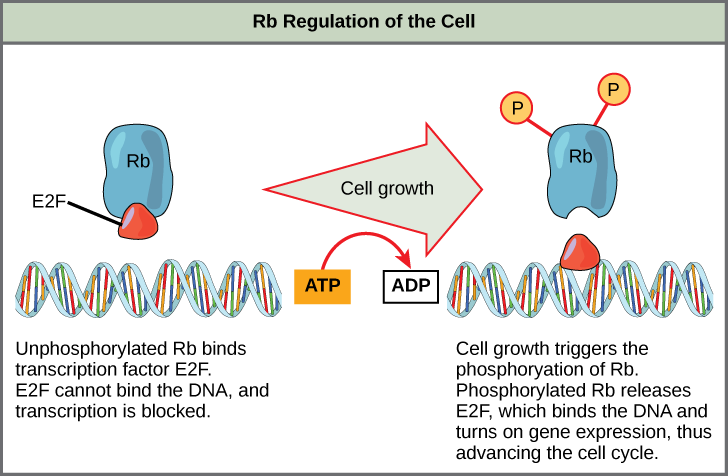
This illustration shows the regulation of the cell cycle by the upper case R lower case b protein. Unphosphorylated upper R lower b binds the transcription factor E 2 F. E 2 F cannot bind the D N A, and transcription is blocked. Cell growth triggers the phosphorylation of R b. Phosphorylated R b releases E 2 F, which binds the D N A and turns on gene expression, thus advancing the cell cycle.
Figure 10.14 — 10.3 Control of the

Figure 10.14 Rb halts the cell cycle and releases its hold in response to cell growth.

Mary Ann Clark, Matthew Douglas, and Jung Choi, Biology 2E, OpenStax. · CC BY-NC-SA 4.0 · Original image ↗ · Access for free ↗

Source: 10.3 Control of the Cell Cycle

229 terms, alphabetically ordered

3′ hydroxyl group · · ·

The at the sugar’s 3′ carbon that attacks the incoming ’s during strand extension.

Read explanation · OpenStax ↗
Acid · ·

In the Brønsted–Lowry model, a substance that can donate a . Acidic have a higher activity than water.

Read explanation · OpenStax ↗
Actin

A that forms cellular filaments and, together with , supports muscle contraction and other movements.

Read explanation · OpenStax ↗
Activation energy ·

The energy barrier that must overcome to reach a and undergo .

Read explanation · OpenStax ↗
Adenosine triphosphate

An energy-carrying ; its powers many cellular processes, including cycles.

Read explanation · OpenStax ↗
Allele

One version or copy of a at a ; a typical cell carries two copies of an autosomal .

Read explanation · NCBI Bookshelf / Sinauer Associates ↗
Alpha carbon · ·

The central carbon next to the carboxyl carbon. In a standard it binds an , hydrogen, and .

Read explanation · OpenStax ↗
Alpha helix · · · · · ·

A helical stabilized by between a oxygen and an hydrogen four later.

Read explanation · OpenStax ↗
Amide · · ·

A group containing a carbon bonded to nitrogen. are amide linkages.

Read explanation · OpenStax ↗
Amino acid

A building block containing amino and and a characteristic .

Read explanation · OpenStax ↗
Amino acid residue · ·

The part of an incorporated into a chain. Residue can also describe another building block within a larger .

Read explanation · OpenStax ↗
Amino group · · ·

A nitrogen-containing . A free ’s amino group is typically (NH₃⁺) near physiological ; ’s ring nitrogen is an exception to this formula.

Read explanation · OpenStax ↗
Amphipathic

Having both a region and a region, as in a or .

Read explanation · OpenStax ↗
Amylose

The largely unbranched component of , made mainly of linked by alpha-1,4 .

Read explanation · OpenStax ↗
anaphase

The division stage in which separated units move toward opposite poles.

Read explanation · OpenStax ↗
aneuploidy

An abnormal number of individual rather than a change in whole -set number.

Read explanation · OpenStax ↗
anomeric carbon

The former carbon of a sugar, which becomes the or hemiketal center upon cyclization.

Read explanation · OpenStax ↗
Antiparallel

Running in opposite directions, as with two strands or neighboring strands in some .

Read explanation · OpenStax ↗
APC/C

The -promoting complex/cyclosome, a regulating sister separation and mitotic exit.

Read explanation · www.ncbi.nlm.nih.gov ↗
Apoptosis

A regulated process of cell death that removes cells in development, tissue maintenance, and responses to damage.

Read explanation · OpenStax ↗
Aqueous solution ·

A in which water is the .

Read explanation · OpenStax ↗
Aromatic

Containing a cyclic, with . , , and have aromatic .

Read explanation · OpenStax ↗
Atom

The smallest unit of an element that retains its chemical identity; it has a surrounded by .

Read explanation · OpenStax ↗
attachment

Enduring emotional bond and expectations about another’s availability and responsiveness.

Read explanation · OpenStax ↗
Benzene ring

A six-carbon ring with .

Read explanation · OpenStax ↗
Benzyl group

A attached through a –CH₂– group; part of ’s .

Read explanation · OpenStax ↗
Beta sheet · · · · · ·

A made of aligned segments connected by ; strands may be parallel or .

Read explanation · OpenStax ↗
Biomolecule

A made by or involved in living systems, including , , , and .

Read explanation · OpenStax ↗
blastula

An early embryonic stage typically organized around a fluid-containing cavity.

Read explanation · OpenStax ↗
Blotting ·

Transfer of separated or to a so a specific target can be detected.

Read explanation · Thermo Fisher ↗
Brønsted–Lowry base · · · · · · · · · · ·

In the Brønsted–Lowry model, a substance that can accept a . Basic solutions have lower activity than water.

Read explanation · OpenStax ↗
Cancer

A group of diseases involving abnormal cells that can proliferate without normal restraints and invade tissues.

Read explanation · OpenStax ↗
Carbohydrate

A class of including sugars and their , used for energy storage, structure, and recognition.

Read explanation · OpenStax ↗
Carbonyl group · ·

A carbon double-bonded to oxygen, C=O; present in and many other .

Read explanation · OpenStax ↗
Carboxyl group · ·

An , COOH. In free it is usually to COO⁻ near physiological .

Read explanation · OpenStax ↗
caspase

A family involved in and selected inflammatory processes.

Read explanation · OpenStax ↗
Catalyst · ·

A substance that speeds a by providing a pathway with lower and is regenerated rather than consumed overall.

Read explanation · OpenStax ↗
Cell cycle

The ordered stages of cell growth, , and division.

Read explanation · OpenStax ↗
Cell proliferation

Increase in cell number through cell division; it should be distinguished experimentally from changes in survival.

Read explanation · OpenStax ↗
Cell-cycle checkpoint · ·

A control system that delays cell-cycle progression when necessary conditions, such as integrity, are not satisfied.

Read explanation · National Cancer Institute ↗
Cellular senescence

A stable state of cell-cycle arrest in which a cell remains metabolically active; it differs from .

Read explanation · NCBI Bookshelf / Sinauer Associates ↗
Cellulose

A structural in plant cell walls, with beta-1,4 linkages; also used as a support.

Read explanation · OpenStax ↗
centriole

A cylindrical structure with nine triplets found in and related basal bodies.

Read explanation · OpenStax ↗
Centromere

A region where the assembles and spindle supports .

Read explanation · OpenStax ↗
centrosome

A major animal-cell -organizing center, usually containing and surrounding nucleating material.

Read explanation · OpenStax ↗
Chemical reaction ·

A process that rearranges and chemical bonds, converting into .

Read explanation · OpenStax ↗
Chromatin

The complex of and associated , including , in .

Read explanation · OpenStax ↗
Chromatography

Separation of mixture components by their different interactions with a and a moving .

Read explanation · Waters ↗
Chromosome ·

An organized with associated that carries . A replicated chromosome has two .

Read explanation · OpenStax ↗
cleavage furrow

The constriction formed by the animal-cell – ring during .

Read explanation · OpenStax ↗
Codon

A sequence of three that specifies an or a stop signal.

Read explanation · OpenStax ↗
cohesin

A that helps hold together before separation.

Read explanation · OpenStax ↗
Concentration

The amount of a substance in a specified volume or mass of mixture.

Read explanation · OpenStax ↗
Configuration

A stereochemical arrangement that cannot be changed into another configuration by ordinary single-bond rotations alone.

Read explanation · OpenStax ↗
Conjugated system ·

Adjacent orbitals that allow to be distributed across several , often through alternating single and multiple bonds.

Read explanation · OpenStax ↗
Copy number ·

The number of copies of a segment in a defined sample or ; it is distinct from or abundance.

Read explanation · NHGRI / NIH ↗
Covalent bond · ·

A bond in which share pairs. and are covalent.

Read explanation · OpenStax ↗
culture

Learned and shared meanings, practices, values, and artifacts.

Read explanation · OpenStax ↗
Cyclin

A regulatory that binds a and helps control its activity and selection.

Read explanation · National Cancer Institute ↗
Cyclin-dependent kinase · · ·

A regulated by and other inputs that transitions in the .

Read explanation · National Cancer Institute ↗
Cysteine ·

An with a . Two cysteines can form a .

Read explanation · OpenStax ↗
cytochrome

A heme-containing involved in transfer.

Read explanation · OpenStax ↗
cytochrome c

A small mobile heme carrying from respiratory complex III to IV and participating in when released into .

Read explanation · OpenStax ↗
cytokinesis

Division of the into daughter cells.

Read explanation · OpenStax ↗
Cytoplasm

Cell contents inside the plasma and outside the , where one is present.

Read explanation · OpenStax ↗
Cytoskeleton

A network of filaments that supports cell shape, organizes components, and enables movement.

Read explanation · OpenStax ↗
Cytosol

The aqueous part of the outside -enclosed .

Read explanation · OpenStax ↗
Dalton

A unit of mass equal to one twelfth of the mass of a carbon-12 ; used to describe .

Read explanation · OpenStax ↗
Dehydration synthesis

Joining with net removal of water; the overall used to depict formation.

Read explanation · OpenStax ↗
Deoxyribonucleic acid

The that stores genetic information used to specify and sequences.

Read explanation · OpenStax ↗
Deprotonation

Removal of a from a or group; this changes its by minus one.

Read explanation · OpenStax ↗
Detergent

An substance that can associate with both water and material; detergents can disrupt or interactions.

Read explanation · Thermo Fisher ↗
differentiation

Acquisition of specialized cellular structures and functions through regulated and other changes.

Read explanation · OpenStax ↗
Diploid

Having two sets; comparisons of must account for and sample composition.

Read explanation · NHGRI / NIH ↗
Dissociation · ·

Separation of a chemical species or molecular complex into smaller components; dissociation releases a .

Read explanation · OpenStax ↗
Disulfide bond · · ·

A covalent sulfur–sulfur bond formed by of two , which can stabilize structure.

Read explanation · NCBI Bookshelf ↗
DNA ligase · · ·

An that seals a by forming a between an adjacent and 5′ .

Read explanation · OpenStax ↗
DNA nick · · ·

A break in one strand’s , with no missing. can seal a nick with suitable ends.

Read explanation · OpenStax ↗
DNA replication

Copying so genetic information can be passed to daughter cells. Each guides a complementary new strand.

Read explanation · OpenStax ↗
E2F

A family of regulating needed for cell-cycle progression, including entry into .

Read explanation · National Cancer Institute ↗
Electric charge · · ·

An electrical property that can be positive or negative. Opposite charges attract and like charges repel; net charge is the sum of all charges on a particle.

Read explanation · OpenStax ↗
Electron

A negatively subatomic particle. Electron sharing and transfer determine much of chemical bonding.

Read explanation · OpenStax ↗
Electron delocalization ·

Distribution of across several rather than confinement to one or bond.

Read explanation · OpenStax ↗
Electronegativity ·

An ’s tendency to attract shared in a chemical bond. Unequal attraction can make a bond .

Read explanation · OpenStax ↗
Endomembrane system

The interacting and compartments involved in processes such as modification, transport, and .

Read explanation · OpenStax ↗
Endoplasmic reticulum

An interconnected network continuous with the ; it includes regions involved in processing and synthesis.

Read explanation · OpenStax ↗
Enzyme

A biological that speeds a without being consumed. Most enzymes are .

Read explanation · OpenStax ↗
Enzyme inhibitor · ·

A substance that reduces activity. The binding mechanism and rate data determine whether inhibition is competitive, uncompetitive, mixed, or another type.

Read explanation · Biology LibreTexts ↗
Eukaryote ·

An organism whose cells have a -enclosed ; animals, plants, fungi, and protists are eukaryotes.

Read explanation · OpenStax ↗
Experimental control ·

A comparison sample or condition used to distinguish the effect being tested from other influences.

Read explanation · Thermo Fisher ↗
Functional group

A group of whose chemical behavior gives a characteristic and properties.

Read explanation · OpenStax ↗
G0 ·

A nondividing state outside the actively progressing ; reversibility depends on cell type and context.

Read explanation · OpenStax ↗
G1 phase · · ·

The cell-cycle period before synthesis; growth and damage signals influence commitment to .

Read explanation · National Cancer Institute ↗
G2 phase ·

The phase after and before , when are available for .

Read explanation · OpenStax ↗
gamete

A reproductive cell that contributes a during fertilization.

Read explanation · OpenStax ↗
Gene

A sequence that contributes to a functional or , together with the information needed to express it.

Read explanation · OpenStax ↗
Gene dosage ·

The number of copies of a or segment; changing dosage can affect expression without altering each copy’s sequence.

Read explanation · NHGRI / NIH ↗
Gene duplication ·

Creation of an additional copy of a or segment, potentially changing or enabling evolutionary divergence.

Read explanation · NHGRI / NIH ↗
Gene expression

Production of a functional from a , including and, for -coding , .

Read explanation · NCBI Bookshelf ↗
Genome

The complete genetic material of an organism or cell.

Read explanation · OpenStax ↗
GH

Growth hormone, an anterior pituitary affecting growth and directly and through .

Read explanation · OpenStax ↗
Glucose

A six-carbon sugar that is a major cellular energy source and a building block of , , and .

Read explanation · OpenStax ↗
glycogen

A highly branched used for storage in animals.

Read explanation · Biology LibreTexts / Oregon State University ↗
Glycosidic bond

A involving a sugar’s . In a , an N-glycosidic bond links the sugar to the .

Read explanation · Biology LibreTexts / Oregon State University ↗
Golgi apparatus ·

A stack of compartments that modifies and sorts and traveling through the .

Read explanation · OpenStax ↗
Haploid

Having one set of , as in a human .

Read explanation · OpenStax ↗
hemiacetal

Carbon bonded to both and alkoxy groups, usually formed by alcohol addition to a .

Read explanation · OpenStax ↗
Histone

A -associated rich in . Core histones form the around which nucleosomal wraps.

Read explanation · OpenStax ↗
Homologous recombination ·

Exchange or copying between with similar sequences. In break repair, an intact homologous template, usually a , guides restoration.

Read explanation · Biology LibreTexts / Oregon State University ↗
Hormone

A chemical messenger released by cells that changes the activity of target cells with matching .

Read explanation · OpenStax ↗
Hydrogen bond ·

An attraction between a hydrogen bonded to an and another . hydrogen bonds stabilize .

Read explanation · OpenStax ↗
Hydrogen ion · ·

A positively hydrogen species. H⁺ denotes a , which is hydrated in water; its activity determines .

Read explanation · OpenStax ↗
Hydrolysis

Breaking a chemical bond by adding water. hydrolysis separates .

Read explanation · OpenStax ↗
Hydrophilic

Interacting favorably with water, typically because a group is or .

Read explanation · OpenStax ↗
Hydrophobic

Interacting poorly with water. often gather in a ’s interior.

Read explanation · OpenStax ↗
Hydroxide ion ·

The negatively OH⁻. Basic contain a higher hydroxide ion activity than water at the same temperature.

Read explanation · OpenStax ↗
Hydroxyl group ·

An oxygen bonded to hydrogen, –OH. Hydroxyl groups can and often increase .

Read explanation · OpenStax ↗
IGF-1

Insulin-like growth factor 1, a growth-promoting signal whose production is stimulated by .

Read explanation · OpenStax ↗
Indole

A fused six-membered and five-membered ring system containing nitrogen; found in .

Read explanation · OpenStax ↗
Interphase

The , S, and portions of the between divisions; occurs in .

Read explanation · OpenStax ↗
Ion · ·

An or with a net because its numbers of and do not balance.

Read explanation · OpenStax ↗
Kinase ·

An that transfers a , often from , to a target .

Read explanation · Thermo Fisher ↗
kinetochore

A assembly at a that binds spindle .

Read explanation · OpenStax ↗
linkage

A tendency of nearby on the same to be inherited together.

Read explanation · OpenStax ↗
Lipid

A broad class of largely or , including fats, , and steroids.

Read explanation · OpenStax ↗
locus

The specific position of a or other feature.

Read explanation · OpenStax ↗
Logarithm · · ·

The exponent needed to obtain a number from a chosen fixed number. For decimal logarithms, log₁₀(100) = 2 because 10² = 100; uses the negative decimal logarithm of activity.

Read explanation · OpenStax ↗
Lysosome

An -enclosed compartment that uses hydrolytic to digest and recycle cellular material.

Read explanation · OpenStax ↗
Membrane · · · ·

A thin boundary or sheet. Cell membranes are bilayers containing ; membranes are synthetic supports that hold or for detection.

Read explanation · OpenStax ↗
Messenger RNA ·

An that carries coding information used by to synthesize a .

Read explanation · OpenStax ↗
Metabolism

The chemical that build , break them down, and transform energy in a living system.

Read explanation · OpenStax ↗
Metaphase

The division stage in which condensed align before separation.

Read explanation · OpenStax ↗
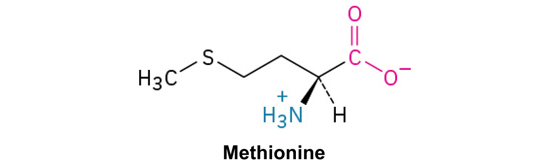
Methionine ·

A with sulfur in a . Unlike , it does not form .

Read explanation · OpenStax ↗
Methyl group ·

A –CH₃ group, consisting of one carbon bonded to three hydrogens.

Read explanation · OpenStax ↗
Microtubule

A hollow tube built from , supporting cell shape, intracellular transport, and separation.

Read explanation · OpenStax ↗
Mitochondrion ·

A with its own , central to aerobic production and several other processes.

Read explanation · OpenStax ↗
mitosis

division that separates duplicated into daughter , ordinarily preserving .

Read explanation · OpenStax ↗
Mobile phase

The moving fluid that carries a sample through a system.

Read explanation · Waters ↗
Molecular chaperone · ·

A that assists other in folding or prevents , without specifying their sequence.

Read explanation · NCBI Bookshelf ↗
Molecular mass · ·

The mass of a , commonly expressed in for . Molecular weight is often used informally for the same quantity.

Read explanation · OpenStax ↗
Molecule

Two or more joined by . A molecule may be electrically or carry a .

Read explanation · OpenStax ↗
Monomer

A small molecular building block that can join other monomers to form a .

Read explanation · OpenStax ↗
motor protein

that converts chemical energy into movement along a track.

Read explanation · Khan Academy ↗
Mutation

A change in a sequence; some mutations alter a ’s sequence.

Read explanation · OpenStax ↗
Myosin

A that uses to move along filaments.

Read explanation · OpenStax ↗
neuron

An electrically excitable cell specialized for receiving and transmitting information.

Read explanation · OpenStax ↗
Neutral

Having no net . For a , neutral instead means equal and activities; its is about 7 at 25 °C.

Read explanation · OpenStax ↗
Nitrogenous base · · ·

A nitrogen-containing ring component of ; its pairing interactions help specify sequences.

Read explanation · OpenStax ↗
Nonpolar ·

Lacking a substantial separation of . Nonpolar groups generally interact poorly with water.

Read explanation · OpenStax ↗
Nuclear envelope

The double enclosing the and continuous with the .

Read explanation · OpenStax ↗
Nucleic acid

A of , such as or , used in information storage, expression, and other cellular functions.

Read explanation · OpenStax ↗
Nucleolus

A region where much is and processed and begin assembly.

Read explanation · OpenStax ↗
Nucleosome

The basic unit of : wrapped around a .

Read explanation · OpenStax ↗
Nucleotide

A joined to a sugar and one or more .

Read explanation · OpenStax ↗
Nucleus ·

A -enclosed compartment containing most of a cell’s .

Read explanation · OpenStax ↗
Octamer

A complex made of eight . A core contains eight .

Read explanation · OpenStax ↗
Oncogene

A whose abnormal activation or excess expression contributes to uncontrolled growth or survival.

Read explanation · OpenStax ↗
Organelle ·

A specialized structure within a cell that carries out particular functions, such as a or .

Read explanation · OpenStax ↗
Oxidation ·

Loss of or an increase in oxidation state. Oxidizing two can form a .

Read explanation · OpenStax ↗
p21 protein

A whose expression can be induced by , helping restrain cell-cycle progression.

Read explanation · National Cancer Institute ↗
p53 protein

A stress-responsive that regulates involved in cell-cycle arrest, repair responses, , and .

Read explanation · NCBI Bookshelf / Sinauer Associates ↗
Peptide

A chain of joined by ; the term commonly describes chains shorter than a typical .

Read explanation · OpenStax ↗
Peptide bond ·

A covalent between the carboxyl carbon of one and the amino nitrogen of the next.

Read explanation · OpenStax ↗
pH

A measure of , defined as the negative of activity. Lower pH means greater ; pH influences .

Read explanation · OpenStax ↗
Phenylalanine ·

A with an .

Read explanation · OpenStax ↗
Phosphate group ·

A phosphorus-containing group bonded to oxygens; phosphate groups occur in , , and modified .

Read explanation · OpenStax ↗
Phosphodiester bond · · ·

A in which connects two sugars. In and it links the 3′ oxygen of one sugar to the 5′ oxygen of the next.

Read explanation · OpenStax ↗
Phospholipid

An with a -containing region and tails; a major component of cellular .

Read explanation · OpenStax ↗
Phosphorylation

Addition of a , often to , , or in ; can regulate activity.

Read explanation · Thermo Fisher ↗
ploidy

The number of complete sets in a cell.

Read explanation · OpenStax ↗
Polar

Having an uneven distribution of . Polar groups often interact favorably with water.

Read explanation · OpenStax ↗
Polymer

A large made from many linked molecular building blocks.

Read explanation · OpenStax ↗
Polypeptide

A chain of connected by . A functional can contain one or several such chains.

Read explanation · OpenStax ↗
Proline ·

An whose loops back to the nitrogen, restricting rotation and often creating bends.

Read explanation · OpenStax ↗
prometaphase

A mitotic stage involving -envelope breakdown and spindle to .

Read explanation · OpenStax ↗
prophase

An early division stage involving and spindle organization.

Read explanation · OpenStax ↗
Protease · ·

An that hydrolyzes in or .

Read explanation · OpenStax ↗
Protein

One or more folded chains that perform biological roles such as , transport, signaling, or structural support.

Read explanation · OpenStax ↗
Protein aggregation · ·

Association of into larger clusters, often when exposed surfaces of unfolded stick together.

Read explanation · NCBI Bookshelf ↗
Protein backbone ·

The repeating nitrogen–alpha-carbon– chain of a , excluding its variable .

Read explanation · OpenStax ↗
Protein complex ·

An assembly of interacting chains that function together.

Read explanation · OpenStax ↗
Proto-oncogene

A normal involved in regulated growth or survival that can become an through an activating alteration.

Read explanation · OpenStax ↗
Proton

A positively subatomic particle. In – chemistry, donating a proton means transferring a .

Read explanation · OpenStax ↗
Protonation

Addition of a to an , , or ; this changes its by plus one.

Read explanation · OpenStax ↗
quiescence

A usually reversible state of cell-cycle , distinct from and terminal .

Read explanation · OpenStax ↗
R group · ·

The variable side chain attached to an ’s . It determines identity, , , and chemical behavior.

Read explanation · OpenStax ↗
RB1 gene

The encoding , a regulator that restrains entry into through .

Read explanation · NCBI Bookshelf / Sinauer Associates ↗
Reactant

A starting substance transformed during a .

Read explanation · OpenStax ↗
Reaction intermediate · ·

A species formed in one step and consumed in another. It occupies a local energy minimum between .

Read explanation · OpenStax ↗
Reaction product ·

A substance formed by a .

Read explanation · OpenStax ↗
Receptor

A that recognizes a particular signal and helps produce a cellular response.

Read explanation · OpenStax ↗
Retinoblastoma protein ·

The -encoded that binds and restrains ; its activity is regulated by .

Read explanation · NCBI Bookshelf / Sinauer Associates ↗
Ribonucleic acid ·

A with roles in , regulation, and ; carries -coding information.

Read explanation · OpenStax ↗
Ribosomal RNA · ·

An component of that contributes to their structure, decoding, and ; it is not into .

Read explanation · OpenStax ↗
Ribosome ·

A complex of and that translates into a .

Read explanation · OpenStax ↗
S phase

The phase of the during which is replicated.

Read explanation · OpenStax ↗
Secondary structure

Local folding, such as and , stabilized mainly by .

Read explanation · OpenStax ↗
Secretory pathway ·

The route through the , , and used to deliver many to the cell surface, outside the cell, or compartments.

Read explanation · OpenStax ↗
securin

An of whose degradation helps initiate sister-chromatid separation.

Read explanation · www.ncbi.nlm.nih.gov ↗
segregation

Separation of paired into different through segregation.

Read explanation · OpenStax ↗
separase

A that cleaves components to permit separation.

Read explanation · www.ncbi.nlm.nih.gov ↗
Serine ·

A , uncharged with a -containing .

Read explanation · OpenStax ↗
Sister chromatid

One of the two copies of a produced by . An intact sister chromatid can supply a template for accurate repair.

Read explanation · Biology LibreTexts / Oregon State University ↗
Solubility ·

The amount of a substance that can dissolve in a under specified conditions. Soluble means able to dissolve; insoluble means only very slightly soluble in that .

Read explanation · OpenStax ↗
Solute

A dissolved component of a .

Read explanation · OpenStax ↗
Solution

A homogeneous mixture in which are dissolved in a .

Read explanation · OpenStax ↗
Solvent

The component of a that dissolves other substances, usually the component present in the greatest amount.

Read explanation · OpenStax ↗
Starch

A plant storage made of chains; its two main forms are and amylopectin.

Read explanation · OpenStax ↗
Stationary phase

The material that remains fixed in a system.

Read explanation · Waters ↗
Substrate

A that an binds and chemically transforms.

Read explanation · OpenStax ↗
Subunit ·

An individual chain within a containing multiple chains.

Read explanation · OpenStax ↗
telophase

A late division stage involving daughter- reassembly and decondensation.

Read explanation · OpenStax ↗
Template strand · · ·

A preexisting strand whose sequence guides complementary nucleic- synthesis.

Read explanation · OpenStax ↗
Thioether

A sulfur bonded to two carbon groups, as in ’s . It has no hydrogen.

Read explanation · OpenStax ↗
Thiol group · · · ·

A sulfur bonded to hydrogen, –SH. thiols can form through .

Read explanation · OpenStax ↗
Threonine ·

A , uncharged with a and a on its .

Read explanation · OpenStax ↗
Transcription

Synthesis of using a template.

Read explanation · OpenStax ↗
Transcription factor

A that helps recruit or regulate machinery, often by binding or other .

Read explanation · OpenStax ↗
Transition state

A fleeting, high-energy arrangement at a barrier. It is an energy maximum along the path, rather than a stable .

Read explanation · OpenStax ↗
Translation ·

Synthesis of a on a using the sequence of in .

Read explanation · OpenStax ↗
Tryptophan ·

An with a bulky ; commonly grouped as .

Read explanation · OpenStax ↗
Tubulin

A that forms alpha–beta dimers; these dimers assemble into .

Read explanation · OpenStax ↗
Tumor · ·

An abnormal mass of tissue; tumors can be benign or malignant.

Read explanation · OpenStax ↗
Tumor suppressor gene · · · ·

A whose normal function restrains formation, for example by controlling , damage responses, or integrity.

Read explanation · OpenStax ↗
Tyrosine ·

An with a phenolic . Usually uncharged near physiological .

Read explanation · OpenStax ↗
Ubiquitin

A small attached to other to regulate their degradation, localization, or interactions.

Read explanation · Thermo Fisher ↗
Vesicle · · ·

A small -enclosed compartment that can transport material between cellular locations.

Read explanation · OpenStax ↗
withdrawal

Symptoms emerging when exposure to an adapted-to substance is reduced or stopped.

Read explanation · OpenStax ↗
zygote

The single cell formed by fusion and union of parental genetic contributions.

Read explanation · OpenStax ↗

10.2 The Cell Cycle

OpenStax · 10.2 The Cell Cycle ↗
Instructional page including learning objectives, examples, figures, source tables and questions. Site navigation and embedded media players are omitted; source media links remain available.

Learning Objectives

By the end of this section, you will be able to do the following:

The is an ordered series of events involving cell growth and cell division that produces two new daughter cells. Cells on the path to cell division proceed through a series of precisely timed and carefully regulated stages of growth, , and and division that ultimately produces two identical (clone) cells. The has two major phases: and the mitotic phase (Figure 10.5). During , the cell grows and is replicated. During the mitotic phase, the replicated and contents are separated, and the cell is typically partitioned by a third process of the called . We should note, however, that and (karyokinesis) may take place without , in which case cells with multiple (multinucleate cells) are produced.

Like a clock, the cell cycles from interphase to the mitotic phase and back to interphase. Most of the cell cycle is spent in interphase, which is subdivided into G subscript 1 baseline, S, and G subscript 2 baseline phases. Cell growth occurs during G subscript 1 baseline, D N A synthesis occurs during S, and more growth occurs during G subscript 2 baseline. The mitotic phase consists of mitosis, in which the nuclear chromatin is divided, and cytokinesis, in which the cytoplasm is divided, resulting in two daughter cells.
Figure 10.5 — 10.2 The

Figure 10.5 The cell cycle in multicellular organisms consists of interphase and the mitotic phase. During interphase, the cell grows and the nuclear DNA is duplicated. Interphase is followed by the mitotic phase. During the mitotic phase, the duplicated chromosomes are segregated and distributed into daughter nuclei. Following mitosis, the cytoplasm is usually divided as well by cytokinesis, resulting in two genetically identical daughter cells.

Mary Ann Clark, Matthew Douglas, and Jung Choi, Biology 2E, OpenStax. · CC BY-NC-SA 4.0 · Original image ↗ · Access for free ↗

During , the cell undergoes normal growth processes while also preparing for cell division. In order for a cell to move from into the mitotic phase, many internal and external conditions must be . The three stages of are called G1, S, and G2.

G1 Phase (First Gap)

The first stage of is called the G1 phase (first gap) because, from a microscopic point of view, little change is visible. However, during the G1 stage, the cell is quite active at the biochemical level. The cell is accumulating the building blocks of and the associated as well as accumulating sufficient energy reserves to complete the task of replicating each in the .

(Synthesis of )

Throughout , remains in a semi-condensed . In the , can proceed through the mechanisms that result in the formation of identical pairs of ——that are firmly attached at the centromeric region. The is also duplicated during the . The two will give rise to the mitotic spindle, the apparatus that orchestrates the movement of during . For example, roughly at the center of each animal cell, the are associated with a pair of rod-like objects, the , which are positioned at right angles to each other. help organize cell division. We should note, however, that are not present in the of other organisms, such as plants and most fungi.

G2 Phase (Second Gap)

In the G2 phase, the cell replenishes its energy stores and synthesizes necessary for manipulation and movement. Some cell are duplicated, and the is dismantled to provide resources for the mitotic phase. There may be additional cell growth during G2. The final preparations for the mitotic phase must be completed before the cell is able to enter the first stage of .

The Mitotic Phase

The mitotic phase is a multistep process during which the duplicated are aligned, separated, and move into two new, identical daughter cells. The first portion of the mitotic phase is called karyokinesis, or division. As we have just seen, the second portion of the mitotic phase (and often viewed as a process separate from and following ) is called —the physical separation of the components into the two daughter cells.

Link to Learning

Revisit the stages of at this site.

Karyokinesis ()

Karyokinesis, also known as , is divided into a series of phases—, , , , and —that result in the division of the cell (Figure 10.6).

Visual Connection

This diagram shows the five phases of mitosis and cytokinesis. During prophase, the chromosomes condense and become visible, spindle fibers emerge from the centrosomes, the nuclear envelope breaks down, and the nucleolus disappears. During prometaphase, the chromosomes continue to condense and kinetochores appear at the centromeres. Mitotic spindle microtubules attach to the kinetochores, and centrosomes move toward opposite poles. During metaphase, the mitotic spindle is fully developed, and centrosomes are at opposite poles of the cell. Chromosomes line up at the metaphase plate and each sister chromatid is attached to a spindle fiber originating from the opposite pole. During anaphase, the cohesin proteins that were binding the sister chromatids together break down. The sister chromatids, which are now called chromosomes, move toward opposite poles of the cell. Non-kinetochore spindle fibers lengthen, elongating the cell. During telophase, chromosomes arrive at the opposite poles and begin to decondense. The nuclear envelope reforms. During cytokinesis in animals, a cleavage furrow separates the two daughter cells. In plants, a cell plate separates the two cells.
Figure 10.6 — 10.2 The

Figure 10.6 G2 of Interphase – The last stage of interphase is the second gap period, G2. During this stage, cells grow, replenish energy and synthesize needed macromolecules, such as proteins and lipids. Mitosis - When G2 is complete, the cell will enter mitosis. Although there are 5 phases in mitosis, with the exception of the metaphase to anaphase transition, these phases are not discrete and happen as a continuous process. Prophase is the first stage in mitosis. The nuclear envelope begins to break down and chromosomes condense and are now visible. Spindle fibers start to appear and centrosomes begin to move towards opposite poles. Prometaphase - Chromosomes continue to condense and are more visible. Kinetochores appear at the centromere and kinetochore microtubules attach. Centrosomes continue to move towards opposite poles. Metaphase – The mitotic spindle is fully developed and centrosomes are at opposite poles. Chromosomes are aligned at the “equatorial plate”, and each sister chromatid rests on one side of the plate, with spindle fibers attached to them. Anaphase - Sister chromatids are pulled apart by spindle fibers and are separated from each other. Each chromatid is now a chromosome. Telophase and Cytokinesis - Chromosomes arrive at opposite poles and start to decondense and become less visible. The nuclear envelope reassembles and begins to surround each new set of chromosomes. The mitotic spindle assembly breaks down and the division of the cytoplasm begins via cytokinesis. This physical separation into two cells are remarkably different processes in plant and animal cells. Credit: Rao, A., Hawkins, A.and Fletcher, S. Department of Biology, Texas A&M University.

Mary Ann Clark, Matthew Douglas, and Jung Choi, Biology 2E, OpenStax. · CC BY-NC-SA 4.0 · Original image ↗ · Access for free ↗

Which of the following is the correct order of events in ?

  1. line up at the plate. The becomes attached to the mitotic spindle. The reforms and the cell divides. break down and the separate.
  2. The becomes attached to the mitotic spindle. break down and the separate. line up at the plate. The reforms and the cell divides.
  3. The becomes attached to the . line up at the plate. The breaks down and the separate. The reforms and the cell divides.
  4. The becomes attached to the mitotic spindle. line up at the plate. break down and the separate. The reforms and the cell divides.

(the “first phase”): the starts to into small , and the membranous (such as the complex [] and the ), fragment and disperse toward the periphery of the cell. The disappears (disperses) as well, and the begin to move to opposite poles of the cell. that will form the mitotic spindle extend between the , pushing them farther apart as the fibers lengthen. The begin to coil more tightly with the aid of condensin and now become visible under a light microscope.

(the “first change phase”): Many processes that began in continue to advance. The remnants of the fragment further, and the mitotic spindle continues to develop as more assemble and stretch across the length of the former area. become even more condensed and discrete. Each develops a structure called a in its centromeric region (Figure 10.7). The of the attract and bind to the mitotic spindle . As the spindle extend from the , some of these come into contact with and firmly bind to the . Once a mitotic fiber attaches to a , the will be oriented until the of face the opposite poles. Eventually, all the will be attached via their to from opposing poles. Spindle that do not engage the are called . These overlap each other midway between the two poles and contribute to cell elongation. Astral are located near the poles, aid in spindle orientation, and are required for the regulation of .

This illustration shows two sister chromatids. Each has a kinetochore at the centromere, and mitotic spindle microtubules radiate from the kinetochore.
Figure 10.7 — 10.2 The

Figure 10.7 During prometaphase, mitotic spindle microtubules from opposite poles attach to each sister chromatid at the kinetochore. In anaphase, the connection between the sister chromatids breaks down, and the microtubules pull the chromosomes toward opposite poles.

Mary Ann Clark, Matthew Douglas, and Jung Choi, Biology 2E, OpenStax. · CC BY-NC-SA 4.0 · Original image ↗ · Access for free ↗

(the “change phase”): All the are aligned in a plane called the plate, or the equatorial plane, roughly midway between the two poles of the cell. The are still tightly attached to each other by . At this time, the are maximally condensed.

(“upward phase”): The degrade, and the separate at the . Each chromatid, now called a single , is pulled rapidly toward the to which its is attached. The cell becomes visibly elongated (oval shaped) as the slide against each other at the plate where they overlap.

(the “distance phase”): the reach the opposite poles and begin to decondense (unravel), relaxing once again into a stretched-out . The mitotic spindles are depolymerized into that will be used to assemble components for each daughter cell. envelopes form around the , and appear within the area.

A cell shows overlapping microtubules extending throughout the cell.  At the center of the cell, sister chomratids line up. At their center are kinetochores. And at the very ends of the cell, away from the center, are centrosomes.
Figure 10.8 — 10.2 The

Figure 10.8 Mitotic Cell at Metaphase. The microtubule spindle has completed the alignment of chromosomes at the metaphase plate in preparation for the separation of sister chromatids during anaphase. Credit: Fletcher, S., Ryan, K. and Rao, A., Department of Biology, Texas A&M University.

Mary Ann Clark, Matthew Douglas, and Jung Choi, Biology 2E, OpenStax. · CC BY-NC-SA 4.0 · Original image ↗ · Access for free ↗

, or “cell motion,” is sometimes viewed as the second main stage of the mitotic phase, during which cell division is completed via the physical separation of the components into two daughter cells However, as we have seen earlier, can also be viewed as a separate phase, which may or may not take place following . If does take place, cell division is not complete until the cell components have been apportioned and completely separated into the two daughter cells. Although the stages of are similar for most , the process of is quite different for that have cell walls, such as plant cells.

In animal cells, typically starts during late . A contractile ring composed of filaments forms just inside the plasma at the former plate. The filaments pull the equator of the cell inward, forming a fissure. This fissure is called the . The furrow deepens as the ring contracts, and eventually the is cleaved in two (Figure 10.9).

In plant cells, a new cell wall must form between the daughter cells. During , the accumulates , structural , and prior to breaking into and dispersing throughout the dividing cell. During , these are transported on to form a phragmoplast (a vesicular structure) at the plate. There, the fuse and coalesce from the center toward the cell walls; this structure is called a cell plate. As more fuse, the cell plate enlarges until it merges with the cell walls at the periphery of the cell. use the that has accumulated between the layers to build a new cell wall. The become parts of the plasma on either side of the new cell wall (Figure 10.9).

Part a: This illustration shows cytokinesis in a typical animal cell. Part b: Cytokinesis is shown in a typical plant cell. In an animal cell, a contractile ring of actin filaments forms a cleavage furrow that divides the cell in two. In a plant cell, Golgi vesicles coalesce at the metaphase plate. A cell plate grows from the center outward, and the vesicles form a plasma membrane that divides the cytoplasm.
Figure 10.9 — 10.2 The

Figure 10.9 Animal cell cleavage - Contraction of actin filaments by myosin pull the “equator” of the cell causing an invagination of the cell (a fissure called a cleavage furrow). The cleavage furrow gets continuously deeper until it eventually divides the cell into two new, independent daughter cells. Plant cell wall formation - A mixture of enzymes, proteins, and glucose molecules are transported via vesicles to the center of the cell. These vesicles continuously build upon each other until a completely new cell wall has emerged and two new cells are formed, independent of one another. Credit: Rao, A., Ryan, K., Hawkins, A.and Fletcher, S. Department of Biology, Texas A&M University.

Mary Ann Clark, Matthew Douglas, and Jung Choi, Biology 2E, OpenStax. · CC BY-NC-SA 4.0 · Original image ↗ · Access for free ↗

G0 Phase

Not all cells adhere to the classic cell-cycle pattern in which a newly formed daughter cell immediately enters the preparatory phases of , closely followed by the mitotic phase, and . Cells in G0 phase are not actively preparing to divide. The cell is in a quiescent (inactive) stage that occurs when cells exit the . Some cells enter G0 temporarily due to environmental conditions such as availability of nutrients, or stimulation by growth factors. The cell will remain in this phase until conditions improve or until an external signal triggers the onset of G1. Other cells that never or rarely divide, such as mature cardiac muscle and nerve cells, remain in G0 permanently.

Scientific Method Connection

Determine the Time Spent in Cell-Cycle Stages

Problem: How long does a cell spend in compared to each stage of ?

Background: A prepared microscope slide of whitefish cross-sections will show cells arrested in various stages of the . (Note: It is not visually possible to separate the stages of from each other, but the mitotic stages are readily identifiable.) If 100 cells are examined, the number of cells in each identifiable cell-cycle stage will give an estimate of the time it takes for the cell to complete that stage.

Problem Statement: Given the events included in all of and those that take place in each stage of , estimate the length of each stage based on a 24-hour . Before proceeding, state your hypothesis.

Test your hypothesis: Test your hypothesis by doing the following:

  1. Place a fixed and stained microscope slide of whitefish cross-sections under the scanning objective of a light microscope.
  2. Locate and focus on one of the sections using the low-power objective of your microscope. Notice that the section is a circle composed of dozens of closely packed individual cells.
  3. Switch to the medium-power objective and refocus. With this objective, individual cells are clearly visible, but the will still be very small.
  4. Switch to the high-power objective and slowly move the slide left to right, and up and down to view all the cells in the section (Figure 10.10). As you scan, you will notice that most of the cells are not undergoing but are in the period of the .

    Left: This figure shows an illustration of whitefish blastula cells with a scanning pattern from right to left, and from top to bottom. Right: A micrograph of whitefish blastula cells in various phases of the cell cycle is shown.
    Figure 10.10 — 10.2 The

    Figure 10.10 Slowly scan whitefish blastula cells with the high-power objective as illustrated in image (a) to identify their mitotic stage. (b) A microscopic image of the scanned cells is shown. (credit “micrograph”: modification of work by Linda Flora; scale-bar data from Matt Russell)

    Mary Ann Clark, Matthew Douglas, and Jung Choi, Biology 2E, OpenStax. (credit “micrograph”: modification of work by Linda Flora; scale-bar data from Matt Russell) · CC BY-NC-SA 4.0 · Original image ↗ · Access for free ↗
  5. Practice identifying the various stages of the , using the drawings of the stages as a guide (Figure 10.6).
  6. Once you are confident about your identification, begin to record the stage of each cell you encounter as you scan left to right, and top to bottom across the section.
  7. Keep a tally of your observations and stop when you reach 100 cells identified.
  8. The larger the sample size (total number of cells counted), the more accurate the results. If possible, gather and record group data prior to calculating percentages and making estimates.

Record your observations: Make a table similar to Table 10.1 within which to record your observations.

Results of Cell Stage Identification
Phase or StageIndividual TotalsGroup TotalsPercent
Totals100100100 percent
Table 10.1

Analyze your data/report your results: To find the length of time whitefish cells spend in each stage, multiply the percent (recorded as a decimal) by 24 hours. Make a table similar to Table 10.2 to illustrate your data.

Estimate of Cell Stage Length
Phase or StagePercentTime in Hours
Table 10.2

Draw a conclusion: Did your results support your estimated times? Were any of the outcomes unexpected? If so, discuss those events in that stage that may have contributed to the calculated time.

10.3 Control of the Cell Cycle

OpenStax · 10.3 Control of the Cell Cycle ↗

Instructional page including learning objectives, examples, figures, source tables and questions. Site navigation and embedded media players are omitted; source media links remain available.

Learning Objectives

By the end of this section, you will be able to do the following:

  • Understand how the is controlled by mechanisms that are both internal and external to the cell
  • Explain how the three internal “control ” occur at the end of G1, at the G2/M transition, and during
  • Describe the that control the through positive and negative regulation

The length of the is highly variable, even within the cells of a single organism. In humans, the frequency of cell turnover ranges from a few hours in early embryonic development, to an average of two to five days for epithelial cells, and to an entire human lifetime spent in G0 by specialized cells, such as cortical or cardiac muscle cells.

There is also variation in the time that a cell spends in each phase of the . When rapidly dividing mammalian cells are grown in a (outside the body under optimal growing conditions), the length of the is about 24 hours. In rapidly dividing human cells with a 24-hour , the G1 phase lasts approximately nine hours, the lasts 10 hours, the G2 phase lasts about four and one-half hours, and the M phase lasts approximately one-half hour. By comparison, in fertilized eggs (and early embryos) of fruit flies, the is completed in about eight minutes. This is because the of the fertilized egg divides many times by but does not go through until a multinucleate “” has been produced, with many located along the periphery of the , thereby shortening the time of the cell division cycle. The timing of events in the of both “invertebrates” and “vertebrates” is controlled by mechanisms that are both internal and external to the cell.

Regulation of the by External Events

Both the initiation and inhibition of cell division are triggered by events external to the cell when it is about to begin the process. An event may be as simple as the death of nearby cells or as sweeping as the release of growth-promoting , such as human (HGH or hGH). A lack of HGH can inhibit cell division, resulting in dwarfism, whereas too much HGH can result in gigantism. Crowding of cells can also inhibit cell division. In contrast, a factor that can initiate cell division is the size of the cell: As a cell grows, it becomes physiologically inefficient due to its decreasing surface-to-volume ratio. The to this problem is to divide.

Whatever the source of the message, the cell receives the signal, and a series of events within the cell allows it to proceed into . Moving forward from this initiation point, every parameter required during each phase must be or the cycle cannot progress.
Regulation at Internal

It is essential that the daughter cells produced be exact duplicates of the parent cell. Mistakes in the or distribution of the lead to that may be passed forward to every new cell produced from an abnormal cell. To prevent a compromised cell from continuing to divide, there are internal control mechanisms that operate at three main : A is one of several points in the at which the progression of a cell to the next stage in the cycle can be halted until conditions are favorable. These occur near the end of G1, at the G2/M transition, and during (Figure 10.11).
This illustration shows the three major checkpoints of the cell cycle: G subscript 1 baseline checkpoint restriction, G subscript 2 baseline checkpoint, and M checkpoint in the mitotic phase.
Figure 10.11 — 10.3 Control of the

Figure 10.11 The cell cycle is controlled at three checkpoints. The integrity of the DNA is assessed at the G 1 checkpoint. Proper chromosome duplication is assessed at the G 2 checkpoint. Attachment of each kinetochore to a spindle fiber is assessed at the M checkpoint.

Mary Ann Clark, Matthew Douglas, and Jung Choi, Biology 2E, OpenStax. · CC BY-NC-SA 4.0 · Original image ↗ · Access for free ↗

The G1

The G1 determines whether all conditions are favorable for cell division to proceed. The G1 , also called the restriction point (in yeast), is a point at which the cell irreversibly commits to the cell division process. External influences, such as growth factors, play a large role in carrying the cell past the G1 . In addition to adequate reserves and cell size, there is a check for damage at the G1 . A cell that does not meet all the requirements will not be allowed to progress into the . The cell can halt the cycle and attempt to remedy the problematic condition, or the cell can advance into G0 and await further signals when conditions improve.

The G2

The G2 bars entry into the mitotic phase if certain conditions are not . As at the G1 , cell size and reserves are assessed. However, the most important role of the G2 is to ensure that all of the have been replicated and that the replicated is not damaged. If the mechanisms detect problems with the , the is halted, and the cell attempts to either complete or repair the damaged .

The M

The M occurs near the end of the stage of karyokinesis. The M is also known as the spindle , because it determines whether all the are correctly attached to the spindle . Because the separation of the during is an irreversible step, the cycle will not proceed until the of each pair of are firmly anchored to at least two spindle fibers arising from opposite poles of the cell.

Link to Learning

Watch what occurs at the G1, G2, and M by visiting this website to see an animation of the .

Regulator of the

In addition to the internally controlled , there are two groups of intracellular that regulate the . These regulatory either promote progress of the cell to the next phase (positive regulation) or halt the cycle (negative regulation). Regulator may act individually, or they can influence the activity or production of other regulatory . Therefore, the failure of a single regulator may have almost no effect on the , especially if more than one mechanism the same event. However, the effect of a deficient or non-functioning regulator can be wide-ranging and possibly fatal to the cell if multiple processes are affected.

Positive Regulation of the

Two groups of , called and (), are termed positive regulators. They are responsible for the progress of the cell through the various . The levels of the four fluctuate throughout the in a predictable pattern (Figure 10.12). Increases in the of are triggered by both external and internal signals. After the cell moves to the next stage of the , the that were active in the previous stage are degraded by , as shown in Figure 10.12 below.

This graph shows the concentrations of different cyclin proteins during various phases of the cell cycle. Cyclin D concentrations increase in G subscript 1 baseline phase, and decrease at the end of mitosis. Cyclin E levels rise during G subscript 1 baseline phase and fall during S phase. Cyclin A levels rise during S phase and fall during mitosis. Cyclin B levels rise in S phase and fall during mitosis.
Figure 10.12 — 10.3 Control of the

Figure 10.12 The concentrations of cyclin proteins change throughout the cell cycle. There is a direct correlation between cyclin accumulation and the three major cell-cycle checkpoints. Also note the sharp decline of cyclin levels following each checkpoint (the transition between phases of the cell cycle), as cyclin is degraded by cytoplasmic enzymes. (credit: modification of work by "WikiMiMa"/Wikimedia Commons)

Mary Ann Clark, Matthew Douglas, and Jung Choi, Biology 2E, OpenStax. (credit: modification of work by "WikiMiMa"/Wikimedia Commons) · CC BY-NC-SA 4.0 · Original image ↗ · Access for free ↗

regulate the only when they are tightly bound to . To be fully active, the / complex must also be in specific locations to activate the complex. Like all , are () that in turn phosphorylate other . activates the by changing its shape. The by are involved in advancing the cell to the next phase. (Figure 10.13). The levels of are relatively stable throughout the ; however, the of fluctuate and determine when / complexes form. The different and bind at specific points in the and thus regulate different .

This illustration shows a cyclin protein binding to a upper case C lower case d lower case k. The cyclin slash upper C lower d lower k complex is activated when a kinase phosphorylates it. The cyclin slash upper C lower d lower k complex, in turn, phosphorylates other proteins, thus advancing the cell cycle.
Figure 10.13 — 10.3 Control of the

Figure 10.13 Cyclin-dependent kinases (Cdks) are protein kinases that, when fully activated, can phosphorylate and thus activate other proteins that advance the cell cycle past a checkpoint. To become fully activated, a Cdk must bind to a cyclin protein and then be phosphorylated by another kinase.

Mary Ann Clark, Matthew Douglas, and Jung Choi, Biology 2E, OpenStax. · CC BY-NC-SA 4.0 · Original image ↗ · Access for free ↗

Because the cyclic fluctuations of levels are largely based on the timing of the and not on specific events, regulation of the usually occurs by either the alone or the / complexes. Without a specific of fully activated / complexes, the cannot proceed through the .

Although the are the main regulatory that determine the forward momentum of the , there are several other mechanisms that fine-tune the progress of the cycle with negative, rather than positive, effects. These mechanisms essentially block the progression of the until problematic conditions are resolved. that prevent the full activation of are called . Many of these directly or indirectly monitor a particular cell-cycle event. The block placed on by will not be removed until the specific event that the monitors is completed.

Negative Regulation of the

The second group of cell-cycle regulatory are negative regulators, which stop the . Remember that in positive regulation, active cause the cycle to progress.

The best understood negative regulatory are (), , and . Retinoblastoma are a group of -suppressor common in many cells. We should note here that the 53 and 21 designations refer to the functional of the (p) in kilodaltons (a is equal to an atomic mass unit, which is equal to one or one neutron or 1 g/mol). Much of what is known about cell-cycle regulation comes from research conducted with cells that have lost regulatory control. All three of these regulatory were discovered to be damaged or non-functional in cells that had begun to replicate uncontrollably (i.e., became ). In each case, the main cause of the unchecked progress through the was a faulty copy of the regulatory .

, , and act primarily at the G1 . is a multi-functional that has a major impact on the commitment of a cell to division because it acts when there is damaged in cells that are undergoing the preparatory processes during G1. If damaged is detected, halts the and then recruits specific to repair the . If the cannot be repaired, can trigger , or cell suicide, to prevent the of damaged . As levels rise, the production of is triggered. enforces the halt in the cycle dictated by by binding to and inhibiting the activity of the / complexes. As a cell is exposed to more stress, higher levels of and accumulate, making it less likely that the cell will move into the .

, which largely monitors cell size, exerts its regulatory influence on other positive regulator . In the active, dephosphorylated state, binds to called , most commonly, (Figure 10.14). “turn on” specific , allowing the production of encoded by that . When is bound to , production of necessary for the G1/S transition is blocked. As the cell increases in size, is slowly until it becomes inactivated. releases , which can now turn on the that produces the transition , and this particular block is removed. For the cell to move past each of the , all positive regulators must be “turned on,” and all negative regulators must be “turned off.”

Visual Connection

This illustration shows the regulation of the cell cycle by the upper case R lower case b protein. Unphosphorylated upper R lower b binds the transcription factor E 2 F. E 2 F cannot bind the D N A, and transcription is blocked. Cell growth triggers the phosphorylation of R b. Phosphorylated R b releases E 2 F, which binds the D N A and turns on gene expression, thus advancing the cell cycle.
Figure 10.14 — 10.3 Control of the

Figure 10.14 Rb halts the cell cycle and releases its hold in response to cell growth.

Mary Ann Clark, Matthew Douglas, and Jung Choi, Biology 2E, OpenStax. · CC BY-NC-SA 4.0 · Original image ↗ · Access for free ↗

and other that negatively regulate the are sometimes called . Why do you think the name might be appropriate for these ?