Foundation 2 · 2B · Prokaryotes and viruses

Viruses, , and subviral agents

Sources ↗

Viruses package a for transfer between host cells; strategy depends on type, entry route, and access to host machinery.

Genomes and protective structures

A contains a or enclosed in a ; some also have a host-derived envelope containing viral . Genomes can be single- or double-stranded, linear or circular, and segmented or unsegmented. can serve as , while requires a complementary before .

often have a -containing head plus a tail and structures, though shapes vary. Typical viruses are tens to hundreds of nanometers across and are smaller than most , with giant-virus exceptions. Envelopes are vulnerable to and drying; the presence of an envelope does not indicate whether the is or .

Source: 21.1 Viral Evolution, Morphology, and Classification · 21.2 Virus Infections and Hosts · 21.4 Other Acellular Entities: Prions and Viroids

Host range and dependence

requires a compatible host surface , helping determine host range and tissue . Entry alone is insufficient: the cell must also supply conditions and factors permitting expression and . Viruses lack cellular and independent systems and rely on host and resources, even when they encode their own polymerases.

A viral may be carried in the particle when the host cannot immediately express the needed machinery. For example, viruses must bring an -dependent because their is not directly readable as . presence and intracellular permissiveness are separate experimental variables.

Source: 21.1 Viral Evolution, Morphology, and Classification · 21.2 Virus Infections and Hosts · 21.4 Other Acellular Entities: Prions and Viroids

and persistence

A productive cycle proceeds through , entry and uncoating, and , assembly, and release. Release can lyse a cell or occur through budding or other routes; an enveloped virus commonly acquires its during budding. Infection does not always produce immediate lysis.

In a temperate ’s state, a is maintained with the host, often integrated into its . Induction can switch it to . Latency in animal viruses also involves persistence with limited expression but is not identical in mechanism to every system. Integration is neither universal for viruses nor required for all persistent infections.

Source: 21.1 Viral Evolution, Morphology, and Classification · 21.2 Virus Infections and Hosts · 21.4 Other Acellular Entities: Prions and Viroids

converts information into integrated

is an enveloped carrying , , , and . involves and a coreceptor, followed by fusion. makes from and then a double-stranded ; inserts it into the host as a . Host produces viral and genomes.

Viral and assemble at the , particles bud, and -mediated processing produces mature infectious particles. is error-prone and contributes to variation. Distinguish stages when interpreting : integration can reduce new formation without directly eliminating already integrated viral .

Source: 21.1 Viral Evolution, Morphology, and Classification · 21.2 Virus Infections and Hosts · 21.4 Other Acellular Entities: Prions and Viroids

and are different infectious entities

are infectious of that promote misfolding of related host ; they have no nucleic- . Propagation depends on templating a shape, not or of a -specific . Associated pathology often involves and neural injury.

are small, typically circular of plants without a . They depend on host processes and can disrupt regulation or other cellular functions. A virus has a packaged and encodes viral ; a lacks a coat, and a lacks . These distinctions predict which kinds of molecular treatments can act directly on each agent.

Source: 21.1 Viral Evolution, Morphology, and Classification · 21.2 Virus Infections and Hosts · 21.4 Other Acellular Entities: Prions and Viroids

Practice: apply the mechanism

Question. A virus enters a cell but its -associated polymerase is inactive. Can host simply translate the incoming ? Compare this with an acting only on .

Show reasoning

No. is complementary to the sequence and must first be copied into positive-sense by a suitable polymerase. acts later in a different strategy: its inhibition prevents efficient integration of newly synthesized viral but does not directly block host or remove a pre-existing .

Source: 21.1 Viral Evolution, Morphology, and Classification · 21.2 Virus Infections and Hosts · 21.4 Other Acellular Entities: Prions and Viroids

21.1 Viral Evolution, Morphology, and Classification — figures and tables
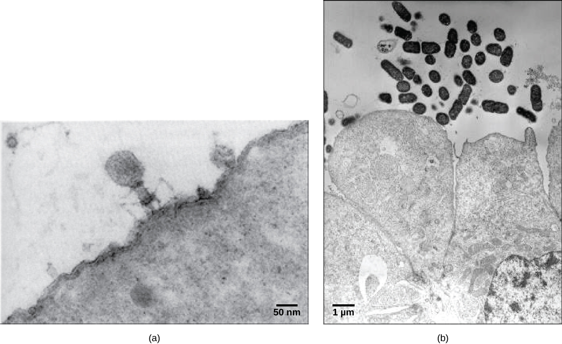
Micrograph a shows a virus with a hexagonal head that stands on thin, bent legs. The virus sits on the surface of a cell that is so large that only a small fraction of its surface is visible. Micrograph b shows small bacterial cells that are about the size of the organelles in the adjacent colon cells.
Figure 21.2 — 21.1 Viral Evolution, Morphology, and Classification

Figure 21.2 Most virus particles are visible only by electron microscopy. In these transmission electron micrographs, (a) a virus is as dwarfed by the bacterial cell it infects, as (b) these E. coli cells are dwarfed by cultured colon cells. (credit a: modification of work by U.S. Dept. of Energy, Office of Science, LBL, PBD; credit b: modification of work by J.P. Nataro and S. Sears, unpub. data, CDC; scale-bar data from Matt Russell)

Mary Ann Clark, Matthew Douglas, and Jung Choi, Biology 2E, OpenStax. (credit a: modification of work by U.S. Dept. of Energy, Office of Science, LBL, PBD; credit b: modification of work by J.P. Nataro and S. Sears, unpub. data, CDC; scale-bar data from Matt Russell) · CC BY-NC-SA 4.0 · Original image ↗ · Access for free ↗
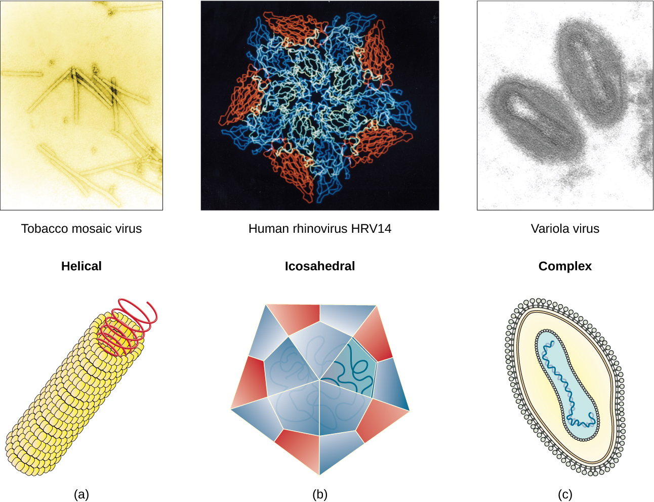
Figure a is a helical virus which has a long linear structure. The outer proteins are small spheres arranged into a long, hollow tube. Inside the tube is the genetic material. Tobacco mosaic virus is an example of a helical virus. Figure b is an icosahedral virus which has a polyhedron structure. The example shown is human rhinovirus which has a pentagon structure. Complex viruses have a more complex structure in figure C. The example is variola which has an ovoid structure.
Figure 21.3 — 21.1 Viral Evolution, Morphology, and Classification

Figure 21.3 Viral capsids can be (a) helical, (b) polyhedral, or (c) have a complex shape. (credit a “micrograph”: modification of work by USDA ARS; credit b “micrograph”: modification of work by U.S. Department of Energy)

Mary Ann Clark, Matthew Douglas, and Jung Choi, Biology 2E, OpenStax. (credit a “micrograph”: modification of work by USDA ARS; credit b “micrograph”: modification of work by U.S. Department of Energy) · CC BY-NC-SA 4.0 · Original image ↗ · Access for free ↗
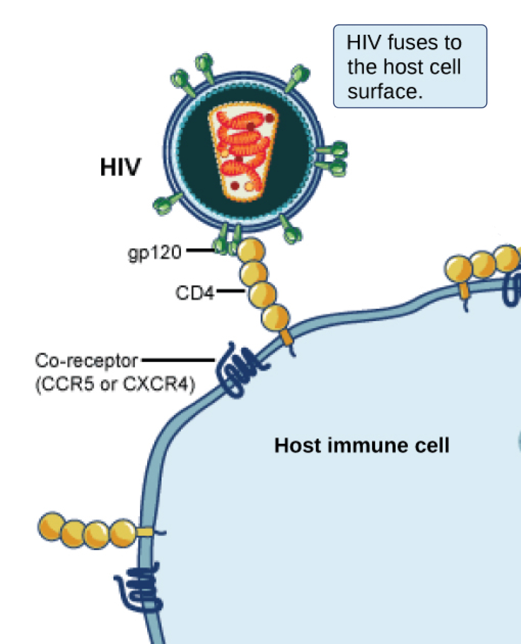
In the illustration an H I V virus attaches to a C D 4 molecule embedded in the plasma membrane of a host immune cell.
Figure 21.4 — 21.1 Viral Evolution, Morphology, and Classification

Figure 21.4 A virus and its host receptor protein. The HIV virus binds the CD4 receptor on the surface of human cells. CD4 receptors help white blood cells to communicate with other cells of the immune system when producing an immune response. (credit: modification of work by NIAID, NIH)

Mary Ann Clark, Matthew Douglas, and Jung Choi, Biology 2E, OpenStax. (credit: modification of work by NIAID, NIH) · CC BY-NC-SA 4.0 · Original image ↗ · Access for free ↗
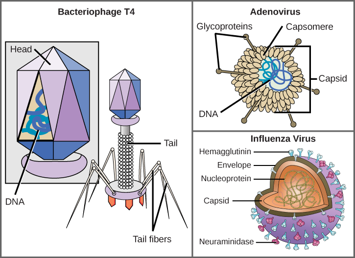
Illustration a shows bacteriophage T 4, which houses its D N A genome in a hexagonal head. A long, straight tail extends from the bottom of the head. Tail fibers attached to the base of the tail are bent, like spider legs. In b, adenovirus houses its D N A genome in a round capsid made from many small capsomere subunits. Glycoproteins extend from the capsomere, like pins from a pincushion. In c, the influenza virus houses its R N A genome and a bullet-shaped capsid. A spherical viral envelope, lined with matrix proteins, surrounds the capsid. Two different varieties of glycoprotein spike are embedded in the envelope. Approximately 80 percent of the spikes are hemagglutinin. The remaining 20 percent or so of the glycoprotein spikes consist of neuraminidase.
Figure 21.5 — 21.1 Viral Evolution, Morphology, and Classification

Figure 21.5 Complex Viruses. Viruses can be either complex or relatively simple in shape. This figure shows three relatively complex virions: the bacteriophage T4, with its DNA-containing head group and tail fibers that attach to host cells; adenovirus, which uses spikes from its capsid to bind to host cells; and the influenza virus, which uses glycoproteins embedded in its envelope to bind to host cells. The influenza virus also has matrix proteins, internal to the envelope, which help stabilize the virion’s shape. (credit “bacteriophage, adenovirus”: modification of work by NCBI, NIH; credit "influenza virus": modification of work by Dan Higgins, Centers for Disease Control and Prevention)

Mary Ann Clark, Matthew Douglas, and Jung Choi, Biology 2E, OpenStax. (credit “bacteriophage, adenovirus”: modification of work by NCBI, NIH; credit "influenza virus": modification of work by Dan Higgins, Centers for Disease Control and Prevention) · CC BY-NC-SA 4.0 · Original image ↗ · Access for free ↗
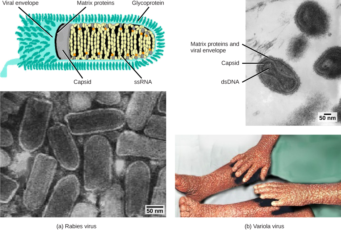
Part a is an illustration of the rabies virus, which is bullet shaped. R N A is coiled inside a capsid, which is encased in a matrix protein lined viral envelope studded with glycoproteins. Part a below is a micrograph of a cluster of bullet shaped rabies viruses. Part b is a micrograph of variola virus, which has D N A encased in a bow shaped capsid. An oval matrix protein-lined envelope surrounds the capsid. Part b below shows irregular, bumpy lesions on the arms and legs of a person with smallpox.
Figure 21.6 — 21.1 Viral Evolution, Morphology, and Classification

Figure 21.6 Viruses can be classified according to their core genetic material and capsid design. (a) Rabies virus has a single-stranded RNA (ssRNA) core and an enveloped helical capsid, whereas (b) variola virus, the causative agent of smallpox, has a double-stranded DNA (dsDNA) core and a complex capsid. Rabies transmission occurs when saliva from an infected mammal enters a wound. The virus travels through neurons in the peripheral nervous system to the central nervous system, where it impairs brain function, and then travels to other tissues. The virus can infect any mammal, and most die within weeks of infection. Smallpox is a human virus transmitted by inhalation of the variola virus, localized in the skin, mouth, and throat, which causes a characteristic rash. Before its eradication in 1979, infection resulted in a 30 to 35 percent mortality rate. (credit “rabies diagram”: modification of work by CDC; “rabies micrograph”: modification of work by Dr. Fred Murphy, CDC; credit “small pox micrograph”: modification of work by Dr. Fred Murphy, Sylvia Whitfield, CDC; credit “smallpox photo”: modification of work by CDC; scale-bar data from Matt Russell)

Mary Ann Clark, Matthew Douglas, and Jung Choi, Biology 2E, OpenStax. (credit “rabies diagram”: modification of work by CDC; “rabies micrograph”: modification of work by Dr. Fred Murphy, CDC; credit “small pox micrograph”: modification of work by Dr. Fred Murphy, Sylvia Whitfield, CDC; credit “smallpox photo”: modification of work by CDC; scale-bar data from Matt Russell) · CC BY-NC-SA 4.0 · Original image ↗ · Access for free ↗
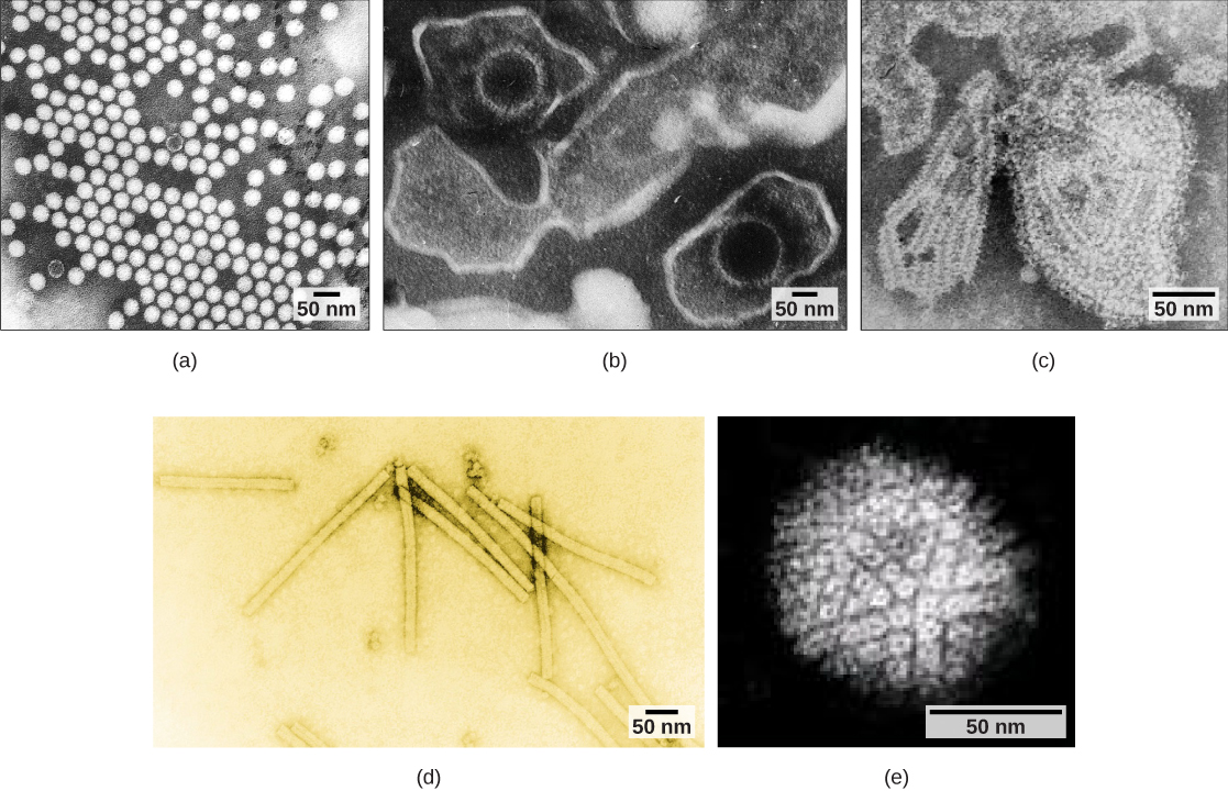
Micrograph a shows icosahedral polioviruses arranged in a grid; micrograph b shows two Epstein-Barr viruses with icosahedral capsids encased in an oval membrane; micrograph c shows a mumps virus capsid encased in an irregular membrane; micrograph d shows rectangular tobacco mosaic virus capsids; and micrograph e shows a spherical herpesvirus envelope studded with glycoproteins.
Figure 21.7 — 21.1 Viral Evolution, Morphology, and Classification

Figure 21.7 Transmission electron micrographs of various viruses show their capsid structures. The capsid of the (a) polio virus is naked icosahedral; (b) the Epstein-Barr virus capsid is enveloped icosahedral; (c) the mumps virus capsid is an enveloped helix; (d) the tobacco mosaic virus capsid is naked helical; and (e) the herpesvirus capsid is complex. (credit a: modification of work by Dr. Fred Murphy, Sylvia Whitfield; credit b: modification of work by Liza Gross; credit c: modification of work by Dr. F. A. Murphy, CDC; credit d: modification of work by USDA ARS; credit e: modification of work by Linda Stannard, Department of Medical Microbiology, University of Cape Town, South Africa, NASA; scale-bar data from Matt Russell)

Mary Ann Clark, Matthew Douglas, and Jung Choi, Biology 2E, OpenStax. (credit a: modification of work by Dr. Fred Murphy, Sylvia Whitfield; credit b: modification of work by Liza Gross; credit c: modification of work by Dr. F. A. Murphy, CDC; credit d: modification of work by USDA ARS; credit e: modification of work by Linda Stannard, Department of Medical Microbiology, University of Cape Town, South Africa, NASA; scale-bar data from Matt Russell) · CC BY-NC-SA 4.0 · Original image ↗ · Access for free ↗
Structure Examples
  • Single-stranded
  • Double-stranded
  • Linear
  • Circular
  • Non-segmented: consists of a single segment of genetic material
  • Segmented: is divided into multiple segments
  • Parainfluenza viruses
  • Influenza viruses

Table 21.1. Mary Ann Clark, Matthew Douglas, and Jung Choi, Biology 2E, OpenStax.

Classification Examples
Naked icosahedral Hepatitis A virus, polioviruses
Enveloped icosahedral Epstein-Barr virus, herpes simplex virus, rubella virus, yellow fever virus, -1
Enveloped helical Influenza viruses, mumps virus, measles virus, rabies virus
Naked helical Tobacco mosaic virus
Complex with many ; some have combinations of icosahedral and helical structures Herpesviruses, smallpox virus, hepatitis B virus, T4

Table 21.2. Mary Ann Clark, Matthew Douglas, and Jung Choi, Biology 2E, OpenStax.

Group Characteristics Mode of Production Example
I Double-stranded is directly from the template Herpes simplex (herpesvirus)
II Single-stranded is converted to double-stranded form before is Canine parvovirus (parvovirus)
III Double-stranded is from the Childhood gastroenteritis (rotavirus)
IV Single stranded (+) functions as Common cold (picornavirus)
V Single stranded (-) is from the Rabies (rhabdovirus)
VI Single stranded viruses with makes from the ; is then incorporated in the host ; is from the incorporated Human virus ()
VII Double stranded viruses with The viral is double-stranded , but viral is replicated through an intermediate; the may serve directly as or as a template to make Hepatitis B virus (hepadnavirus)

Table 21.3. Mary Ann Clark, Matthew Douglas, and Jung Choi, Biology 2E, OpenStax.

Source: 21.1 Viral Evolution, Morphology, and Classification

21.2 Virus Infections and Hosts — figures and tables
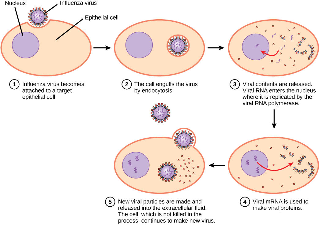
The illustration shows the steps of an influenza virus infection. In step 1, influenza virus becomes attached to a target epithelial cell. In step 2, the cell engulfs the virus by endocytosis, and the virus becomes encased in the cell's plasma membrane. In step 3, the membrane dissolves, and the viral contents are released into the cytoplasm. Viral m R N A enters the nucleus, where it is replicated by viral R N A polymerase. In step 4, viral m R N A exits to the cytoplasm, where it is used to make viral proteins. In step 5, new viral particles are released into the extracellular fluid. The cell, which is not killed in the process, continues to make new virus.
Figure 21.8 — 21.2 Virus Infections and Hosts

Figure 21.8 The influenza reproductive cycle. In influenza virus infection, glycoproteins on the capsid attach to a host epithelial cell. Following this, the virus is engulfed. RNA and proteins are then made and assembled into new virions.

Mary Ann Clark, Matthew Douglas, and Jung Choi, Biology 2E, OpenStax. · CC BY-NC-SA 4.0 · Original image ↗ · Access for free ↗
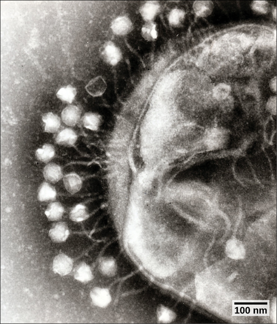
The micrograph shows hexagonal bacteriophage capsids attached to a host bacterial cell by slender stalks.
Figure 21.9 — 21.2 Virus Infections and Hosts

Figure 21.9 Bacteriophages attached to a host cell (transmission electron micrograph). In bacteriophage with tails, like the one shown here, the tails serve as a passageway for transmission of the phage genome. (credit: modification of work by Dr. Graham Beards; scale-bar data from Matt Russell)

Mary Ann Clark, Matthew Douglas, and Jung Choi, Biology 2E, OpenStax. (credit: modification of work by Dr. Graham Beards; scale-bar data from Matt Russell) · CC BY-NC-SA 4.0 · Original image ↗ · Access for free ↗
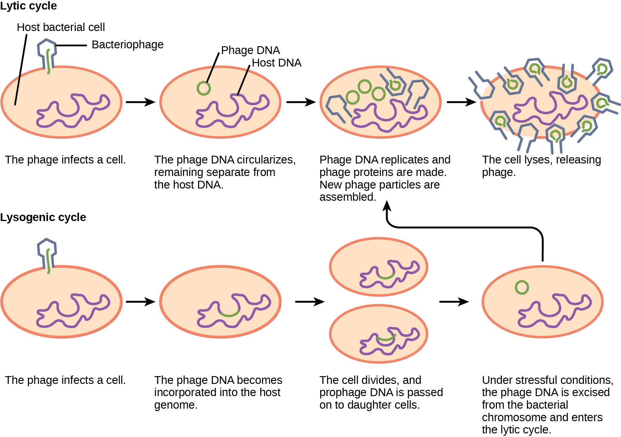
The bacteriophage lytic cycle begins when the phage attaches via a slender stalk to the host cell. Linear DNA from the viral head is injected into the host cell. The phage DNA circularizes, remaining separate from the host DNA. The phage DNA replicates, and new phage proteins are made. New phage particles are assembled. The cell lyses, releasing the phage. The bacteriophage lysogenic cycle begins the same way as the lytic cycle, with phage infecting a host cell. However, the phage DNA becomes incorporated into the host genome. The cell divides, and phage DNA is passed on to daughter cells. Under stressful conditions, the phage DNA is excised from the bacterial chromosome and enters the lytic cycle.
Figure 21.10 — 21.2 Virus Infections and Hosts

Figure 21.10 A temperate bacteriophage has both lytic and lysogenic cycles. In the lytic cycle, the phage replicates and lyses the host cell. In the lysogenic cycle, phage DNA is incorporated into the host genome, where it is passed on to subsequent generations. Environmental stressors such as starvation or exposure to toxic chemicals may cause the prophage to excise and enter the lytic cycle.

Mary Ann Clark, Matthew Douglas, and Jung Choi, Biology 2E, OpenStax. · CC BY-NC-SA 4.0 · Original image ↗ · Access for free ↗
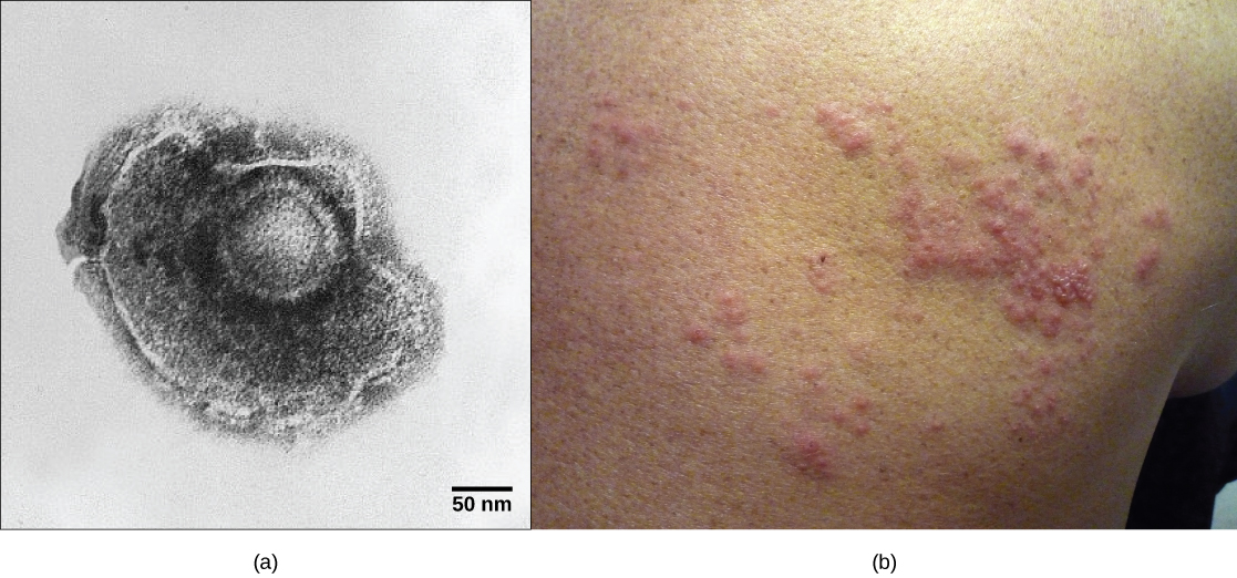
Part a shows a micrograph of the varicella zoster virus, which has an icosahedral capsid surrounded by an irregularly shaped envelope. Part b shows a red, bumpy shingles rash on a person's face.
Figure 21.11 — 21.2 Virus Infections and Hosts

Figure 21.11 A latent virus infection. (a) Varicella-zoster, the virus that causes chickenpox, has an enveloped icosahedral capsid visible in this transmission electron micrograph. Its double-stranded DNA genome becomes incorporated in the host DNA and can reactivate after latency in the form of (b) shingles, often exhibiting a rash. (credit a: modification of work by Dr. Erskine Palmer, B. G. Martin, CDC; credit b: modification of work by “rosmary”/Flickr; scale-bar data from Matt Russell)

Mary Ann Clark, Matthew Douglas, and Jung Choi, Biology 2E, OpenStax. (credit a: modification of work by Dr. Erskine Palmer, B. G. Martin, CDC; credit b: modification of work by “rosmary”/Flickr; scale-bar data from Matt Russell) · CC BY-NC-SA 4.0 · Original image ↗ · Access for free ↗
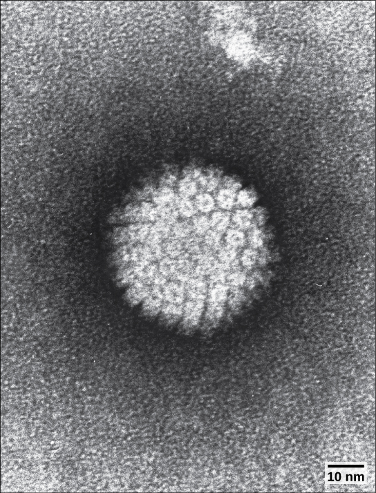
The micrograph shows an icosahedral virus with glycoproteins protruding from its capsid.
Figure 21.12 — 21.2 Virus Infections and Hosts

Figure 21.12 HPV, or human papillomavirus, has a naked icosahedral capsid visible in this transmission electron micrograph and a double-stranded DNA genome that is incorporated into the host DNA. The virus, which is sexually transmitted, is oncogenic and can lead to cervical cancer. (credit: modification of work by NCI, NIH; scale-bar data from Matt Russell)

Mary Ann Clark, Matthew Douglas, and Jung Choi, Biology 2E, OpenStax. (credit: modification of work by NCI, NIH; scale-bar data from Matt Russell) · CC BY-NC-SA 4.0 · Original image ↗ · Access for free ↗
Symptom Appears as
Hyperplasia Galls ()
Hypoplasia Thinned, yellow splotches on leaves
Cell necrosis Dead, blackened stems, leaves, or fruit
Abnormal growth patterns Malformed stems, leaves, or fruit
Discoloration Yellow, red, or black lines, or rings in stems, leaves, or fruit

Table 21.4. Mary Ann Clark, Matthew Douglas, and Jung Choi, Biology 2E, OpenStax.

Source: 21.2 Virus Infections and Hosts

21.4 Other Acellular Entities: and — figures and tables

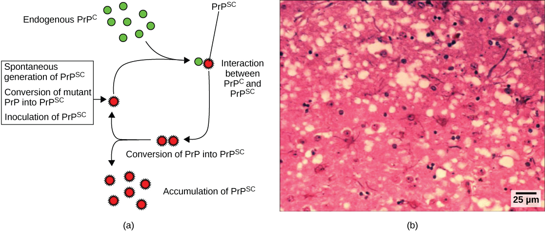
Part A illustrates how normal prion protein, called P r P, is converted into the disease causing form  P r P. P r P superscript lowercase s lowercase c may spontaneously form in brain tissue, may be introduced when a mutant form of the protein misfolds, or may introduced into the brain tissue by inoculation. The misfolded protein causes normal P r P already present in the brain to misfold. A chain reaction occurs, leading to a large amount of misfolded protein. Part B is a light microscopy image of prion-infected brain tissue.
Figure 21.17 — 21.4 Other Acellular Entities: and

Figure 21.17 Mad Cow Disease in humans. (a) Endogenous normal prion protein (PrP c ) is converted into the disease-causing form (PrP sc ) when it encounters this variant form of the protein. PrP sc may arise spontaneously in brain tissue, especially if a mutant form of the protein is present, or it may occur via the spread of misfolded prions consumed in food into brain tissue. (b) This prion-infected brain tissue, visualized using light microscopy, shows the vacuoles that give it a spongy texture, typical of transmissible spongiform encephalopathies. (credit b: modification of work by Dr. Al Jenny, USDA APHIS; scale-bar data from Matt Russell)

Mary Ann Clark, Matthew Douglas, and Jung Choi, Biology 2E, OpenStax. (credit b: modification of work by Dr. Al Jenny, USDA APHIS; scale-bar data from Matt Russell) · CC BY-NC-SA 4.0 · Original image ↗ · Access for free ↗
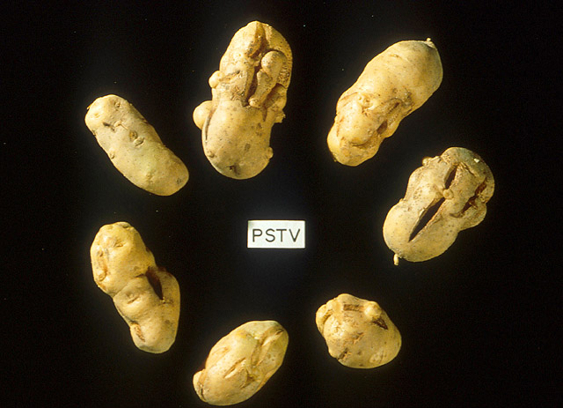
The photo shows shriveled, cracked potatoes.
Figure 21.18 — 21.4 Other Acellular Entities: and

Figure 21.18 These potatoes have been infected by the potato spindle tuber viroid (PSTV), which is typically spread when infected knives are used to cut healthy potatoes, which are then planted. (credit: Pamela Roberts, University of Florida Institute of Food and Agricultural Sciences, USDA ARS)

Mary Ann Clark, Matthew Douglas, and Jung Choi, Biology 2E, OpenStax. (credit: Pamela Roberts, University of Florida Institute of Food and Agricultural Sciences, USDA ARS) · CC BY-NC-SA 4.0 · Original image ↗ · Access for free ↗
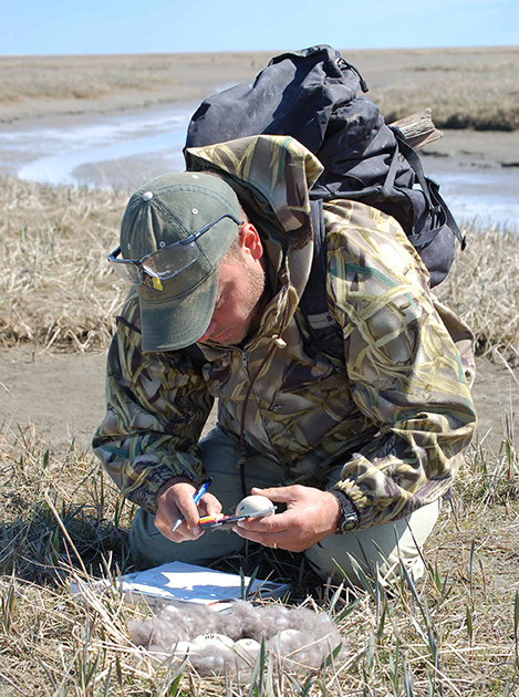
This photo shows a scientist in a field, measuring the length of an egg.
Figure 21.19 — 21.4 Other Acellular Entities: and

Figure 21.19 This virologist is engaged in fieldwork, sampling eggs from this nest for avian influenza. (credit: Don Becker, USGS EROS, U.S. Fish and Wildlife Service)

Mary Ann Clark, Matthew Douglas, and Jung Choi, Biology 2E, OpenStax. (credit: Don Becker, USGS EROS, U.S. Fish and Wildlife Service) · CC BY-NC-SA 4.0 · Original image ↗ · Access for free ↗


Source: 21.4 Other Acellular Entities: Prions and Viroids

174 terms, alphabetically ordered

3′ end · ·

The end of a nucleic- strand associated with the sugar’s 3′ position. extends a strand at a free .

Read explanation · OpenStax ↗
3′ hydroxyl group · · ·

The at the sugar’s 3′ carbon that attacks the incoming ’s during strand extension.

Read explanation · OpenStax ↗
A site · ·

The site that accepts most incoming during elongation and at termination.

Read explanation · OpenStax ↗
Acid · ·

In the Brønsted–Lowry model, a substance that can donate a . Acidic have a higher activity than water.

Read explanation · OpenStax ↗
Activation energy ·

The energy barrier that must overcome to reach a and undergo .

Read explanation · OpenStax ↗
Adenosine triphosphate

An energy-carrying ; its powers many cellular processes, including cycles.

Read explanation · OpenStax ↗
Alpha carbon · ·

The central carbon next to the carboxyl carbon. In a standard it binds an , hydrogen, and .

Read explanation · OpenStax ↗
Alpha helix · · · · · ·

A helical stabilized by between a oxygen and an hydrogen four later.

Read explanation · OpenStax ↗
Amide · · ·

A group containing a carbon bonded to nitrogen. are amide linkages.

Read explanation · OpenStax ↗
Amino acid

A building block containing amino and and a characteristic .

Read explanation · OpenStax ↗
Amino acid residue · ·

The part of an incorporated into a chain. Residue can also describe another building block within a larger .

Read explanation · OpenStax ↗
Amino group · · ·

A nitrogen-containing . A free ’s amino group is typically (NH₃⁺) near physiological ; ’s ring nitrogen is an exception to this formula.

Read explanation · OpenStax ↗
Aminoacyl-tRNA · ·

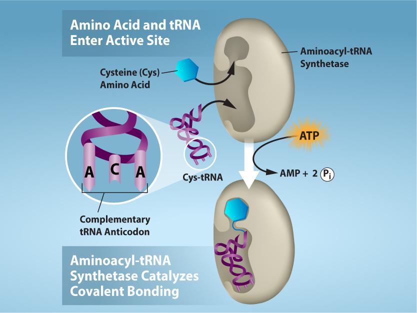
A covalently linked to an at its , ready to deliver that during .

Read explanation · OpenStax ↗
Amphipathic

Having both a region and a region, as in a or .

Read explanation · OpenStax ↗
Anticodon

A triplet that pairs to an . The anticodon for 5′-AUG-3′ is aligned 3′-UAC-5′, or written 5′-CAU-3′.

Read explanation · NCBI Bookshelf / Garland Science ↗
Antiparallel

Running in opposite directions, as with two strands or neighboring strands in some .

Read explanation · OpenStax ↗
Apoptosis

A regulated process of cell death that removes cells in development, tissue maintenance, and responses to damage.

Read explanation · OpenStax ↗
Aqueous solution ·

A in which water is the .

Read explanation · OpenStax ↗
Archaea ·

A of distinct from , with characteristic chemistry and -expression machinery.

Read explanation · OpenStax ↗
Assay

A procedure used to detect a substance or measure its amount, activity, or another property.

Read explanation · Thermo Fisher ↗
Atom

The smallest unit of an element that retains its chemical identity; it has a surrounded by .

Read explanation · OpenStax ↗
attachment

Enduring emotional bond and expectations about another’s availability and responsiveness.

Read explanation · OpenStax ↗
Bacteria ·

A of single-celled . Bacterial cells lack a -bound and are distinct from .

Read explanation · OpenStax ↗
bacteriophage · ·

A virus that infects .

Read explanation · OpenStax ↗
Beta sheet · · · · · ·

A made of aligned segments connected by ; strands may be parallel or .

Read explanation · OpenStax ↗
Binding site

A region of a where another interacts selectively.

Read explanation · OpenStax ↗
Biomolecule

A made by or involved in living systems, including , , , and .

Read explanation · OpenStax ↗
Blocking

Covering unoccupied on a or plate to reduce interactions.

Read explanation · Thermo Fisher ↗
Blotting ·

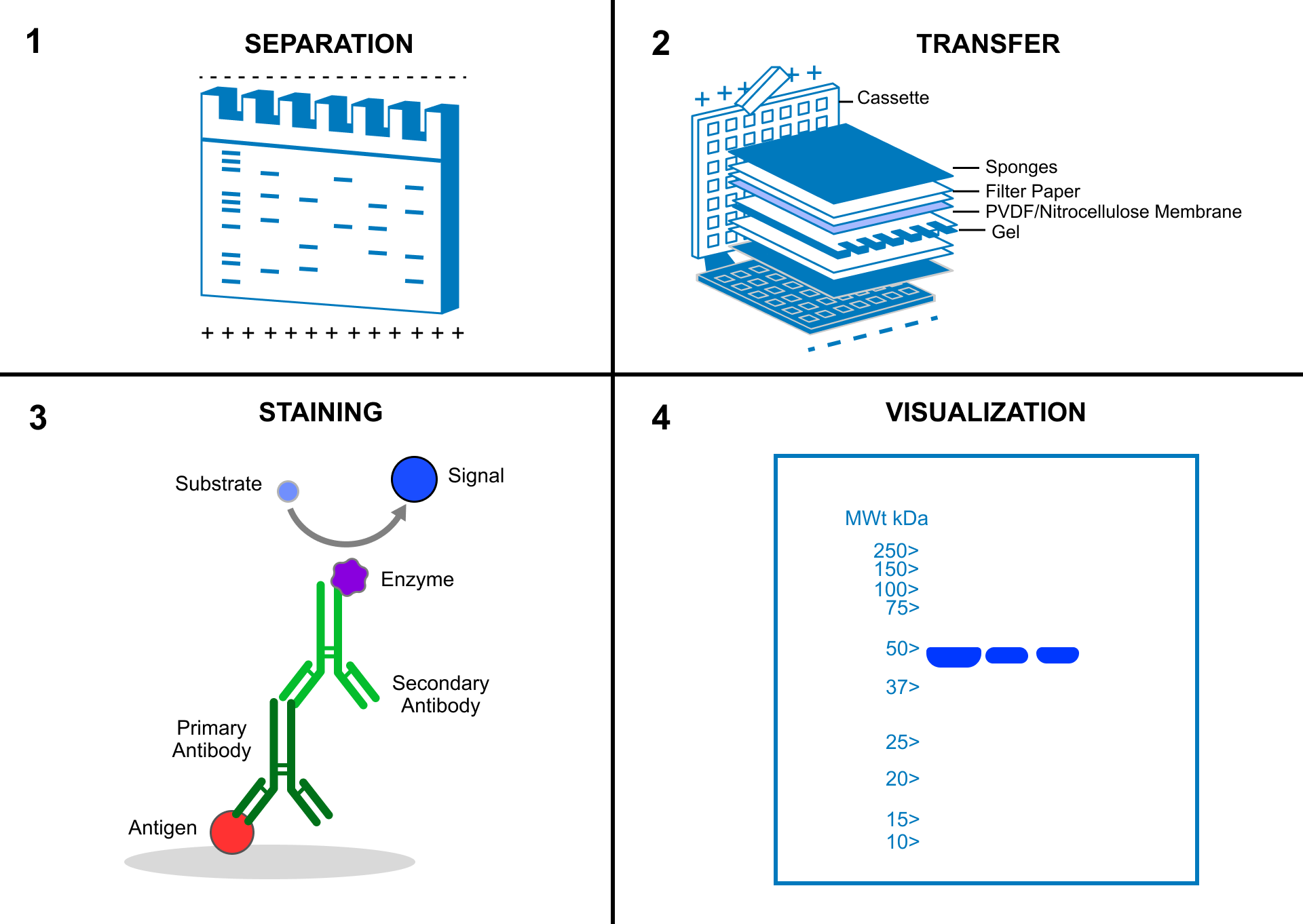
Transfer of separated or to a so a specific target can be detected.

Read explanation · Thermo Fisher ↗
Brønsted–Lowry base · · · · · · · · · · ·

In the Brønsted–Lowry model, a substance that can accept a . Basic solutions have lower activity than water.

Read explanation · OpenStax ↗
Cancer

A group of diseases involving abnormal cells that can proliferate without normal restraints and invade tissues.

Read explanation · OpenStax ↗
capsid

The shell enclosing a viral .

Read explanation · OpenStax ↗
Carbohydrate

A class of including sugars and their , used for energy storage, structure, and recognition.

Read explanation · OpenStax ↗
Carbonyl group · ·

A carbon double-bonded to oxygen, C=O; present in and many other .

Read explanation · OpenStax ↗
Carboxyl group · ·

An , COOH. In free it is usually to COO⁻ near physiological .

Read explanation · OpenStax ↗
Catalyst · ·

A substance that speeds a by providing a pathway with lower and is regenerated rather than consumed overall.

Read explanation · OpenStax ↗
CD4

A cell-surface immune coreceptor used with additional coreceptors for and entry.

Read explanation · OpenStax ↗
Cell cycle

The ordered stages of cell growth, , and division.

Read explanation · OpenStax ↗
Cell proliferation

Increase in cell number through cell division; it should be distinguished experimentally from changes in survival.

Read explanation · OpenStax ↗
Chemical element

A substance defined by having the same number of , such as carbon or oxygen.

Read explanation · OpenStax ↗
Chemical reaction ·

A process that rearranges and chemical bonds, converting into .

Read explanation · OpenStax ↗
Chromatin

The complex of and associated , including , in .

Read explanation · OpenStax ↗
Chromatography

Separation of mixture components by their different interactions with a and a moving .

Read explanation · Waters ↗
Chromosome ·

An organized with associated that carries . A replicated chromosome has two .

Read explanation · OpenStax ↗
Codon

A sequence of three that specifies an or a stop signal.

Read explanation · OpenStax ↗
Conformation ·

A spatial arrangement accessible by rotations and other motions that do not require breaking .

Read explanation · NCBI Bookshelf ↗
Copy number ·

The number of copies of a segment in a defined sample or ; it is distinct from or abundance.

Read explanation · NHGRI / NIH ↗
Covalent bond · ·

A bond in which share pairs. and are covalent.

Read explanation · OpenStax ↗
Cysteine ·

An with a . Two cysteines can form a .

Read explanation · OpenStax ↗
Cytoplasm

Cell contents inside the plasma and outside the , where one is present.

Read explanation · OpenStax ↗
Deoxyribonucleic acid

The that stores genetic information used to specify and sequences.

Read explanation · OpenStax ↗
Deprotonation

Removal of a from a or group; this changes its by minus one.

Read explanation · OpenStax ↗
Detergent

An substance that can associate with both water and material; detergents can disrupt or interactions.

Read explanation · Thermo Fisher ↗
Diploid

Having two sets; comparisons of must account for and sample composition.

Read explanation · NHGRI / NIH ↗
Disulfide bond · · ·

A covalent sulfur–sulfur bond formed by of two , which can stabilize structure.

Read explanation · NCBI Bookshelf ↗
DNA lesion ·

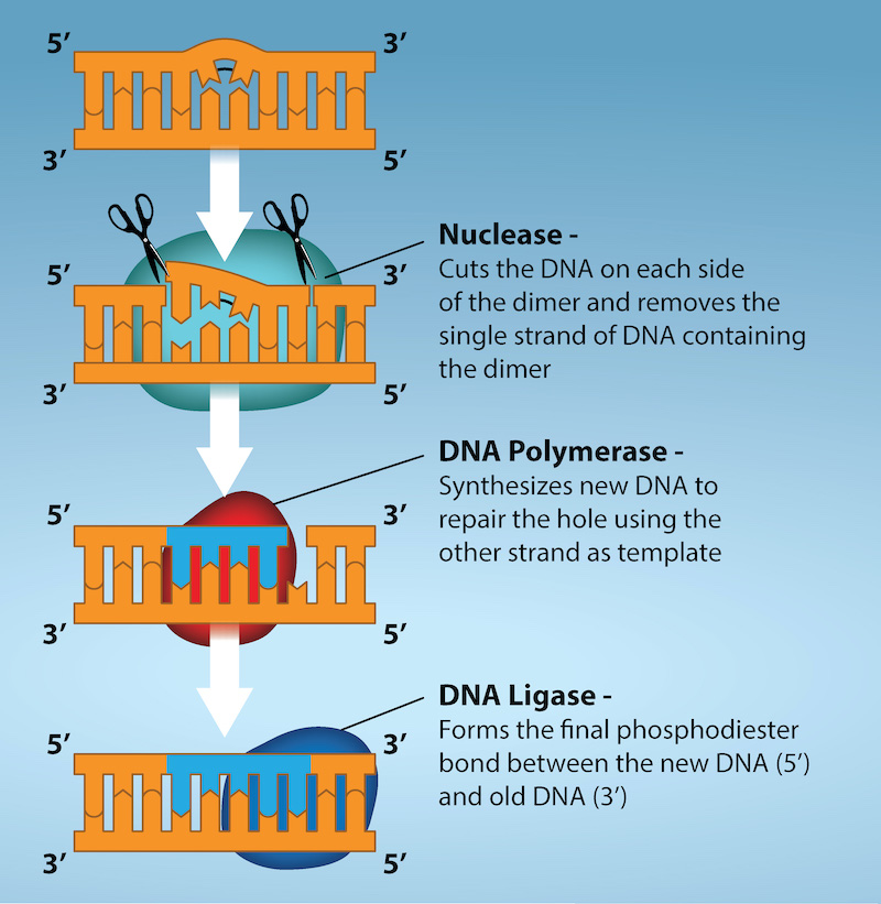
of molecular damage in . A lesion can cause a if it is copied or repaired incorrectly, but damage and sequence change are distinct.

Read explanation · OpenStax ↗
DNA polymerase ·

An that synthesizes 5′ to 3′ using a template and a with a free .

Read explanation · OpenStax ↗
DNA replication

Copying so genetic information can be passed to daughter cells. Each guides a complementary new strand.

Read explanation · OpenStax ↗
Electric charge · · ·

An electrical property that can be positive or negative. Opposite charges attract and like charges repel; net charge is the sum of all charges on a particle.

Read explanation · OpenStax ↗
Electron

A negatively subatomic particle. Electron sharing and transfer determine much of chemical bonding.

Read explanation · OpenStax ↗
Electronegativity ·

An ’s tendency to attract shared in a chemical bond. Unequal attraction can make a bond .

Read explanation · OpenStax ↗
endocytosis

Uptake of extracellular material or surface through inward budding of .

Read explanation · OpenStax ↗
Enzyme

A biological that speeds a without being consumed. Most enzymes are .

Read explanation · OpenStax ↗
Enzyme inhibitor · ·

A substance that reduces activity. The binding mechanism and rate data determine whether inhibition is competitive, uncompetitive, mixed, or another type.

Read explanation · Biology LibreTexts ↗
Eukaryote ·

An organism whose cells have a -enclosed ; animals, plants, fungi, and protists are eukaryotes.

Read explanation · OpenStax ↗
FACT complex ·

A -associated complex that helps machinery traverse and supports reorganization.

Read explanation · OpenStax ↗
Functional group

A group of whose chemical behavior gives a characteristic and properties.

Read explanation · OpenStax ↗
Gene

A sequence that contributes to a functional or , together with the information needed to express it.

Read explanation · OpenStax ↗
Gene expression

Production of a functional from a , including and, for -coding , .

Read explanation · NCBI Bookshelf ↗
Genome

The complete genetic material of an organism or cell.

Read explanation · OpenStax ↗
Histone

A -associated rich in . Core histones form the around which nucleosomal wraps.

Read explanation · OpenStax ↗
HIV

Human virus, an enveloped that uses and coreceptors for entry.

Read explanation · OpenStax ↗
Human papillomavirus

A group of viruses; some high-risk types produce that disrupt host -suppressor pathways.

Read explanation · NCBI Bookshelf / Sinauer Associates ↗
Hydrogen bond ·

An attraction between a hydrogen bonded to an and another . hydrogen bonds stabilize .

Read explanation · OpenStax ↗
Hydrogen ion · ·

A positively hydrogen species. H⁺ denotes a , which is hydrated in water; its activity determines .

Read explanation · OpenStax ↗
Hydrolysis

Breaking a chemical bond by adding water. hydrolysis separates .

Read explanation · OpenStax ↗
Hydrophilic

Interacting favorably with water, typically because a group is or .

Read explanation · OpenStax ↗
Hydrophobic

Interacting poorly with water. often gather in a ’s interior.

Read explanation · OpenStax ↗
Hydroxide ion ·

The negatively OH⁻. Basic contain a higher hydroxide ion activity than water at the same temperature.

Read explanation · OpenStax ↗
Hydroxyl group ·

An oxygen bonded to hydrogen, –OH. Hydroxyl groups can and often increase .

Read explanation · OpenStax ↗
immunodeficiency

Inadequate immune function that compromises defense or regulation.

Read explanation · OpenStax ↗
integrase

A viral inserting newly synthesized viral into host .

Read explanation · OpenStax ↗
Ion · ·

An or with a net because its numbers of and do not balance.

Read explanation · OpenStax ↗
linkage

A tendency of nearby on the same to be inherited together.

Read explanation · OpenStax ↗
Lipid

A broad class of largely or , including fats, , and steroids.

Read explanation · OpenStax ↗
locus

The specific position of a or other feature.

Read explanation · OpenStax ↗
Logarithm · · ·

The exponent needed to obtain a number from a chosen fixed number. For decimal logarithms, log₁₀(100) = 2 because 10² = 100; uses the negative decimal logarithm of activity.

Read explanation · OpenStax ↗
lysogeny

Maintenance of a temperate with the host rather than immediate productive lysis.

Read explanation · OpenStax ↗
Lysosome

An -enclosed compartment that uses hydrolytic to digest and recycle cellular material.

Read explanation · OpenStax ↗
lytic cycle

A productive infection ending in host lysis and release of progeny .

Read explanation · OpenStax ↗
Matrix

A supporting material, such as a gel or the solid support in a column. Its properties influence movement and binding of sample .

Read explanation · Thermo Fisher ↗
Membrane · · · ·

A thin boundary or sheet. Cell membranes are bilayers containing ; membranes are synthetic supports that hold or for detection.

Read explanation · OpenStax ↗
Messenger RNA ·

An that carries coding information used by to synthesize a .

Read explanation · OpenStax ↗
Metabolism

The chemical that build , break them down, and transform energy in a living system.

Read explanation · OpenStax ↗
Mitochondrion ·

A with its own , central to aerobic production and several other processes.

Read explanation · OpenStax ↗
Mobile phase

The moving fluid that carries a sample through a system.

Read explanation · Waters ↗
Molecular chaperone · ·

A that assists other in folding or prevents , without specifying their sequence.

Read explanation · NCBI Bookshelf ↗
Molecule

Two or more joined by . A molecule may be electrically or carry a .

Read explanation · OpenStax ↗
Mutation

A change in a sequence; some mutations alter a ’s sequence.

Read explanation · OpenStax ↗
negative-sense RNA

Viral complementary to that must first be copied into readable .

Read explanation · OpenStax ↗
Neutral

Having no net . For a , neutral instead means equal and activities; its is about 7 at 25 °C.

Read explanation · OpenStax ↗
Nitrogenous base · · ·

A nitrogen-containing ring component of ; its pairing interactions help specify sequences.

Read explanation · OpenStax ↗
Noncoding RNA · · · ·

An whose functional is rather than a , including , , and regulatory .

Read explanation · NCBI Bookshelf / Landes Bioscience ↗
Nonpolar ·

Lacking a substantial separation of . Nonpolar groups generally interact poorly with water.

Read explanation · OpenStax ↗
Nonspecific binding · ·

Unwanted binding outside the intended target interaction, often contributing background signal in an .

Read explanation · Thermo Fisher ↗
Nucleic acid

A of , such as or , used in information storage, expression, and other cellular functions.

Read explanation · OpenStax ↗
Nucleosome

The basic unit of : wrapped around a .

Read explanation · OpenStax ↗
Nucleotide

A joined to a sugar and one or more .

Read explanation · OpenStax ↗
Nucleus ·

A -enclosed compartment containing most of a cell’s .

Read explanation · OpenStax ↗
Octamer

A complex made of eight . A core contains eight .

Read explanation · OpenStax ↗
Oncogene

A whose abnormal activation or excess expression contributes to uncontrolled growth or survival.

Read explanation · OpenStax ↗
Organelle ·

A specialized structure within a cell that carries out particular functions, such as a or .

Read explanation · OpenStax ↗
Oxidation ·

Loss of or an increase in oxidation state. Oxidizing two can form a .

Read explanation · OpenStax ↗
Pathogen

An infectious agent capable of causing disease in a host.

Read explanation · OpenStax ↗
Peptide

A chain of joined by ; the term commonly describes chains shorter than a typical .

Read explanation · OpenStax ↗
Peptide bond ·

A covalent between the carboxyl carbon of one and the amino nitrogen of the next.

Read explanation · OpenStax ↗
pH

A measure of , defined as the negative of activity. Lower pH means greater ; pH influences .

Read explanation · OpenStax ↗
Phosphate group ·

A phosphorus-containing group bonded to oxygens; phosphate groups occur in , , and modified .

Read explanation · OpenStax ↗
Phospholipid

An with a -containing region and tails; a major component of cellular .

Read explanation · OpenStax ↗
ploidy

The number of complete sets in a cell.

Read explanation · OpenStax ↗
Polar

Having an uneven distribution of . Polar groups often interact favorably with water.

Read explanation · OpenStax ↗
Polymer

A large made from many linked molecular building blocks.

Read explanation · OpenStax ↗
Polypeptide

A chain of connected by . A functional can contain one or several such chains.

Read explanation · OpenStax ↗
Porous material · · · ·

A material containing small spaces that can admit of suitable size.

Read explanation · Cytiva ↗
positive-sense RNA

Viral with the same coding sense as and potentially readable directly by .

Read explanation · OpenStax ↗
Primer · ·

A short nucleic- strand paired with a template that provides a free for polymerase extension.

Read explanation · OpenStax ↗
prion

An infectious capable of promoting misfolding of related host .

Read explanation · OpenStax ↗
Prokaryote ·

An organism whose cells lack a -bound ; and are prokaryotes.

Read explanation · OpenStax ↗
Proline ·

An whose loops back to the nitrogen, restricting rotation and often creating bends.

Read explanation · OpenStax ↗
prophage

A maintained within a host, often integrated into its .

Read explanation · OpenStax ↗
Protease · ·

An that hydrolyzes in or .

Read explanation · OpenStax ↗
Protein

One or more folded chains that perform biological roles such as , transport, signaling, or structural support.

Read explanation · OpenStax ↗
Protein aggregation · ·

Association of into larger clusters, often when exposed surfaces of unfolded stick together.

Read explanation · NCBI Bookshelf ↗
Protein backbone ·

The repeating nitrogen–alpha-carbon– chain of a , excluding its variable .

Read explanation · OpenStax ↗
Protein domain · ·

A region of a that forms a recognizable structural or functional unit.

Read explanation · OpenStax ↗
Proton

A positively subatomic particle. In – chemistry, donating a proton means transferring a .

Read explanation · OpenStax ↗
Protonation

Addition of a to an , , or ; this changes its by plus one.

Read explanation · OpenStax ↗
provirus

A viral integrated into a host .

Read explanation · OpenStax ↗
R group · ·

The variable side chain attached to an ’s . It determines identity, , , and chemical behavior.

Read explanation · OpenStax ↗
Reactant

A starting substance transformed during a .

Read explanation · OpenStax ↗
Reaction intermediate · ·

A species formed in one step and consumed in another. It occupies a local energy minimum between .

Read explanation · OpenStax ↗
Reaction product ·

A substance formed by a .

Read explanation · OpenStax ↗
Receptor

A that recognizes a particular signal and helps produce a cellular response.

Read explanation · OpenStax ↗
Release factor ·

A that recognizes a stop signal and promotes of the – to release the .

Read explanation · OpenStax ↗
retrovirus

An virus replicating through a intermediate produced by .

Read explanation · OpenStax ↗
Reverse transcriptase

An that makes using an template. is a specialized example.

Read explanation · OpenStax ↗
Reverse transcription

Synthesis of from an template by ; it transfers information between .

Read explanation · OpenStax ↗
Ribonucleic acid ·

A with roles in , regulation, and ; carries -coding information.

Read explanation · OpenStax ↗
Ribosomal RNA · ·

An component of that contributes to their structure, decoding, and ; it is not into .

Read explanation · OpenStax ↗
Ribosome ·

A complex of and that translates into a .

Read explanation · OpenStax ↗
RNA polymerase

An that synthesizes 5′ → 3′. Cellular uses a template and can begin without a .

Read explanation · OpenStax ↗
Secondary structure

Local folding, such as and , stabilized mainly by .

Read explanation · OpenStax ↗
Sister chromatid

One of the two copies of a produced by . An intact sister chromatid can supply a template for accurate repair.

Read explanation · Biology LibreTexts / Oregon State University ↗
Solubility ·

The amount of a substance that can dissolve in a under specified conditions. Soluble means able to dissolve; insoluble means only very slightly soluble in that .

Read explanation · OpenStax ↗
Solute

A dissolved component of a .

Read explanation · OpenStax ↗
Solution

A homogeneous mixture in which are dissolved in a .

Read explanation · OpenStax ↗
Solvent

The component of a that dissolves other substances, usually the component present in the greatest amount.

Read explanation · OpenStax ↗
Stationary phase

The material that remains fixed in a system.

Read explanation · Waters ↗
Subunit ·

An individual chain within a containing multiple chains.

Read explanation · OpenStax ↗
Telomerase

A ribonucleoprotein that uses its own template to extend a ’s 3′ end.

Read explanation · OpenStax ↗
Template strand · · ·

A preexisting strand whose sequence guides complementary nucleic- synthesis.

Read explanation · OpenStax ↗
Thiol group · · · ·

A sulfur bonded to hydrogen, –SH. thiols can form through .

Read explanation · OpenStax ↗
Transcript

An produced by of a sequence.

Read explanation · OpenStax ↗
Transcription

Synthesis of using a template.

Read explanation · OpenStax ↗
Transfer RNA ·

An adaptor that carries an and pairs its with a on .

Read explanation · OpenStax ↗
Transition state

A fleeting, high-energy arrangement at a barrier. It is an energy maximum along the path, rather than a stable .

Read explanation · OpenStax ↗
Translation ·

Synthesis of a on a using the sequence of in .

Read explanation · OpenStax ↗
transposon · ·

A element capable of moving between genomic locations using mobility machinery.

Read explanation · OpenStax ↗
tropism

A ’s preference or capacity to infect particular tissues or cell types.

Read explanation · OpenStax ↗
Tumor · ·

An abnormal mass of tissue; tumors can be benign or malignant.

Read explanation · OpenStax ↗
Vesicle · · ·

A small -enclosed compartment that can transport material between cellular locations.

Read explanation · OpenStax ↗
viral envelope

A surrounding some and containing viral or entry .

Read explanation · OpenStax ↗
virion

A complete extracellular virus particle containing a and protective structures.

Read explanation · OpenStax ↗
viroid

A small infectious , generally circular, that causes disease in plants without a .

Read explanation · OpenStax ↗

21.1 Viral Evolution, Morphology, and Classification

OpenStax · 21.1 Viral Evolution, Morphology, and Classification ↗
Instructional page including learning objectives, examples, figures, source tables and questions. Site navigation and embedded media players are omitted; source media links remain available.

Learning Objectives

By the end of this section, you will be able to do the following:

  • Describe how viruses were first discovered and how they are detected
  • Discuss three hypotheses about how viruses evolved
  • Describe the general structure of a virus
  • Recognize the basic shapes of viruses
  • Understand past and emerging classification systems for viruses
  • Describe the basis for the Baltimore classification system

Viruses are diverse entities: They vary in structure, methods of , and the hosts they infect. Nearly all forms of life—from and archaeans, to such as plants, animals, and fungi—have viruses that infect them. While most biological diversity can be understood through evolutionary history (such as how species have adapted to changing environmental conditions and how different species are related to one another through common descent), much about virus origins and evolution remains unknown.

Discovery and Detection

Viruses were first discovered after the development of a porcelain filter—the Chamberland-Pasteur filter—that could remove all visible in the microscope from any liquid sample. In 1886, Adolph Meyer demonstrated that a disease of tobacco plants—tobacco mosaic disease—could be transferred from a diseased plant to a healthy one via liquid plant extracts. In 1892, Dmitri Ivanowski showed that this disease could be transmitted in this way even after the Chamberland-Pasteur filter had removed all viable from the extract. Still, it was many years before it was proved that these “filterable” infectious agents were not simply very small but were a new type of very small, disease-causing particle.

Most , or single virus particles, are very small, about 20 to 250 nanometers in diameter. However, some recently discovered viruses from amoebae range up to 1000 nm in diameter. With the exception of large , like the poxvirus and other large viruses, viruses cannot be seen with a light microscope. It was not until the development of the microscope in the late 1930s that scientists got their first good view of the structure of the tobacco mosaic virus (TMV) (Figure 21.1), discussed above, and other viruses (Figure 21.2). The surface structure of can be observed by both scanning and transmission microscopy, whereas the internal structures of the virus can only be observed in images from a transmission microscope. The use of microscopy and other technologies has allowed for the discovery of many viruses of all types of living organisms.

Micrograph a shows a virus with a hexagonal head that stands on thin, bent legs. The virus sits on the surface of a cell that is so large that only a small fraction of its surface is visible. Micrograph b shows small bacterial cells that are about the size of the organelles in the adjacent colon cells.
Figure 21.2 — 21.1 Viral Evolution, Morphology, and Classification

Figure 21.2 Most virus particles are visible only by electron microscopy. In these transmission electron micrographs, (a) a virus is as dwarfed by the bacterial cell it infects, as (b) these E. coli cells are dwarfed by cultured colon cells. (credit a: modification of work by U.S. Dept. of Energy, Office of Science, LBL, PBD; credit b: modification of work by J.P. Nataro and S. Sears, unpub. data, CDC; scale-bar data from Matt Russell)

Mary Ann Clark, Matthew Douglas, and Jung Choi, Biology 2E, OpenStax. (credit a: modification of work by U.S. Dept. of Energy, Office of Science, LBL, PBD; credit b: modification of work by J.P. Nataro and S. Sears, unpub. data, CDC; scale-bar data from Matt Russell) · CC BY-NC-SA 4.0 · Original image ↗ · Access for free ↗

Evolution of Viruses

Although biologists have a significant amount of knowledge about how present-day viruses mutate and adapt, much less is known about how viruses originated in the first place. When exploring the evolutionary history of most organisms, scientists can look at fossil records and similar historic evidence. However, viruses do not fossilize, as far as we know, so researchers must extrapolate from investigations of how today’s viruses evolve and by using biochemical and genetic information to create speculative virus histories.

Most scholars agree that viruses don’t have a single common ancestor, nor is there a single reasonable hypothesis about virus origins. There are current evolutionary scenarios that may explain the origin of viruses. One such hypothesis, the “devolution” or the regressive hypothesis, suggests that viruses evolved from free-living cells, or from intracellular parasites. However, many components of how this process might have occurred remain a mystery. A second hypothesis, the escapist or the progressive hypothesis, suggests that viruses originated from and , or self-replicating entities similar to or other mobile genetic , that escaped from a host cell with the ability to enter another. A third hypothesis, the virus first hypothesis, suggests that viruses may have been the first self-replicating entities before the first cells. In all cases, viruses are probably continuing to evolve along with the cells on which they rely on as hosts.

As technology advances, scientists may develop and refine additional hypotheses to explain the origins of viruses. The emerging field called virus molecular systematics attempts to do just that through comparisons of sequenced genetic material. These researchers hope one day to better understand the origin of viruses—a discovery that could lead to advances in the treatments for the ailments they produce.

Viral Morphology

Viruses are noncellular, meaning they are biological entities that do not have a cellular structure. They therefore lack most of the components of cells, such as , , and the plasma . A consists of a core, an outer coating or , and sometimes an outer envelope made of and derived from the host cell. Viruses may also contain additional , such as , within the or attached to the viral . The most obvious difference between members of different viral families is the variation in their morphology, which is quite diverse. An interesting feature of viral complexity is that the complexity of the host does not necessarily correlate with the complexity of the . In , some of the most complex structures are found in the —viruses that infect the simplest living organisms, .

Morphology

Viruses come in many shapes and sizes, but these features are consistent for each viral family. As we have seen, all have a covered by a protective . The of the are encoded in the viral , and are called capsomeres. Some viral are simple helices or polyhedral “spheres,” whereas others are quite complex in structure (Figure 21.3).

Figure a is a helical virus which has a long linear structure. The outer proteins are small spheres arranged into a long, hollow tube. Inside the tube is the genetic material. Tobacco mosaic virus is an example of a helical virus. Figure b is an icosahedral virus which has a polyhedron structure. The example shown is human rhinovirus which has a pentagon structure. Complex viruses have a more complex structure in figure C. The example is variola which has an ovoid structure.
Figure 21.3 — 21.1 Viral Evolution, Morphology, and Classification

Figure 21.3 Viral capsids can be (a) helical, (b) polyhedral, or (c) have a complex shape. (credit a “micrograph”: modification of work by USDA ARS; credit b “micrograph”: modification of work by U.S. Department of Energy)

Mary Ann Clark, Matthew Douglas, and Jung Choi, Biology 2E, OpenStax. (credit a “micrograph”: modification of work by USDA ARS; credit b “micrograph”: modification of work by U.S. Department of Energy) · CC BY-NC-SA 4.0 · Original image ↗ · Access for free ↗

In general, the of viruses are classified into four groups: helical, icosahedral, enveloped, and head-and-tail. Helical are long and cylindrical. Many plant viruses are helical, including TMV. Icosahedral viruses have shapes that are roughly spherical, such as those of poliovirus or herpesviruses. Enveloped viruses have derived from the host cell that surrounds the . Animal viruses, such as , are frequently enveloped. Head-and-tail viruses infect and have a head that is similar to icosahedral viruses and a tail shaped like helical viruses.

Many viruses use some sort of glycoprotein to attach to their host cells via on the cell called viral . For these viruses, is required for later penetration of the ; only after penetration takes place can the virus complete its inside the cell. The that viruses use are that are normally found on cell surfaces and have their own physiological functions. It appears that viruses have simply evolved to make use of these for their own . For example, uses the on T lymphocytes as one of its (Figure 21.4). is a type of called a cell adhesion , which functions to keep different types of immune cells in close proximity to each other during the generation of a T lymphocyte immune response.

In the illustration an H I V virus attaches to a C D 4 molecule embedded in the plasma membrane of a host immune cell.
Figure 21.4 — 21.1 Viral Evolution, Morphology, and Classification

Figure 21.4 A virus and its host receptor protein. The HIV virus binds the CD4 receptor on the surface of human cells. CD4 receptors help white blood cells to communicate with other cells of the immune system when producing an immune response. (credit: modification of work by NIAID, NIH)

Mary Ann Clark, Matthew Douglas, and Jung Choi, Biology 2E, OpenStax. (credit: modification of work by NIAID, NIH) · CC BY-NC-SA 4.0 · Original image ↗ · Access for free ↗

One of the most complex known, the T4 (which infects the Escherichia coli) , has a tail structure that the virus uses to attach to host cells and a head structure that houses its .

Adenovirus, a non-enveloped animal virus that causes respiratory illnesses in humans, uses glycoprotein spikes protruding from its capsomeres to attach to host cells. Non-enveloped viruses also include those that cause polio (poliovirus), plantar warts (papillomavirus), and hepatitis A (hepatitis A virus).

Enveloped , such as the influenza virus, consist of ( in the case of influenza) and surrounded by a bilayer envelope that contains virus-encoded . Glycoproteins embedded in the are used to attach to host cells. Other envelope are the that stabilize the envelope and often play a role in the assembly of progeny . Chicken pox, , and mumps are other examples of diseases caused by viruses with envelopes. Because of the fragility of the envelope, non-enveloped viruses are more resistant to changes in temperature, , and some disinfectants than enveloped viruses.

Overall, the shape of the and the presence or absence of an envelope tell us little about what disease the virus may cause or what species it might infect, but they are still useful means to begin viral classification (Figure 21.5).

Visual Connection

Illustration a shows bacteriophage T 4, which houses its D N A genome in a hexagonal head. A long, straight tail extends from the bottom of the head. Tail fibers attached to the base of the tail are bent, like spider legs. In b, adenovirus houses its D N A genome in a round capsid made from many small capsomere subunits. Glycoproteins extend from the capsomere, like pins from a pincushion. In c, the influenza virus houses its R N A genome and a bullet-shaped capsid. A spherical viral envelope, lined with matrix proteins, surrounds the capsid. Two different varieties of glycoprotein spike are embedded in the envelope. Approximately 80 percent of the spikes are hemagglutinin. The remaining 20 percent or so of the glycoprotein spikes consist of neuraminidase.
Figure 21.5 — 21.1 Viral Evolution, Morphology, and Classification

Figure 21.5 Complex Viruses. Viruses can be either complex or relatively simple in shape. This figure shows three relatively complex virions: the bacteriophage T4, with its DNA-containing head group and tail fibers that attach to host cells; adenovirus, which uses spikes from its capsid to bind to host cells; and the influenza virus, which uses glycoproteins embedded in its envelope to bind to host cells. The influenza virus also has matrix proteins, internal to the envelope, which help stabilize the virion’s shape. (credit “bacteriophage, adenovirus”: modification of work by NCBI, NIH; credit "influenza virus": modification of work by Dan Higgins, Centers for Disease Control and Prevention)

Mary Ann Clark, Matthew Douglas, and Jung Choi, Biology 2E, OpenStax. (credit “bacteriophage, adenovirus”: modification of work by NCBI, NIH; credit "influenza virus": modification of work by Dan Higgins, Centers for Disease Control and Prevention) · CC BY-NC-SA 4.0 · Original image ↗ · Access for free ↗

Which of the following statements about virus structure is true?

  1. All viruses are encased in a viral .
  2. The capsomere is made up of small called .
  3. is the genetic material in all viruses.
  4. Glycoproteins help the virus attach to the host cell.

Types of

Unlike nearly all living organisms that use as their genetic material, viruses may use either or . The virus core contains the —the total genetic content of the virus. Viral genomes tend to be small, containing only those that encode which the virus cannot get from the host cell. This genetic material may be single- or double-stranded. It may also be linear or circular. While most viruses contain a single , others have genomes divided into several segments. The of the influenza virus is segmented, which contributes to its variability and continuous evolution, and explains why it is difficult to develop a vaccine against it.

In viruses, the viral directs the host cell’s to synthesize new copies of the viral and to transcribe and translate that into viral . Human diseases caused by viruses include chickenpox, hepatitis B, and adenoviruses. Sexually transmitted viruses include the herpes virus and the human papilloma virus (), which has been associated with cervical and genital warts.

viruses contain only as their genetic material. To replicate their genomes in the host cell, the viruses must encode their own that can replicate into or, in the , into . These are more likely to make copying errors than , and therefore often make mistakes during . For this reason, in viruses occur more frequently than in viruses. This causes them to change and adapt more rapidly to their host. Human diseases caused by viruses include influenza, hepatitis C, measles, and rabies. The virus, which is sexually transmitted, is an .

The Challenge of Virus Classification

Because most viruses probably evolved from different ancestors, the systematic methods that scientists have used to classify and cells are not very useful. If viruses represent “remnants” of different organisms, then even genomic or analysis is not useful. Why?, Because viruses have no common genomic sequence that they all share. For example, the 16S sequence so useful for constructing phylogenies is of no use for a creature with no ! Biologists have used several classification systems in the past. Viruses were initially grouped by shared morphology. Later, groups of viruses were classified by the type of they contained, or , and whether their was single- or double-stranded. However, these earlier classification methods grouped viruses differently, because they were based on different sets of characters of the virus. The most commonly used classification method today is called the Baltimore classification scheme, and is based on how () is generated in each particular type of virus.

Past Systems of Classification

Viruses contain only a few by which they can be classified: the viral , the type of , and the envelope structure for the enveloped viruses. All of these have been used in the past for viral classification (Table 21.1 and Figure 21.6). Viral genomes may vary in the type of genetic material ( or ) and its organization (single- or double-stranded, linear or circular, and segmented or non-segmented). In some viruses, additional needed for are associated directly with the or contained within the viral .

Virus Classification by Structure
Structure Examples
  • Single-stranded
  • Double-stranded
  • Linear
  • Circular
  • Non-segmented: consists of a single segment of genetic material
  • Segmented: is divided into multiple segments
  • Parainfluenza viruses
  • Influenza viruses
Table 21.1
Part a is an illustration of the rabies virus, which is bullet shaped. R N A is coiled inside a capsid, which is encased in a matrix protein lined viral envelope studded with glycoproteins. Part a below is a micrograph of a cluster of bullet shaped rabies viruses. Part b is a micrograph of variola virus, which has D N A encased in a bow shaped capsid. An oval matrix protein-lined envelope surrounds the capsid. Part b below shows irregular, bumpy lesions on the arms and legs of a person with smallpox.
Figure 21.6 — 21.1 Viral Evolution, Morphology, and Classification

Figure 21.6 Viruses can be classified according to their core genetic material and capsid design. (a) Rabies virus has a single-stranded RNA (ssRNA) core and an enveloped helical capsid, whereas (b) variola virus, the causative agent of smallpox, has a double-stranded DNA (dsDNA) core and a complex capsid. Rabies transmission occurs when saliva from an infected mammal enters a wound. The virus travels through neurons in the peripheral nervous system to the central nervous system, where it impairs brain function, and then travels to other tissues. The virus can infect any mammal, and most die within weeks of infection. Smallpox is a human virus transmitted by inhalation of the variola virus, localized in the skin, mouth, and throat, which causes a characteristic rash. Before its eradication in 1979, infection resulted in a 30 to 35 percent mortality rate. (credit “rabies diagram”: modification of work by CDC; “rabies micrograph”: modification of work by Dr. Fred Murphy, CDC; credit “small pox micrograph”: modification of work by Dr. Fred Murphy, Sylvia Whitfield, CDC; credit “smallpox photo”: modification of work by CDC; scale-bar data from Matt Russell)

Mary Ann Clark, Matthew Douglas, and Jung Choi, Biology 2E, OpenStax. (credit “rabies diagram”: modification of work by CDC; “rabies micrograph”: modification of work by Dr. Fred Murphy, CDC; credit “small pox micrograph”: modification of work by Dr. Fred Murphy, Sylvia Whitfield, CDC; credit “smallpox photo”: modification of work by CDC; scale-bar data from Matt Russell) · CC BY-NC-SA 4.0 · Original image ↗ · Access for free ↗

Viruses can also be classified by the design of their (Table 21.2 and Figure 21.7). are classified as naked icosahedral, enveloped icosahedral, enveloped helical, naked helical, and complex. The type of genetic material ( or ) and its structure (single- or double-stranded, linear or circular, and segmented or non-segmented) are used to classify the virus core structures (Table 21.2).

Virus Classification by Structure
Classification Examples
Naked icosahedral Hepatitis A virus, polioviruses
Enveloped icosahedral Epstein-Barr virus, herpes simplex virus, rubella virus, yellow fever virus, -1
Enveloped helical Influenza viruses, mumps virus, measles virus, rabies virus
Naked helical Tobacco mosaic virus
Complex with many ; some have combinations of icosahedral and helical structures Herpesviruses, smallpox virus, hepatitis B virus, T4
Table 21.2
Micrograph a shows icosahedral polioviruses arranged in a grid; micrograph b shows two Epstein-Barr viruses with icosahedral capsids encased in an oval membrane; micrograph c shows a mumps virus capsid encased in an irregular membrane; micrograph d shows rectangular tobacco mosaic virus capsids; and micrograph e shows a spherical herpesvirus envelope studded with glycoproteins.
Figure 21.7 — 21.1 Viral Evolution, Morphology, and Classification

Figure 21.7 Transmission electron micrographs of various viruses show their capsid structures. The capsid of the (a) polio virus is naked icosahedral; (b) the Epstein-Barr virus capsid is enveloped icosahedral; (c) the mumps virus capsid is an enveloped helix; (d) the tobacco mosaic virus capsid is naked helical; and (e) the herpesvirus capsid is complex. (credit a: modification of work by Dr. Fred Murphy, Sylvia Whitfield; credit b: modification of work by Liza Gross; credit c: modification of work by Dr. F. A. Murphy, CDC; credit d: modification of work by USDA ARS; credit e: modification of work by Linda Stannard, Department of Medical Microbiology, University of Cape Town, South Africa, NASA; scale-bar data from Matt Russell)

Mary Ann Clark, Matthew Douglas, and Jung Choi, Biology 2E, OpenStax. (credit a: modification of work by Dr. Fred Murphy, Sylvia Whitfield; credit b: modification of work by Liza Gross; credit c: modification of work by Dr. F. A. Murphy, CDC; credit d: modification of work by USDA ARS; credit e: modification of work by Linda Stannard, Department of Medical Microbiology, University of Cape Town, South Africa, NASA; scale-bar data from Matt Russell) · CC BY-NC-SA 4.0 · Original image ↗ · Access for free ↗

Baltimore Classification

The most commonly and currently used system of virus classification was first developed by Nobel Prize-winning biologist David Baltimore in the early 1970s. In addition to the differences in morphology and genetics mentioned above, the Baltimore classification scheme groups viruses according to how the is produced during the replicative cycle of the virus.

Group I viruses contain double-stranded (dsDNA) as their . Their is produced by in much the same way as with cellular , using the of the host cell.

Group II viruses have single-stranded (ssDNA) as their . They convert their single-stranded genomes into a dsDNA intermediate before to can occur.

Group III viruses use dsRNA as their . The strands separate, and one of them is used as a template for the generation of using the -dependent encoded by the virus.

Group IV viruses have ssRNA as their with a positive , which means that the genomic can serve directly as . of dsRNA, called replicative , are made in the process of copying the genomic . Multiple, full-length strands of negative (complementary to the positive-stranded genomic ) are formed from these , which may then serve as templates for the production of with positive , including both full-length genomic and shorter viral .

Group V viruses contain ssRNA genomes with a negative , meaning that their sequence is complementary to the . As with Group IV viruses, dsRNA are used to make copies of the and produce . In this case, the negative-stranded can be converted directly to . Additionally, full-length positive strands are made to serve as templates for the production of the negative-stranded .

Group VI viruses have (two copies) ssRNA genomes that must be converted, using the , to dsDNA; the dsDNA is then transported to the of the host cell and inserted into the host . Then, can be produced by of the viral that was integrated into the host .

Group VII viruses have partial dsDNA genomes and make ssRNA that act as , but are also converted back into dsDNA genomes by , necessary for .

The characteristics of each group in the Baltimore classification are summarized in Table 21.3 with examples of each group.

Baltimore Classification
Group Characteristics Mode of Production Example
I Double-stranded is directly from the template Herpes simplex (herpesvirus)
II Single-stranded is converted to double-stranded form before is Canine parvovirus (parvovirus)
III Double-stranded is from the Childhood gastroenteritis (rotavirus)
IV Single stranded (+) functions as Common cold (picornavirus)
V Single stranded (-) is from the Rabies (rhabdovirus)
VI Single stranded viruses with makes from the ; is then incorporated in the host ; is from the incorporated Human virus ()
VII Double stranded viruses with The viral is double-stranded , but viral is replicated through an intermediate; the may serve directly as or as a template to make Hepatitis B virus (hepadnavirus)
Table 21.3

21.2 Virus Infections and Hosts

OpenStax · 21.2 Virus Infections and Hosts ↗
Instructional page including learning objectives, examples, figures, source tables and questions. Site navigation and embedded media players are omitted; source media links remain available.

Learning Objectives

By the end of this section, you will be able to do the following:

  • List the steps of and explain what occurs at each step
  • Describe the and cycles of virus
  • Explain the transmission of plant and animal viruses
  • Discuss some of the diseases caused by plant and animal viruses
  • Discuss the economic impact of plant and animal viruses

Viruses are obligate, intracellular parasites. A virus must first recognize and attach to a specific living cell prior to entering it. After penetration, the invading virus must copy its and manufacture its own . Finally, the progeny must escape the host cell so that they can infect other cells. Viruses can infect only certain species of hosts and only certain cells within that host. Specific host cells that a virus must occupy and use to replicate are called permissive. In most cases, the molecular basis for this specificity is due to a particular surface known as the viral on the host cell surface. A specific viral is required for the virus to attach. In addition, differences in and host-cell immune responses (based on differential ) are a likely factor in determining which cells a virus may target for .

Steps of Virus Infections

A virus must use its host-cell processes to replicate. The viral cycle can produce dramatic biochemical and structural changes in the host cell, which may cause cell damage. These changes, called cytopathic effects, can change cell functions or even destroy the cell. Some infected cells, such as those infected by the common cold virus known as rhinovirus, die through lysis (bursting) or ( or “cell suicide”), releasing all progeny at once. The symptoms of viral diseases result both from such cell damage caused by the virus and from the immune response to the virus, which attempts to control and eliminate the virus from the body.

Many animal viruses, such as (human virus), leave the infected cells of the immune system by a process known as budding, where leave the cell individually. During the budding process, the cell does not undergo lysis and is not immediately killed. However, the damage to the cells that the virus infects may make it impossible for the cells to function normally, even though the cells remain alive for a period of time. Most productive viral infections follow similar steps in the virus cycle: , penetration, uncoating, , assembly, and release (Figure 21.8).

A virus attaches to a specific site on the host through in the or via glycoproteins embedded in the . The specificity of this interaction determines the host—and the cells within the host—that can be infected by a particular virus. This can be illustrated by thinking of several keys and several locks, where each key will fit only one specific lock.

Link to Learning

This video explains how influenza attacks the body.

Entry

Viruses may enter a host cell either with or without the viral . The of enters the host cell “naked,” leaving the outside the cell. Plant and animal viruses can enter through (as you may recall, the surrounds and engulfs the entire virus). Some enveloped viruses enter the cell when the fuses directly with the . Once inside the cell, the viral degrades, and then the viral is released and becomes available for and .

and Assembly

The mechanism depends on the viral . viruses usually use host-cell and to replicate the viral and to transcribe viral , which is then used to direct viral . viruses usually use the core as a template for synthesis of viral genomic and . The viral directs the host cell to synthesize viral and , and assemble new .

Of course, there are exceptions to this pattern. If a host cell does not provide the necessary for viral , viral supply the information to direct synthesis of the missing . , such as (group VI of the Baltimore classification scheme), have an that must be reverse into , which then is incorporated into the host cell . To convert into , must contain that encode the virus-specific that transcribes an template to . never occurs in uninfected host cells—the is only derived from the expression of viral within the infected host cells. The that produces some of its own not found in the host has allowed researchers to develop drugs that inhibit these without affecting the host’s .

This approach has led to the development of a variety of drugs used to treat and has been effective at reducing the number of infectious (copies of viral ) in the blood to non-detectable levels in many -infected individuals.

Egress

The last stage of viral is the release of the new produced in the host organism, where they are able to infect adjacent cells and repeat the cycle. As you’ve learned, some viruses are released when the host cell dies, and other viruses can leave infected cells by budding through the without directly killing the cell.

Visual Connection

The illustration shows the steps of an influenza virus infection. In step 1, influenza virus becomes attached to a target epithelial cell. In step 2, the cell engulfs the virus by endocytosis, and the virus becomes encased in the cell's plasma membrane. In step 3, the membrane dissolves, and the viral contents are released into the cytoplasm. Viral m R N A enters the nucleus, where it is replicated by viral R N A polymerase. In step 4, viral m R N A exits to the cytoplasm, where it is used to make viral proteins. In step 5, new viral particles are released into the extracellular fluid. The cell, which is not killed in the process, continues to make new virus.
Figure 21.8 — 21.2 Virus Infections and Hosts

Figure 21.8 The influenza reproductive cycle. In influenza virus infection, glycoproteins on the capsid attach to a host epithelial cell. Following this, the virus is engulfed. RNA and proteins are then made and assembled into new virions.

Mary Ann Clark, Matthew Douglas, and Jung Choi, Biology 2E, OpenStax. · CC BY-NC-SA 4.0 · Original image ↗ · Access for free ↗

Influenza virus is packaged in a that fuses with the plasma . This way, the virus can exit the host cell without killing it. What advantage does the virus gain by keeping the host cell alive?

Link to Learning

Watch a video on viruses, identifying structures, modes of transmission, , and more.

Different Hosts and Their Viruses

As you’ve learned, viruses often infect very specific hosts, as well as specific cells within the host. This feature of a virus makes it specific to one or a few species of life on Earth. On the other hand, so many different types of viruses exist on Earth that nearly every living organism has its own set of viruses trying to infect its cells. Even , the smallest and simplest of cells, may be attacked by specific types of viruses. In the following section, we will look at some of the features of viral infection of cells. As we have learned, viruses that infect are called (Figure 21.9). have their own similar viruses.

The micrograph shows hexagonal bacteriophage capsids attached to a host bacterial cell by slender stalks.
Figure 21.9 — 21.2 Virus Infections and Hosts

Figure 21.9 Bacteriophages attached to a host cell (transmission electron micrograph). In bacteriophage with tails, like the one shown here, the tails serve as a passageway for transmission of the phage genome. (credit: modification of work by Dr. Graham Beards; scale-bar data from Matt Russell)

Mary Ann Clark, Matthew Douglas, and Jung Choi, Biology 2E, OpenStax. (credit: modification of work by Dr. Graham Beards; scale-bar data from Matt Russell) · CC BY-NC-SA 4.0 · Original image ↗ · Access for free ↗

Most are dsDNA viruses, which use host for and . particles must bind to specific surface and actively insert the into the host cell. (The complex tail structures seen in many are actively involved in getting the viral across the cell wall.) When infection of a cell by a results in the production of new , the infection is said to be productive. If the are released by bursting the cell, the virus replicates by means of a (Figure 21.10). An example of a is T4, which infects Escherichia coli found in the human intestinal tract. Sometimes, however, a virus can remain within the cell without being released. For example, when a temperate infects a cell, it replicates by means of a cycle (Figure 21.10), and the viral is incorporated into the of the host cell. When the is incorporated into the host-cell , it is called a . An example of a is the λ (lambda) virus, which also infects the E. coli . Viruses that infect plant or animal cells may sometimes undergo infections where they are not producing for long periods. An example is the animal herpesviruses, including herpes simplex viruses, the cause of oral and genital herpes in humans. In a process called latency, these viruses can exist in nervous tissue for long periods of time without producing new , only to leave latency periodically and cause in the skin where the virus replicates. Even though there are similarities between and latency, the term cycle is usually reserved to describe . Latency will be described in more detail in the next section.

Visual Connection

The bacteriophage lytic cycle begins when the phage attaches via a slender stalk to the host cell. Linear DNA from the viral head is injected into the host cell. The phage DNA circularizes, remaining separate from the host DNA. The phage DNA replicates, and new phage proteins are made. New phage particles are assembled. The cell lyses, releasing the phage. The bacteriophage lysogenic cycle begins the same way as the lytic cycle, with phage infecting a host cell. However, the phage DNA becomes incorporated into the host genome. The cell divides, and phage DNA is passed on to daughter cells. Under stressful conditions, the phage DNA is excised from the bacterial chromosome and enters the lytic cycle.
Figure 21.10 — 21.2 Virus Infections and Hosts

Figure 21.10 A temperate bacteriophage has both lytic and lysogenic cycles. In the lytic cycle, the phage replicates and lyses the host cell. In the lysogenic cycle, phage DNA is incorporated into the host genome, where it is passed on to subsequent generations. Environmental stressors such as starvation or exposure to toxic chemicals may cause the prophage to excise and enter the lytic cycle.

Mary Ann Clark, Matthew Douglas, and Jung Choi, Biology 2E, OpenStax. · CC BY-NC-SA 4.0 · Original image ↗ · Access for free ↗

Which of the following statements is false?

  1. In the , new are produced and released into the environment.
  2. In the cycle, is incorporated into the host .
  3. An environmental stressor can cause the to initiate the cycle.
  4. Cell lysis only occurs in the .

Plant Viruses

Most plant viruses, like the tobacco mosaic virus, have single-stranded (+) genomes. However, there are also plant viruses in most other virus categories. Unlike , plant viruses do not have active mechanisms for delivering the viral across the protective cell wall. For a plant virus to enter a new host plant, some type of mechanical damage must occur. This damage is often caused by weather, insects, animals, fire, or human activities like farming or landscaping. Movement from cell to cell within a plant can be facilitated by viral modification of plasmodesmata ( threads that pass from one plant cell to the next). Additionally, plant offspring may inherit viral diseases from parent plants. Plant viruses can be transmitted by a variety of vectors, through contact with an infected plant’s sap, by living organisms such as insects and nematodes, and through pollen. The transfer of a virus from one plant to another is known as horizontal transmission, whereas the inheritance of a virus from a parent is called vertical transmission.

Symptoms of viral diseases vary according to the virus and its host (Table 21.4). One common symptom is hyperplasia, the abnormal of cells that causes the appearance of plant known as galls. Other viruses induce hypoplasia, or decreased cell growth, in the leaves of plants, causing thin, yellow areas to appear. Still other viruses affect the plant by directly killing plant cells, a process known as cell necrosis. Other symptoms of plant viruses include malformed leaves; black streaks on the stems of the plants; altered growth of stems, leaves, or fruits; and ring spots, which are circular or linear areas of discoloration found in a leaf.

Some Common Symptoms of Plant Viral Diseases
Symptom Appears as
Hyperplasia Galls ()
Hypoplasia Thinned, yellow splotches on leaves
Cell necrosis Dead, blackened stems, leaves, or fruit
Abnormal growth patterns Malformed stems, leaves, or fruit
Discoloration Yellow, red, or black lines, or rings in stems, leaves, or fruit
Table 21.4

Plant viruses can seriously disrupt crop growth and development, significantly affecting our food supply. They are responsible for poor crop quality and quantity globally, and can bring about huge economic losses annually. Other viruses may damage plants used in landscaping. Some viruses that infect agricultural food plants include the name of the plant they infect, such as tomato spotted wilt virus, bean common mosaic virus, and cucumber mosaic virus. In plants used for landscaping, two of the most common viruses are peony ring spot and rose mosaic virus. There are far too many plant viruses to discuss each in detail, but symptoms of bean common mosaic virus result in lowered bean production and stunted, unproductive plants. In the ornamental rose, the rose mosaic disease causes wavy yellow lines and colored splotches on the leaves of the plant.

Animal Viruses

Animal viruses, unlike the viruses of plants and , do not have to penetrate a cell wall to gain access to the host cell. The virus may even induce the host cell to cooperate in the infection process. Non-enveloped or “naked” animal viruses may enter cells in two different ways. As a in the viral binds to its on the host cell, the virus may be taken inside the cell via a during the normal cell process of -mediated . An alternative method of cell penetration used by non-enveloped viruses is for to undergo shape changes after binding to the , creating channels in the host . The viral is then “injected” into the host cell through these channels in a manner analogous to that used by many .

Enveloped viruses also have two ways of entering cells after binding to their : -mediated , or fusion. Many enveloped viruses enter the cell by -mediated in a fashion similar to that seen in some non-enveloped viruses. On the other hand, fusion only occurs with enveloped . These viruses, which include among others, use special fusion in their envelopes to cause the envelope to fuse with the plasma of the cell, thus releasing the and of the virus into the cell .

After making their and copying their genomes, animal viruses complete the assembly of new and exit the cell. As we have already discussed using the example the influenza virus, enveloped animal viruses may bud from the as they assemble themselves, taking a piece of the cell’s plasma in the process. On the other hand, non-enveloped viral progeny, such as rhinoviruses, accumulate in infected cells until there is a signal for lysis or , and all are released together.

As you will learn in the next module, animal viruses are associated with a variety of human diseases. Some of them follow the classic pattern of acute disease, where symptoms get increasingly worse for a short period followed by the elimination of the virus from the body by the immune system and eventual recovery from the infection. Examples of acute viral diseases are the common cold and influenza. Other viruses cause long-term chronic infections, such as the virus causing hepatitis C, whereas others, like herpes simplex virus, only cause intermittent symptoms. Still other viruses, such as human herpesviruses 6 and 7, which in some cases can cause the minor childhood disease roseola, often successfully cause productive infections without causing any symptoms at all in the host, and thus we say these patients have an asymptomatic infection.

In hepatitis C infections, the virus grows and reproduces in liver cells, causing low levels of liver damage. The damage is so low that infected individuals are often unaware that they are infected, and many infections are detected only by routine blood work on patients with risk factors such as intravenous drug use. On the other hand, since many of the symptoms of viral diseases are caused by immune responses, a lack of symptoms is an indication of a weak immune response to the virus. This allows the virus to escape elimination by the immune system and persist in individuals for years, all the while producing low levels of progeny in what is known as a chronic viral disease. Chronic infection of the liver by this virus leads to a much greater chance of developing liver , sometimes as much as 30 years after the initial infection.

As already discussed, herpes simplex virus can remain in a state of latency in nervous tissue for months, even years. As the virus “hides” in the tissue and makes few if any viral , there is nothing for the immune response to act against, and immunity to the virus slowly declines. Under certain conditions, including various types of physical and psychological stress, the latent herpes simplex virus may be reactivated and undergo a cycle in the skin, causing the associated with the disease. Once are produced in the skin and viral are synthesized, the immune response is again stimulated and resolves the skin in a few days or weeks by destroying viruses in the skin. As a result of this type of replicative cycle, appearances of cold sores and genital herpes outbreaks only occur intermittently, even though the viruses remain in the nervous tissue for life. Latent infections are common with other herpesviruses as well, including the varicella-zoster virus that causes chickenpox. After having a chickenpox infection in childhood, the varicella-zoster virus can remain latent for many years and reactivate in adults to cause the painful condition known as “shingles” (Figure 21.11).

Part a shows a micrograph of the varicella zoster virus, which has an icosahedral capsid surrounded by an irregularly shaped envelope. Part b shows a red, bumpy shingles rash on a person's face.
Figure 21.11 — 21.2 Virus Infections and Hosts

Figure 21.11 A latent virus infection. (a) Varicella-zoster, the virus that causes chickenpox, has an enveloped icosahedral capsid visible in this transmission electron micrograph. Its double-stranded DNA genome becomes incorporated in the host DNA and can reactivate after latency in the form of (b) shingles, often exhibiting a rash. (credit a: modification of work by Dr. Erskine Palmer, B. G. Martin, CDC; credit b: modification of work by “rosmary”/Flickr; scale-bar data from Matt Russell)

Mary Ann Clark, Matthew Douglas, and Jung Choi, Biology 2E, OpenStax. (credit a: modification of work by Dr. Erskine Palmer, B. G. Martin, CDC; credit b: modification of work by “rosmary”/Flickr; scale-bar data from Matt Russell) · CC BY-NC-SA 4.0 · Original image ↗ · Access for free ↗

Some animal-infecting viruses, including the hepatitis C virus discussed above, are known as oncogenic viruses: They have the ability to cause . These viruses interfere with the normal regulation of the host either by introducing that stimulate unregulated cell growth () or by interfering with the expression of that inhibit cell growth. Oncogenic viruses can be either or viruses. Cancers known to be associated with viral infections include cervical , caused by () (Figure 21.12), liver caused by hepatitis C virus, T-cell leukemia, and several types of lymphoma.

The micrograph shows an icosahedral virus with glycoproteins protruding from its capsid.
Figure 21.12 — 21.2 Virus Infections and Hosts

Figure 21.12 HPV, or human papillomavirus, has a naked icosahedral capsid visible in this transmission electron micrograph and a double-stranded DNA genome that is incorporated into the host DNA. The virus, which is sexually transmitted, is oncogenic and can lead to cervical cancer. (credit: modification of work by NCI, NIH; scale-bar data from Matt Russell)

Mary Ann Clark, Matthew Douglas, and Jung Choi, Biology 2E, OpenStax. (credit: modification of work by NCI, NIH; scale-bar data from Matt Russell) · CC BY-NC-SA 4.0 · Original image ↗ · Access for free ↗

Link to Learning

This video shows the life cycle of a virus.

21.4 Other Acellular Entities: Prions and Viroids

OpenStax · 21.4 Other Acellular Entities: Prions and Viroids ↗
Instructional page including learning objectives, examples, figures, source tables and questions. Site navigation and embedded media players are omitted; source media links remain available.

Learning Objectives

By the end of this section, you will be able to do the following:

  • Describe and their basic properties
  • Define and their targets of infection

and are (agents with the ability to cause disease) that have simpler structures than viruses but, in the case of , still can produce deadly diseases.

, so-called because they are proteinaceous, are infectious particles—smaller than viruses—that contain no (neither nor ). Historically, the idea of an infectious agent that did not use was considered impossible, but pioneering work by Nobel Prize-winning biologist Stanley Prusiner has convinced the majority of biologists that such agents do indeed exist.

Fatal neurodegenerative diseases, such as kuru in humans and bovine spongiform encephalopathy (BSE) in cattle (commonly known as “mad cow disease”) were shown to be transmitted by . The disease was spread by the consumption of meat, nervous tissue, or internal organs between members of the same species. Kuru, native to humans in Papua New Guinea, was spread from human to human via ritualistic cannibalism. BSE, originally detected in the United Kingdom, was spread between cattle by the practice of including cattle nervous tissue in feed for other cattle. Individuals with kuru and BSE show symptoms of loss of motor control and unusual behaviors, such as uncontrolled bursts of laughter with kuru, followed by death. Kuru was controlled by inducing the population to abandon its ritualistic cannibalism.

On the other hand, BSE was initially thought to only affect cattle. Cattle dying of the disease were shown to have developed or “holes” in the brain, causing the brain tissue to resemble a sponge. Later on in the outbreak, however, it was shown that a similar encephalopathy in humans, known as variant Creutzfeldt-Jakob disease (CJD), could be acquired from eating beef from animals infected with BSE, sparking bans by various countries on the importation of British beef and causing considerable economic damage to the British beef industry (Figure 21.17). BSE still exists in various areas, and although a rare disease, individuals that acquire CJD are difficult to treat. The disease can be spread from human to human by blood, so many countries have banned blood donation from regions associated with BSE.

The cause of spongiform encephalopathies, such as kuru and BSE, is an infectious structural variant of a normal cellular called PrP ( ). It is this variant that constitutes the particle. PrP exists in two forms, PrPc, the normal form of the , and PrPsc, the infectious form. Once introduced into the body, the PrPsc contained within the binds to PrPc and converts it to PrPsc. This leads to an exponential increase of the PrPsc , which . PrPsc is folded abnormally, and the resulting (shape) is directly responsible for the seen in the brains of infected cattle. Thus, although not without some detractors among scientists, the seems likely to be an entirely new form of infectious agent, the first one found whose transmission is not reliant upon made of or .

Part A illustrates how normal prion protein, called P r P, is converted into the disease causing form  P r P. P r P superscript lowercase s lowercase c may spontaneously form in brain tissue, may be introduced when a mutant form of the protein misfolds, or may introduced into the brain tissue by inoculation. The misfolded protein causes normal P r P already present in the brain to misfold. A chain reaction occurs, leading to a large amount of misfolded protein. Part B is a light microscopy image of prion-infected brain tissue.
Figure 21.17 — 21.4 Other Acellular Entities: and

Figure 21.17 Mad Cow Disease in humans. (a) Endogenous normal prion protein (PrP c ) is converted into the disease-causing form (PrP sc ) when it encounters this variant form of the protein. PrP sc may arise spontaneously in brain tissue, especially if a mutant form of the protein is present, or it may occur via the spread of misfolded prions consumed in food into brain tissue. (b) This prion-infected brain tissue, visualized using light microscopy, shows the vacuoles that give it a spongy texture, typical of transmissible spongiform encephalopathies. (credit b: modification of work by Dr. Al Jenny, USDA APHIS; scale-bar data from Matt Russell)

Mary Ann Clark, Matthew Douglas, and Jung Choi, Biology 2E, OpenStax. (credit b: modification of work by Dr. Al Jenny, USDA APHIS; scale-bar data from Matt Russell) · CC BY-NC-SA 4.0 · Original image ↗ · Access for free ↗

are plant : small, single-stranded, circular particles that are much simpler than a virus. They do not have a or outer envelope, but like viruses can reproduce only within a host cell. do not, however, manufacture any , and they only produce a single, specific . Human diseases caused by have yet to be identified.

are known to infect plants (Figure 21.18) and are responsible for crop failures and the loss of millions of dollars in agricultural revenue each year. Some of the plants they infect include potatoes, cucumbers, tomatoes, chrysanthemums, avocados, and coconut palms.

The photo shows shriveled, cracked potatoes.
Figure 21.18 — 21.4 Other Acellular Entities: and

Figure 21.18 These potatoes have been infected by the potato spindle tuber viroid (PSTV), which is typically spread when infected knives are used to cut healthy potatoes, which are then planted. (credit: Pamela Roberts, University of Florida Institute of Food and Agricultural Sciences, USDA ARS)

Mary Ann Clark, Matthew Douglas, and Jung Choi, Biology 2E, OpenStax. (credit: Pamela Roberts, University of Florida Institute of Food and Agricultural Sciences, USDA ARS) · CC BY-NC-SA 4.0 · Original image ↗ · Access for free ↗

Career Connection

Virologist

Virology is the study of viruses, and a virologist is an individual trained in this discipline. Training in virology can lead to many different career paths. Virologists are actively involved in academic research and teaching in colleges and medical schools. Some virologists treat patients or are involved in the generation and production of vaccines. They might participate in epidemiologic studies (Figure 21.19) or become science writers, to name just a few possible careers.

This photo shows a scientist in a field, measuring the length of an egg.
Figure 21.19 — 21.4 Other Acellular Entities: and

Figure 21.19 This virologist is engaged in fieldwork, sampling eggs from this nest for avian influenza. (credit: Don Becker, USGS EROS, U.S. Fish and Wildlife Service)

Mary Ann Clark, Matthew Douglas, and Jung Choi, Biology 2E, OpenStax. (credit: Don Becker, USGS EROS, U.S. Fish and Wildlife Service) · CC BY-NC-SA 4.0 · Original image ↗ · Access for free ↗

If you think you may be interested in a career in virology, find a mentor in the field. Many large medical centers have departments of virology, and smaller hospitals usually have virology labs within their microbiology departments. Volunteer in a virology lab for a semester or work in one over the summer. Discussing the profession and getting a first-hand look at the work will help you decide whether a career in virology is right for you. The American Society of Virology’s website is a good resource for information regarding training and careers in virology.