Foundation 3 · 3A · Nervous and endocrine coordination

Nervous-system organization and feedback

Sources ↗

Follow information from sensors through integration to , and distinguish fast neural signals from circulating .

Classify the pathway

The comprises brain and spinal cord; the peripheral system comprises nerves and ganglia outside them. Afferent sensory pathways carry information toward the ; efferent motor pathways carry commands away. motor control skeletal muscle. Autonomic pathways regulate smooth muscle, cardiac muscle, and glands, commonly through preganglionic and postganglionic . Sensory input can be visceral or . A pathway’s direction is defined relative to the , not whether its signal excites or inhibits a target.

Source: 12.1 Basic Structure and Function of the Nervous System · 15.1 Divisions of the Autonomic Nervous System · 14.3 Motor Responses · 1.5 Homeostasis

Coordinate sympathetic and parasympathetic output

Sympathetic output generally raises heart rate, redistributes blood flow, and supports fuel mobilization; parasympathetic output supports digestion and lowers pacemaker rate. Both use acetylcholine at autonomic ganglia. Most sympathetic postganglionic release norepinephrine, with exceptions such as cholinergic sweat-gland fibers; parasympathetic postganglionic fibers release acetylcholine. subtype determines the target response. The adrenal medulla releases into blood after sympathetic stimulation. The systems can oppose, , or independently control organs rather than acting as a universal on/off pair.

Source: 12.1 Basic Structure and Function of the Nervous System · 15.1 Divisions of the Autonomic Nervous System · 14.3 Motor Responses · 1.5 Homeostasis

Trace a

In a spinal reflex, a stimulus activates a sensory , an afferent enters the spinal cord, and integration activates an efferent pathway to an . The stretch reflex has a direct excitatory connection from a muscle-spindle afferent to a motor for that muscle; interneurons support reciprocal inhibition of antagonists. reflexes use multiple and can coordinate the opposite limb. A spinal reflex can occur before conscious awareness, while descending pathways alter its gain. Damage to sensory, central, or motor components can disrupt different parts of the arc.

Source: 12.1 Basic Structure and Function of the Nervous System · 15.1 Divisions of the Autonomic Nervous System · 14.3 Motor Responses · 1.5 Homeostasis

Combine neural and hormonal feedback

A homeostatic loop contains a regulated variable, sensor, integrating system, and . opposes deviations; amplifies a process until a terminating event. The integrates neural inputs and signals, controlling autonomic output and pituitary . Falling pressure can increase sympathetic activity rapidly, while renal and hormonal responses restore volume more slowly. A can change under regulated conditions; an increased output does not necessarily imply failure of .

Source: 12.1 Basic Structure and Function of the Nervous System · 15.1 Divisions of the Autonomic Nervous System · 14.3 Motor Responses · 1.5 Homeostasis

Practice and reasoning

Question. A tap stretches a muscle. Which pathway permits contraction before conscious awareness? During acute blood loss, why can heart rate change before red-cell mass increases?

Show reasoning

The muscle-spindle afferent and spinal motor circuit provide the short-latency response; ascending signals permit later awareness. Neural changes occur within seconds, while -driven production of new takes much longer. Distinguish rapid compensation from restoration of the lost material.

Source: 12.1 Basic Structure and Function of the Nervous System · 15.1 Divisions of the Autonomic Nervous System · 14.3 Motor Responses · 1.5 Homeostasis

12.1 Basic Structure and Function of the Nervous System — figures and tables
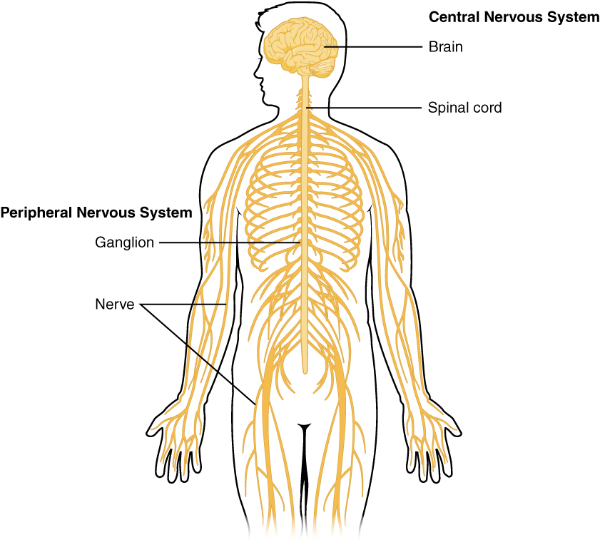
This diagram shows a silhouette of a human highlighting the nervous system. The central nervous system is composed of the brain and spinal cord. The brain is a large mass of ridged and striated tissue within the head. The spinal cord extends down from the brain and travels through the torso, ending in the pelvis. Pairs of enlarged nervous tissue, labeled ganglia, flank the spinal cord as it travels through the rib area. The ganglia are part of the peripheral nervous system, along with the many thread-like nerves that radiate from the spinal cord and ganglia through the arms, abdomen and legs.
Figure 12.2 — 12.1 Basic Structure and Function of the Nervous System

Figure 12.2 Central and Peripheral Nervous System The structures of the PNS are referred to as ganglia and nerves, which can be seen as distinct structures. The equivalent structures in the CNS are not obvious from this overall perspective and are best examined in prepared tissue under the microscope.

J. Gordon Betts and colleagues, Anatomy And Physiology 2E, OpenStax. · CC BY-NC-SA 4.0 · Original image ↗ · Access for free ↗
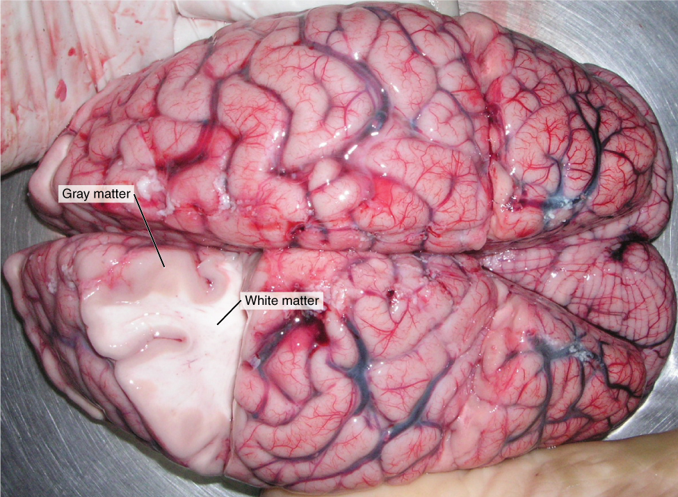
This photo shows an enlarged view of the dorsal side of a human brain. The right side of the occipital lobe has been shaved to reveal the white and gray matter beneath the surface blood vessels. The white matter branches though the shaved section like the limbs of a tree. The gray matter branches and curves on outside of the white matter, creating a buffer between the outer edges of the occipital lobe and the internal white matter.
Figure 12.3 — 12.1 Basic Structure and Function of the Nervous System

Figure 12.3 Gray Matter and White Matter A brain removed during an autopsy, with a partial section removed, shows white matter surrounded by gray matter. Gray matter makes up the outer cortex of the brain. (credit: modification of work by “Suseno”/Wikimedia Commons)

J. Gordon Betts and colleagues, Anatomy And Physiology 2E, OpenStax. (credit: modification of work by “Suseno”/Wikimedia Commons) · CC BY-NC-SA 4.0 · Original image ↗ · Access for free ↗
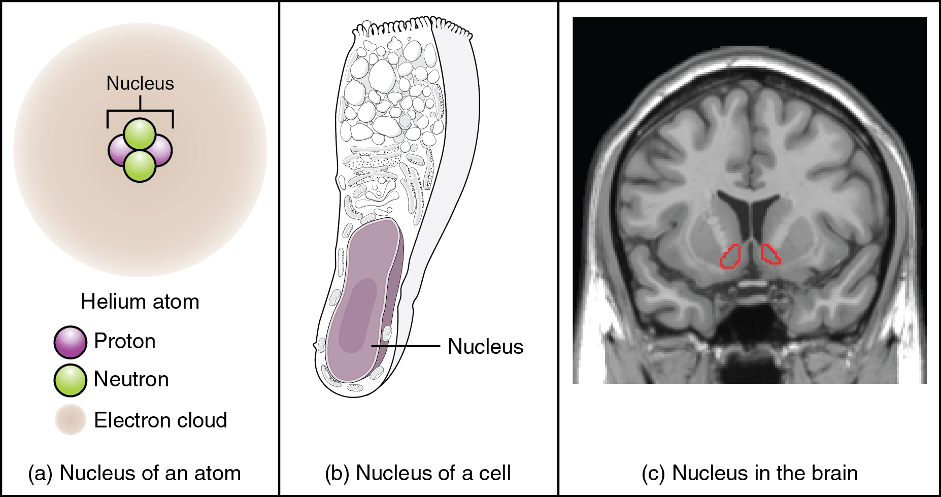
This figure shows two diagrams and a photo, labeled A, B, and C. Image A shows an atom composed of two neutrons and two protons surrounded by a hazy electron cloud. The nucleus of the atom is where the protons and neutrons are located. Image B shows a trumpet shaped cell with a large, oval nucleus near its narrow end. This is the nucleus of a cell. Image C shows an MRI capture of the brain. Two red areas near the center of the brain are highlighted in red. These are the nuclei within the brain.
Figure 12.4 — 12.1 Basic Structure and Function of the Nervous System

Figure 12.4 What Is a Nucleus? (a) The nucleus of an atom contains its protons and neutrons. (b) The nucleus of a cell is the organelle that contains DNA. (c) A nucleus in the CNS is a localized center of function with the cell bodies of several neurons, shown here circled in red. (credit c: “Was a bee”/Wikimedia Commons)

J. Gordon Betts and colleagues, Anatomy And Physiology 2E, OpenStax. (credit c: “Was a bee”/Wikimedia Commons) · CC BY-NC-SA 4.0 · Original image ↗ · Access for free ↗
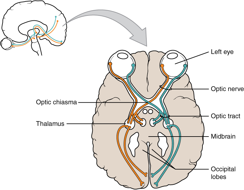
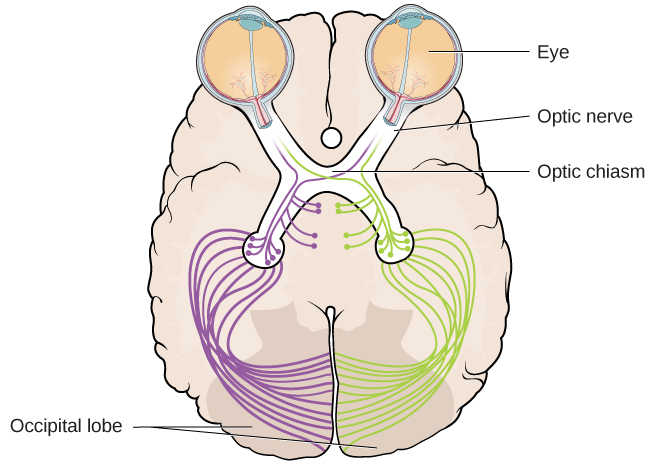
This illustration shows a superior view of a cross section of the brain. The anterior side of the brain is at the top of the diagram with the two eyes clearly visible. Each eye contains a left nerve tract and a right nerve tract. In the left eye, the left nerve tract travels straight back to the right side of the thalamus. It then enters the left occipital lobe. Conversely, the right nerve tract crosses to the right side of the brain through the optic chiasma. It travels through the right side of the thalamus and enters the right occipital lobe. In the right eye, the opposite is true. The left nerve tract crosses over to the left side of the brain at the optic chiasma, traveling into the left side of the thalamus and the left side of the occipital lobe. However, the right nerve tract leads straight back to the right side of the thalamus and the right occipital lobe. Therefore, the optic chiasma is where the right nerve tract from the right eye crosses over the left nerve tract from the left eye.
Figure 12.5 — 12.1 Basic Structure and Function of the Nervous System

Figure 12.5 Optic Nerve Versus Optic Tract This drawing of the connections of the eye to the brain shows the optic nerve extending from the eye to the chiasm, where the structure continues as the optic tract. The same axons extend from the eye to the brain through these two bundles of fibers, but the chiasm represents the border between peripheral and central.

J. Gordon Betts and colleagues, Anatomy And Physiology 2E, OpenStax. · CC BY-NC-SA 4.0 · Original image ↗ · Access for free ↗
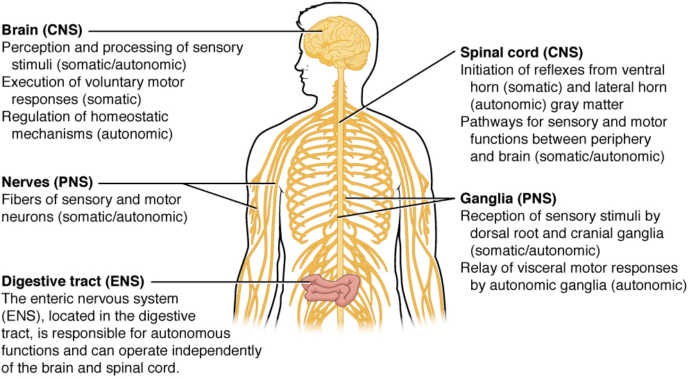
This illustration shows a silhouette of a human with only the brain, spinal cord, PNS ganglia, nerves and a section of the digestive tract visible. The brain, which is part of the CNS, is the area of perception and processing of sensory stimuli (somatic/autonomic), the execution of voluntary motor responses (somatic), and the regulation of homeostatic mechanisms (autonomic). The spinal cord, which is part of the CNS, is the area where reflexes are initiated. The gray matter of the ventral horn initiates somatic reflexes while the gray matter of the lateral horn initiates autonomic reflexes. The spinal cord is also the somatic and autonomic pathway for sensory and motor functions between the PNS and the brain. The nerves, which are part of the PNS, are the fibers of sensory and motor neurons, which can be either somatic or autonomic. The ganglia, which are part of the PNS, are the areas for the reception of somatic and autonomic sensory stimuli. These are received by the dorsal root ganglia and cranial ganglia. The autonomic ganglia are also the relay for visceral motor responses. The digestive tract is part of the enteric nervous system, the ENS, which is located in the digestive tract and is responsible for autonomous function. The ENS can operate independent of the brain and spinal cord.
Figure 12.6 — 12.1 Basic Structure and Function of the Nervous System

Figure 12.6 Somatic, Autonomic, and Enteric Structures of the Nervous System Somatic structures include the spinal nerves, both motor and sensory fibers, as well as the sensory ganglia (posterior root ganglia and cranial nerve ganglia). Autonomic structures are found in the nerves also, but include the sympathetic and parasympathetic ganglia. The enteric nervous system includes the nervous tissue within the organs of the digestive tract.

J. Gordon Betts and colleagues, Anatomy And Physiology 2E, OpenStax. · CC BY-NC-SA 4.0 · Original image ↗ · Access for free ↗
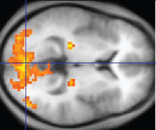
This MRI image shows a grainy computer readout of a cross section of the brain. The anterior side of the brain, located on the right hand side of the image, has a large area lighting up with yellow, indicating neural stimulation. Two smaller regions at the center of the brain are also yellow. The two small areas are in the same relative location but in opposite hemispheres of the brain.
Figure 12.7 — 12.1 Basic Structure and Function of the Nervous System

Figure 12.7 fMRI This fMRI shows activation of the visual cortex in response to visual stimuli. (credit: “Superborsuk”/Wikimedia Commons)

J. Gordon Betts and colleagues, Anatomy And Physiology 2E, OpenStax. (credit: “Superborsuk”/Wikimedia Commons) · CC BY-NC-SA 4.0 · Original image ↗ · Access for free ↗
Group of Cell Bodies (i.e., gray matter) Ganglion
Bundle of Axons (i.e., white matter) Tract Nerve

Table 12.1. J. Gordon Betts and colleagues, Anatomy And Physiology 2E, OpenStax.

Source: 12.1 Basic Structure and Function of the Nervous System

15.1 Divisions of the — figures and tables

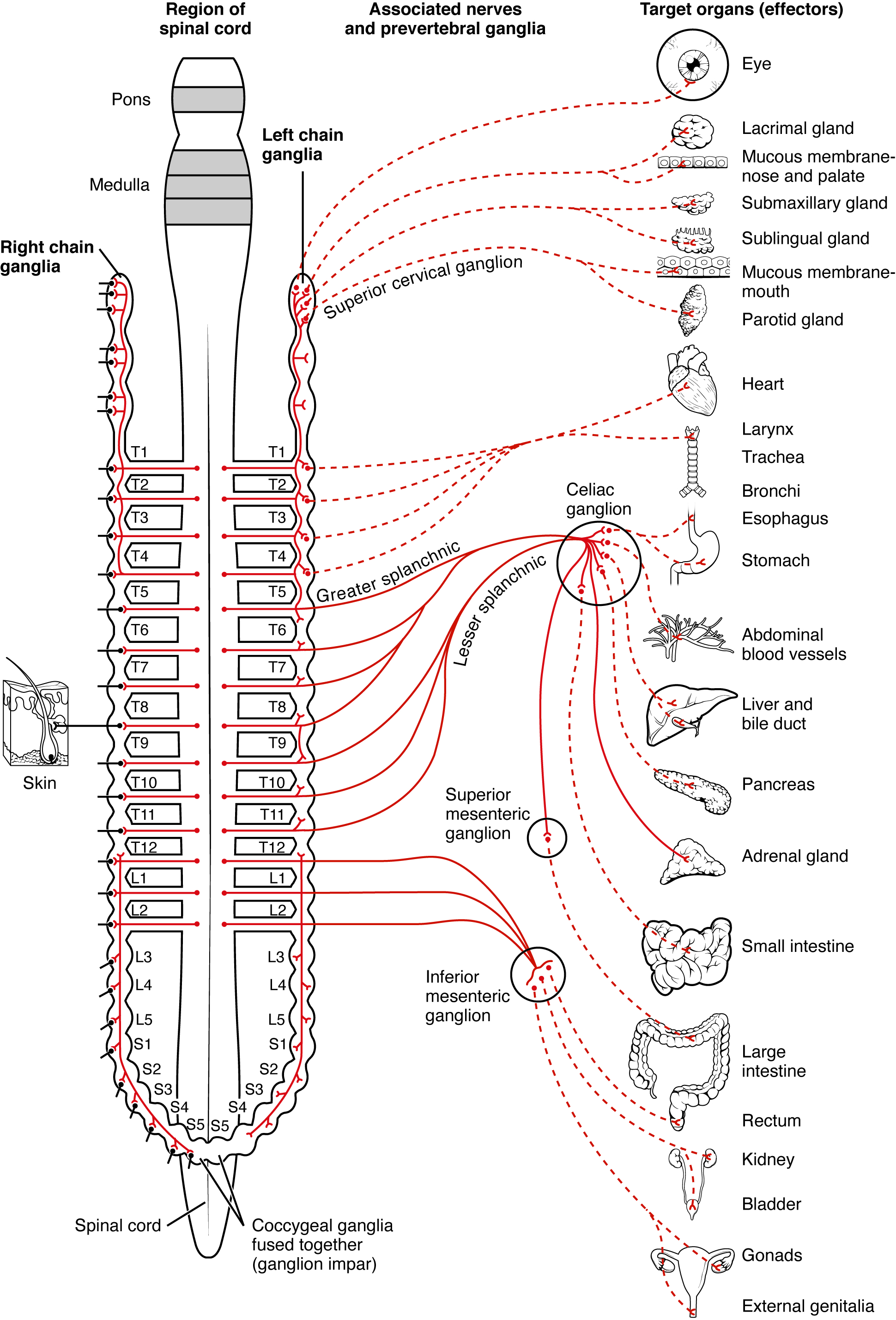
This diagram shows the spinal cord, and the connections from the spinal cord to the different target organs. The target organs are listed on the right.
Figure 15.2 — 15.1 Divisions of the

Figure 15.2 Connections of Sympathetic Division of the Autonomic Nervous System Neurons from the lateral horn of the spinal cord (preganglionic nerve fibers - solid lines) project to the chain ganglia on either side of the vertebral column or to collateral (prevertebral) ganglia that are anterior to the vertebral column in the abdominal cavity. Axons from these ganglionic neurons (postganglionic nerve fibers - dotted lines) then project to target effectors throughout the body.

J. Gordon Betts and colleagues, Anatomy And Physiology 2E, OpenStax. · CC BY-NC-SA 4.0 · Original image ↗ · Access for free ↗
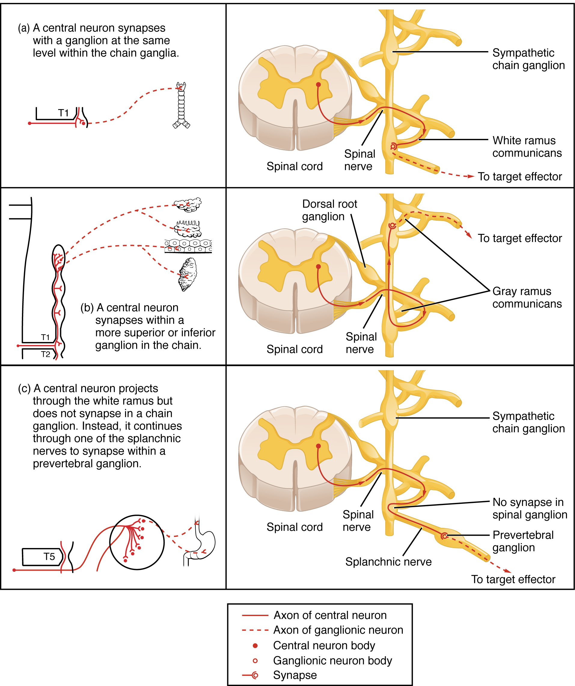
This table shows the connections between the spinal cord and the ganglia. The top panel shows the connection between a central neuron and a chain ganglion at the same lever. The center panel shows the connection between a central neuron and a synapse with a superior or inferior ganglion. The bottom panel shows the projection of a central neuron into the white ramus.
Figure 15.3 — 15.1 Divisions of the

Figure 15.3 Sympathetic Connections and Chain Ganglia The axon from a central sympathetic neuron in the spinal cord can project to the periphery in a number of different ways. (a) The fiber can project out to the ganglion at the same level and synapse on a ganglionic neuron. (b) A branch can project to more superior or inferior ganglion in the chain. (c) A branch can project through the white ramus communicans, but not terminate on a ganglionic neuron in the chain. Instead, it projects through one of the splanchnic nerves to a collateral ganglion or the adrenal medulla (not pictured).

J. Gordon Betts and colleagues, Anatomy And Physiology 2E, OpenStax. · CC BY-NC-SA 4.0 · Original image ↗ · Access for free ↗
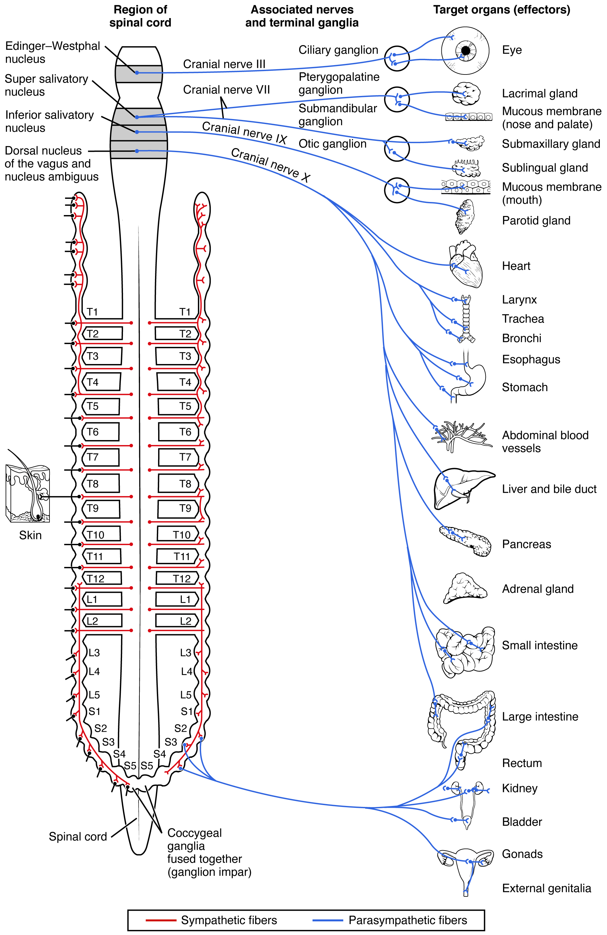
This diagram shows the spinal cord and has different central nerves emerging from it. The central nerves target different effector organs that are listed on the right.
Figure 15.4 — 15.1 Divisions of the

Figure 15.4 Connections of Parasympathetic Division of the Autonomic Nervous System Neurons from brain-stem nuclei, or from the lateral horn of the sacral spinal cord, project to terminal ganglia near or within the various organs of the body. Axons from these ganglionic neurons then project the short distance to those target effectors.

J. Gordon Betts and colleagues, Anatomy And Physiology 2E, OpenStax. · CC BY-NC-SA 4.0 · Original image ↗ · Access for free ↗
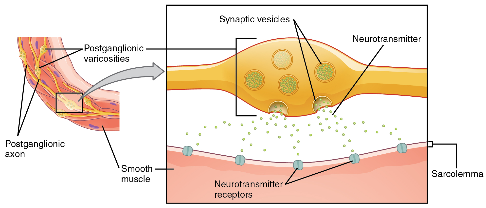
This figure shows the connection between autonomic fibers and the target effectors. The left image shows a slice of smooth muscle with the postganglionic varicosities and the postganglionic axons labeled. The right panel shows a magnified view of the synaptic vesicles, neurotransmitters, and the sarcolemma.
Figure 15.5 — 15.1 Divisions of the

Figure 15.5 Autonomic Varicosities The connection between autonomic fibers and target effectors is not the same as the typical synapse, such as the neuromuscular junction. Instead of a synaptic end bulb, a neurotransmitter is released from swellings along the length of a fiber that makes an extended network of connections in the target effector.

J. Gordon Betts and colleagues, Anatomy And Physiology 2E, OpenStax. · CC BY-NC-SA 4.0 · Original image ↗ · Access for free ↗
Sympathetic Parasympathetic
Preganglionic Acetylcholine → nicotinic Acetylcholine → nicotinic
Postganglionic Norepinephrine → α- or β-adrenergic
Acetylcholine → muscarinic (associated with sweat glands and the blood vessels associated with skeletal muscles only
Acetylcholine → muscarinic

Table 15.1. J. Gordon Betts and colleagues, Anatomy And Physiology 2E, OpenStax.

Source: 15.1 Divisions of the Autonomic Nervous System

14.3 Motor Responses — figures and tables
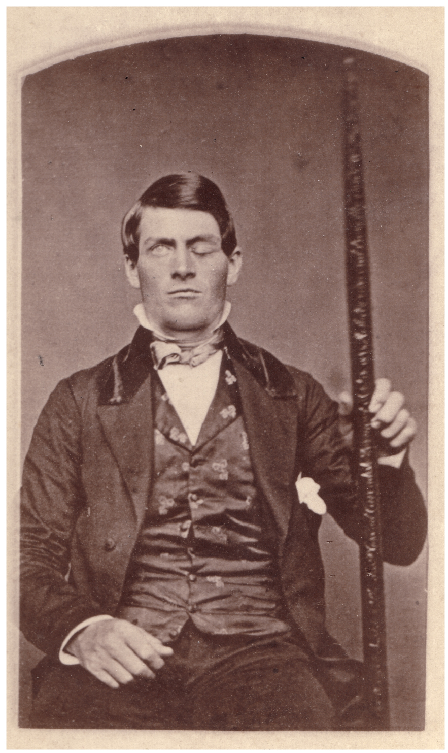
This photo shows Phineas Gage holding the metal spike that impaled his prefrontal cortex.
Figure 14.27 — 14.3 Motor Responses

Figure 14.27 Phineas Gage The victim of an accident while working on a railroad in 1848, Phineas Gage had a large iron rod impaled through the prefrontal cortex of his frontal lobe. After the accident, his personality appeared to change, but he eventually learned to cope with the trauma and lived as a coach driver even after such a traumatic event. (credit b: John M. Harlow, MD)

J. Gordon Betts and colleagues, Anatomy And Physiology 2E, OpenStax. (credit b: John M. Harlow, MD) · CC BY-NC-SA 4.0 · Original image ↗ · Access for free ↗
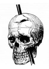
The image on the right shows a drawing of the skull with the metal spike inserted like it would have been when he was injured.
Figure 14.27 — 14.3 Motor Responses

Figure 14.27 Phineas Gage The victim of an accident while working on a railroad in 1848, Phineas Gage had a large iron rod impaled through the prefrontal cortex of his frontal lobe. After the accident, his personality appeared to change, but he eventually learned to cope with the trauma and lived as a coach driver even after such a traumatic event. (credit b: John M. Harlow, MD)

J. Gordon Betts and colleagues, Anatomy And Physiology 2E, OpenStax. (credit b: John M. Harlow, MD) · CC BY-NC-SA 4.0 · Original image ↗ · Access for free ↗
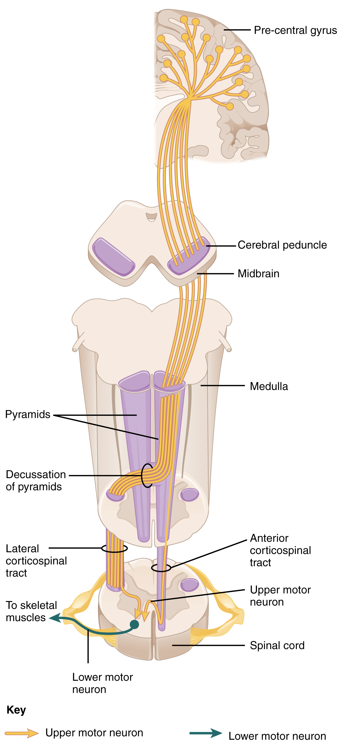
This diagram shows how the motor neurons thread their way through the spinal cord and into the brain. It also shows the different connections they make along the way.
Figure 14.28 — 14.3 Motor Responses

Figure 14.28 Corticospinal Tract The major descending tract that controls skeletal muscle movements is the corticospinal tract. It is composed of two neurons, the upper motor neuron and the lower motor neuron. The upper motor neuron has its cell body in the primary motor cortex of the frontal lobe and synapses on the lower motor neuron, which is in the ventral horn of the spinal cord and projects to the skeletal muscle in the periphery.

J. Gordon Betts and colleagues, Anatomy And Physiology 2E, OpenStax. · CC BY-NC-SA 4.0 · Original image ↗ · Access for free ↗
Source: 14.3 Motor Responses

1.5 — figures and tables

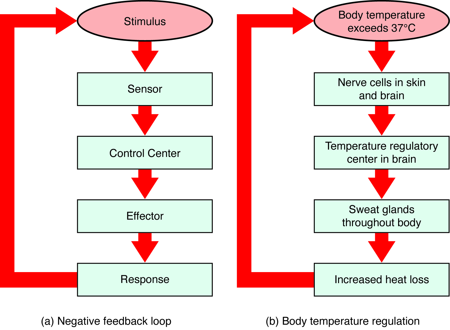
This figure shows two flow charts labeled A and B. Chart A shows a general negative feedback loop. The loop starts with a stimulus. Information about the stimulus is perceived by a sensor which sends that information to a control center. The control center sends a signal to an effector, which creates a response. That then feeds back to the top of the flow chart by inhibiting the stimulus. Part B shows body temperature regulation as an example of negative feedback system. Here, the stimulus is body temperature exceeding 37 degrees Celsius. The sensor is a set of nerve cells in the skin and brain and the control center is the temperature regulatory center of the brain. The effectors are sweat glands throughout the body which lead to increased heat loss and inhibit the rising body temperature.
Figure 1.10 — 1.5

Figure 1.10 Negative Feedback System In a negative feedback system, a stimulus—a deviation from a set point—is resisted through a physiological process that returns the body to homeostasis. (a) A negative feedback system has five basic steps. (b) Body temperature is regulated by negative feedback.

J. Gordon Betts and colleagues, Anatomy And Physiology 2E, OpenStax. · CC BY-NC-SA 4.0 · Original image ↗ · Access for free ↗
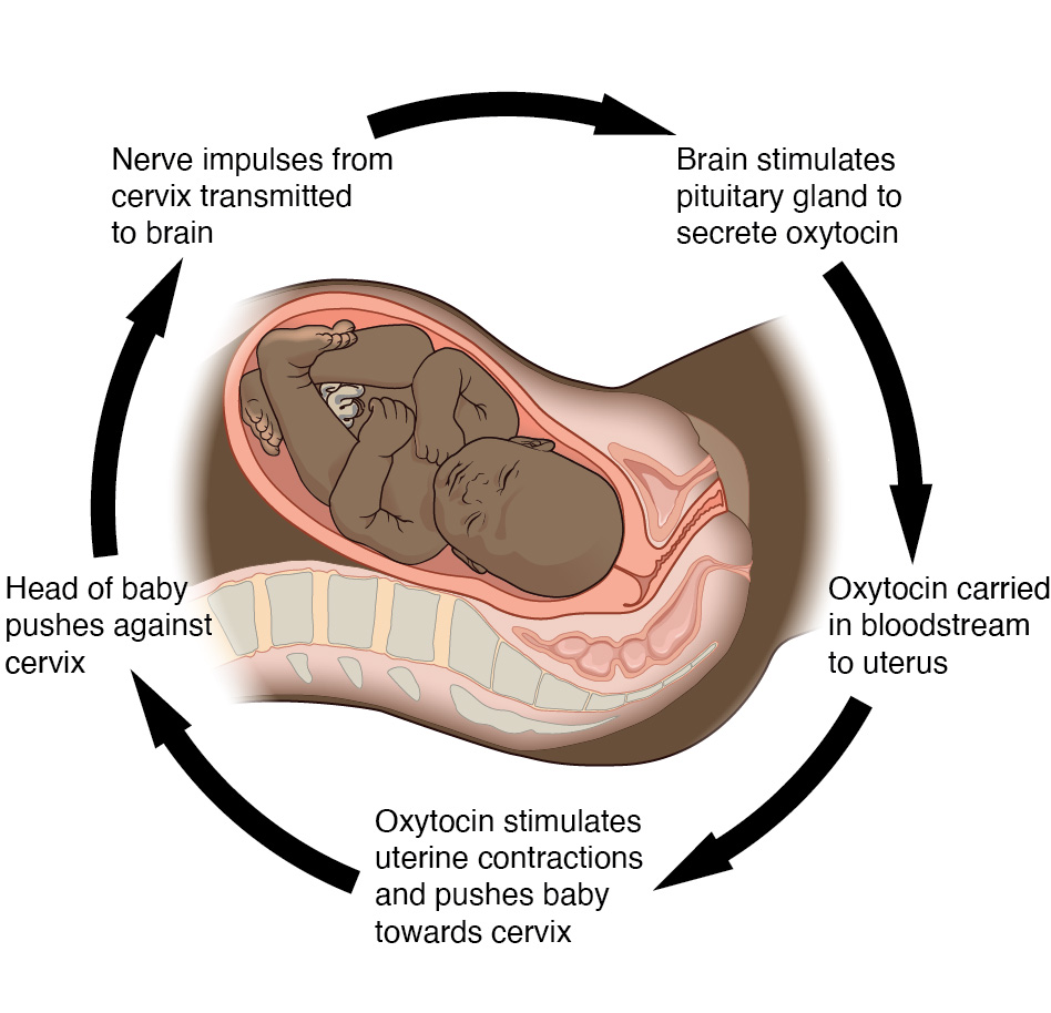
This diagram shows the steps of a positive feedback loop as a series of stepwise arrows looping around a diagram of an infant within the uterus of a pregnant woman. Initially the head of the baby pushes against the cervix, transmitting nerve impulses from the cervix to the brain. Next the brain stimulates the pituitary gland to secrete oxytocin which is carried in the bloodstream to the uterus. Finally, the oxytocin simulates uterine contractions and pushes the baby harder into the cervix. As the head of the baby pushes against the cervix with greater and greater force, the uterine contractions grow stronger and more frequent. This mechanism is a positive feedback loop.
Figure 1.11 — 1.5

Figure 1.11 Positive Feedback Loop Normal childbirth is driven by a positive feedback loop. A positive feedback loop results in a change in the body’s status, rather than a return to homeostasis.

J. Gordon Betts and colleagues, Anatomy And Physiology 2E, OpenStax. · CC BY-NC-SA 4.0 · Original image ↗ · Access for free ↗

Source: 1.5 Homeostasis

220 terms, alphabetically ordered

Acid · ·

In the Brønsted–Lowry model, a substance that can donate a . Acidic have a higher activity than water.

Read explanation · OpenStax ↗
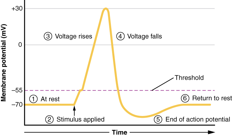
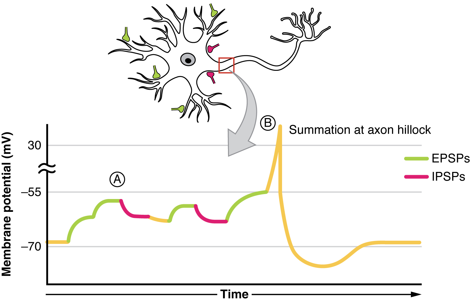
action potential

A regenerative -voltage change produced by voltage-dependent -channel activity.

Read explanation · OpenStax ↗
Activation energy ·

The energy barrier that must overcome to reach a and undergo .

Read explanation · OpenStax ↗
Adenosine triphosphate

An energy-carrying ; its powers many cellular processes, including cycles.

Read explanation · OpenStax ↗
Alpha carbon · ·

The central carbon next to the carboxyl carbon. In a standard it binds an , hydrogen, and .

Read explanation · OpenStax ↗
Alpha helix · · · · · ·

A helical stabilized by between a oxygen and an hydrogen four later.

Read explanation · OpenStax ↗
Amide · · ·

A group containing a carbon bonded to nitrogen. are amide linkages.

Read explanation · OpenStax ↗
Amino acid

A building block containing amino and and a characteristic .

Read explanation · OpenStax ↗
Amino acid residue · ·

The part of an incorporated into a chain. Residue can also describe another building block within a larger .

Read explanation · OpenStax ↗
Amino group · · ·

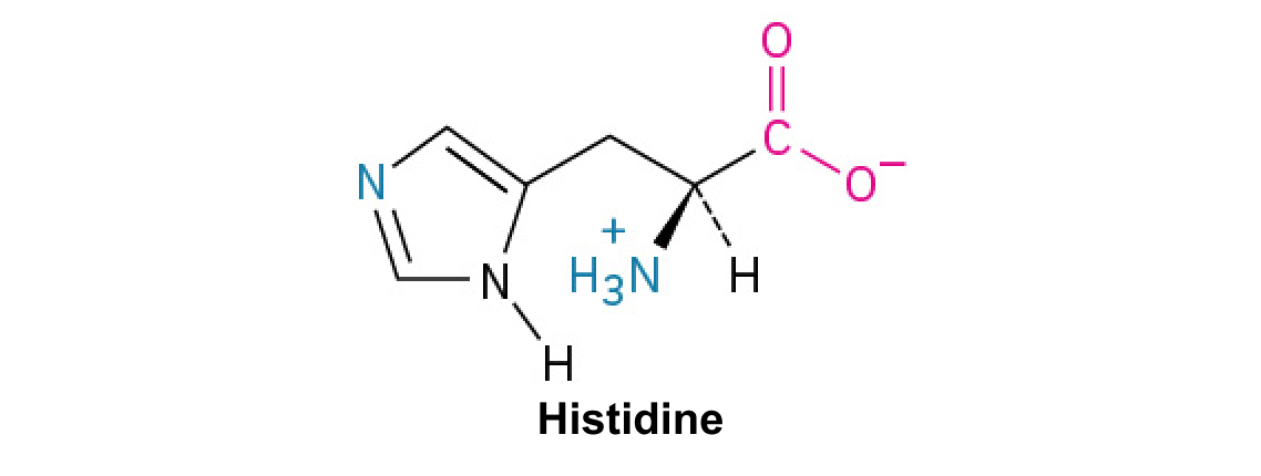
A nitrogen-containing . A free ’s amino group is typically (NH₃⁺) near physiological ; ’s ring nitrogen is an exception to this formula.

Read explanation · OpenStax ↗
Amphipathic

Having both a region and a region, as in a or .

Read explanation · OpenStax ↗
Amylose

The largely unbranched component of , made mainly of linked by alpha-1,4 .

Read explanation · OpenStax ↗
anomeric carbon

The former carbon of a sugar, which becomes the or hemiketal center upon cyclization.

Read explanation · OpenStax ↗
Antibody

An that recognizes a specific region () on an . Antibodies help immune defenses and are used as detection tools in .

Read explanation · OpenStax ↗
Antigen

A or molecular structure specifically recognized by an or other adaptive immune .

Read explanation · OpenStax ↗
Antiparallel

Running in opposite directions, as with two strands or neighboring strands in some .

Read explanation · OpenStax ↗
Aqueous solution ·

A in which water is the .

Read explanation · OpenStax ↗
Aromatic

Containing a cyclic, with . , , and have aromatic .

Read explanation · OpenStax ↗
Assay

A procedure used to detect a substance or measure its amount, activity, or another property.

Read explanation · Thermo Fisher ↗
Atom

The smallest unit of an element that retains its chemical identity; it has a surrounded by .

Read explanation · OpenStax ↗
attachment

Enduring emotional bond and expectations about another’s availability and responsiveness.

Read explanation · OpenStax ↗
autonomic nervous system

Neural pathways regulating visceral organs through sympathetic, parasympathetic, and enteric systems.

Read explanation · OpenStax ↗
axon

Neuronal process that usually conducts toward terminals.

Read explanation · OpenStax ↗
baroreflex

Rapid autonomic adjustment of cardiovascular function in response to arterial-pressure sensing.

Read explanation · OpenStax ↗
basal ganglia

Interconnected subcortical involved in action selection and learning.

Read explanation · OpenStax ↗
Benzene ring

A six-carbon ring with .

Read explanation · OpenStax ↗
Benzyl group

A attached through a –CH₂– group; part of ’s .

Read explanation · OpenStax ↗
Beta sheet · · · · · ·

A made of aligned segments connected by ; strands may be parallel or .

Read explanation · OpenStax ↗
Big Five

Trait framework describing openness, conscientiousness, extraversion, agreeableness, and neuroticism.

Read explanation · OpenStax ↗
Biomolecule

A made by or involved in living systems, including , , , and .

Read explanation · OpenStax ↗
Biosynthesis

Formation of biological through cellular , such as synthesis of .

Read explanation · OpenStax ↗
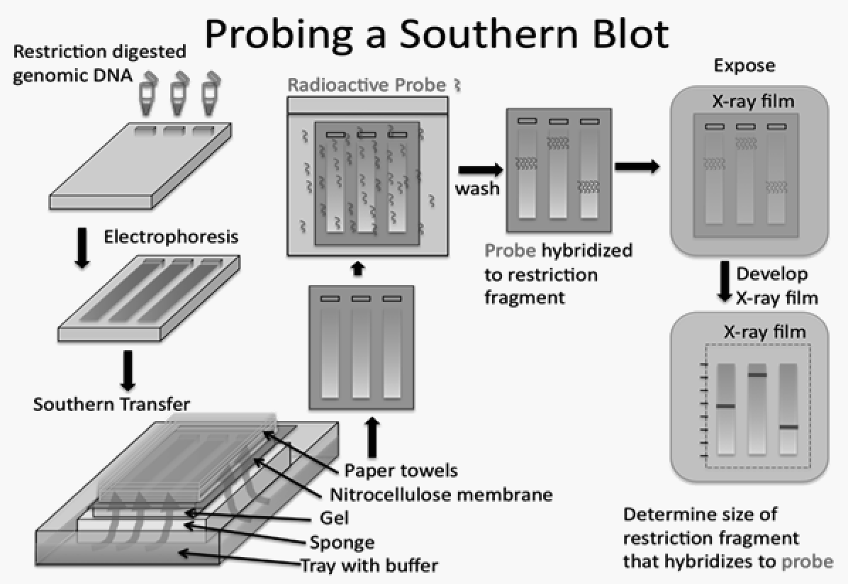
Blotting ·

Transfer of separated or to a so a specific target can be detected.

Read explanation · Thermo Fisher ↗
Brønsted–Lowry base · · · · · · · · · · ·

In the Brønsted–Lowry model, a substance that can accept a . Basic solutions have lower activity than water.

Read explanation · OpenStax ↗
Carbohydrate

A class of including sugars and their , used for energy storage, structure, and recognition.

Read explanation · OpenStax ↗
Carbonyl group · ·

A carbon double-bonded to oxygen, C=O; present in and many other .

Read explanation · OpenStax ↗
Carboxyl group · ·

An , COOH. In free it is usually to COO⁻ near physiological .

Read explanation · OpenStax ↗
Catalyst · ·

A substance that speeds a by providing a pathway with lower and is regenerated rather than consumed overall.

Read explanation · OpenStax ↗
catecholamine

A -derived chemical class including dopamine, norepinephrine, and epinephrine.

Read explanation · OpenStax ↗
Cation ·

An with a positive net .

Read explanation · OpenStax ↗
Cellulose

A structural in plant cell walls, with beta-1,4 linkages; also used as a support.

Read explanation · OpenStax ↗
central nervous system

Brain and spinal cord.

Read explanation · OpenStax ↗
cerebellum

structure important for coordination, timing, and motor learning.

Read explanation · OpenStax ↗
Chemical compound ·

A substance containing of two or more different chemically combined in a definite composition.

Read explanation · OpenStax ↗
Chemical element

A substance defined by having the same number of , such as carbon or oxygen.

Read explanation · OpenStax ↗
Chemical reaction ·

A process that rearranges and chemical bonds, converting into .

Read explanation · OpenStax ↗
Chromatin

The complex of and associated , including , in .

Read explanation · OpenStax ↗
Chromatography

Separation of mixture components by their different interactions with a and a moving .

Read explanation · Waters ↗
Chromosome ·

An organized with associated that carries . A replicated chromosome has two .

Read explanation · OpenStax ↗
Codon

A sequence of three that specifies an or a stop signal.

Read explanation · OpenStax ↗
Collagen

A major structural in , with a characteristic triple-helical arrangement.

Read explanation · OpenStax ↗
complement

A system that can promote , , and attack against targets.

Read explanation · OpenStax ↗
Concentration

The amount of a substance in a specified volume or mass of mixture.

Read explanation · OpenStax ↗
conduction

Thermal energy transfer through microscopic interactions without bulk matter flow.

Read explanation · OpenStax ↗
Conformation ·

A spatial arrangement accessible by rotations and other motions that do not require breaking .

Read explanation · NCBI Bookshelf ↗
Conjugated system ·

Adjacent orbitals that allow to be distributed across several , often through alternating single and multiple bonds.

Read explanation · OpenStax ↗
connective tissue

A tissue category generally characterized by cells distributed within substantial .

Read explanation · OpenStax ↗
cornea

Transparent anterior surface of the eye providing substantial refraction of incoming light.

Read explanation · OpenStax ↗
cortisol

An adrenal-cortex glucocorticoid regulating , stress responses, and immune activity.

Read explanation · OpenStax ↗
Covalent bond · ·

A bond in which share pairs. and are covalent.

Read explanation · OpenStax ↗
Cross-reactivity

Binding of a detection reagent, such as an , to another target with a similar recognizable structure.

Read explanation · OpenStax ↗
Cysteine ·

An with a . Two cysteines can form a .

Read explanation · OpenStax ↗
dendrite

Branching neuronal process receiving many synaptic inputs.

Read explanation · OpenStax ↗
Deoxyribonucleic acid

The that stores genetic information used to specify and sequences.

Read explanation · OpenStax ↗
depolarization

A shift in voltage toward a less negative value.

Read explanation · OpenStax ↗
Deprotonation

Removal of a from a or group; this changes its by minus one.

Read explanation · OpenStax ↗
Detergent

An substance that can associate with both water and material; detergents can disrupt or interactions.

Read explanation · Thermo Fisher ↗
Disulfide bond · · ·

A covalent sulfur–sulfur bond formed by of two , which can stabilize structure.

Read explanation · NCBI Bookshelf ↗
DNA replication

Copying so genetic information can be passed to daughter cells. Each guides a complementary new strand.

Read explanation · OpenStax ↗
ectoderm

The embryonic germ layer contributing and nervous-system lineages.

Read explanation · OpenStax ↗
Effector

A that changes a ’s activity or binding behavior, often by stabilizing a particular .

Read explanation · Biology LibreTexts ↗
Electric charge · · ·

An electrical property that can be positive or negative. Opposite charges attract and like charges repel; net charge is the sum of all charges on a particle.

Read explanation · OpenStax ↗
Electron

A negatively subatomic particle. Electron sharing and transfer determine much of chemical bonding.

Read explanation · OpenStax ↗
Electron delocalization ·

Distribution of across several rather than confinement to one or bond.

Read explanation · OpenStax ↗
Electronegativity ·

An ’s tendency to attract shared in a chemical bond. Unequal attraction can make a bond .

Read explanation · OpenStax ↗
Endocrine signaling ·

Communication in which enter the circulation and act on distant target cells.

Read explanation · OpenStax ↗
Endomembrane system

The interacting and compartments involved in processes such as modification, transport, and .

Read explanation · OpenStax ↗
Endoplasmic reticulum

An interconnected network continuous with the ; it includes regions involved in processing and synthesis.

Read explanation · OpenStax ↗
enteric nervous system

Neural networks in the gastrointestinal wall coordinating motility, , and local responses.

Read explanation · OpenStax ↗
epidermis

The outer epithelial layer of skin, dominated by .

Read explanation · OpenStax ↗
Epitope

The particular part of an recognized by an or immune .

Read explanation · OpenStax ↗
Equilibrium

A state where opposing processes occur at equal rates, so macroscopic or other properties remain constant.

Read explanation · OpenStax ↗
erythropoietin

A produced mainly by the adult kidney that stimulates production in marrow.

Read explanation · OpenStax ↗
Eukaryote ·

An organism whose cells have a -enclosed ; animals, plants, fungi, and protists are eukaryotes.

Read explanation · OpenStax ↗
Experimental control ·

A comparison sample or condition used to distinguish the effect being tested from other influences.

Read explanation · Thermo Fisher ↗
extracellular matrix

The network of and surrounding cells and providing structural and signaling functions.

Read explanation · OpenStax ↗
FACT complex ·

A -associated complex that helps machinery traverse and supports reorganization.

Read explanation · OpenStax ↗
forebrain

Major developmental brain division including cerebral hemispheres, , and .

Read explanation · OpenStax ↗
Functional group

A group of whose chemical behavior gives a characteristic and properties.

Read explanation · OpenStax ↗
functional magnetic resonance imaging

Imaging method estimating neural activity indirectly through blood-oxygenation changes.

Read explanation · OpenStax ↗
gamete

A reproductive cell that contributes a during fertilization.

Read explanation · OpenStax ↗
Gene

A sequence that contributes to a functional or , together with the information needed to express it.

Read explanation · OpenStax ↗
Gene expression

Production of a functional from a , including and, for -coding , .

Read explanation · NCBI Bookshelf ↗
Genome

The complete genetic material of an organism or cell.

Read explanation · OpenStax ↗
Germline · · ·

The cell lineage that produces . A germline mutation can be inherited if the altered contributes to an offspring.

Read explanation · OpenStax ↗
Glucagon

A pancreatic that helps raise , especially by promoting release from the liver.

Read explanation · OpenStax ↗
Glucose

A six-carbon sugar that is a major cellular energy source and a building block of , , and .

Read explanation · OpenStax ↗
glycogen

A highly branched used for storage in animals.

Read explanation · Biology LibreTexts / Oregon State University ↗
Glycosidic bond

A involving a sugar’s . In a , an N-glycosidic bond links the sugar to the .

Read explanation · Biology LibreTexts / Oregon State University ↗
Golgi apparatus ·

A stack of compartments that modifies and sorts and traveling through the .

Read explanation · OpenStax ↗
Haploid

Having one set of , as in a human .

Read explanation · OpenStax ↗
hemiacetal

Carbon bonded to both and alkoxy groups, usually formed by alcohol addition to a .

Read explanation · OpenStax ↗
Hemoglobin

The oxygen-carrying in , composed of four with heme groups.

Read explanation · OpenStax ↗
hindbrain

Developmental brain division including medulla, pons, and .

Read explanation · OpenStax ↗
Histidine ·

A with an . Mostly uncharged near physiological , it can accept or donate .

Read explanation · OpenStax ↗
Histone

A -associated rich in . Core histones form the around which nucleosomal wraps.

Read explanation · OpenStax ↗
homeostasis

Dynamic regulation that maintains physiological variables within functional ranges despite disturbances.

Read explanation · OpenStax ↗
Hormone

A chemical messenger released by cells that changes the activity of target cells with matching .

Read explanation · OpenStax ↗
Hydrogen bond ·

An attraction between a hydrogen bonded to an and another . hydrogen bonds stabilize .

Read explanation · OpenStax ↗
Hydrogen ion · ·

A positively hydrogen species. H⁺ denotes a , which is hydrated in water; its activity determines .

Read explanation · OpenStax ↗
Hydrolysis

Breaking a chemical bond by adding water. hydrolysis separates .

Read explanation · OpenStax ↗
Hydrophilic

Interacting favorably with water, typically because a group is or .

Read explanation · OpenStax ↗
Hydrophobic

Interacting poorly with water. often gather in a ’s interior.

Read explanation · OpenStax ↗
Hydroxide ion ·

The negatively OH⁻. Basic contain a higher hydroxide ion activity than water at the same temperature.

Read explanation · OpenStax ↗
Hydroxyl group ·

An oxygen bonded to hydrogen, –OH. Hydroxyl groups can and often increase .

Read explanation · OpenStax ↗
hypothalamus

region coordinating homeostatic, , and autonomic functions.

Read explanation · OpenStax ↗
Imidazole

A five-membered ring containing two nitrogens; ’s contains this ring.

Read explanation · OpenStax ↗
Immunoglobulin

An or the -bound form of the same type of -binding .

Read explanation · OpenStax ↗
Indole

A fused six-membered and five-membered ring system containing nitrogen; found in .

Read explanation · OpenStax ↗
inflammation

A coordinated tissue response recruiting defense and repair mechanisms, often involving increased blood flow and permeability.

Read explanation · OpenStax ↗
Insulin

A from that lowers by promoting uptake and storage and reducing production.

Read explanation · OpenStax ↗
Ion · ·

An or with a net because its numbers of and do not balance.

Read explanation · OpenStax ↗
iris

Pigmented muscular structure controlling pupil size.

Read explanation · OpenStax ↗
Keratin

A fibrous structural found in tissues such as hair, nails, and the outer skin.

Read explanation · OpenStax ↗
keratinocyte

The main epidermal cell type producing and contributing to the protective barrier.

Read explanation · OpenStax ↗
ligament

A connective-tissue structure commonly linking bones and stabilizing joints.

Read explanation · OpenStax ↗
Ligand

A or that binds to a particular site on another , such as a or metal .

Read explanation · Cytiva ↗
linkage

A tendency of nearby on the same to be inherited together.

Read explanation · OpenStax ↗
Lipid

A broad class of largely or , including fats, , and steroids.

Read explanation · OpenStax ↗
locus

The specific position of a or other feature.

Read explanation · OpenStax ↗
Logarithm · · ·

The exponent needed to obtain a number from a chosen fixed number. For decimal logarithms, log₁₀(100) = 2 because 10² = 100; uses the negative decimal logarithm of activity.

Read explanation · OpenStax ↗
magnetic resonance imaging

Imaging method using magnetic fields and radiofrequency signals to reveal tissue structure.

Read explanation · OpenStax ↗
mean arterial pressure

The time-averaged pressure driving systemic circulation over a cardiac cycle.

Read explanation · OpenStax ↗
Membrane · · · ·

A thin boundary or sheet. Cell membranes are bilayers containing ; membranes are synthetic supports that hold or for detection.

Read explanation · OpenStax ↗
Messenger RNA ·

An that carries coding information used by to synthesize a .

Read explanation · OpenStax ↗
Metabolism

The chemical that build , break them down, and transform energy in a living system.

Read explanation · OpenStax ↗
Methyl group ·

A –CH₃ group, consisting of one carbon bonded to three hydrogens.

Read explanation · OpenStax ↗
midbrain

Brainstem division containing orienting circuits and important motor and neuromodulatory .

Read explanation · OpenStax ↗
Mobile phase

The moving fluid that carries a sample through a system.

Read explanation · Waters ↗
Molecular chaperone · ·

A that assists other in folding or prevents , without specifying their sequence.

Read explanation · NCBI Bookshelf ↗
Molecule

Two or more joined by . A molecule may be electrically or carry a .

Read explanation · OpenStax ↗
muscle spindle

Muscle signaling length and changes in length.

Read explanation · OpenStax ↗
Mutation

A change in a sequence; some mutations alter a ’s sequence.

Read explanation · OpenStax ↗
Myelin

A -rich insulating sheath around many nerve axons that supports efficient signal .

Read explanation · NHGRI / NIH ↗
negative feedback

A control arrangement in which a response opposes the initiating deviation.

Read explanation · OpenStax ↗
neural crest

A migratory -derived cell population contributing peripheral nervous structures and several other lineages.

Read explanation · OpenStax ↗
neuromuscular junction

The connecting a motor to a skeletal-muscle fiber.

Read explanation · OpenStax ↗
neuron

An electrically excitable cell specialized for receiving and transmitting information.

Read explanation · OpenStax ↗
Neutral

Having no net . For a , neutral instead means equal and activities; its is about 7 at 25 °C.

Read explanation · OpenStax ↗
Nitrogenous base · · ·

A nitrogen-containing ring component of ; its pairing interactions help specify sequences.

Read explanation · OpenStax ↗
Nonpolar ·

Lacking a substantial separation of . Nonpolar groups generally interact poorly with water.

Read explanation · OpenStax ↗
Nuclear envelope

The double enclosing the and continuous with the .

Read explanation · OpenStax ↗
Nucleic acid

A of , such as or , used in information storage, expression, and other cellular functions.

Read explanation · OpenStax ↗
Nucleosome

The basic unit of : wrapped around a .

Read explanation · OpenStax ↗
Nucleotide

A joined to a sugar and one or more .

Read explanation · OpenStax ↗
Nucleus ·

A -enclosed compartment containing most of a cell’s .

Read explanation · OpenStax ↗
Octamer

A complex made of eight . A core contains eight .

Read explanation · OpenStax ↗
opsonization

Coating a target with that enhance its recognition and uptake by phagocytes.

Read explanation · OpenStax ↗
optic chiasm

Junction where axons from nasal halves of the retinas cross.

Read explanation · OpenStax ↗
Oxidation ·

Loss of or an increase in oxidation state. Oxidizing two can form a .

Read explanation · OpenStax ↗
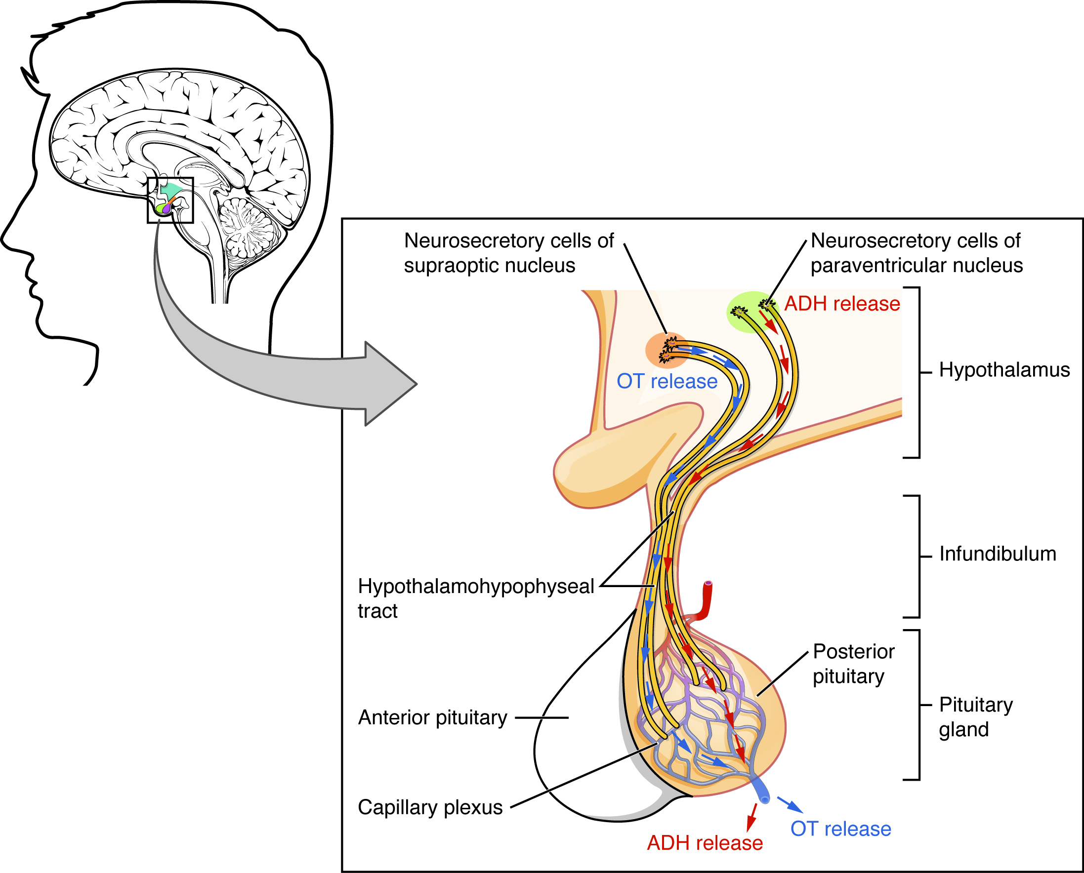
oxytocin

A hypothalamic released by the posterior pituitary, promoting uterine contraction and milk ejection.

Read explanation · OpenStax ↗
Pancreas

An organ with digestive and functions; its islets contain cells that secrete and .

Read explanation · OpenStax ↗
Pancreatic beta cell ·

An cell in a pancreatic islet that secretes .

Read explanation · OpenStax ↗
Peptide

A chain of joined by ; the term commonly describes chains shorter than a typical .

Read explanation · OpenStax ↗
Peptide bond ·

A covalent between the carboxyl carbon of one and the amino nitrogen of the next.

Read explanation · OpenStax ↗
Peptide hormone

An amino--based chemical messenger released by ; and are examples.

Read explanation · OpenStax ↗
perfusion

Blood flow through a tissue or vascular region.

Read explanation · OpenStax ↗
peripheral nervous system

Nerves and ganglia outside the brain and spinal cord.

Read explanation · OpenStax ↗
pH

A measure of , defined as the negative of activity. Lower pH means greater ; pH influences .

Read explanation · OpenStax ↗
Phenylalanine ·

A with an .

Read explanation · OpenStax ↗
Phosphate group ·

A phosphorus-containing group bonded to oxygens; phosphate groups occur in , , and modified .

Read explanation · OpenStax ↗
Phospholipid

An with a -containing region and tails; a major component of cellular .

Read explanation · OpenStax ↗
Polar

Having an uneven distribution of . Polar groups often interact favorably with water.

Read explanation · OpenStax ↗
Polymer

A large made from many linked molecular building blocks.

Read explanation · OpenStax ↗
Polypeptide

A chain of connected by . A functional can contain one or several such chains.

Read explanation · OpenStax ↗
positive feedback

A control arrangement in which a response reinforces the initiating process until a limiting event intervenes.

Read explanation · OpenStax ↗
Probe

A used to detect a target by binding to it. In , a labeled or probe binds a complementary sequence; arrays can use or other binding .

Read explanation · NHGRI ↗
procedural memory

Memory expressed through learned skills and procedures.

Read explanation · OpenStax ↗
Proline ·

An whose loops back to the nitrogen, restricting rotation and often creating bends.

Read explanation · OpenStax ↗
Protein

One or more folded chains that perform biological roles such as , transport, signaling, or structural support.

Read explanation · OpenStax ↗
Protein aggregation · ·

Association of into larger clusters, often when exposed surfaces of unfolded stick together.

Read explanation · NCBI Bookshelf ↗
Protein backbone ·

The repeating nitrogen–alpha-carbon– chain of a , excluding its variable .

Read explanation · OpenStax ↗
Protein domain · ·

A region of a that forms a recognizable structural or functional unit.

Read explanation · OpenStax ↗
Proton

A positively subatomic particle. In – chemistry, donating a proton means transferring a .

Read explanation · OpenStax ↗
Protonation

Addition of a to an , , or ; this changes its by plus one.

Read explanation · OpenStax ↗
R group · ·

The variable side chain attached to an ’s . It determines identity, , , and chemical behavior.

Read explanation · OpenStax ↗
Reactant

A starting substance transformed during a .

Read explanation · OpenStax ↗
Reaction intermediate · ·

A species formed in one step and consumed in another. It occupies a local energy minimum between .

Read explanation · OpenStax ↗
Reaction product ·

A substance formed by a .

Read explanation · OpenStax ↗
Receptor

A that recognizes a particular signal and helps produce a cellular response.

Read explanation · OpenStax ↗
Red blood cell · ·

A blood cell that transports oxygen using ; mature human red blood cells lack a .

Read explanation · OpenStax ↗
reflex arc

Circuit that links sensory input to a relatively rapid patterned response.

Read explanation · OpenStax ↗
Ribonucleic acid ·

A with roles in , regulation, and ; carries -coding information.

Read explanation · OpenStax ↗
Ribosome ·

A complex of and that translates into a .

Read explanation · OpenStax ↗
Secondary structure

Local folding, such as and , stabilized mainly by .

Read explanation · OpenStax ↗
Secretory pathway ·

The route through the , , and used to deliver many to the cell surface, outside the cell, or compartments.

Read explanation · OpenStax ↗
set point

A reference value or range around which a physiological control system operates.

Read explanation · OpenStax ↗
Sister chromatid

One of the two copies of a produced by . An intact sister chromatid can supply a template for accurate repair.

Read explanation · Biology LibreTexts / Oregon State University ↗
Solubility ·

The amount of a substance that can dissolve in a under specified conditions. Soluble means able to dissolve; insoluble means only very slightly soluble in that .

Read explanation · OpenStax ↗
Solute

A dissolved component of a .

Read explanation · OpenStax ↗
Solution

A homogeneous mixture in which are dissolved in a .

Read explanation · OpenStax ↗
Solvent

The component of a that dissolves other substances, usually the component present in the greatest amount.

Read explanation · OpenStax ↗
soma

The neuronal cell body containing the and much machinery.

Read explanation · OpenStax ↗
Somatic cell ·

A body cell outside the . A in it can spread to descendant cells without ordinarily being inherited by offspring.

Read explanation · OpenStax ↗
somatic nervous system

Peripheral pathways carrying sensation and motor commands to skeletal muscle.

Read explanation · OpenStax ↗
Starch

A plant storage made of chains; its two main forms are and amylopectin.

Read explanation · OpenStax ↗
Stationary phase

The material that remains fixed in a system.

Read explanation · Waters ↗
Subunit ·

An individual chain within a containing multiple chains.

Read explanation · OpenStax ↗
synapse

A specialized connection through which a cell communicates with another electrically or chemically.

Read explanation · OpenStax ↗
Template strand · · ·

A preexisting strand whose sequence guides complementary nucleic- synthesis.

Read explanation · OpenStax ↗
tendon

A -rich structure transmitting muscle force to bone or another .

Read explanation · OpenStax ↗
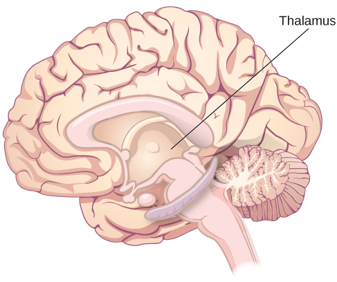
thalamus

structure containing relays and circuits connecting sensory, motor, and cortical systems.

Read explanation · OpenStax ↗
Thiol group · · · ·

A sulfur bonded to hydrogen, –SH. thiols can form through .

Read explanation · OpenStax ↗
Transcription

Synthesis of using a template.

Read explanation · OpenStax ↗
Transition state

A fleeting, high-energy arrangement at a barrier. It is an energy maximum along the path, rather than a stable .

Read explanation · OpenStax ↗
Translation ·

Synthesis of a on a using the sequence of in .

Read explanation · OpenStax ↗
Tryptophan ·

An with a bulky ; commonly grouped as .

Read explanation · OpenStax ↗
Tyrosine ·

An with a phenolic . Usually uncharged near physiological .

Read explanation · OpenStax ↗
Vesicle · · ·

A small -enclosed compartment that can transport material between cellular locations.

Read explanation · OpenStax ↗
withdrawal

Symptoms emerging when exposure to an adapted-to substance is reduced or stopped.

Read explanation · OpenStax ↗
working memory

Limited-capacity system for maintaining and manipulating information during current tasks.

Read explanation · OpenStax ↗

12.1 Basic Structure and Function of the Nervous System

OpenStax · 12.1 Basic Structure and Function of the Nervous System ↗
Instructional page including learning objectives, examples, figures, source tables and questions. Site navigation and embedded media players are omitted; source media links remain available.

Learning Objectives

By the end of this section, you will be able to:

  • Identify the anatomical and functional divisions of the nervous system
  • Relate the functional and structural differences between gray matter and white matter structures of the nervous system to the structure of
  • List the basic functions of the nervous system

The picture you have in your mind of the nervous system probably includes the brain, the nervous tissue contained within the cranium, and the spinal cord, the extension of nervous tissue within the vertebral column. That suggests it is made of two organs—and you may not even think of the spinal cord as an organ—but the nervous system is a very complex structure. Within the brain, many different and separate regions are responsible for many different and separate functions. It is as if the nervous system is composed of many organs that all look similar and can only be differentiated using tools such as the microscope or electrophysiology. In comparison, it is easy to see that the stomach is different than the esophagus or the liver, so you can imagine the digestive system as a collection of specific organs.

The Central and Peripheral Nervous Systems

The nervous system can be divided into two major regions: the central and peripheral nervous systems. The () is the brain and spinal cord, and the () is everything else (Figure 12.2). The brain is contained within the cranial cavity of the skull, and the spinal cord is contained within the vertebral cavity of the vertebral column. It is a bit of an oversimplification to say that the is what is inside these two cavities and the is outside of them, but that is one way to start to think about it. In actuality, there are some of the that are within the cranial or vertebral cavities. The is so named because it is on the periphery—meaning beyond the brain and spinal cord. Depending on different aspects of the nervous system, the dividing line between central and peripheral is not necessarily universal.

This diagram shows a silhouette of a human highlighting the nervous system. The central nervous system is composed of the brain and spinal cord. The brain is a large mass of ridged and striated tissue within the head. The spinal cord extends down from the brain and travels through the torso, ending in the pelvis. Pairs of enlarged nervous tissue, labeled ganglia, flank the spinal cord as it travels through the rib area. The ganglia are part of the peripheral nervous system, along with the many thread-like nerves that radiate from the spinal cord and ganglia through the arms, abdomen and legs.
Figure 12.2 — 12.1 Basic Structure and Function of the Nervous System

Figure 12.2 Central and Peripheral Nervous System The structures of the PNS are referred to as ganglia and nerves, which can be seen as distinct structures. The equivalent structures in the CNS are not obvious from this overall perspective and are best examined in prepared tissue under the microscope.

J. Gordon Betts and colleagues, Anatomy And Physiology 2E, OpenStax. · CC BY-NC-SA 4.0 · Original image ↗ · Access for free ↗

Nervous tissue, present in both the and , contains two basic types of cells: and glial cells. A glial cell is one of a variety of cells that provide a framework of tissue that supports the and their activities. The is the more functionally important of the two, in terms of the communicative function of the nervous system. To describe the functional divisions of the nervous system, it is important to understand the structure of a . are cells and therefore have a , or cell body, but they also have extensions of the cell; each extension is generally referred to as a process. There is one important process that every has called an , which is the fiber that connects a with its target. Another type of process that branches off from the is the . are responsible for receiving most of the input from other . Looking at nervous tissue, there are regions that predominantly contain cell bodies and regions that are largely composed of just axons. These two regions within nervous system structures are often referred to as gray matter (the regions with many cell bodies and ) or white matter (the regions with many axons). Figure 12.3 demonstrates the appearance of these regions in the brain and spinal cord. The colors ascribed to these regions are what would be seen in “fresh,” or unstained, nervous tissue. Gray matter is not necessarily gray. It can be pinkish because of blood content, or even slightly tan, depending on how long the tissue has been preserved. But white matter is white because axons are insulated by a -rich substance called . can appear as white (“fatty”) material, much like the fat on a raw piece of chicken or beef. Actually, gray matter may have that color ascribed to it because next to the white matter, it is just darker—hence, gray.

The distinction between gray matter and white matter is most often applied to central nervous tissue, which has large regions that can be seen with the unaided eye. When looking at peripheral structures, often a microscope is used and the tissue is stained with artificial colors. That is not to say that central nervous tissue cannot be stained and viewed under a microscope, but unstained tissue is most likely from the —for example, a frontal section of the brain or cross section of the spinal cord.

This photo shows an enlarged view of the dorsal side of a human brain. The right side of the occipital lobe has been shaved to reveal the white and gray matter beneath the surface blood vessels. The white matter branches though the shaved section like the limbs of a tree. The gray matter branches and curves on outside of the white matter, creating a buffer between the outer edges of the occipital lobe and the internal white matter.
Figure 12.3 — 12.1 Basic Structure and Function of the Nervous System

Figure 12.3 Gray Matter and White Matter A brain removed during an autopsy, with a partial section removed, shows white matter surrounded by gray matter. Gray matter makes up the outer cortex of the brain. (credit: modification of work by “Suseno”/Wikimedia Commons)

J. Gordon Betts and colleagues, Anatomy And Physiology 2E, OpenStax. (credit: modification of work by “Suseno”/Wikimedia Commons) · CC BY-NC-SA 4.0 · Original image ↗ · Access for free ↗

Regardless of the appearance of stained or unstained tissue, the cell bodies of or axons can be located in discrete anatomical structures that need to be named. Those names are specific to whether the structure is central or peripheral. A localized collection of cell bodies in the is referred to as a . In the , a cluster of cell bodies is referred to as a ganglion. Figure 12.4 indicates how the term has a few different meanings within anatomy and physiology. It is the center of an , where and neutrons are found; it is the center of a cell, where the is found; and it is a center of some function in the . There is also a potentially confusing use of the word ganglion (plural = ganglia) that has a historical explanation. In the , there is a group of that are connected together and were once called the before “ganglion” became accepted as a description for a peripheral structure. Some sources refer to this group of as the “basal ” to avoid confusion.

This figure shows two diagrams and a photo, labeled A, B, and C. Image A shows an atom composed of two neutrons and two protons surrounded by a hazy electron cloud. The nucleus of the atom is where the protons and neutrons are located. Image B shows a trumpet shaped cell with a large, oval nucleus near its narrow end. This is the nucleus of a cell. Image C shows an MRI capture of the brain. Two red areas near the center of the brain are highlighted in red. These are the nuclei within the brain.
Figure 12.4 — 12.1 Basic Structure and Function of the Nervous System

Figure 12.4 What Is a Nucleus? (a) The nucleus of an atom contains its protons and neutrons. (b) The nucleus of a cell is the organelle that contains DNA. (c) A nucleus in the CNS is a localized center of function with the cell bodies of several neurons, shown here circled in red. (credit c: “Was a bee”/Wikimedia Commons)

J. Gordon Betts and colleagues, Anatomy And Physiology 2E, OpenStax. (credit c: “Was a bee”/Wikimedia Commons) · CC BY-NC-SA 4.0 · Original image ↗ · Access for free ↗

Terminology applied to bundles of axons also differs depending on location. A bundle of axons, or fibers, found in the is called a tract whereas the same thing in the would be called a nerve. There is an important point to make about these terms, which is that they can both be used to refer to the same bundle of axons. When those axons are in the , the term is nerve, but if they are , the term is tract. The most obvious example of this is the axons that project from the retina into the brain. Those axons are called the optic nerve as they leave the eye, but when they are inside the cranium, they are referred to as the optic tract. There is a specific place where the name changes, which is the , but they are still the same axons (Figure 12.5). A similar situation outside of science can be described for some roads. Imagine a road called “Broad Street” in a town called “Anyville.” The road leaves Anyville and goes to the next town over, called “Hometown.” When the road crosses the line between the two towns and is in Hometown, its name changes to “Main Street.” That is the idea behind the naming of the retinal axons. In the , they are called the optic nerve, and in the , they are the optic tract. Table 12.1 helps to clarify which of these terms apply to the central or peripheral nervous systems.

This illustration shows a superior view of a cross section of the brain. The anterior side of the brain is at the top of the diagram with the two eyes clearly visible. Each eye contains a left nerve tract and a right nerve tract. In the left eye, the left nerve tract travels straight back to the right side of the thalamus. It then enters the left occipital lobe. Conversely, the right nerve tract crosses to the right side of the brain through the optic chiasma. It travels through the right side of the thalamus and enters the right occipital lobe. In the right eye, the opposite is true. The left nerve tract crosses over to the left side of the brain at the optic chiasma, traveling into the left side of the thalamus and the left side of the occipital lobe. However, the right nerve tract leads straight back to the right side of the thalamus and the right occipital lobe. Therefore, the optic chiasma is where the right nerve tract from the right eye crosses over the left nerve tract from the left eye.
Figure 12.5 — 12.1 Basic Structure and Function of the Nervous System

Figure 12.5 Optic Nerve Versus Optic Tract This drawing of the connections of the eye to the brain shows the optic nerve extending from the eye to the chiasm, where the structure continues as the optic tract. The same axons extend from the eye to the brain through these two bundles of fibers, but the chiasm represents the border between peripheral and central.

J. Gordon Betts and colleagues, Anatomy And Physiology 2E, OpenStax. · CC BY-NC-SA 4.0 · Original image ↗ · Access for free ↗

Interactive Link

In 2003, the Nobel Prize in Physiology or Medicine was awarded to Paul C. Lauterbur and Sir Peter Mansfield for discoveries related to (). This is a tool to see the structures of the body (not just the nervous system) that depends on magnetic fields associated with certain atomic . The utility of this technique in the nervous system is that fat tissue and water appear as different shades between black and white. Because white matter is fatty (from ) and gray matter is not, they can be easily distinguished in images. Try this PhET simulation that demonstrates the use of this technology and compares it with other types of imaging technologies. Also, the results from an session are compared with images obtained from X-ray or computed tomography. How do the imaging techniques shown in this game indicate the separation of white and gray matter compared with the freshly dissected tissue shown earlier?

Structures of the and
Group of Cell Bodies (i.e., gray matter) Ganglion
Bundle of Axons (i.e., white matter) Tract Nerve
Table 12.1

Functional Divisions of the Nervous System

The nervous system can also be divided on the basis of its functions, but anatomical divisions and functional divisions are different. The and the both contribute to the same functions, but those functions can be attributed to different regions of the brain (such as the cerebral cortex or the ) or to different ganglia in the periphery. The problem with trying to fit functional differences into anatomical divisions is that sometimes the same structure can be part of several functions. For example, the optic nerve carries signals from the retina that are either used for the conscious perception of visual stimuli, which takes place in the cerebral cortex, or for the reflexive responses of smooth muscle tissue that are processed through the .

There are two ways to consider how the nervous system is divided functionally. First, the basic functions of the nervous system are sensation, integration, and response. Secondly, control of the body can be or autonomic—divisions that are largely defined by the structures that are involved in the response. There is also a region of the that is called the that is responsible for a specific set of the functions within the realm of autonomic control related to gastrointestinal functions.

Basic Functions

The nervous system is involved in receiving information about the environment around us (sensation) and generating responses to that information (motor responses). The nervous system can be divided into regions that are responsible for sensation (sensory functions) and for the response (motor functions). But there is a third function that needs to be included. Sensory input needs to be integrated with other sensations, as well as with memories, emotional state, or learning (cognition). Some regions of the nervous system are termed integration or association areas. The process of integration combines sensory perceptions and higher cognitive functions such as memories, learning, and emotion to produce a response.

Sensation. The first major function of the nervous system is sensation—receiving information about the environment to gain input about what is happening outside the body (or, sometimes, within the body). The sensory functions of the nervous system register the presence of a change from or a particular event in the environment, known as a stimulus. The senses we think of most are the “”: taste, smell, touch, sight, and hearing. The stimuli for taste and smell are both chemical substances (, , , etc.), touch is physical or mechanical stimuli that interact with the skin, sight is light stimuli, and hearing is the perception of sound, which is a physical stimulus similar to some aspects of touch. There are actually more senses than just those, but that list represents the major senses. Those five are all senses that receive stimuli from the outside world, and of which there is conscious perception. Additional sensory stimuli might be from the internal environment (inside the body), such as the stretch of an organ wall or the of certain in the blood.

Response. The nervous system produces a response on the basis of the stimuli perceived by sensory structures. An obvious response would be the movement of muscles, such as withdrawing a hand from a hot stove, but there are broader uses of the term. The nervous system can cause the contraction of all three types of muscle tissue. For example, skeletal muscle contracts to move the skeleton, cardiac muscle is influenced as heart rate increases during exercise, and smooth muscle contracts as the digestive system moves food along the digestive tract. Responses also include the neural control of glands in the body as well, such as the production and of sweat by the eccrine and merocrine sweat glands found in the skin to lower body temperature.

Responses can be divided into those that are voluntary or conscious (contraction of skeletal muscle) and those that are involuntary (contraction of smooth muscles, regulation of cardiac muscle, activation of glands). Voluntary responses are governed by the and involuntary responses are governed by the , which are discussed in the next section.

Integration. Stimuli that are received by sensory structures are communicated to the nervous system where that information is processed. This is called integration. Stimuli are compared with, or integrated with, other stimuli, memories of previous stimuli, or the state of a person at a particular time. This leads to the specific response that will be generated. Seeing a baseball pitched to a batter will not automatically cause the batter to swing. The trajectory of the ball and its speed will need to be considered. Maybe the count is three balls and one strike, and the batter wants to let this pitch go by in the hope of getting a walk to first . Or maybe the batter’s team is so far ahead, it would be fun to just swing away.

Controlling the Body

The nervous system can be divided into two parts mostly on the basis of a functional difference in responses. The (SNS) is responsible for conscious perception and voluntary motor responses. Voluntary motor response means the contraction of skeletal muscle, but those contractions are not always voluntary in the sense that you have to want to perform them. Some motor responses are reflexes, and often happen without a conscious decision to perform them. If your friend jumps out from behind a corner and yells “Boo!” you will be startled and you might scream or leap back. You didn’t decide to do that, and you may not have wanted to give your friend a reason to laugh at your expense, but it is a reflex involving skeletal muscle contractions. Other motor responses become automatic (in other words, unconscious) as a person learns motor skills (referred to as “habit learning” or “”).

The () is responsible for involuntary control of the body, usually for the sake of (regulation of the internal environment). Sensory input for autonomic functions can be from sensory structures tuned to external or internal environmental stimuli. The motor output extends to smooth and cardiac muscle as well as glandular tissue. The role of the autonomic system is to regulate the organ systems of the body, which usually means to control . Sweat glands, for example, are controlled by the autonomic system. When you are hot, sweating helps cool your body down. That is a homeostatic mechanism. But when you are nervous, you might start sweating also. That is not homeostatic, it is the physiological response to an emotional state.

There is another division of the nervous system that describes functional responses. The (ENS) is responsible for controlling the smooth muscle and glandular tissue in your digestive system. It is a large part of the , and is not dependent on the . It is sometimes valid, however, to consider the enteric system to be a part of the autonomic system because the neural structures that make up the enteric system are a component of the autonomic output that regulates digestion. There are some differences between the two, but for our purposes here there will be a good bit of overlap. See Figure 12.6 for examples of where these divisions of the nervous system can be found.

This illustration shows a silhouette of a human with only the brain, spinal cord, PNS ganglia, nerves and a section of the digestive tract visible. The brain, which is part of the CNS, is the area of perception and processing of sensory stimuli (somatic/autonomic), the execution of voluntary motor responses (somatic), and the regulation of homeostatic mechanisms (autonomic). The spinal cord, which is part of the CNS, is the area where reflexes are initiated. The gray matter of the ventral horn initiates somatic reflexes while the gray matter of the lateral horn initiates autonomic reflexes. The spinal cord is also the somatic and autonomic pathway for sensory and motor functions between the PNS and the brain. The nerves, which are part of the PNS, are the fibers of sensory and motor neurons, which can be either somatic or autonomic. The ganglia, which are part of the PNS, are the areas for the reception of somatic and autonomic sensory stimuli. These are received by the dorsal root ganglia and cranial ganglia. The autonomic ganglia are also the relay for visceral motor responses. The digestive tract is part of the enteric nervous system, the ENS, which is located in the digestive tract and is responsible for autonomous function. The ENS can operate independent of the brain and spinal cord.
Figure 12.6 — 12.1 Basic Structure and Function of the Nervous System

Figure 12.6 Somatic, Autonomic, and Enteric Structures of the Nervous System Somatic structures include the spinal nerves, both motor and sensory fibers, as well as the sensory ganglia (posterior root ganglia and cranial nerve ganglia). Autonomic structures are found in the nerves also, but include the sympathetic and parasympathetic ganglia. The enteric nervous system includes the nervous tissue within the organs of the digestive tract.

J. Gordon Betts and colleagues, Anatomy And Physiology 2E, OpenStax. · CC BY-NC-SA 4.0 · Original image ↗ · Access for free ↗

Interactive Link

Visit this site to read about a woman that notices that her daughter is having trouble walking up the stairs. This leads to the discovery of a hereditary condition that affects the brain and spinal cord. The electromyography and tests indicated deficiencies in the spinal cord and , both of which are responsible for controlling coordinated movements. To what functional division of the nervous system would these structures belong?

Everyday Connection

How Much of Your Brain Do You Use?

Have you ever heard the claim that humans only use 10 percent of their brains? Maybe you have seen an advertisement on a website saying that there is a secret to unlocking the full potential of your mind—as if there were 90 percent of your brain sitting idle, just waiting for you to use it. If you see an ad like that, don’t click. It isn’t true.

An easy way to see how much of the brain a person uses is to take measurements of brain activity while performing a task. An example of this kind of measurement is (), which generates a of the most active areas and can be generated and presented in three dimensions (Figure 12.7). This procedure is different from the standard technique because it is measuring changes in the tissue in time with an experimental condition or event.

This MRI image shows a grainy computer readout of a cross section of the brain. The anterior side of the brain, located on the right hand side of the image, has a large area lighting up with yellow, indicating neural stimulation. Two smaller regions at the center of the brain are also yellow. The two small areas are in the same relative location but in opposite hemispheres of the brain.
Figure 12.7 — 12.1 Basic Structure and Function of the Nervous System

Figure 12.7 fMRI This fMRI shows activation of the visual cortex in response to visual stimuli. (credit: “Superborsuk”/Wikimedia Commons)

J. Gordon Betts and colleagues, Anatomy And Physiology 2E, OpenStax. (credit: “Superborsuk”/Wikimedia Commons) · CC BY-NC-SA 4.0 · Original image ↗ · Access for free ↗

The underlying assumption is that active nervous tissue will have greater blood flow. By having the subject perform a visual task, activity all over the brain can be measured. Consider this possible experiment: the subject is told to look at a screen with a black dot in the middle (a fixation point). A photograph of a face is projected on the screen away from the center. The subject has to look at the photograph and decipher what it is. The subject has been instructed to push a button if the photograph is of someone they recognize. The photograph might be of a celebrity, so the subject would press the button, or it might be of a random person unknown to the subject, so the subject would not press the button.

In this task, visual sensory areas would be active, integrating areas would be active, motor areas responsible for moving the eyes would be active, and motor areas for pressing the button with a finger would be active. Those areas are distributed all around the brain and the images would show activity in more than just 10 percent of the brain (some evidence suggests that about 80 percent of the brain is using energy—based on blood flow to the tissue—during well-defined tasks similar to the one suggested above). This task does not even include all of the functions the brain performs. There is no language response, the body is mostly lying still in the machine, and it does not consider the autonomic functions that would be ongoing in the background.

15.1 Divisions of the Autonomic Nervous System

OpenStax · 15.1 Divisions of the Autonomic Nervous System ↗
Instructional page including learning objectives, examples, figures, source tables and questions. Site navigation and embedded media players are omitted; source media links remain available.

Learning Objectives

By the end of this section, you will be able to:

The nervous system can be divided into two functional parts: the and the . The major differences between the two systems are evident in the responses that each produces. The causes contraction of skeletal muscles. The cardiac and smooth muscle, as well as glandular tissue. The is associated with voluntary responses (though many can happen without conscious awareness, like breathing), and the is associated with involuntary responses, such as those related to .

The regulates many of the internal organs through a balance of two aspects, or divisions. In addition to the system, the is instrumental in homeostatic mechanisms in the body. The two divisions of the are the sympathetic division and the parasympathetic division. The sympathetic system is associated with the fight-or-flight response, and parasympathetic activity is referred to by the epithet of rest and digest. is the balance between the two systems. At each target , dual innervation determines activity. For example, the heart receives connections from both the sympathetic and parasympathetic divisions. One causes heart rate to increase, whereas the other causes heart rate to decrease.

Interactive Link

Watch this video to learn more about adrenaline and the fight-or-flight response. When someone is said to have a rush of adrenaline, the image of bungee jumpers or skydivers usually comes to mind. But adrenaline, also known as epinephrine, is an important chemical in coordinating the body’s fight-or-flight response. In this video, you look inside the physiology of the fight-or-flight response, as envisioned for a firefighter. body’s is the result of the sympathetic division of the causing system-wide changes as it prepares for extreme responses. What two changes does adrenaline bring about to help the skeletal muscle response?

Sympathetic Division of the

To respond to a threat—to fight or to run away—the sympathetic system causes divergent effects as many different organs are activated together for a common purpose. More oxygen needs to be inhaled and delivered to skeletal muscle. The respiratory, cardiovascular, and musculoskeletal systems are all activated together. Additionally, sweating keeps the excess heat that comes from muscle contraction from causing the body to overheat. The digestive system shuts down so that blood is not absorbing nutrients when it should be delivering oxygen to skeletal muscles. To coordinate all these responses, the connections in the sympathetic system diverge from a limited region of the () to a wide array of ganglia that project to the many organs simultaneously. The complex set of structures that compose the output of the sympathetic system make it possible for these disparate to come together in a coordinated, systemic change.

The sympathetic division of the influences the various organ systems of the body through connections emerging from the thoracic and upper lumbar spinal cord. It is referred to as the thoracolumbar system to reflect this anatomical basis. A central in the lateral horn of any of these spinal regions projects to ganglia adjacent to the vertebral column through the ventral spinal roots. The majority of ganglia of the sympathetic system belong to a network of sympathetic chain ganglia that runs alongside the vertebral column. The ganglia appear as a series of clusters of linked by axonal bridges. There are typically 23 ganglia in the chain on either side of the spinal column. Three correspond to the cervical region, 12 are in the thoracic region, four are in the lumbar region, and four correspond to the sacral region. The cervical and sacral levels are not connected to the spinal cord directly through the spinal roots, but through ascending or descending connections through the bridges within the chain.

A diagram that shows the connections of the sympathetic system is somewhat like a circuit diagram that shows the electrical connections between different receptacles and devices. In Figure 15.2, the “circuits” of the sympathetic system are intentionally simplified.

This diagram shows the spinal cord, and the connections from the spinal cord to the different target organs. The target organs are listed on the right.
Figure 15.2 — 15.1 Divisions of the

Figure 15.2 Connections of Sympathetic Division of the Autonomic Nervous System Neurons from the lateral horn of the spinal cord (preganglionic nerve fibers - solid lines) project to the chain ganglia on either side of the vertebral column or to collateral (prevertebral) ganglia that are anterior to the vertebral column in the abdominal cavity. Axons from these ganglionic neurons (postganglionic nerve fibers - dotted lines) then project to target effectors throughout the body.

J. Gordon Betts and colleagues, Anatomy And Physiology 2E, OpenStax. · CC BY-NC-SA 4.0 · Original image ↗ · Access for free ↗

To continue with the analogy of the circuit diagram, there are three different types of “junctions” that operate within the sympathetic system (Figure 15.3). The first type is most direct: the sympathetic nerve projects to the chain ganglion at the same level as the target (the organ, tissue, or gland to be innervated). An example of this type is spinal nerve T1 that with the T1 chain ganglion to innervate the trachea. The fibers of this branch are called white rami communicantes (singular = ramus communicans); they are myelinated and therefore referred to as white (see Figure 15.3a). The from the central (the preganglionic fiber shown as a solid line) with the ganglionic (with the postganglionic fiber shown as a dashed line). This then projects to a target —in this case, the trachea—via gray rami communicantes, which are unmyelinated axons.

In some cases, the target are located superior or inferior to the spinal segment at which the preganglionic fiber emerges. With respect to the “wiring” involved, the with the ganglionic occurs at chain ganglia superior or inferior to the location of the central . An example of this is spinal nerve T1 that innervates the eye. The spinal nerve tracks up through the chain until it reaches the superior cervical ganglion, where it with the postganglionic (see Figure 15.3b). Ganglia, which are part of the sympathetic chain ganglia, are also called the paravertebral ganglia.

Not all axons from the central terminate in the chain ganglia. Additional branches from the ventral nerve root continue through the chain and on to one of the collateral ganglia as the greater splanchnic nerve or lesser splanchnic nerve. For example, the greater splanchnic nerve at the level of T5 with a collateral ganglion outside the chain before making the connection to the postganglionic nerves that innervate the stomach (see Figure 15.3c).

Collateral ganglia, also called prevertebral ganglia, are situated anterior to the vertebral column and receive inputs from splanchnic nerves as well as central sympathetic . They are associated with controlling organs in the abdominal cavity, and are also considered part of the . The three collateral ganglia are the celiac ganglion, the superior mesenteric ganglion, and the inferior mesenteric ganglion (see Figure 15.2). The word celiac is derived from the Latin word “coelom,” which refers to a body cavity (in this case, the abdominal cavity), and the word mesenteric refers to the digestive system.

This table shows the connections between the spinal cord and the ganglia. The top panel shows the connection between a central neuron and a chain ganglion at the same lever. The center panel shows the connection between a central neuron and a synapse with a superior or inferior ganglion. The bottom panel shows the projection of a central neuron into the white ramus.
Figure 15.3 — 15.1 Divisions of the

Figure 15.3 Sympathetic Connections and Chain Ganglia The axon from a central sympathetic neuron in the spinal cord can project to the periphery in a number of different ways. (a) The fiber can project out to the ganglion at the same level and synapse on a ganglionic neuron. (b) A branch can project to more superior or inferior ganglion in the chain. (c) A branch can project through the white ramus communicans, but not terminate on a ganglionic neuron in the chain. Instead, it projects through one of the splanchnic nerves to a collateral ganglion or the adrenal medulla (not pictured).

J. Gordon Betts and colleagues, Anatomy And Physiology 2E, OpenStax. · CC BY-NC-SA 4.0 · Original image ↗ · Access for free ↗

An from the central that projects to a sympathetic ganglion is referred to as a preganglionic fiber or , and represents the output from the to the ganglion. Because the sympathetic ganglia are adjacent to the vertebral column, preganglionic sympathetic fibers are relatively short, and they are myelinated. A postganglionic fiber—the from a ganglionic that projects to the target —represents the output of a ganglion that directly influences the organ. Compared with the preganglionic fibers, postganglionic sympathetic fibers are long because of the relatively greater distance from the ganglion to the target . These fibers are unmyelinated. (Note that the term “postganglionic ” may be used to describe the projection from a ganglion to the target. The problem with that usage is that the cell body is in the ganglion, and only the fiber is postganglionic. Typically, the term applies to the entire cell.)

One type of preganglionic sympathetic fiber does not terminate in a ganglion. These are the axons from central sympathetic that project to the adrenal medulla, the interior portion of the adrenal gland. These axons are still referred to as preganglionic fibers, but the target is not a ganglion. The adrenal medulla releases signaling into the bloodstream, rather than using axons to communicate with target structures. The cells in the adrenal medulla that are contacted by the preganglionic fibers are called chromaffin cells. These cells are neurosecretory cells that develop from the along with the sympathetic ganglia, reinforcing the idea that the gland is, functionally, a sympathetic ganglion.

The projections of the sympathetic division of the diverge widely, resulting in a broad influence of the system throughout the body. As a response to a threat, the sympathetic system would increase heart rate and breathing rate and cause blood flow to the skeletal muscle to increase and blood flow to the digestive system to decrease. Sweat gland should also increase as part of an integrated response. All of those physiological changes are going to be required to occur together to run away from the hunting lioness, or the modern equivalent. This divergence is seen in the branching patterns of preganglionic sympathetic —a single preganglionic sympathetic may have 10–20 targets. An that leaves a central of the lateral horn in the thoracolumbar spinal cord will pass through the white ramus communicans and enter the sympathetic chain, where it will branch toward a variety of targets. At the level of the spinal cord at which the preganglionic sympathetic fiber exits the spinal cord, a branch will on a in the adjacent chain ganglion. Some branches will extend up or down to a different level of the chain ganglia. Other branches will pass through the chain ganglia and project through one of the splanchnic nerves to a collateral ganglion. Finally, some branches may project through the splanchnic nerves to the adrenal medulla. All of these branches mean that one preganglionic can influence different regions of the sympathetic system very broadly, by acting on widely distributed organs.

Parasympathetic Division of the

The parasympathetic system can also be referred to as the craniosacral system (or outflow) because the preganglionic are located in of the brain stem and the lateral horn of the sacral spinal cord.

The connections, or “circuits,” of the parasympathetic division are similar to the general layout of the sympathetic division with a few specific differences (Figure 15.4). The preganglionic fibers from the cranial region travel in cranial nerves, whereas preganglionic fibers from the sacral region travel in spinal nerves. The targets of these fibers are terminal ganglia, which are located near—or even within—the target . These ganglia are often referred to as intramural ganglia when they are found within the walls of the target organ. The postganglionic fiber projects from the terminal ganglia a short distance to the target , or to the specific target tissue within the organ. Comparing the relative lengths of axons in the parasympathetic system, the preganglionic fibers are long and the postganglionic fibers are short because the ganglia are close to—and sometimes within—the target .

The cranial component of the parasympathetic system is based in particular of the brain stem. In the , the Edinger–Westphal is part of the oculomotor complex, and axons from those travel with the fibers in the oculomotor nerve (cranial nerve III) that innervate the extraocular muscles. The preganglionic parasympathetic fibers within cranial nerve III terminate in the ciliary ganglion, which is located in the posterior orbit. The postganglionic parasympathetic fibers then project to the smooth muscle of the to control pupillary size. In the upper medulla, the salivatory contain with axons that project through the facial and glossopharyngeal nerves to ganglia that control salivary glands. Tear production is influenced by parasympathetic fibers in the facial nerve, which activate a ganglion, and ultimately the lacrimal (tear) gland. in the dorsal of the vagus nerve and the ambiguus project through the vagus nerve (cranial nerve X) to the terminal ganglia of the thoracic and abdominal cavities. Parasympathetic preganglionic fibers primarily influence the heart, bronchi, and esophagus in the thoracic cavity and the stomach, liver, , gall bladder, and small intestine of the abdominal cavity. The postganglionic fibers from the ganglia activated by the vagus nerve are often incorporated into the structure of the organ, such as the mesenteric plexus of the digestive tract organs and the intramural ganglia.

This diagram shows the spinal cord and has different central nerves emerging from it. The central nerves target different effector organs that are listed on the right.
Figure 15.4 — 15.1 Divisions of the

Figure 15.4 Connections of Parasympathetic Division of the Autonomic Nervous System Neurons from brain-stem nuclei, or from the lateral horn of the sacral spinal cord, project to terminal ganglia near or within the various organs of the body. Axons from these ganglionic neurons then project the short distance to those target effectors.

J. Gordon Betts and colleagues, Anatomy And Physiology 2E, OpenStax. · CC BY-NC-SA 4.0 · Original image ↗ · Access for free ↗

Chemical Signaling in the

Where an autonomic connects with a target, there is a . The electrical signal of the causes the release of a signaling , which will bind to on the target cell. of the autonomic system are classified as either cholinergic, meaning that acetylcholine (ACh) is released, or adrenergic, meaning that norepinephrine is released. The terms cholinergic and adrenergic refer not only to the signaling that is released but also to the class of that each binds.

The cholinergic system includes two classes of : the nicotinic and the muscarinic . Both types bind to ACh and cause changes in the target cell. The nicotinic is a -gated channel and the muscarinic is a G –coupled . The are named for, and differentiated by, other that bind to them. Whereas nicotine will bind to the nicotinic , and muscarine will bind to the muscarinic , there is no between the . The situation is similar to locks and keys. Imagine two locks—one for a classroom and the other for an office—that are opened by two separate keys. The classroom key will not open the office door and the office key will not open the classroom door. This is similar to the specificity of nicotine and muscarine for their . However, a master key can open multiple locks, such as a master key for the Biology Department that opens both the classroom and the office doors. This is similar to ACh that binds to both types of . The that define these are not crucial—they are simply tools for researchers to use in the laboratory. These are exogenous, meaning that they are made outside of the human body, so a researcher can use them without any confounding endogenous results (results caused by the produced in the body).

The adrenergic system also has two types of , named the alpha (α)-adrenergic and beta (β)-adrenergic . Unlike cholinergic , these types are not classified by which drugs can bind to them. All of them are G –coupled . There are two types of α-adrenergic , termed α1, and α2, and there are three types of β-adrenergic , termed β1, β2 and β3. An additional aspect of the adrenergic system is that there is a second signaling called epinephrine. The chemical difference between norepinephrine and epinephrine is the addition of a (CH3) in epinephrine. The prefix “nor-” actually refers to this chemical difference, in which a is missing.

The term adrenergic should remind you of the word adrenaline, which is associated with the fight-or-flight response described at the beginning of the chapter. Adrenaline and epinephrine are two names for the same . The adrenal gland (in Latin, ad- = “on top of”; renal = “kidney”) secretes adrenaline. The ending “-ine” refers to the chemical being derived, or extracted, from the adrenal gland. A similar construction from Greek instead of Latin results in the word epinephrine (epi- = “above”; nephr- = “kidney”). In scientific usage, epinephrine is preferred in the United States, whereas adrenaline is preferred in Great Britain, because “adrenalin” was once a registered, proprietary drug name in the United States. Though the drug is no longer sold, the convention of referring to this by the two different names persists. Similarly, norepinephrine and noradrenaline are two names for the same .

Having understood the cholinergic and adrenergic systems, their role in the autonomic system is relatively simple to understand. All preganglionic fibers, both sympathetic and parasympathetic, release ACh. All ganglionic —the targets of these preganglionic fibers—have nicotinic in their . The nicotinic is a -gated channel that results in of the postsynaptic . The postganglionic parasympathetic fibers also release ACh, but the on their targets are muscarinic , which are G –coupled and do not exclusively cause of the postsynaptic . Postganglionic sympathetic fibers release norepinephrine, except for fibers that project to sweat glands and to blood vessels associated with skeletal muscles, which release ACh (Table 15.1).

Autonomic System Signaling
Sympathetic Parasympathetic
Preganglionic Acetylcholine → nicotinic Acetylcholine → nicotinic
Postganglionic Norepinephrine → α- or β-adrenergic
Acetylcholine → muscarinic (associated with sweat glands and the blood vessels associated with skeletal muscles only
Acetylcholine → muscarinic
Table 15.1

Signaling can belong to two broad groups. Neurotransmitters are released at , whereas are released into the bloodstream. These are simplistic definitions, but they can help to clarify this point. Acetylcholine can be considered a neurotransmitter because it is released by axons at . The adrenergic system, however, presents a challenge. Postganglionic sympathetic fibers release norepinephrine, which can be considered a neurotransmitter. But the adrenal medulla releases epinephrine and norepinephrine into circulation, so they should be considered .

What are referred to here as may not fit the strictest definition of . Some sources will refer to the connection between a postganglionic fiber and a target as neuroeffector junctions; neurotransmitters, as defined above, would be called neuromodulators. The structure of postganglionic connections are not the typical synaptic end bulb that is found at the , but rather are chains of swellings along the length of a postganglionic fiber called a varicosity (Figure 15.5).

This figure shows the connection between autonomic fibers and the target effectors. The left image shows a slice of smooth muscle with the postganglionic varicosities and the postganglionic axons labeled. The right panel shows a magnified view of the synaptic vesicles, neurotransmitters, and the sarcolemma.
Figure 15.5 — 15.1 Divisions of the

Figure 15.5 Autonomic Varicosities The connection between autonomic fibers and target effectors is not the same as the typical synapse, such as the neuromuscular junction. Instead of a synaptic end bulb, a neurotransmitter is released from swellings along the length of a fiber that makes an extended network of connections in the target effector.

J. Gordon Betts and colleagues, Anatomy And Physiology 2E, OpenStax. · CC BY-NC-SA 4.0 · Original image ↗ · Access for free ↗

Everyday Connection

Fight or Flight? What About Fright and Freeze?

The original usage of the epithet “fight or flight” comes from a scientist named Walter Cannon who worked at Harvard in 1915. The concept of and the functioning of the sympathetic system had been introduced in France in the previous century. Cannon expanded the idea, and introduced the idea that an animal responds to a threat by preparing to stand and fight or run away. The nature of this response was thoroughly explained in a book on the physiology of pain, hunger, fear, and rage.

When students learn about the sympathetic system and the fight-or-flight response, they often stop and wonder about other responses. If you were faced with a lioness running toward you as pictured at the beginning of this chapter, would you run or would you stand your ground? Some people would say that they would freeze and not know what to do. So isn’t there really more to what the autonomic system does than fight, flight, rest, or digest. What about fear and paralysis in the face of a threat?

The common epithet of “fight or flight” is being enlarged to be “fight, flight, or fright” or even “fight, flight, fright, or freeze.” Cannon’s original contribution was a catchy phrase to express some of what the nervous system does in response to a threat, but it is incomplete. The sympathetic system is responsible for the physiological responses to emotional states. The name “sympathetic” can be said to mean that (sym- = “together”; -pathos = “pain,” “suffering,” or “emotion”).

Interactive Link

Watch this video to learn more about the nervous system. As described in this video, the nervous system has a way to deal with threats and stress that is separate from the conscious control of the . The system comes from a time when threats were about survival, but in the modern age, these responses become part of stress and anxiety. This video describes how the autonomic system is only part of the response to threats, or stressors. What other organ system gets involved, and what part of the brain coordinates the two systems for the entire response, including epinephrine (adrenaline) and ?

14.3 Motor Responses

OpenStax · 14.3 Motor Responses ↗
Instructional page including learning objectives, examples, figures, source tables and questions. Site navigation and embedded media players are omitted; source media links remain available.

Learning Objectives

By the end of this section, you will be able to:

  • List the components of the basic processing stream for the motor system
  • Describe the pathway of descending motor commands from the cortex to the skeletal muscles
  • Compare different descending pathways, both by structure and function
  • Explain the initiation of movement from the neurological connections
  • Describe several reflex arcs and their functional roles

The defining characteristic of the is that it skeletal muscles. senses inform the nervous system about the external environment, but the response to that is through voluntary muscle movement. The term “voluntary” suggests that there is a conscious decision to make a movement. However, some aspects of the system use voluntary muscles without conscious control. One example is the ability of our breathing to switch to unconscious control while we are focused on another task. However, the muscles that are responsible for the basic process of breathing are also utilized for speech, which is entirely voluntary.

Cortical Responses

Let’s start with sensory stimuli that have been registered through cells and the information relayed to the along ascending pathways. In the cerebral cortex, the initial processing of sensory perception progresses to associative processing and then integration in multimodal areas of cortex. These levels of processing can lead to the incorporation of sensory perceptions into memory, but more importantly, they lead to a response. The completion of cortical processing through the primary, associative, and integrative sensory areas initiates a similar progression of motor processing, usually in different cortical areas.

Whereas the sensory cortical areas are located in the occipital, temporal, and parietal lobes, motor functions are largely controlled by the frontal lobe (See Figure 13.7). The most anterior regions of the frontal lobe—the prefrontal areas—are important for executive functions, which are those cognitive functions that lead to goal-directed behaviors. These higher cognitive processes include , which has been called a “mental scratch pad,” that can help organize and represent information that is not in the immediate environment. The prefrontal lobe is responsible for aspects of attention, such as inhibiting distracting thoughts and actions so that a person can focus on a goal and direct behavior toward achieving that goal.

The functions of the prefrontal cortex are integral to the personality of an individual, because it is largely responsible for what a person intends to do and how they accomplish those plans. A famous case of damage to the prefrontal cortex is that of Phineas Gage, dating back to 1848. He was a railroad worker who had a metal spike impale prefrontal cortex (Figure 14.27). He survived the accident, but according to second-hand accounts, personality changed drastically. Friends described him as no longer acting like himself. Whereas he was a hardworking, amiable man before the accident, he turned into an irritable, temperamental, and lazy man after the accident. Many of the accounts of change may have been inflated in the retelling, and some behavior was likely attributable to alcohol used as a pain medication. However, the accounts suggest that some aspects of personality did change. Also, there is new evidence that though life changed dramatically, he was able to become a functioning stagecoach driver, suggesting that the brain has the ability to recover even from major trauma such as this.

This photo shows Phineas Gage holding the metal spike that impaled his prefrontal cortex.
Figure 14.27 — 14.3 Motor Responses

Figure 14.27 Phineas Gage The victim of an accident while working on a railroad in 1848, Phineas Gage had a large iron rod impaled through the prefrontal cortex of his frontal lobe. After the accident, his personality appeared to change, but he eventually learned to cope with the trauma and lived as a coach driver even after such a traumatic event. (credit b: John M. Harlow, MD)

J. Gordon Betts and colleagues, Anatomy And Physiology 2E, OpenStax. (credit b: John M. Harlow, MD) · CC BY-NC-SA 4.0 · Original image ↗ · Access for free ↗
The image on the right shows a drawing of the skull with the metal spike inserted like it would have been when he was injured.
Figure 14.27 — 14.3 Motor Responses

Figure 14.27 Phineas Gage The victim of an accident while working on a railroad in 1848, Phineas Gage had a large iron rod impaled through the prefrontal cortex of his frontal lobe. After the accident, his personality appeared to change, but he eventually learned to cope with the trauma and lived as a coach driver even after such a traumatic event. (credit b: John M. Harlow, MD)

J. Gordon Betts and colleagues, Anatomy And Physiology 2E, OpenStax. (credit b: John M. Harlow, MD) · CC BY-NC-SA 4.0 · Original image ↗ · Access for free ↗

Secondary Motor Cortices

In generating motor responses, the executive functions of the prefrontal cortex will need to initiate actual movements. One way to define the prefrontal area is any region of the frontal lobe that does not elicit movement when electrically stimulated. These are primarily in the anterior part of the frontal lobe. The regions of the frontal lobe that remain are the regions of the cortex that produce movement. The prefrontal areas project into the secondary motor cortices, which include the premotor cortex and the supplemental motor area.

Two important regions that assist in planning and coordinating movements are located adjacent to the primary motor cortex. The premotor cortex is more lateral, whereas the supplemental motor area is more medial and superior. The premotor area aids in controlling movements of the core muscles to maintain posture during movement, whereas the supplemental motor area is hypothesized to be responsible for planning and coordinating movement. The supplemental motor area also manages sequential movements that are based on prior experience (that is, learned movements). in these areas are most active leading up to the initiation of movement. For example, these areas might prepare the body for the movements necessary to drive a car in anticipation of a traffic light changing.

Adjacent to these two regions are two specialized motor planning centers. The frontal eye fields are responsible for moving the eyes in response to visual stimuli. There are direct connections between the frontal eye fields and the superior colliculus. Also, anterior to the premotor cortex and primary motor cortex is Broca’s area. The premotor cortex is also superior to Broca's area. This area is responsible for controlling movements of the structures of speech production. The area is named after a French surgeon and anatomist who studied patients who could not produce speech. They did not have impairments to understanding speech, only to producing speech sounds, suggesting a damaged or underdeveloped Broca’s area.

Primary Motor Cortex

The primary motor cortex is located in the precentral gyrus of the frontal lobe. A neurosurgeon, Walter Penfield, described much of the basic understanding of the primary motor cortex by electrically stimulating the surface of the cerebrum. Penfield would the surface of the cortex while the patient was only under local anesthesia so that he could observe responses to the stimulation. This led to the belief that the precentral gyrus directly stimulated muscle movement. We now know that the primary motor cortex receives input from several areas that aid in planning movement, and its principal output stimulates spinal cord to stimulate skeletal muscle contraction.

The primary motor cortex is arranged in a similar fashion to the primary somatosensory cortex, in that it has a topographical of the body, creating a motor homunculus (see Figure 14.23). The responsible for musculature in the feet and lower legs are in the medial wall of the precentral gyrus, with the thighs, trunk, and shoulder at the crest of the longitudinal fissure. The hand and face are in the lateral face of the gyrus. Also, the relative space allotted for the different regions is exaggerated in muscles that have greater innervation. The greatest amount of cortical space is given to muscles that perform fine, agile movements, such as the muscles of the fingers and the lower face. The “power muscles” that perform coarser movements, such as the buttock and back muscles, occupy much less space on the motor cortex.

Descending Pathways

The motor output from the cortex descends into the brain stem and to the spinal cord to control the musculature through motor . located in the primary motor cortex, named Betz cells, are large cortical that with lower motor in the brain stem or in the spinal cord. The two descending pathways travelled by the axons of Betz cells are the corticobulbar tract and the corticospinal tract, respectively. Both tracts are named for their origin in the cortex and their targets—either the brain stem (the term “bulbar” refers to the brain stem as the bulb, or enlargement, at the top of the spinal cord) or the spinal cord.

These two descending pathways are responsible for the conscious or voluntary movements of skeletal muscles. Any motor command from the primary motor cortex is sent down the axons of the Betz cells to activate lower motor in either the cranial motor or in the ventral horn of the spinal cord. The axons of the corticobulbar tract are ipsilateral, meaning they project from the cortex to the motor on the same side of the nervous system. Conversely, the axons of the corticospinal tract are largely contralateral, meaning that they cross the midline of the brain stem or spinal cord and on the opposite side of the body. Therefore, the right motor cortex of the cerebrum muscles on the left side of the body, and vice versa.

The corticospinal tract descends from the cortex through the deep white matter of the cerebrum. It then passes between the caudate and putamen of the basal as a bundle called the internal capsule. The tract then passes through the as the cerebral peduncles, after which it burrows through the pons. Upon entering the medulla, the tracts make up the large white matter tract referred to as the pyramids (Figure 14.28). The defining landmark of the medullary-spinal border is the pyramidal decussation, which is where most of the fibers in the corticospinal tract cross over to the opposite side of the brain. At this point, the tract separates into two parts, which have control over different of the musculature.

This diagram shows how the motor neurons thread their way through the spinal cord and into the brain. It also shows the different connections they make along the way.
Figure 14.28 — 14.3 Motor Responses

Figure 14.28 Corticospinal Tract The major descending tract that controls skeletal muscle movements is the corticospinal tract. It is composed of two neurons, the upper motor neuron and the lower motor neuron. The upper motor neuron has its cell body in the primary motor cortex of the frontal lobe and synapses on the lower motor neuron, which is in the ventral horn of the spinal cord and projects to the skeletal muscle in the periphery.

J. Gordon Betts and colleagues, Anatomy And Physiology 2E, OpenStax. · CC BY-NC-SA 4.0 · Original image ↗ · Access for free ↗

Appendicular Control

The lateral corticospinal tract is composed of the fibers that cross the midline at the pyramidal decussation (see Figure 14.28). The axons cross over from the anterior position of the pyramids in the medulla to the lateral column of the spinal cord. These axons are responsible for controlling appendicular muscles.

This influence over the appendicular muscles means that the lateral corticospinal tract is responsible for moving the muscles of the arms and legs. The ventral horn in both the lower cervical spinal cord and the lumbar spinal cord both have wider ventral horns, representing the greater number of muscles controlled by these motor . The cervical enlargement is particularly large because there is greater control over the fine musculature of the upper limbs, particularly of the fingers. The lumbar enlargement is not as significant in appearance because there is less fine motor control of the lower limbs.

Axial Control

The anterior corticospinal tract is responsible for controlling the muscles of the body trunk (see Figure 14.28). These axons do not decussate in the medulla. Instead, they remain in an anterior position as they descend the brain stem and enter the spinal cord. These axons then travel to the spinal cord level at which they with a lower motor . Upon reaching the appropriate level, the axons decussate, entering the ventral horn on the opposite side of the spinal cord from which they entered. In the ventral horn, these axons with their corresponding lower motor . The lower motor are located in the medial regions of the ventral horn, because they control the axial muscles of the trunk.

Because movements of the body trunk involve both sides of the body, the anterior corticospinal tract is not entirely contralateral. Some collateral branches of the tract will project into the ipsilateral ventral horn to control synergistic muscles on that side of the body, or to inhibit antagonistic muscles through interneurons within the ventral horn. Through the influence of both sides of the body, the anterior corticospinal tract can coordinate postural muscles in broad movements of the body. These coordinating axons in the anterior corticospinal tract are often considered bilateral, as they are both ipsilateral and contralateral.

Interactive Link

Watch this video to learn more about the descending motor pathway for the . The autonomic connections are mentioned, which are covered in another chapter. From this brief video, only some of the descending motor pathway of the is described. Which division of the pathway is described and which division is left out?

Extrapyramidal

Other descending connections between the brain and the spinal cord are called the extrapyramidal system. The name comes from the that this system is outside the corticospinal pathway, which includes the pyramids in the medulla. A few pathways originating from the brain stem contribute to this system.

The tectospinal tract projects from the to the spinal cord and is important for postural movements that are driven by the superior colliculus. The name of the tract comes from an alternate name for the superior colliculus, which is the tectum. The reticulospinal tract connects the reticular system, a diffuse region of gray matter in the brain stem, with the spinal cord. This tract influences trunk and proximal limb muscles related to posture and locomotion. The reticulospinal tract also contributes to muscle tone and influences autonomic functions. The vestibulospinal tract connects the brain stem of the vestibular system with the spinal cord. This allows posture, movement, and balance to be modulated on the basis of information provided by the vestibular system.

The pathways of the extrapyramidal system are influenced by subcortical structures. For example, connections between the secondary motor cortices and the extrapyramidal system modulate spine and cranium movements. The basal , which are important for regulating movement initiated by the , influence the extrapyramidal system as well as its thalamic feedback to the motor cortex.

The conscious movement of our muscles is more complicated than simply sending a single command from the precentral gyrus down to the proper motor . During the movement of any body part, our muscles relay information back to the brain, and the brain is constantly sending “revised” instructions back to the muscles. The is important in contributing to the motor system because it compares cerebral motor commands with proprioceptive feedback. The corticospinal fibers that project to the ventral horn of the spinal cord have branches that also in the pons, which project to the . Also, the proprioceptive sensations of the dorsal column system have a collateral projection to the medulla that projects to the . These two streams of information are compared in the cerebellar cortex. Conflicts between the motor commands sent by the cerebrum and body position information provided by the proprioceptors cause the to stimulate the red of the . The red then sends corrective commands to the spinal cord along the rubrospinal tract. The name of this tract comes from the word for red that is seen in the English word “ruby.”

A good example of how the corrects cerebral motor commands can be illustrated by walking in water. An original motor command from the cerebrum to walk will result in a highly coordinated set of learned movements. However, in water, the body cannot actually perform a typical walking movement as instructed. The can alter the motor command, stimulating the leg muscles to take larger steps to overcome the water resistance. The can make the necessary changes through the rubrospinal tract. Modulating the basic command to walk also relies on spinal reflexes, but the is responsible for calculating the appropriate response. When the does not work properly, coordination and balance are severely affected. The most dramatic example of this is during the overconsumption of alcohol. Alcohol inhibits the ability of the to interpret proprioceptive feedback, making it more difficult to coordinate body movements, such as walking a straight line, or guide the movement of the hand to touch the tip of the nose.

Interactive Link

Visit this site to read about an elderly woman who starts to lose the ability to control fine movements, such as speech and the movement of limbs. Many of the usual causes were ruled out. It was not a stroke, Parkinson’s disease, diabetes, or thyroid dysfunction. The next most obvious cause was medication, so her pharmacist had to be consulted. The side effect of a drug meant to help her sleep had resulted in changes in motor control. What regions of the nervous system are likely to be the focus of haloperidol side effects?

Ventral Horn Output

The provides output strictly to skeletal muscles. The lower motor , which are responsible for the contraction of these muscles, are found in the ventral horn of the spinal cord. These large, multipolar have a corona of surrounding the cell body and an that extends out of the ventral horn. This travels through the ventral nerve root to join the emerging spinal nerve. The is relatively long because it needs to reach muscles in the periphery of the body. The diameters of cell bodies may be on the order of hundreds of micrometers to support the long ; some axons are a meter in length, such as the lumbar motor that innervate muscles in the first digits of the feet.

The axons will also branch to innervate multiple muscle fibers. Together, the motor and all the muscle fibers that it make up a motor unit. Motor units vary in size. Some may contain up to 1000 muscle fibers, such as in the quadriceps, or they may only have 10 fibers, such as in an extraocular muscle. The number of muscle fibers that are part of a motor unit corresponds to the precision of control of that muscle. Also, muscles that have finer motor control have more motor units connecting to them, and this requires a larger topographical field in the primary motor cortex.

Motor axons connect to muscle fibers at a . This is a specialized synaptic structure at which multiple terminals with the muscle fiber sarcolemma. The synaptic end bulbs of the motor secrete acetylcholine, which binds to on the sarcolemma. The binding of acetylcholine opens -gated channels, increasing the movement of across the sarcolemma. This depolarizes the sarcolemma, initiating muscle contraction. Whereas other result in graded potentials that must reach a threshold in the postsynaptic target, activity at the reliably leads to muscle fiber contraction with every nerve impulse received from a motor . However, the strength of contraction and the number of fibers that contract can be affected by the frequency of the motor impulses.

Reflexes

This chapter began by introducing reflexes as an example of the basic of the . Simple reflexes do not include the higher centers discussed for conscious or voluntary aspects of movement. Reflexes can be spinal or cranial, depending on the nerves and central components that are involved. The example described at the beginning of the chapter involved heat and pain sensations from a hot stove causing of the arm through a connection in the spinal cord that leads to contraction of the biceps brachii. The description of this reflex was simplified, for the sake of the introduction, to emphasize the parts of the . But to consider reflexes fully, more attention needs to be given to this example.

As you withdraw your hand from the stove, you do not want to slow that reflex down. As the biceps brachii contracts, the antagonistic triceps brachii needs to relax. Because the is strictly excitatory, the biceps will contract when the motor nerve is active. Skeletal muscles do not actively relax. Instead the motor needs to “quiet down,” or be inhibited. In the hot-stove reflex, this occurs through an interneuron in the spinal cord. The interneuron’s cell body is located in the dorsal horn of the spinal cord. The interneuron receives a from the of the sensory that detects that the hand is being burned. In response to this stimulation from the sensory , the interneuron then inhibits the motor that the triceps brachii. This is done by releasing a neurotransmitter or other signal that hyperpolarizes the motor connected to the triceps brachii, making it less likely to initiate an . With this motor being inhibited, the triceps brachii relaxes. Without the antagonistic contraction, from the hot stove is faster and keeps further tissue damage from occurring.

Another example of a reflex occurs when you step on a painful stimulus, like a tack or a sharp rock. The nociceptors that are activated by the painful stimulus activate the motor responsible for contraction of the tibialis anterior muscle. This causes dorsiflexion of the foot. An inhibitory interneuron, activated by a collateral branch of the nociceptor fiber, will inhibit the motor of the gastrocnemius and soleus muscles to cancel plantar flexion. An important difference in this reflex is that plantar flexion is most likely in progress as the foot is pressing down onto the tack. Contraction of the tibialis anterior is not the most important aspect of the reflex, as continuation of plantar flexion will result in further damage from stepping onto the tack.

Another type of reflex is a stretch reflex. In this reflex, when a skeletal muscle is stretched, a is activated. The from this structure will cause direct contraction of the muscle. A collateral of the fiber will also inhibit the motor of the antagonist muscles. The reflex helps to maintain muscles at a constant length. A common example of this reflex is the knee jerk that is elicited by a rubber hammer struck against the patellar in a physical exam.

A specialized reflex to protect the surface of the eye is the corneal reflex, or the eye blink reflex. When the is stimulated by a tactile stimulus, or even by bright light in a related reflex, blinking is initiated. The sensory component travels through the trigeminal nerve, which carries somatosensory information from the face, or through the optic nerve, if the stimulus is bright light. The motor response travels through the facial nerve and innervates the orbicularis oculi bilaterally. This reflex is commonly tested during a physical exam using an air puff or a gentle touch of a cotton-tipped applicator.

Interactive Link

Watch this video to learn more about the of the corneal reflex. When the right senses a tactile stimulus, what happens to the left eye? Explain your answer.

Interactive Link

Watch this video to learn more about newborn reflexes. Newborns have a set of reflexes that are expected to have been crucial to survival before the modern age. These reflexes disappear as the baby grows, as some of them may be unnecessary as they age. The video demonstrates a reflex called the Babinski reflex, in which the foot flexes dorsally and the toes splay out when the sole of the foot is lightly scratched. This is normal for newborns, but it is a sign of reduced myelination of the spinal tract in adults. Why would this reflex be a problem for an adult?

1.5 Homeostasis
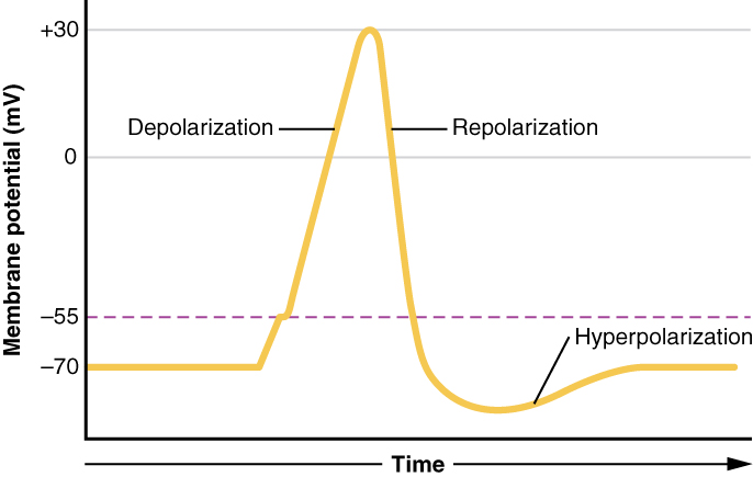
OpenStax · 1.5 Homeostasis ↗

Instructional page including learning objectives, examples, figures, source tables and questions. Site navigation and embedded media players are omitted; source media links remain available.

Learning Objectives

By the end of this section, you will be able to:

  • Discuss the role of in healthy functioning
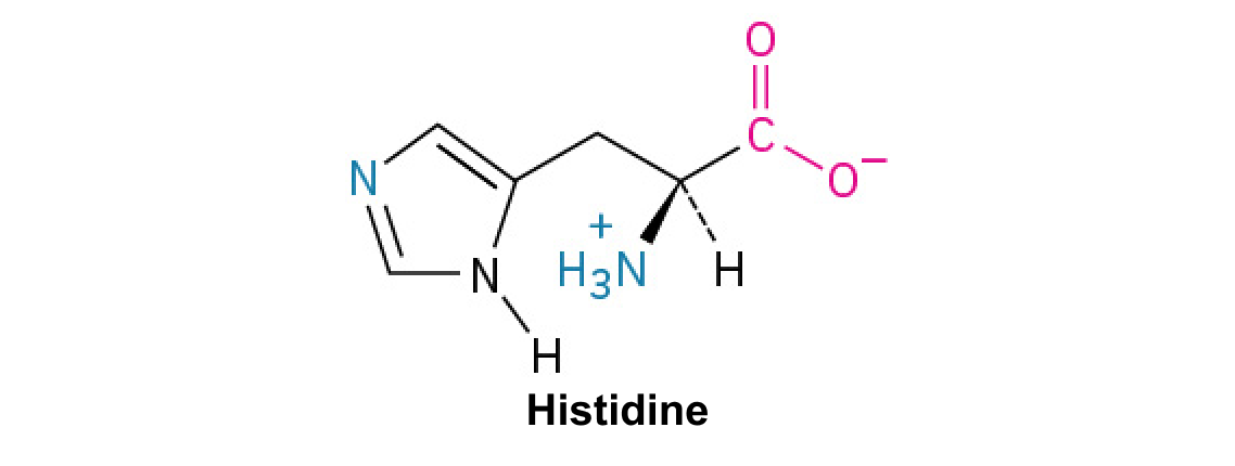
  • Contrast negative and , giving one physiologic example of each mechanism

Maintaining requires that the body continuously monitor its internal conditions. From body temperature to blood pressure to levels of certain nutrients, each physiological condition has a particular . A is the physiological value around which the normal range fluctuates. A normal range is the restricted set of values that is optimally healthful and stable. For example, the for normal human body temperature is approximately 37°C (98.6°F) Physiological parameters, such as body temperature and blood pressure, tend to fluctuate within a normal range a few degrees above and below that point. Control centers in the brain and other parts of the body monitor and react to deviations from using . is a mechanism that reverses a deviation from the . Therefore, maintains body parameters within their normal range. The maintenance of by goes on throughout the body at all times, and an understanding of is thus fundamental to an understanding of human physiology.

A system has three basic components (Figure 1.10a). A sensor, also referred to a , is a component of a feedback system that monitors a physiological value. This value is reported to the control center. The control center is the component in a feedback system that compares the value to the normal range. If the value deviates too much from the , then the control center activates an . An is the component in a feedback system that causes a change to reverse the situation and return the value to the normal range.

This figure shows two flow charts labeled A and B. Chart A shows a general negative feedback loop. The loop starts with a stimulus. Information about the stimulus is perceived by a sensor which sends that information to a control center. The control center sends a signal to an effector, which creates a response. That then feeds back to the top of the flow chart by inhibiting the stimulus. Part B shows body temperature regulation as an example of negative feedback system. Here, the stimulus is body temperature exceeding 37 degrees Celsius. The sensor is a set of nerve cells in the skin and brain and the control center is the temperature regulatory center of the brain. The effectors are sweat glands throughout the body which lead to increased heat loss and inhibit the rising body temperature.
Figure 1.10 — 1.5

Figure 1.10 Negative Feedback System In a negative feedback system, a stimulus—a deviation from a set point—is resisted through a physiological process that returns the body to homeostasis. (a) A negative feedback system has five basic steps. (b) Body temperature is regulated by negative feedback.

J. Gordon Betts and colleagues, Anatomy And Physiology 2E, OpenStax. · CC BY-NC-SA 4.0 · Original image ↗ · Access for free ↗

In order to set the system in motion, a stimulus must drive a physiological parameter beyond its normal range (that is, beyond ). This stimulus is “heard” by a specific sensor. For example, in the control of , specific in the detect excess (the stimulus) in the bloodstream. These respond to the increased level of by releasing the into the bloodstream. The signals skeletal muscle fibers, fat cells (adipocytes), and liver cells to take up the excess , removing it from the bloodstream. As in the bloodstream drops, the decrease in —the actual —is detected by pancreatic alpha cells, and release stops. This prevents blood sugar levels from continuing to drop below the normal range.

Humans have a similar temperature regulation feedback system that works by promoting either heat loss or heat gain (Figure 1.10b). When the brain’s temperature regulation center receives data from the sensors indicating that the body’s temperature exceeds its normal range, it stimulates a cluster of brain cells referred to as the “heat-loss center.” This stimulation has three major effects:

  • Blood vessels in the skin begin to dilate allowing more blood from the body core to flow to the surface of the skin allowing the heat to radiate into the environment.
  • As blood flow to the skin increases, sweat glands are activated to increase their output. As the sweat evaporates from the skin surface into the surrounding air, it takes heat with it.
  • The depth of respiration increases, and a person may breathe through an open mouth instead of through the nasal passageways. This further increases heat loss from the lungs.

In contrast, activation of the brain’s heat-gain center by exposure to cold reduces blood flow to the skin, and blood returning from the limbs is diverted into a network of deep veins. This arrangement traps heat closer to the body core and restricts heat loss. If heat loss is severe, the brain triggers an increase in random signals to skeletal muscles, causing them to contract and producing shivering. The muscle contractions of shivering release heat while using up . The brain triggers the thyroid gland in the system to release thyroid , which increases activity and heat production in cells throughout the body. The brain also signals the adrenal glands to release epinephrine (adrenaline), a that causes the breakdown of into , which can be used as an energy source. The breakdown of into also results in increased and heat production.

Interactive Link

Water in the body is critical for proper functioning. A person’s body retains very tight control on water levels without conscious control by the person. Watch this video to learn more about water in the body. Which organ has primary control over the amount of water in the body?

intensifies a change in the body’s physiological condition rather than reversing it. A deviation from the normal range results in more change, and the system moves farther away from the normal range. in the body is normal only when there is a definite end point. Childbirth and the body’s response to blood loss are two examples of loops that are normal but are activated only when needed.

Childbirth at full term is an example of a situation in which the maintenance of the existing body state is not desired. Enormous changes in a person’s body are required to expel the baby at the end of pregnancy. And the events of childbirth, once begun, must progress rapidly to a conclusion or the life of a person giving birth and the baby are at risk. The extreme muscular work of labor and delivery are the result of a system (Figure 1.11).

This diagram shows the steps of a positive feedback loop as a series of stepwise arrows looping around a diagram of an infant within the uterus of a pregnant woman. Initially the head of the baby pushes against the cervix, transmitting nerve impulses from the cervix to the brain. Next the brain stimulates the pituitary gland to secrete oxytocin which is carried in the bloodstream to the uterus. Finally, the oxytocin simulates uterine contractions and pushes the baby harder into the cervix. As the head of the baby pushes against the cervix with greater and greater force, the uterine contractions grow stronger and more frequent. This mechanism is a positive feedback loop.
Figure 1.11 — 1.5

Figure 1.11 Positive Feedback Loop Normal childbirth is driven by a positive feedback loop. A positive feedback loop results in a change in the body’s status, rather than a return to homeostasis.

J. Gordon Betts and colleagues, Anatomy And Physiology 2E, OpenStax. · CC BY-NC-SA 4.0 · Original image ↗ · Access for free ↗

The first contractions of labor (the stimulus) push the baby toward the cervix (the lowest part of the uterus). The cervix contains stretch-sensitive nerve cells that monitor the degree of stretching (the sensors). These nerve cells send messages to the brain, which in turn causes the pituitary gland at the of the brain to release the into the bloodstream. causes stronger contractions of the smooth muscles in the uterus (the ), pushing the baby further down the birth canal. This causes even greater stretching of the cervix. The cycle of stretching, release, and increasingly more forceful contractions stops only when the baby is born. At this point, the stretching of the cervix halts, stopping the release of .

A second example of centers on reversing extreme damage to the body. Following a penetrating wound, the most immediate threat is excessive blood loss. Less blood circulating means reduced blood pressure and reduced (penetration of blood) to the brain and other vital organs. If is severely reduced, vital organs will shut down and the person will die. The body responds to this potential catastrophe by releasing substances in the injured blood vessel wall that begin the process of blood clotting. As each step of clotting occurs, it stimulates the release of more clotting substances. This accelerates the processes of clotting and sealing off the damaged area. Clotting is contained in a local area based on the tightly controlled availability of clotting . This is an adaptive, life-saving cascade of events.