Ion gradients store potential energy; selective channels convert it into membrane signals.
Cell compartments and glial support
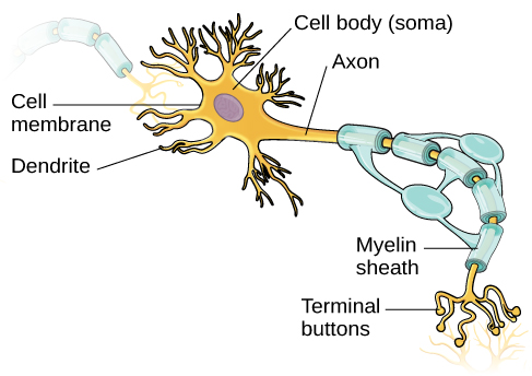
Dendrites receive many inputs; the soma integrates them and maintains cellular machinery; the axon initial segment commonly initiates action potentials; terminals release transmitter. Schwann cells myelinate peripheral axon segments, whereas oligodendrocytes myelinate CNS segments. Astrocytes support extracellular ion balance, metabolism, and blood–brain barrier function. Microglia provide immune surveillance; ependymal cells line fluid spaces and participate in cerebrospinal-fluid functions. Myelin reduces current leakage and membrane capacitance, allowing efficient spread between nodes of Ranvier.
Source: 12.2 Nervous Tissue · 12.4 The Action Potential · 12.5 Communication Between Neurons
Resting potential and the Nernst relationship
Ion pumps maintain concentration differences; the Na⁺/K⁺ ATPase exports three Na⁺ and imports two K⁺ per ATP. Resting permeability is often dominated by K⁺ leak channels, placing the membrane near the K⁺ equilibrium potential. For one ion, Eion = (RT/zF) ln([ion]out/[ion]in); at 37°C this is approximately (61.5 mV/z) log₁₀(out/in). Real resting potential reflects several permeant ions, so it is not exactly one Nernst potential. Opening a channel drives voltage toward that ion’s reversal potential.
Source: 12.2 Nervous Tissue · 12.4 The Action Potential · 12.5 Communication Between Neurons
Action potentials and propagation
Depolarization to threshold triggers regenerative opening of voltage-gated Na⁺ channels in a typical axon. Na⁺ channel inactivation and delayed K⁺ conductance repolarize the membrane; continued K⁺ efflux can produce an afterhyperpolarization. The absolute refractory period reflects unavailable Na⁺ channels; the relative period requires a stronger stimulus. Local current brings neighboring membrane to threshold. Myelinated axons regenerate spikes at nodes, producing saltatory conduction. Individual spikes are approximately all-or-none; stimulus intensity is encoded by firing frequency and recruitment, not unlimited spike height.
Source: 12.2 Nervous Tissue · 12.4 The Action Potential · 12.5 Communication Between Neurons
Release, receptors, and summation
An arriving spike opens presynaptic voltage-gated Ca²⁺ channels; Ca²⁺ promotes vesicle fusion and neurotransmitter release. Postsynaptic receptors can be ion channels or signaling receptors. Excitatory and inhibitory effects depend on reversal potentials and conductances, not the transmitter name alone. Spatial summation combines different inputs; temporal summation combines closely spaced inputs. Inhibition can hyperpolarize or shunt depolarizing current. Reuptake, enzymatic breakdown, and diffusion terminate signals. Stronger sustained net excitation usually increases spike frequency within refractory limits.
Source: 12.2 Nervous Tissue · 12.4 The Action Potential · 12.5 Communication Between Neurons
Practice and reasoning
Question. For K⁺ at 37°C, [K⁺]out = 5 mM and [K⁺]in = 150 mM. Estimate EK. What does raising extracellular K⁺ generally do initially?
Show reasoning
EK ≈ 61.5 log₁₀(5/150) ≈ −91 mV. Raising extracellular K⁺ makes the ratio larger and EK less negative, tending to depolarize a K⁺-permeable membrane. Sustained strong depolarization can inactivate Na⁺ channels, so increased extracellular K⁺ does not guarantee persistently increased firing.
Source: 12.2 Nervous Tissue · 12.4 The Action Potential · 12.5 Communication Between Neurons
12.2 Nervous Tissue — figures and tables
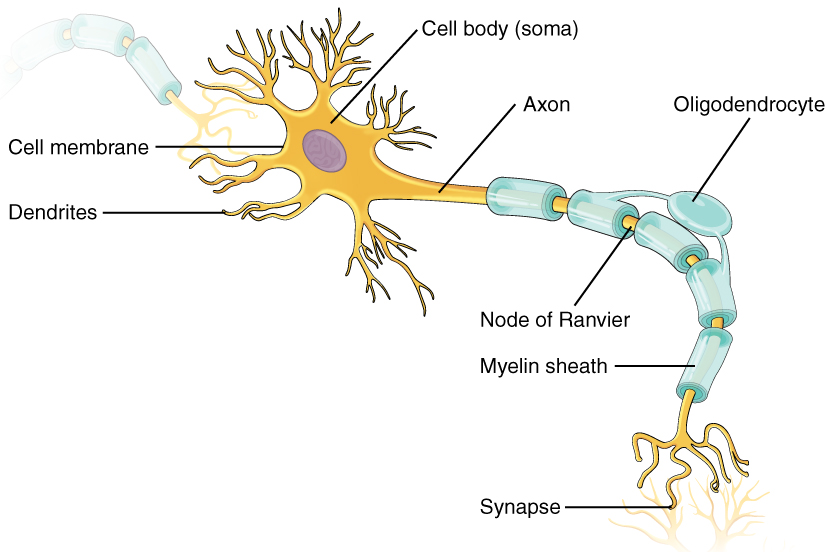
Figure 12.8 Parts of a Neuron The major parts of the neuron are labeled on a multipolar neuron from the CNS.
J. Gordon Betts and colleagues, Anatomy And Physiology 2E, OpenStax. · CC BY-NC-SA 4.0 · Original image ↗ · Access for free ↗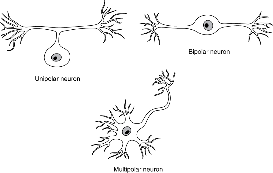
Figure 12.9 Neuron Classification by Shape Unipolar cells have one process that includes both the axon and dendrite. Bipolar cells have two processes, the axon and a dendrite. Multipolar cells have more than two processes, the axon and two or more dendrites.
J. Gordon Betts and colleagues, Anatomy And Physiology 2E, OpenStax. · CC BY-NC-SA 4.0 · Original image ↗ · Access for free ↗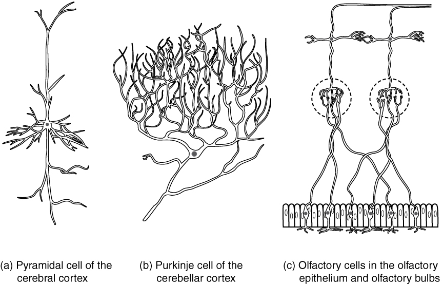
Figure 12.10 Other Neuron Classifications Three examples of neurons that are classified on the basis of other criteria. (a) The pyramidal cell is a multipolar cell with a cell body that is shaped something like a pyramid. (b) The Purkinje cell in the cerebellum was named after the scientist who originally described it. (c) Olfactory neurons are named for the functional group with which they belong.
J. Gordon Betts and colleagues, Anatomy And Physiology 2E, OpenStax. · CC BY-NC-SA 4.0 · Original image ↗ · Access for free ↗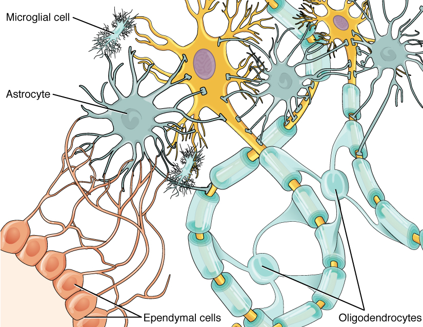
Figure 12.11 Glial Cells of the CNS The CNS has astrocytes, oligodendrocytes, microglia, and ependymal cells that support the neurons of the CNS in several ways.
J. Gordon Betts and colleagues, Anatomy And Physiology 2E, OpenStax. · CC BY-NC-SA 4.0 · Original image ↗ · Access for free ↗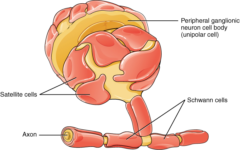
Figure 12.12 Glial Cells of the PNS The PNS has satellite cells and Schwann cells.
J. Gordon Betts and colleagues, Anatomy And Physiology 2E, OpenStax. · CC BY-NC-SA 4.0 · Original image ↗ · Access for free ↗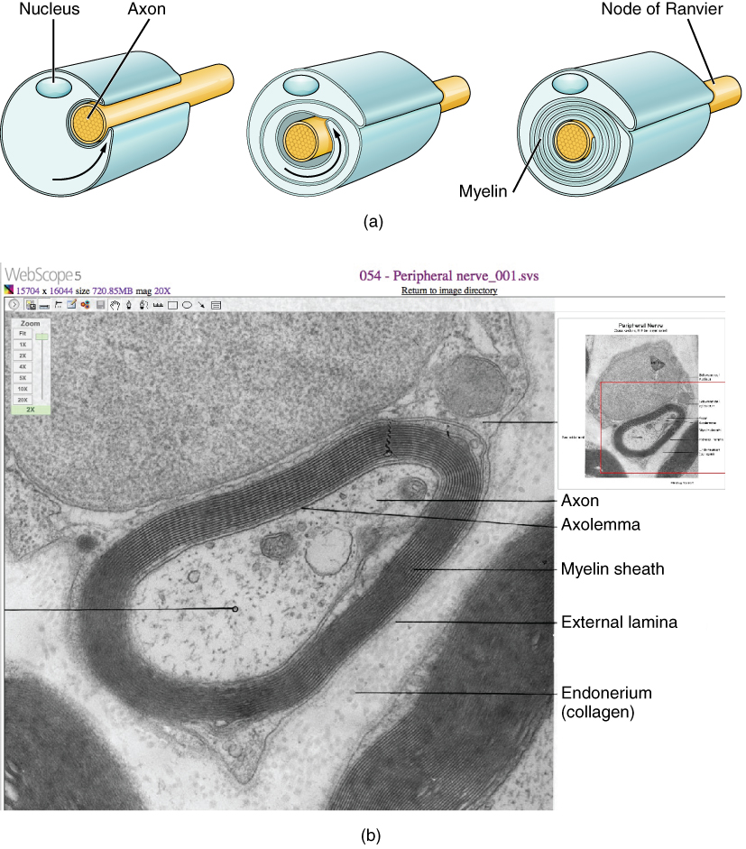
Figure 12.13 The Process of Myelination Myelinating glia wrap several layers of cell membrane around the cell membrane of an axon segment. A single Schwann cell insulates a segment of a peripheral nerve, whereas in the CNS, an oligodendrocyte may provide insulation for a few separate axon segments. EM × 1,460,000. (Micrograph provided by the Regents of University of Michigan Medical School © 2012)
J. Gordon Betts and colleagues, Anatomy And Physiology 2E, OpenStax. · CC BY-NC-SA 4.0 · Original image ↗ · Access for free ↗| CNS glia | PNS glia | Basic function |
|---|---|---|
| Astrocyte | Satellite cell | Support |
| Oligodendrocyte | Schwann cell | Insulation, myelination |
| Microglia | - | Immune surveillance and phagocytosis |
| Ependymal cell | - | Creating CSF |
Table 12.2. J. Gordon Betts and colleagues, Anatomy And Physiology 2E, OpenStax.
Source: 12.2 Nervous Tissue
12.4 The Action Potential — figures and tables
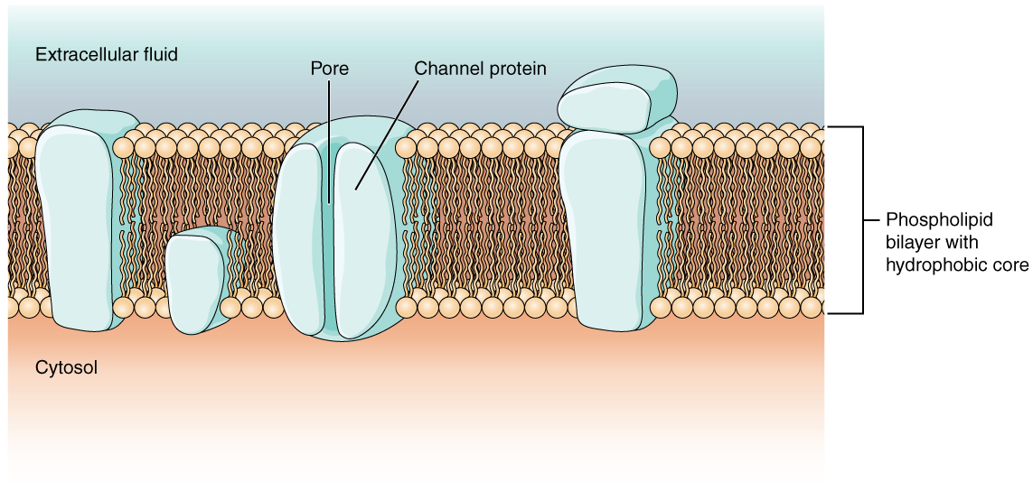
Figure 12.17 Cell Membrane and Transmembrane Proteins The cell membrane is composed of a phospholipid bilayer and has many transmembrane proteins, including different types of channel proteins that serve as ion channels.
J. Gordon Betts and colleagues, Anatomy And Physiology 2E, OpenStax. · CC BY-NC-SA 4.0 · Original image ↗ · Access for free ↗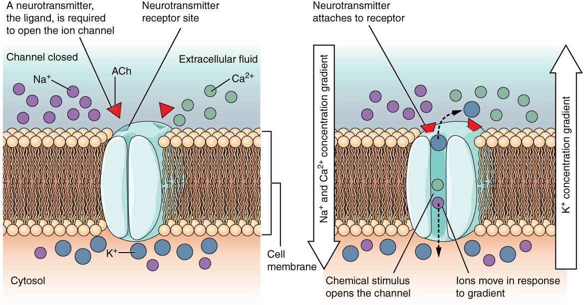
Figure 12.18 Ligand-Gated Channels When the ligand, in this case the neurotransmitter acetylcholine, binds to a specific location on the extracellular surface of the channel protein, the pore opens to allow select ions through. The ions, in this case, are cations of sodium, calcium, and potassium.
J. Gordon Betts and colleagues, Anatomy And Physiology 2E, OpenStax. · CC BY-NC-SA 4.0 · Original image ↗ · Access for free ↗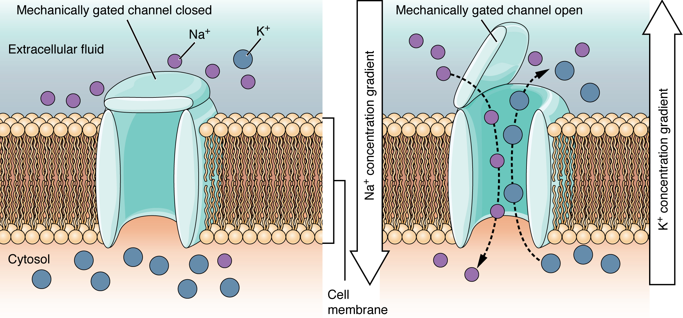
Figure 12.19 Mechanically Gated Channels When a mechanical change occurs in the surrounding tissue, such as pressure or touch, the channel is physically opened. Thermoreceptors work on a similar principle. When the local tissue temperature changes, the protein reacts by physically opening the channel.
J. Gordon Betts and colleagues, Anatomy And Physiology 2E, OpenStax. · CC BY-NC-SA 4.0 · Original image ↗ · Access for free ↗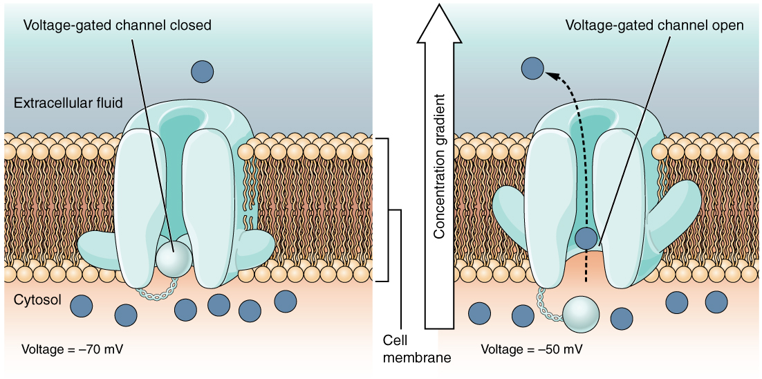
Figure 12.20 Voltage-Gated Channels Voltage-gated channels open when the transmembrane voltage changes around them. Amino acids in the structure of the protein are sensitive to charge and cause the pore to open to the selected ion.
J. Gordon Betts and colleagues, Anatomy And Physiology 2E, OpenStax. · CC BY-NC-SA 4.0 · Original image ↗ · Access for free ↗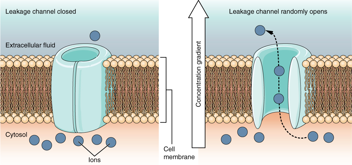
Figure 12.21 Leakage Channels In certain situations, ions need to move across the membrane randomly. The particular electrical properties of certain cells are modified by the presence of this type of channel.
J. Gordon Betts and colleagues, Anatomy And Physiology 2E, OpenStax. · CC BY-NC-SA 4.0 · Original image ↗ · Access for free ↗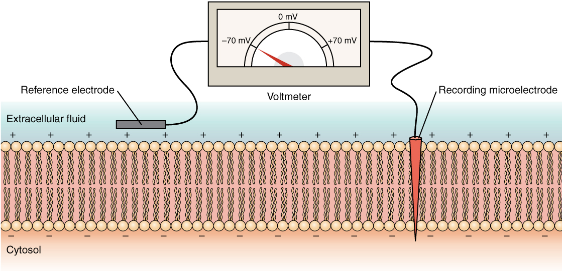
Figure 12.22 Measuring Charge across a Membrane with a Voltmeter A recording electrode is inserted into the cell and a reference electrode is outside the cell. By comparing the charge measured by these two electrodes, the transmembrane voltage is determined. It is conventional to express that value for the cytosol relative to the outside.
J. Gordon Betts and colleagues, Anatomy And Physiology 2E, OpenStax. · CC BY-NC-SA 4.0 · Original image ↗ · Access for free ↗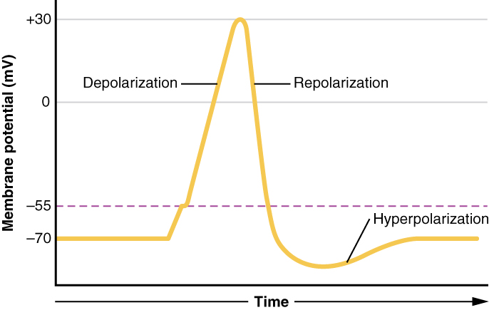
Figure 12.23 Graph of Action Potential Plotting voltage measured across the cell membrane against time, the action potential begins with depolarization, followed by repolarization, which goes past the resting potential into hyperpolarization, and finally the membrane returns to rest.
J. Gordon Betts and colleagues, Anatomy And Physiology 2E, OpenStax. · CC BY-NC-SA 4.0 · Original image ↗ · Access for free ↗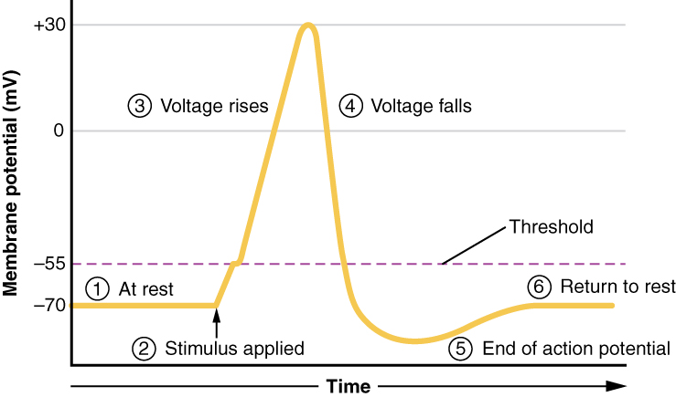
Figure 12.24 Stages of an Action Potential Plotting voltage measured across the cell membrane against time, the events of the action potential can be related to specific changes in the membrane voltage. (1) At rest, the membrane voltage is -70 mV. (2) The membrane begins to depolarize when an external stimulus is applied. (3) The membrane voltage begins a rapid rise toward +30 mV. (4) The membrane voltage starts to return to a negative value. (5) Repolarization continues past the resting membrane voltage, resulting in hyperpolarization. (6) The membrane voltage returns to the resting value shortly after hyperpolarization.
J. Gordon Betts and colleagues, Anatomy And Physiology 2E, OpenStax. · CC BY-NC-SA 4.0 · Original image ↗ · Access for free ↗Source: 12.4 The Action Potential
12.5 Communication Between Neurons — figures and tables
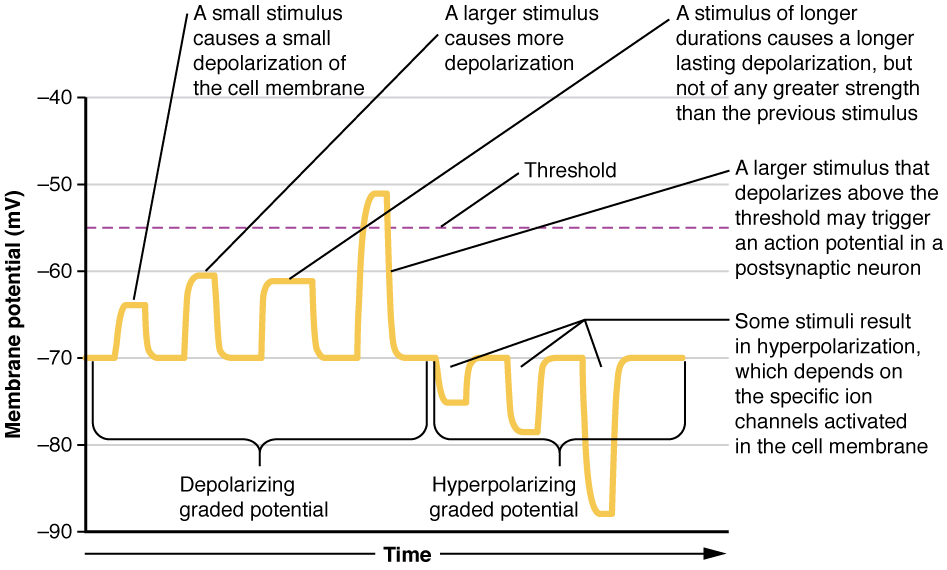
Figure 12.25 Graded Potentials Graded potentials are temporary changes in the membrane voltage, the characteristics of which depend on the size of the stimulus. Some types of stimuli cause depolarization of the membrane, whereas others cause hyperpolarization. It depends on the specific ion channels that are activated in the cell membrane.
J. Gordon Betts and colleagues, Anatomy And Physiology 2E, OpenStax. · CC BY-NC-SA 4.0 · Original image ↗ · Access for free ↗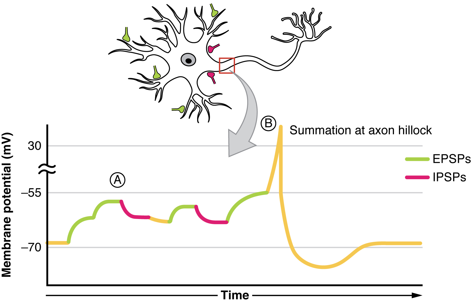
Figure 12.26 Postsynaptic Potential Summation The result of summation of postsynaptic potentials is the overall change in the membrane potential. At point A, several different excitatory postsynaptic potentials add up to a large depolarization. At point B, a mix of excitatory and inhibitory postsynaptic potentials result in a different end result for the membrane potential.
J. Gordon Betts and colleagues, Anatomy And Physiology 2E, OpenStax. · CC BY-NC-SA 4.0 · Original image ↗ · Access for free ↗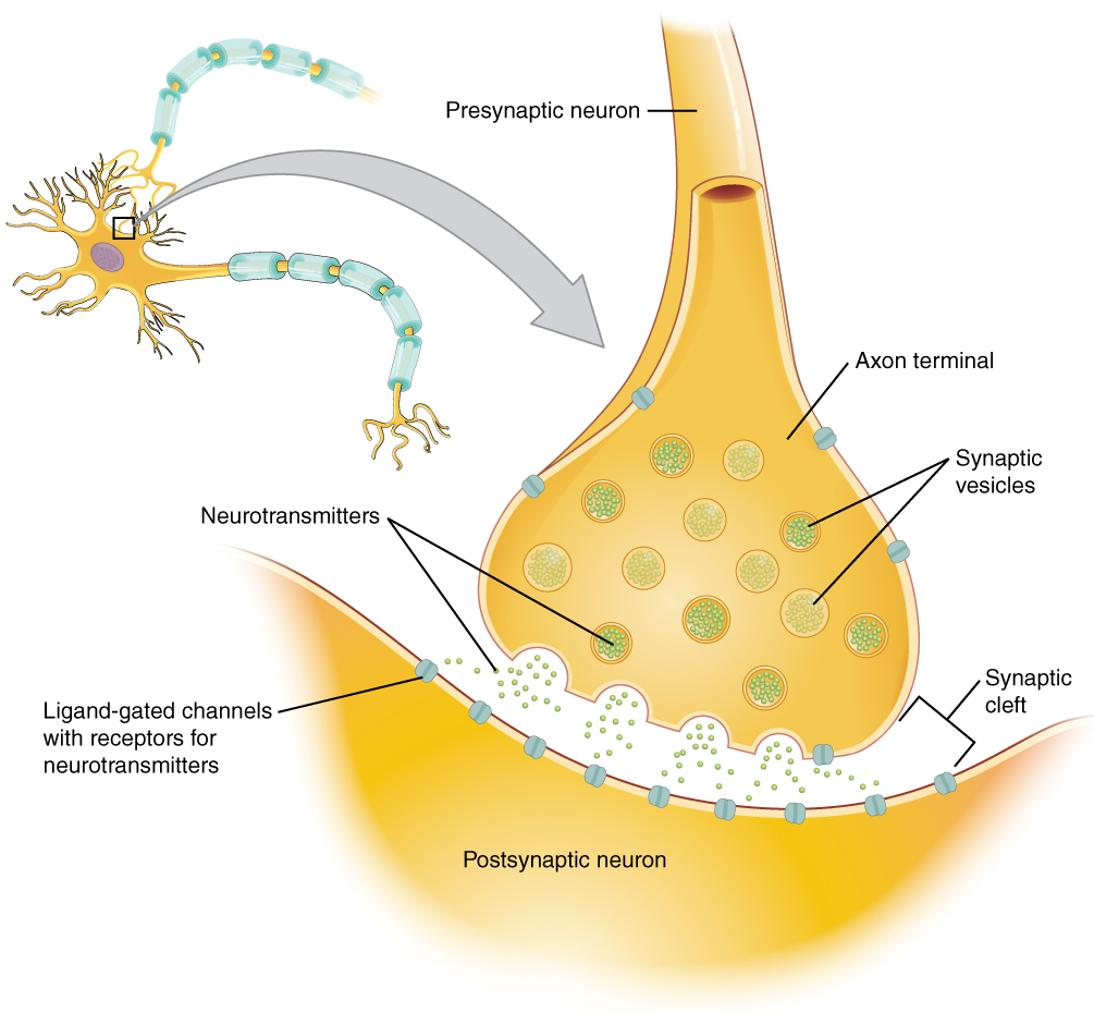
Figure 12.27 The Synapse The synapse is a connection between a neuron and its target cell (which is not necessarily a neuron). The presynaptic element is the synaptic end bulb of the axon where Ca 2+ enters the bulb to cause vesicle fusion and neurotransmitter release. The neurotransmitter diffuses across the synaptic cleft to bind to its receptor. The neurotransmitter is cleared from the synapse either by enzymatic degradation, neuronal reuptake, or glial reuptake.
J. Gordon Betts and colleagues, Anatomy And Physiology 2E, OpenStax. · CC BY-NC-SA 4.0 · Original image ↗ · Access for free ↗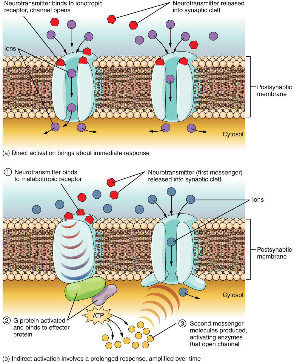
Figure 12.28 Receptor Types (a) An ionotropic receptor is a channel that opens when the neurotransmitter binds to it. (b) A metabotropic receptor is a complex that causes metabolic changes in the cell when the neurotransmitter binds to it (1). After binding, the G protein hydrolyzes ATP and moves to the effector protein (2). When the G protein contacts the effector protein, the latter is activated. In the case shown, the effector protein then acts on ATP to generate a second messenger, cAMP (3). The second messenger can then go on to cause changes in the neuron, such as opening or closing ion channels, metabolic changes, and changes in gene transcription.
J. Gordon Betts and colleagues, Anatomy And Physiology 2E, OpenStax. · CC BY-NC-SA 4.0 · Original image ↗ · Access for free ↗| System | Cholinergic | Amino acids | Biogenic amines | Neuropeptides |
|---|---|---|---|---|
| Neurotransmitters | Acetylcholine | Glutamate, glycine, GABA | Serotonin (5-HT), dopamine, norepinephrine, (epinephrine) | Met-enkephalin, beta-endorphin, VIP, Substance P, etc. |
| Receptors | Nicotinic and muscarinic receptors | Glu receptors, gly receptors, GABA receptors | 5-HT receptors, D1 and D2 receptors, α-adrenergic and β-adrenergic receptors | Receptors are too numerous to list, but are specific to the peptides. |
| Elimination | Degradation by acetylcholinesterase | Reuptake by neurons or glia | Reuptake by neurons | Degradation by enzymes called peptidases |
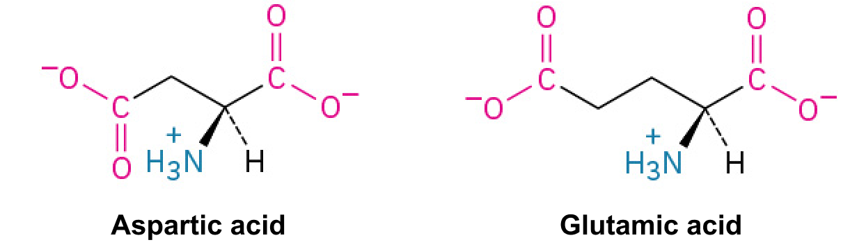
| Postsynaptic effect | Nicotinic receptor causes depolarization. Muscarinic receptors can cause both depolarization or hyperpolarization depending on the subtype. | Glu receptors cause depolarization. Gly and GABA receptors cause hyperpolarization. | Depolarization or hyperpolarization depends on the specific receptor. For example, D1 receptors cause depolarization and D2 receptors cause hyperpolarization. | Depolarization or hyperpolarization depends on the specific receptor. |
Table 12.3. J. Gordon Betts and colleagues, Anatomy And Physiology 2E, OpenStax.