Foundation 4 · 4D · Sound, light, optics, and spectroscopy

Molecular absorption and

Sources ↗

Use each for the structural question it can answer; one rarely identifies an entire .

Infrared detects changing

An infrared absorption excites molecular vibration; a vibration is -active when it changes the molecular . is inverse , commonly ⁻¹. A broad alcohol O–H band commonly appears near 3200–3600 ⁻¹; carboxylic- O–H is broader around 2500–3300 ⁻¹. A strong band is often near 1700 ⁻¹, with shifted by and . C≡N occurs near 2250 ⁻¹. The below roughly 1500 ⁻¹ helps compare whole molecular patterns. Absence of an expected strong can exclude a proposed group.

Source: 12.8 Infrared Spectra of Some Common Functional Groups · 13.6 Spin–Spin Splitting in 1 H NMR Spectra · 14.8 Interpreting Ultraviolet Spectra: The Effect of Conjugation

Electronic transitions and color

–visible absorption promotes between molecular energy levels. Greater usually reduces the relevant energy gap and shifts absorption toward longer . A colored sample transmits or reflects the of strongly absorbed visible light, so an absorption maximum is not necessarily its observed color. – indicators change , which changes distribution and absorption. A = εℓc relates to in dilute, non-scattering samples; ε depends on and chemical species. , scattering, or chemical equilibria can make nonlinear.

Source: 12.8 Infrared Spectra of Some Common Functional Groups · 13.6 Spin–Spin Splitting in 1 H NMR Spectra · 14.8 Interpreting Ultraviolet Spectra: The Effect of Conjugation

Read in three passes

In magnetic , chemically equivalent generally share a signal. δ, in ppm relative to a reference, reports ; -withdrawing groups usually move nearby signals downfield to larger δ. Integration gives relative counts. For simple coupling to n equivalent neighboring spin-½ , a signal splits into n + 1 lines; equivalent do not split one another. Exchangeable O–H or N–H signals may broaden or lose coupling. Complex nonequivalent neighbors require more than the simple n + 1 rule.

Source: 12.8 Infrared Spectra of Some Common Functional Groups · 13.6 Spin–Spin Splitting in 1 H NMR Spectra · 14.8 Interpreting Ultraviolet Spectra: The Effect of Conjugation

Apply it

Try before revealing the answer. A has a strong band near 1715 ⁻¹ and a singlet integrating to six hydrogens near 2 ppm. Is acetone or ethanol more consistent?

Worked answer

Acetone is consistent: its gives the absorption, and its two equivalent give six equivalent with no adjacent carbon-bound hydrogens to split them. Ethanol instead gives a triplet and methylene quartet in the simple coupling model, plus a variable O–H signal and an alcohol O–H band.

Source: 12.8 Infrared Spectra of Some Common Functional Groups · 13.6 Spin–Spin Splitting in 1 H NMR Spectra · 14.8 Interpreting Ultraviolet Spectra: The Effect of Conjugation

12.8 Infrared of Some Common — figures and tables

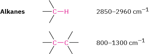
Characteristic alkane bonds and their absorption values. Alkane CH bonds absorb at 2850 to 2960 inverse centimeters and alkane CC bonds absorb at 800 to 1300 inverse centimeters.
12.8 Infrared of Some Common — illustration 1 John McMurry, Organic Chemistry, OpenStax. · CC BY-NC-SA 4.0 · Original image ↗ · Access for free ↗
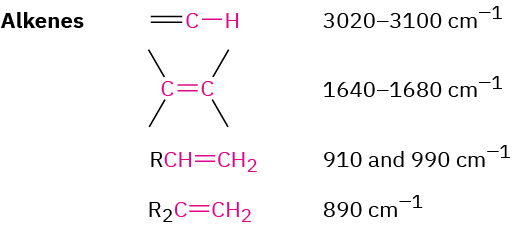
Characteristic alkene bonds and their absorption values. Various alkene CC and CH bond types absorb just below 3100, below 1680, below 990, and at 890 inverse centimeters.
12.8 Infrared of Some Common — illustration 2 John McMurry, Organic Chemistry, OpenStax. · CC BY-NC-SA 4.0 · Original image ↗ · Access for free ↗
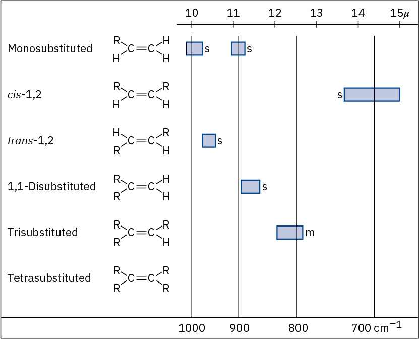
Characteristic alkene bonds and their absorption values, separated by level of substitution and cis or trans.
Figure 12.23 — 12.8 Infrared of Some Common

Figure 12.23 C–H out-of-plane bending vibrations for substituted alkenes.

John McMurry, Organic Chemistry, OpenStax. · CC BY-NC-SA 4.0 · Original image ↗ · Access for free ↗

Characteristic alkyne bonds and their absorption values. Alkyne CC bonds absorb at 2100 to 2260 inverse centimeters and alkyne CH bonds absorb at 3300 inverse centimeters.
12.8 Infrared of Some Common — illustration 4 John McMurry, Organic Chemistry, OpenStax. · CC BY-NC-SA 4.0 · Original image ↗ · Access for free ↗
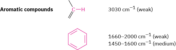
Characteristic aromatic bonds and their absorption values. Aromatic CH bonds absorb weakly at 3030 inverse centimeters and aromatic CC bonds absorb at in regions below 2000 and 1600 inverse centimeters.
12.8 Infrared of Some Common — illustration 5 John McMurry, Organic Chemistry, OpenStax. · CC BY-NC-SA 4.0 · Original image ↗ · Access for free ↗
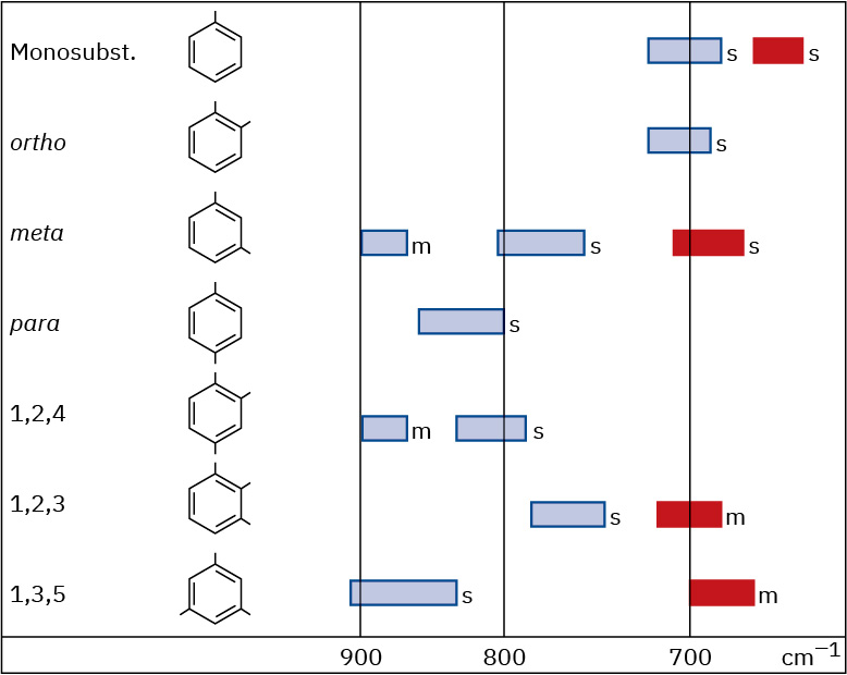
Characteristic aromatic bonds and their absorption values, separated by number of substituents and ortho, meta, or para.
Figure 12.24 — 12.8 Infrared of Some Common

Figure 12.24 C–H out-of-plane bending vibrations for substituted benzenes.

John McMurry, Organic Chemistry, OpenStax. · CC BY-NC-SA 4.0 · Original image ↗ · Access for free ↗
A characteristic alcohol bond and its absorption value, indicated as a broad intense band at 3400 to 3650 inverse centimeters.
12.8 Infrared of Some Common — illustration 7 John McMurry, Organic Chemistry, OpenStax. · CC BY-NC-SA 4.0 · Original image ↗ · Access for free ↗
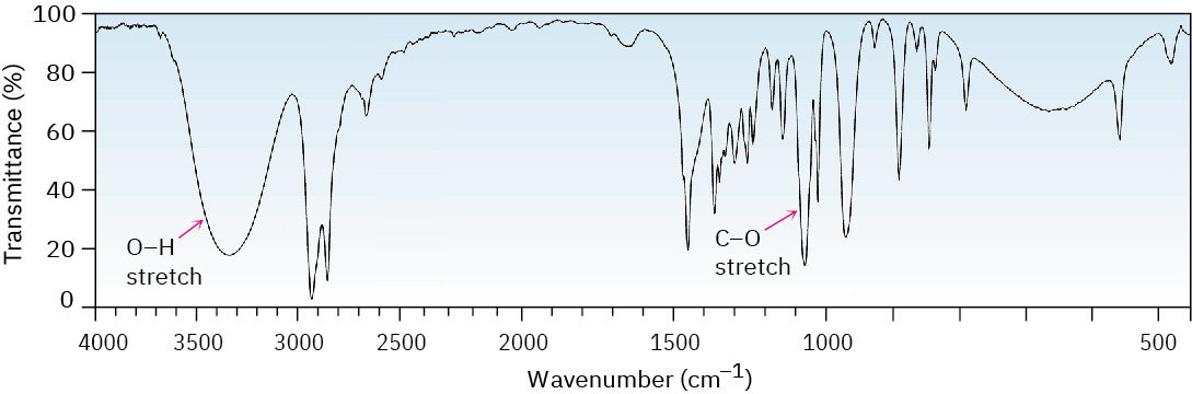
An infrared spectrum in which a wide absorption band near 3300 inverse centimeters is labeled O H, and a sharp band below 1100 is labeled C single bond O.
Figure 12.25 — 12.8 Infrared of Some Common

Figure 12.25 IR spectrum of cyclohexanol.

John McMurry, Organic Chemistry, OpenStax. · CC BY-NC-SA 4.0 · Original image ↗ · Access for free ↗
A characteristic amine bond and its absorption value, indicated as a sharp band at 3300 to 3500 inverse centimeters.
12.8 Infrared of Some Common — illustration 9 John McMurry, Organic Chemistry, OpenStax. · CC BY-NC-SA 4.0 · Original image ↗ · Access for free ↗
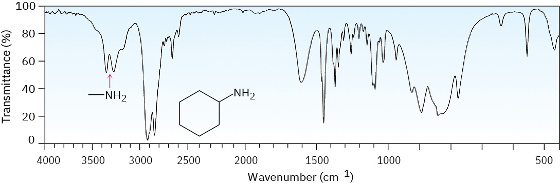
An infrared spectrum in which a sharp, two-pronged absorption band near 3300 inverse centimeters is labeled N H 2.
Figure 12.26 — 12.8 Infrared of Some Common

Figure 12.26 IR spectrum of cyclohexylamine.

John McMurry, Organic Chemistry, OpenStax. · CC BY-NC-SA 4.0 · Original image ↗ · Access for free ↗
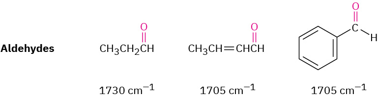
Characteristic aldehyde C O bonds and their absorption values, indicated at 1730 and 1705 inverse centimeters for alkyl and allyl or benzyl aldehydes respectively.
12.8 Infrared of Some Common — illustration 11 John McMurry, Organic Chemistry, OpenStax. · CC BY-NC-SA 4.0 · Original image ↗ · Access for free ↗
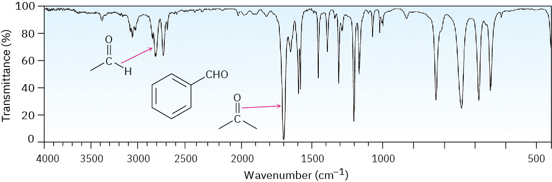
An infrared spectrum in which a small absorption band near 2800 inverse centimeters is labeled aldehyde C H, and a strong band near 1700 is labeled aldehyde C O.
Figure 12.27 — 12.8 Infrared of Some Common

Figure 12.27 The IR spectrum of benzaldehyde.

John McMurry, Organic Chemistry, OpenStax. · CC BY-NC-SA 4.0 · Original image ↗ · Access for free ↗
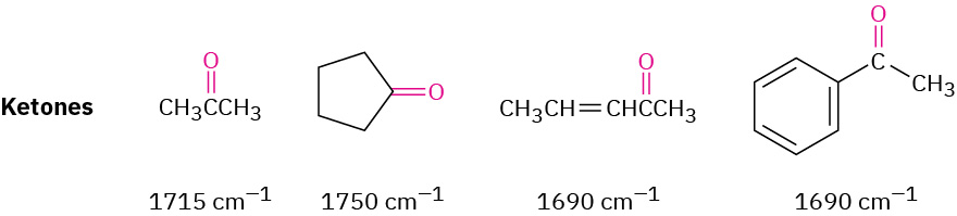
Characteristic ketone C O bonds and their absorption values, indicated at values just above 1700 inverse centimeters for alkyl ketones and values just below 1700 for allyl and benzyl ketones.
12.8 Infrared of Some Common — illustration 13 John McMurry, Organic Chemistry, OpenStax. · CC BY-NC-SA 4.0 · Original image ↗ · Access for free ↗
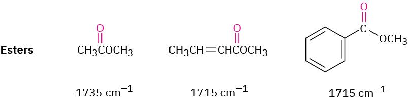
Characteristic ester C O bonds and their absorption values, indicated at 1735 inverse centimeters for alkyl esters and 1715 for allyl and benzyl esters.
12.8 Infrared of Some Common — illustration 14 John McMurry, Organic Chemistry, OpenStax. · CC BY-NC-SA 4.0 · Original image ↗ · Access for free ↗
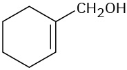
A chemical structure of 1-cyclohexen-1-ylmethanol
12.8 Infrared of Some Common — illustration 15 John McMurry, Organic Chemistry, OpenStax. · CC BY-NC-SA 4.0 · Original image ↗ · Access for free ↗
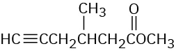
A chemical structure of a methyl ester with a five-carbon chain off the carbonyl. There is a terminal alkyne and a methyl on the beta C.
12.8 Infrared of Some Common — illustration 16 John McMurry, Organic Chemistry, OpenStax. · CC BY-NC-SA 4.0 · Original image ↗ · Access for free ↗
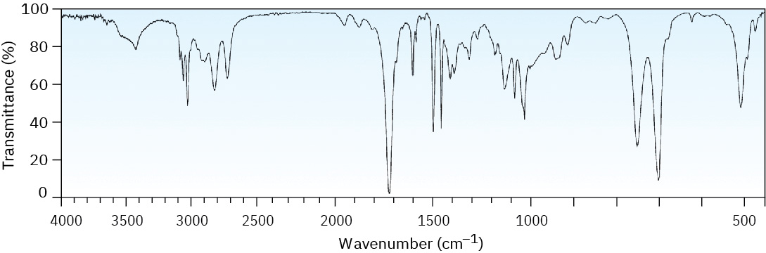
An infrared spectrum of an unknown compound. Significant absorption bands are at 700,1000 and 1700 centimeter inverse.
Figure 12.28 — 12.8 Infrared of Some Common

Figure 12.28 IR spectrum for Worked Example 12.6 .

John McMurry, Organic Chemistry, OpenStax. · CC BY-NC-SA 4.0 · Original image ↗ · Access for free ↗
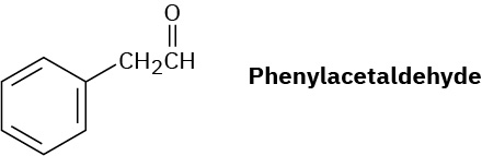
Chemical structure structure of phenylacetaldehyde.
12.8 Infrared of Some Common — illustration 18 John McMurry, Organic Chemistry, OpenStax. · CC BY-NC-SA 4.0 · Original image ↗ · Access for free ↗
An infrared spectrum with xignificant absorption bands near 3300 and 1500 inverse centimeters, and in the fingerprint region.
Figure 12.29 — 12.8 Infrared of Some Common

Figure 12.29 The IR spectrum of phenylacetylene, Problem 12-9.

John McMurry, Organic Chemistry, OpenStax. · CC BY-NC-SA 4.0 · Original image ↗ · Access for free ↗
Chemical structure of a cyclohexene with a C O O C H 3 substituent.
12.8 Infrared of Some Common — illustration 20 John McMurry, Organic Chemistry, OpenStax. · CC BY-NC-SA 4.0 · Original image ↗ · Access for free ↗
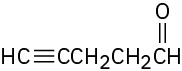
A five-carbon chain with a triple bond on C4 and a C H O group on C1.
12.8 Infrared of Some Common — illustration 21 John McMurry, Organic Chemistry, OpenStax. · CC BY-NC-SA 4.0 · Original image ↗ · Access for free ↗
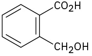
A benzene ring with a C O O H  on C1 and C H 2 O H on C2.
12.8 Infrared of Some Common — illustration 22 John McMurry, Organic Chemistry, OpenStax. · CC BY-NC-SA 4.0 · Original image ↗ · Access for free ↗
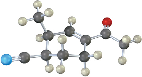
A ball and stick model of a cyclopentane ring linked to methyl and acetate groups, and triple bonded to nitrogen. The red and blue spheres represent oxygen and nitrogen, respectively.
12.8 Infrared of Some Common — illustration 23 John McMurry, Organic Chemistry, OpenStax. · CC BY-NC-SA 4.0 · Original image ↗ · Access for free ↗

Source: 12.8 Infrared Spectra of Some Common Functional Groups

13.6 in 1 H — figures and tables

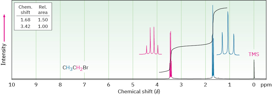
1 H N M R spectrum of bromoethane shows singlet peak at 0 (T M S), triplet peak at 1.8, and quartet peak at 3.4 parts per million.
Figure 13.8 — 13.6 in 1 H

Figure 13.8 The 1 H NMR spectrum of bromoethane, CH 3 CH 2 Br. The –CH 2 Br protons appear as a quartet at 3.42 δ , and the –CH 3 protons appear as a triplet at 1.68 δ .

John McMurry, Organic Chemistry, OpenStax. · CC BY-NC-SA 4.0 · Original image ↗ · Access for free ↗

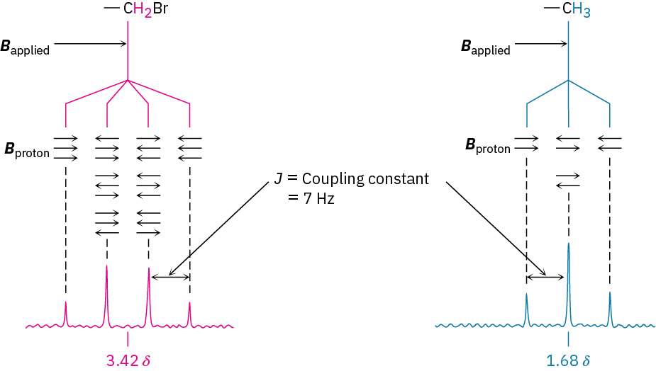
C H 2 B r splits into four branches and C H 3 splits into three branches, each with arrows labeled B proton and J values equals 7 Hertz.
Figure 13.9 — 13.6 in 1 H

Figure 13.9 The origin of spin–spin splitting in bromoethane. The nuclear spins of neighboring protons, indicated by horizontal arrows, align either with or against the applied field, causing the splitting of absorptions into multiplets.

John McMurry, Organic Chemistry, OpenStax. · CC BY-NC-SA 4.0 · Original image ↗ · Access for free ↗

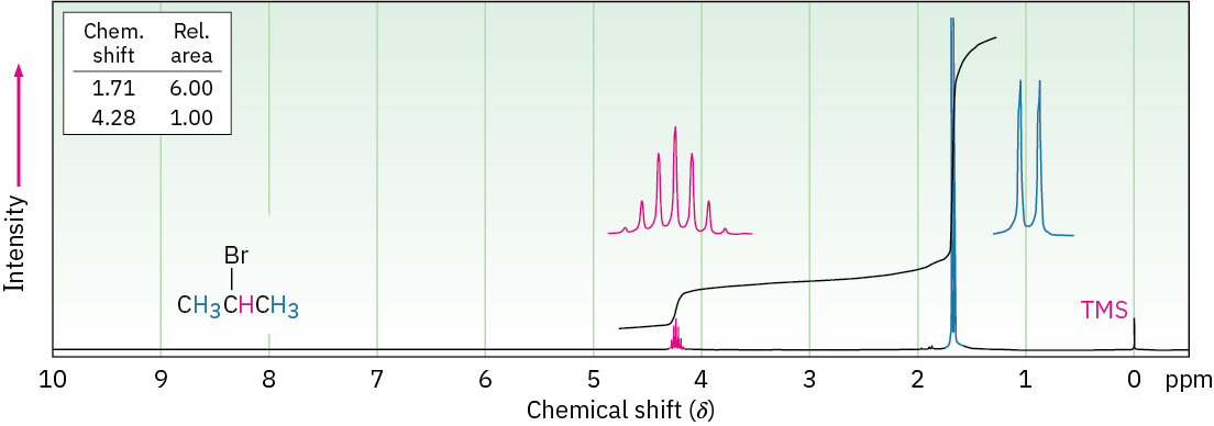
1 H N M R spectrum of 2-bromopropane shows singlet peak at 0 (T M S), doublet peak at 1.8, and septet peak at 4.2 parts per million.
Figure 13.10 — 13.6 in 1 H

Figure 13.10 The 1 H NMR spectrum of 2-bromopropane. The –CH 3 proton signal at 1.71 δ is split into a doublet, and the –CHBr– proton signal at 4.28 δ is split into a septet. Note that the distance between peaks is the same in both multiplets. Note also that the two outer peaks of the septet are small enough to be nearly missed.

John McMurry, Organic Chemistry, OpenStax. · CC BY-NC-SA 4.0 · Original image ↗ · Access for free ↗
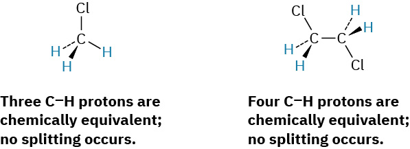
A structure of chloromethane with text indicating all protons are chemically equivalent, no splitting. A structure of 1,2-dichloroethane with text indicating all protons are chemically equivalent, no splitting.
13.6 in 1 H — illustration 4 John McMurry, Organic Chemistry, OpenStax. · CC BY-NC-SA 4.0 · Original image ↗ · Access for free ↗
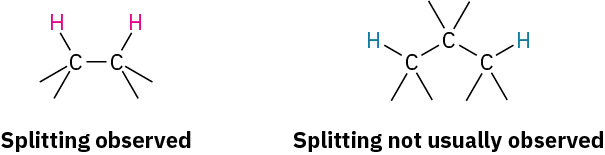
First structure labeled splitting observed shows adjacent carbon atoms, each bonded to hydrogen. Second structure labeled splitting not usually observed has 3-carbon chain, hydrogens on non-adjacent carbons.
13.6 in 1 H — illustration 5 John McMurry, Organic Chemistry, OpenStax. · CC BY-NC-SA 4.0 · Original image ↗ · Access for free ↗
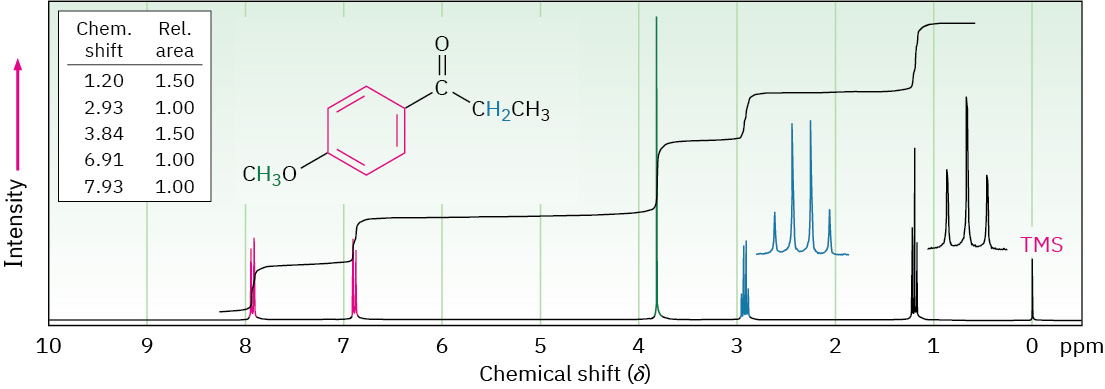
The H N M R spectrum of para-methoxypropiophenone shows peaks at 0 (T M S), 1.2 (triplet, methyl), 2.9 (quartet, methylene), 3.9 (singlet, methoxy), 6.9 and 7.9 (both doublets, benzene).
Figure 13.11 — 13.6 in 1 H

Figure 13.11 The 1 H NMR spectrum of para -methoxypropiophenone.

John McMurry, Organic Chemistry, OpenStax. · CC BY-NC-SA 4.0 · Original image ↗ · Access for free ↗
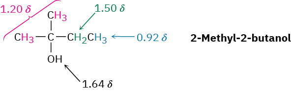
Structure of 2-methyl-2-butanol with shifts for hydrogens on: C 1 and methyl (1.20), O H (1.64), C 3 (1.50), and C 4 (0.92).
13.6 in 1 H — illustration 7 John McMurry, Organic Chemistry, OpenStax. · CC BY-NC-SA 4.0 · Original image ↗ · Access for free ↗
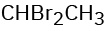
The condensed structural formula reads, C H B r 2 C H 3.
13.6 in 1 H — illustration 8 John McMurry, Organic Chemistry, OpenStax. · CC BY-NC-SA 4.0 · Original image ↗ · Access for free ↗
The condensed structural formula reads, C H 3 O C H 2 C H 2 B r.
13.6 in 1 H — illustration 9 John McMurry, Organic Chemistry, OpenStax. · CC BY-NC-SA 4.0 · Original image ↗ · Access for free ↗
The condensed structural formula reads, C l C H 2 C H 2 C H 2 C l.
13.6 in 1 H — illustration 10 John McMurry, Organic Chemistry, OpenStax. · CC BY-NC-SA 4.0 · Original image ↗ · Access for free ↗
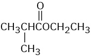
The condensed structural formula of ethyl isobutyrate.
13.6 in 1 H — illustration 11 John McMurry, Organic Chemistry, OpenStax. · CC BY-NC-SA 4.0 · Original image ↗ · Access for free ↗
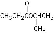
The condensed structural formula of isopropyl propanoate.
13.6 in 1 H — illustration 12 John McMurry, Organic Chemistry, OpenStax. · CC BY-NC-SA 4.0 · Original image ↗ · Access for free ↗
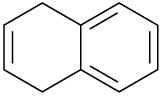
The structure has a cyclohexene ring fused to a benzene ring; double bond in cyclohexene is opposite fusion.
13.6 in 1 H — illustration 13 John McMurry, Organic Chemistry, OpenStax. · CC BY-NC-SA 4.0 · Original image ↗ · Access for free ↗
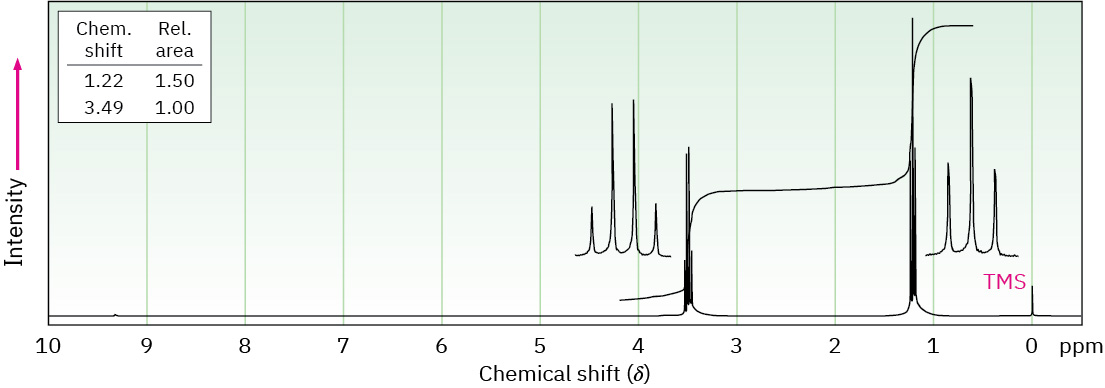
The 1 H N M R spectrum of a compound has peaks at 0 (T M S), 1.2 (triplet), and 3.5 (quartet).
Figure 13.12 — 13.6 in 1 H

Figure 13.12 An integrated 1 H NMR spectrum for Problem 11.

John McMurry, Organic Chemistry, OpenStax. · CC BY-NC-SA 4.0 · Original image ↗ · Access for free ↗
Number of equivalent adjacent Multiplet Ratio of
0 Singlet 1
1 Doublet 1 : 1
2 Triplet 1 : 2 : 1
3 Quartet 1 : 3 : 3 : 1
4 Quintet 1 : 4 : 6 : 4 : 1
6 Septet 1 : 6 : 15 : 20 : 15 : 6 : 1

Table 13.4 Some Common Spin Multiplicities. John McMurry, Organic Chemistry, OpenStax.

Source: 13.6 Spin–Spin Splitting in 1 H NMR Spectra

14.8 Interpreting : The Effect of — figures and tables

In a cyclohexane ring, C 1 is double bonded to C 2. C 4 is double bonded to C 5.
14.8 Interpreting : The Effect of — illustration 5 John McMurry, Organic Chemistry, OpenStax. · CC BY-NC-SA 4.0 · Original image ↗ · Access for free ↗
In a cyclohexane ring, C 1 is double bonded to C 2. C 3 is double bonded to C 4.
14.8 Interpreting : The Effect of — illustration 6 John McMurry, Organic Chemistry, OpenStax. · CC BY-NC-SA 4.0 · Original image ↗ · Access for free ↗
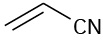
Two carbons double-bonded to each other. At C 1 there is a cyano group.
14.8 Interpreting : The Effect of — illustration 7 John McMurry, Organic Chemistry, OpenStax. · CC BY-NC-SA 4.0 · Original image ↗ · Access for free ↗
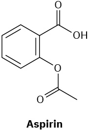
The structure of Aspirin has a benzene ring. C 1 is bonded to a carboxylic acid group. C 2 is bonded to acetoxyl group.
14.8 Interpreting : The Effect of — illustration 8 John McMurry, Organic Chemistry, OpenStax. · CC BY-NC-SA 4.0 · Original image ↗ · Access for free ↗
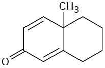
A cyclohexadiene ring with double bonded oxygen at C 3 is fused to a cyclohexane ring. Both rings share a methyl group.
14.8 Interpreting : The Effect of — illustration 9 John McMurry, Organic Chemistry, OpenStax. · CC BY-NC-SA 4.0 · Original image ↗ · Access for free ↗
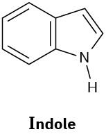
Indole has a benzene ring fused to a 5-membered ring made of  four carbons, in which C 2 is double bonded to C 3, and an N-H group at position 1.
14.8 Interpreting : The Effect of — illustration 10 John McMurry, Organic Chemistry, OpenStax. · CC BY-NC-SA 4.0 · Original image ↗ · Access for free ↗
Name Structure λmax (nm)
2--1,3-butadiene 220
1,3-Cyclohexadiene 256
1,3,5-Hexatriene H2C=CH―CH=CH–CH=CH2 258
1,3,5,7-Octatetraene H2C=CH–CH=CH–CH=CH–CH=CH2 290
3-Buten-2-one 219
254

Table 14.2 Ultraviolet Absorptions of Some Conjugated Molecules. John McMurry, Organic Chemistry, OpenStax.

Source: 14.8 Interpreting Ultraviolet Spectra: The Effect of Conjugation

185 terms, alphabetically ordered

Absorbance

A measure of how much incident light a sample absorbs at a particular .

Read explanation · Waters ↗
Acid · ·

In the Brønsted–Lowry model, a substance that can donate a . Acidic have a higher activity than water.

Read explanation · OpenStax ↗
Activation energy ·

The energy barrier that must overcome to reach a and undergo .

Read explanation · OpenStax ↗
Active catalytic-site concentration ·

The total of active sites used to calculate from . It equals active when each has one site.

Read explanation · Biology LibreTexts ↗
Active site

The region of an that binds its and carries out .

Read explanation · OpenStax ↗
Adenosine triphosphate

An energy-carrying ; its powers many cellular processes, including cycles.

Read explanation · OpenStax ↗
Alpha carbon · ·

The central carbon next to the carboxyl carbon. In a standard it binds an , hydrogen, and .

Read explanation · OpenStax ↗
Alpha helix · · · · · ·

A helical stabilized by between a oxygen and an hydrogen four later.

Read explanation · OpenStax ↗
Amide · · ·

A group containing a carbon bonded to nitrogen. are amide linkages.

Read explanation · OpenStax ↗
Amine ·

An organic derivative of ammonia containing a nitrogen with a lone pair; many amines can accept a .

Read explanation · OpenStax ↗
Amino acid

A building block containing amino and and a characteristic .

Read explanation · OpenStax ↗
Amino acid residue · ·

The part of an incorporated into a chain. Residue can also describe another building block within a larger .

Read explanation · OpenStax ↗
Amino group · · ·

A nitrogen-containing . A free ’s amino group is typically (NH₃⁺) near physiological ; ’s ring nitrogen is an exception to this formula.

Read explanation · OpenStax ↗
Amphipathic

Having both a region and a region, as in a or .

Read explanation · OpenStax ↗
amplitude

Maximum from an oscillation’s position.

Read explanation · OpenStax ↗
Analytical standard ·

A material with a known identity or amount used to compare or calibrate measurements.

Read explanation · Agilent ↗
Anticodon

A triplet that pairs to an . The anticodon for 5′-AUG-3′ is aligned 3′-UAC-5′, or written 5′-CAU-3′.

Read explanation · NCBI Bookshelf / Garland Science ↗
Antiparallel

Running in opposite directions, as with two strands or neighboring strands in some .

Read explanation · OpenStax ↗
Aqueous solution ·

A in which water is the .

Read explanation · OpenStax ↗
Aromatic

Containing a cyclic, with . , , and have aromatic .

Read explanation · OpenStax ↗
Atom

The smallest unit of an element that retains its chemical identity; it has a surrounded by .

Read explanation · OpenStax ↗
Beer–Lambert law

Relationship A = εℓc between , molar absorptivity, path length, and .

Read explanation · OpenStax ↗
Benzene ring

A six-carbon ring with .

Read explanation · OpenStax ↗
Benzyl group

A attached through a –CH₂– group; part of ’s .

Read explanation · OpenStax ↗
Beta sheet · · · · · ·

A made of aligned segments connected by ; strands may be parallel or .

Read explanation · OpenStax ↗
Biomolecule

A made by or involved in living systems, including , , , and .

Read explanation · OpenStax ↗
Blotting ·

Transfer of separated or to a so a specific target can be detected.

Read explanation · Thermo Fisher ↗
Brønsted–Lowry base · · · · · · · · · · ·

In the Brønsted–Lowry model, a substance that can accept a . Basic solutions have lower activity than water.

Read explanation · OpenStax ↗
Calibration

Establishing the relation between a measured signal and known values or .

Read explanation · Agilent ↗
Carbohydrate

A class of including sugars and their , used for energy storage, structure, and recognition.

Read explanation · OpenStax ↗
Carbonyl group · ·

A carbon double-bonded to oxygen, C=O; present in and many other .

Read explanation · OpenStax ↗
Carboxyl group · ·

An , COOH. In free it is usually to COO⁻ near physiological .

Read explanation · OpenStax ↗
Catalyst · ·

A substance that speeds a by providing a pathway with lower and is regenerated rather than consumed overall.

Read explanation · OpenStax ↗
Centimeter

A unit of length equal to one hundredth of a meter.

Read explanation · NIST ↗
Chemical compound ·

A substance containing of two or more different chemically combined in a definite composition.

Read explanation · OpenStax ↗
Chemical element

A substance defined by having the same number of , such as carbon or oxygen.

Read explanation · OpenStax ↗
Chemical reaction ·

A process that rearranges and chemical bonds, converting into .

Read explanation · OpenStax ↗
chemical shift

signal position relative to a reference, normally reported in ppm.

Read explanation · OpenStax ↗
Chromatin

The complex of and associated , including , in .

Read explanation · OpenStax ↗
Chromatographic peak · ·

A rise and fall in detector signal as one or more components pass through the detector; a peak does not guarantee a single pure .

Read explanation · Agilent ↗
Chromosome ·

An organized with associated that carries . A replicated chromosome has two .

Read explanation · OpenStax ↗
Codon

A sequence of three that specifies an or a stop signal.

Read explanation · OpenStax ↗
complement

A system that can promote , , and attack against targets.

Read explanation · OpenStax ↗
Concentration

The amount of a substance in a specified volume or mass of mixture.

Read explanation · OpenStax ↗
Conjugated system ·

Adjacent orbitals that allow to be distributed across several , often through alternating single and multiple bonds.

Read explanation · OpenStax ↗
conjugation

Overlap of adjacent p orbitals that delocalizes across multiple .

Read explanation · OpenStax ↗
Cooperativity · ·

An interaction in which binding at one site changes binding at other sites. Positive cooperativity can produce a response.

Read explanation · Biology LibreTexts ↗
Covalent bond · ·

A bond in which share pairs. and are covalent.

Read explanation · OpenStax ↗
Cysteine ·

An with a . Two cysteines can form a .

Read explanation · OpenStax ↗
Deoxyribonucleic acid

The that stores genetic information used to specify and sequences.

Read explanation · OpenStax ↗
Deprotonation

Removal of a from a or group; this changes its by minus one.

Read explanation · OpenStax ↗
Detergent

An substance that can associate with both water and material; detergents can disrupt or interactions.

Read explanation · Thermo Fisher ↗
determination

Developmental commitment to a cell fate before all specialized features necessarily appear.

Read explanation · OpenStax ↗
Dipole ·

A separation of positive and negative within a bond or .

Read explanation · OpenStax ↗
dipole moment

measure of separation in a bond or .

Read explanation · OpenStax ↗
Disulfide bond · · ·

A covalent sulfur–sulfur bond formed by of two , which can stabilize structure.

Read explanation · NCBI Bookshelf ↗
DNA replication

Copying so genetic information can be passed to daughter cells. Each guides a complementary new strand.

Read explanation · OpenStax ↗
Electric charge · · ·

An electrical property that can be positive or negative. Opposite charges attract and like charges repel; net charge is the sum of all charges on a particle.

Read explanation · OpenStax ↗
Electron

A negatively subatomic particle. Electron sharing and transfer determine much of chemical bonding.

Read explanation · OpenStax ↗
Electron delocalization ·

Distribution of across several rather than confinement to one or bond.

Read explanation · OpenStax ↗
Electronegativity ·

An ’s tendency to attract shared in a chemical bond. Unequal attraction can make a bond .

Read explanation · OpenStax ↗
Enzyme

A biological that speeds a without being consumed. Most enzymes are .

Read explanation · OpenStax ↗
Enzyme saturation · ·

The high- limit in which sites spend almost all their time processing and the rate approaches .

Read explanation · Biology LibreTexts ↗
Equilibrium

A state where opposing processes occur at equal rates, so macroscopic or other properties remain constant.

Read explanation · OpenStax ↗
Eukaryote ·

An organism whose cells have a -enclosed ; animals, plants, fungi, and protists are eukaryotes.

Read explanation · OpenStax ↗
Exon

A segment retained in a mature spliced . In an exon may contain coding sequence, untranslated sequence, or both.

Read explanation · OpenStax ↗
FACT complex ·

A -associated complex that helps machinery traverse and supports reorganization.

Read explanation · OpenStax ↗
fingerprint region

Complex lower- region useful for comparing molecular identity.

Read explanation · OpenStax ↗
First-order kinetics

A rate dependence proportional to the first of a . At low , a is approximately first-order in .

Read explanation · Biology LibreTexts ↗
Functional group

A group of whose chemical behavior gives a characteristic and properties.

Read explanation · OpenStax ↗
Gene

A sequence that contributes to a functional or , together with the information needed to express it.

Read explanation · OpenStax ↗
Gene expression

Production of a functional from a , including and, for -coding , .

Read explanation · NCBI Bookshelf ↗
Half-life

The time required for half of a population or amount to remain during decay; a constant half-life is characteristic of decay.

Read explanation · OpenStax ↗
Histone

A -associated rich in . Core histones form the around which nucleosomal wraps.

Read explanation · OpenStax ↗
Hydrocarbon · ·

A made only of carbon and hydrogen. Hydrocarbon regions are generally .

Read explanation · OpenStax ↗
Hydrogen bond ·

An attraction between a hydrogen bonded to an and another . hydrogen bonds stabilize .

Read explanation · OpenStax ↗
Hydrogen ion · ·

A positively hydrogen species. H⁺ denotes a , which is hydrated in water; its activity determines .

Read explanation · OpenStax ↗
Hydrolysis

Breaking a chemical bond by adding water. hydrolysis separates .

Read explanation · OpenStax ↗
Hydrophilic

Interacting favorably with water, typically because a group is or .

Read explanation · OpenStax ↗
Hydrophobic

Interacting poorly with water. often gather in a ’s interior.

Read explanation · OpenStax ↗
Hydroxide ion ·

The negatively OH⁻. Basic contain a higher hydroxide ion activity than water at the same temperature.

Read explanation · OpenStax ↗
Hydroxyl group ·

An oxygen bonded to hydrogen, –OH. Hydroxyl groups can and often increase .

Read explanation · OpenStax ↗
Hyperbolic curve ·

A curve that approaches an asymptote. The standard rate curve is a rectangular hyperbola approaching .

Read explanation · Biology LibreTexts ↗
Indole

A fused six-membered and five-membered ring system containing nitrogen; found in .

Read explanation · OpenStax ↗
inflammation

A coordinated tissue response recruiting defense and repair mechanisms, often involving increased blood flow and permeability.

Read explanation · OpenStax ↗
infrared spectroscopy ·

Method probing vibrational transitions through absorption of infrared light.

Read explanation · OpenStax ↗
Initial reaction rate · · · ·

The early rate of formation, measured before substantial depletion or accumulation, usually after the brief binding transient.

Read explanation · Biology LibreTexts ↗
Intron ·

A segment removed from a precursor during . Intronic signals or regulatory sequences can affect .

Read explanation · OpenStax ↗
Ion · ·

An or with a net because its numbers of and do not balance.

Read explanation · OpenStax ↗
ketone bodies

Acetoacetate, β-hydroxybutyrate, and acetone, produced mainly by liver during increased fatty- .

Read explanation · Biology LibreTexts / Oregon State University ↗
linkage

A tendency of nearby on the same to be inherited together.

Read explanation · OpenStax ↗
Lipid

A broad class of largely or , including fats, , and steroids.

Read explanation · OpenStax ↗
locus

The specific position of a or other feature.

Read explanation · OpenStax ↗
Logarithm · · ·

The exponent needed to obtain a number from a chosen fixed number. For decimal logarithms, log₁₀(100) = 2 because 10² = 100; uses the negative decimal logarithm of activity.

Read explanation · OpenStax ↗
magnetic field

field that exerts forces on moving and magnetic moments.

Read explanation · OpenStax ↗
Mass spectrometry ·

Analysis of according to their mass-to- ratios, used to identify and or .

Read explanation · Thermo Fisher ↗
Mass-to-charge ratio

The mass of an divided by its number; this is the quantity measured by a .

Read explanation · Thermo Fisher ↗
Maximum reaction rate · ·

The limiting as becomes saturating. For a fixed under given conditions, Vmax = times .

Read explanation · Biology LibreTexts ↗
mechanical power

Rate of energy transfer, measured in watts.

Read explanation · OpenStax ↗
mechanical work

Energy transferred by a force acting through ; the force component parallel to motion contributes.

Read explanation · OpenStax ↗
Membrane · · · ·

A thin boundary or sheet. Cell membranes are bilayers containing ; membranes are synthetic supports that hold or for detection.

Read explanation · OpenStax ↗
Messenger RNA ·

An that carries coding information used by to synthesize a .

Read explanation · OpenStax ↗
Methyl group ·

A –CH₃ group, consisting of one carbon bonded to three hydrogens.

Read explanation · OpenStax ↗
Michaelis–Menten kinetics · ·

A model with = [S]/(Km + ). Its curve is ; may require a different model.

Read explanation · Biology LibreTexts ↗
Missense mutation ·

A coding-sequence that changes the specified ; its effect depends on the and context.

Read explanation · OpenStax ↗
Molecular chaperone · ·

A that assists other in folding or prevents , without specifying their sequence.

Read explanation · NCBI Bookshelf ↗
Molecule

Two or more joined by . A molecule may be electrically or carry a .

Read explanation · OpenStax ↗
Mutation

A change in a sequence; some mutations alter a ’s sequence.

Read explanation · OpenStax ↗
Neutral

Having no net . For a , neutral instead means equal and activities; its is about 7 at 25 °C.

Read explanation · OpenStax ↗
Nitrogenous base · · ·

A nitrogen-containing ring component of ; its pairing interactions help specify sequences.

Read explanation · OpenStax ↗
Nonpolar ·

Lacking a substantial separation of . Nonpolar groups generally interact poorly with water.

Read explanation · OpenStax ↗
Nonsense mutation ·

A that converts an amino- into a premature , potentially truncating the or destabilizing its .

Read explanation · OpenStax ↗
Nuclear magnetic resonance spectroscopy

A method using spin behavior in a to study molecular structure and dynamics, including in .

Read explanation · OpenStax ↗
Nucleic acid

A of , such as or , used in information storage, expression, and other cellular functions.

Read explanation · OpenStax ↗
Nucleosome

The basic unit of : wrapped around a .

Read explanation · OpenStax ↗
Nucleotide

A joined to a sugar and one or more .

Read explanation · OpenStax ↗
Nucleus ·

A -enclosed compartment containing most of a cell’s .

Read explanation · OpenStax ↗
Octamer

A complex made of eight . A core contains eight .

Read explanation · OpenStax ↗
opsonization

Coating a target with that enhance its recognition and uptake by phagocytes.

Read explanation · OpenStax ↗
orbital

Quantum state describing an ’s spatial probability distribution.

Read explanation · OpenStax ↗
oscillation frequency ·

Number of repeated cycles per unit time, measured in hertz.

Read explanation · OpenStax ↗
oscillation phase ·

Position within an oscillatory cycle relative to a reference.

Read explanation · OpenStax ↗
Oxidation ·

Loss of or an increase in oxidation state. Oxidizing two can form a .

Read explanation · OpenStax ↗
Peptide

A chain of joined by ; the term commonly describes chains shorter than a typical .

Read explanation · OpenStax ↗
Peptide bond ·

A covalent between the carboxyl carbon of one and the amino nitrogen of the next.

Read explanation · OpenStax ↗
pH

A measure of , defined as the negative of activity. Lower pH means greater ; pH influences .

Read explanation · OpenStax ↗
Phenylalanine ·

A with an .

Read explanation · OpenStax ↗
Phosphate group ·

A phosphorus-containing group bonded to oxygens; phosphate groups occur in , , and modified .

Read explanation · OpenStax ↗
Phospholipid

An with a -containing region and tails; a major component of cellular .

Read explanation · OpenStax ↗
photon

Quantum of electromagnetic radiation with energy hf.

Read explanation · OpenStax ↗
physical displacement ·

Signed or difference between final and initial position.

Read explanation · OpenStax ↗
Polar

Having an uneven distribution of . Polar groups often interact favorably with water.

Read explanation · OpenStax ↗
Polymer

A large made from many linked molecular building blocks.

Read explanation · OpenStax ↗
Polypeptide

A chain of connected by . A functional can contain one or several such chains.

Read explanation · OpenStax ↗
Proline ·

An whose loops back to the nitrogen, restricting rotation and often creating bends.

Read explanation · OpenStax ↗
Protein

One or more folded chains that perform biological roles such as , transport, signaling, or structural support.

Read explanation · OpenStax ↗
Protein aggregation · ·

Association of into larger clusters, often when exposed surfaces of unfolded stick together.

Read explanation · NCBI Bookshelf ↗
Protein backbone ·

The repeating nitrogen–alpha-carbon– chain of a , excluding its variable .

Read explanation · OpenStax ↗
Proton

A positively subatomic particle. In – chemistry, donating a proton means transferring a .

Read explanation · OpenStax ↗
Protonation

Addition of a to an , , or ; this changes its by plus one.

Read explanation · OpenStax ↗
Quantification · ·

Estimating the amount or of a substance from measured signals and suitable or .

Read explanation · Waters ↗
R group · ·

The variable side chain attached to an ’s . It determines identity, , , and chemical behavior.

Read explanation · OpenStax ↗
Reactant

A starting substance transformed during a .

Read explanation · OpenStax ↗
Reaction intermediate · ·

A species formed in one step and consumed in another. It occupies a local energy minimum between .

Read explanation · OpenStax ↗
Reaction product ·

A substance formed by a .

Read explanation · OpenStax ↗
Reaction rate · ·

How quickly are consumed or form, usually expressed as a change per unit time.

Read explanation · OpenStax ↗
Reduction

Gain of or a decrease in state. Reducing a restores two .

Read explanation · OpenStax ↗
Release factor ·

A that recognizes a stop signal and promotes of the – to release the .

Read explanation · OpenStax ↗
resonance

Large driven response near a system’s natural .

Read explanation · OpenStax ↗
Ribonucleic acid ·

A with roles in , regulation, and ; carries -coding information.

Read explanation · OpenStax ↗
Ribosome ·

A complex of and that translates into a .

Read explanation · OpenStax ↗
RNA splicing · ·

Removal of and joining of in a precursor .

Read explanation · NCBI Bookshelf / Garland Science ↗
RNA stability

Persistence of an before degradation; greater stability generally means a longer lifetime or under specified conditions.

Read explanation · OpenStax ↗
Secondary structure

Local folding, such as and , stabilized mainly by .

Read explanation · OpenStax ↗
shielding

of the external experienced by a due to its electronic environment.

Read explanation · OpenStax ↗
Sigmoidal curve ·

An S-shaped curve. A sigmoidal rate-versus- curve can indicate rather than simple behavior.

Read explanation · Biology LibreTexts ↗
Sister chromatid

One of the two copies of a produced by . An intact sister chromatid can supply a template for accurate repair.

Read explanation · Biology LibreTexts / Oregon State University ↗
Solubility ·

The amount of a substance that can dissolve in a under specified conditions. Soluble means able to dissolve; insoluble means only very slightly soluble in that .

Read explanation · OpenStax ↗
Solute

A dissolved component of a .

Read explanation · OpenStax ↗
Solution

A homogeneous mixture in which are dissolved in a .

Read explanation · OpenStax ↗
Solvent

The component of a that dissolves other substances, usually the component present in the greatest amount.

Read explanation · OpenStax ↗
spatial vector ·

Quantity with both magnitude and direction, combined by adding components.

Read explanation · OpenStax ↗
Spectrum ·

A distribution of measured signal over a variable such as or .

Read explanation · Thermo Fisher ↗
spin–spin splitting

Division of an into lines by magnetic coupling to neighboring spins.

Read explanation · OpenStax ↗
standing wave

Stationary interference pattern with fixed and antinodes.

Read explanation · OpenStax ↗
Stop codon · · ·

An that recruits a to end : UAA, UAG, or UGA in the standard code.

Read explanation · OpenStax ↗
Substitution · ·

Replacement of one by another. In a coding region it can be , , or .

Read explanation · OpenStax ↗
Substrate

A that an binds and chemically transforms.

Read explanation · OpenStax ↗
Substrate concentration

The amount of per volume. Square brackets denote ; [S] is varied to measure an curve.

Read explanation · Biology LibreTexts ↗
Subunit ·

An individual chain within a containing multiple chains.

Read explanation · OpenStax ↗
Synonymous mutation · · · · ·

A coding-sequence that leaves the specified unchanged. It can still affect , , or .

Read explanation · OpenStax ↗
Template strand · · ·

A preexisting strand whose sequence guides complementary nucleic- synthesis.

Read explanation · OpenStax ↗
Thiol group · · · ·

A sulfur bonded to hydrogen, –SH. thiols can form through .

Read explanation · OpenStax ↗
Transcription

Synthesis of using a template.

Read explanation · OpenStax ↗
Transfer RNA ·

An adaptor that carries an and pairs its with a on .

Read explanation · OpenStax ↗
Transition state

A fleeting, high-energy arrangement at a barrier. It is an energy maximum along the path, rather than a stable .

Read explanation · OpenStax ↗
Translation ·

Synthesis of a on a using the sequence of in .

Read explanation · OpenStax ↗
Tryptophan ·

An with a bulky ; commonly grouped as .

Read explanation · OpenStax ↗
Turnover number · · ·

The maximum number of cycles per per unit time, calculated as /. Units are inverse time, commonly s⁻¹.

Read explanation · IUBMB ↗
Tyrosine ·

An with a phenolic . Usually uncharged near physiological .

Read explanation · OpenStax ↗
Ultraviolet

Light with shorter than visible light. near 280 nm is mainly due to and .

Read explanation · Waters ↗
wave intensity ·

crossing unit area, measured in W/m².

Read explanation · OpenStax ↗
wave node ·

Point of zero in a .

Read explanation · OpenStax ↗
Wavelength

The distance between repeating points of a wave, such as neighboring crests; light wavelength determines energy.

Read explanation · JASCO ↗
wavenumber

Inverse , commonly expressed in ⁻¹ for .

Read explanation · OpenStax ↗

12.8 Infrared Spectra of Some Common Functional Groups

OpenStax · 12.8 Infrared Spectra of Some Common Functional Groups ↗
Instructional page including learning objectives, examples, figures, source tables and questions. Site navigation and embedded media players are omitted; source media links remain available.

12.8 • Infrared of Some Common

As each is discussed in future chapters, the spectroscopic properties of that group will be described. For the present, we’ll point out some distinguishing features of the already studied and briefly preview some other common . We should also point out, however, that in addition to interpreting absorptions that are present in an , it’s also possible to get structural information by noticing which absorptions are not present. If the of a has no absorptions at 3300 and 2150 –1, the is not a terminal alkyne; if the has no absorption near 3400 –1, the is not an alcohol; and so on.

Alkanes

The of an alkane is fairly uninformative because no are present and all absorptions are due to C–H and C–C bonds. Alkane C–H bonds show a strong absorption from 2850 to 2960 –1, and saturated C–C bonds show a number of bands in the 800 to 1300 –1 range. Since most organic contain saturated alkane-like portions, most organic have these characteristic absorptions. The C–H and C–C bands are clearly visible in the three shown previously in Figure 12.21.

Characteristic alkane bonds and their absorption values. Alkane CH bonds absorb at 2850 to 2960 inverse centimeters and alkane CC bonds absorb at 800 to 1300 inverse centimeters.
12.8 Infrared of Some Common — illustration 1 John McMurry, Organic Chemistry, OpenStax. · CC BY-NC-SA 4.0 · Original image ↗ · Access for free ↗

Alkenes

Alkenes show several characteristic stretching absorptions. Vinylic =C–H bonds absorb from 3020 to 3100 –1, and alkene C═C bonds usually absorb near 1650 –1, although in some cases their can be rather small and difficult to see clearly when the alkene is symmetric, or nearly so. Both absorptions are visible in the 1-hexene in Figure 12.21b.

Alkenes have characteristic =C–H out-of-plane bending absorptions in the 700 to 1000 –1 range, thereby allowing the pattern on a double bond to be determined (Figure 12.23). For example, monosubstituted alkenes such as 1-hexene show strong characteristic bands at 910 and 990 –1, and 1,1-disubstituted alkenes (R2C═CH2) have an intense band at 890 –1.

Characteristic alkene bonds and their absorption values. Various alkene CC and CH bond types absorb just below 3100, below 1680, below 990, and at 890 inverse centimeters.
12.8 Infrared of Some Common — illustration 2 John McMurry, Organic Chemistry, OpenStax. · CC BY-NC-SA 4.0 · Original image ↗ · Access for free ↗
Characteristic alkene bonds and their absorption values, separated by level of substitution and cis or trans.
Figure 12.23 — 12.8 Infrared of Some Common

Figure 12.23 C–H out-of-plane bending vibrations for substituted alkenes.

John McMurry, Organic Chemistry, OpenStax. · CC BY-NC-SA 4.0 · Original image ↗ · Access for free ↗
Alkynes

Alkynes show a C≡C stretching absorption at 2100 to 2260 –1, an absorption that is much more intense for terminal alkynes than for internal alkynes. Terminal alkynes such as 1-hexyne also have a characteristic ≡C–H stretching absorption at 3300 –1 (Figure 12.21c). This band is diagnostic for terminal alkynes because it is fairly intense and quite sharp.

Characteristic alkyne bonds and their absorption values. Alkyne CC bonds absorb at 2100 to 2260 inverse centimeters and alkyne CH bonds absorb at 3300 inverse centimeters.
12.8 Infrared of Some Common — illustration 4 John McMurry, Organic Chemistry, OpenStax. · CC BY-NC-SA 4.0 · Original image ↗ · Access for free ↗

, such as , have a weak C–H stretching absorption at 3030 –1, just to the left of a typical saturated C–H band. In addition, they have a series of weak absorptions in the 1660 to 2000 –1 range and a series of medium- absorptions in the 1450 to 1600 –1 region. These latter absorptions are due to complex molecular motions of the entire ring. The C–H out-of-plane bending region for derivatives, between 650 to 1000 –1, gives valuable information about the ring’s pattern, as it does for the pattern of alkenes (Figure 12.24).

Characteristic aromatic bonds and their absorption values. Aromatic CH bonds absorb weakly at 3030 inverse centimeters and aromatic CC bonds absorb at in regions below 2000 and 1600 inverse centimeters.
12.8 Infrared of Some Common — illustration 5 John McMurry, Organic Chemistry, OpenStax. · CC BY-NC-SA 4.0 · Original image ↗ · Access for free ↗
Characteristic aromatic bonds and their absorption values, separated by number of substituents and ortho, meta, or para.
Figure 12.24 — 12.8 Infrared of Some Common

Figure 12.24 C–H out-of-plane bending vibrations for substituted benzenes.

John McMurry, Organic Chemistry, OpenStax. · CC BY-NC-SA 4.0 · Original image ↗ · Access for free ↗
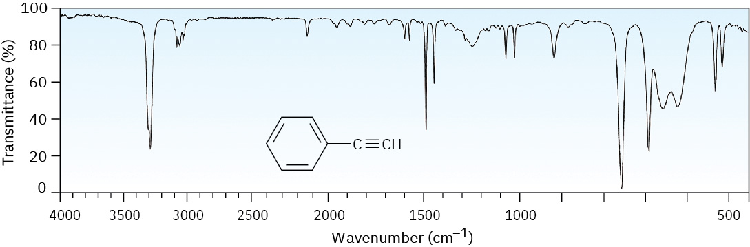
The of phenylacetylene, shown in Figure 12.29 at the end of this section, gives an example, clearly showing the following absorbances: ≡C–H stretch at 3300 –1, C–H stretches from the at 3000 to 3100 –1, C═C stretches of the between 1450 and 1600 –1, and out-of-plane bending of the ring’s C–H groups, indicating monosubstitution at 750 –1.

Alcohols

The O–H of alcohols is easy to spot. Alcohols have a characteristic band in the range 3400 to 3650 –1 that is usually broad and intense. between O–H groups is responsible for making the so broad. If an O–H stretch is present, it’s hard to miss this band or to confuse it with anything else.

A characteristic alcohol bond and its absorption value, indicated as a broad intense band at 3400 to 3650 inverse centimeters.
12.8 Infrared of Some Common — illustration 7 John McMurry, Organic Chemistry, OpenStax. · CC BY-NC-SA 4.0 · Original image ↗ · Access for free ↗

Cyclohexanol (Figure 12.25) is a good example.

An infrared spectrum in which a wide absorption band near 3300 inverse centimeters is labeled O H, and a sharp band below 1100 is labeled C single bond O.
Figure 12.25 — 12.8 Infrared of Some Common

Figure 12.25 IR spectrum of cyclohexanol.

John McMurry, Organic Chemistry, OpenStax. · CC BY-NC-SA 4.0 · Original image ↗ · Access for free ↗
The N–H of is also easy to spot in the , with a characteristic absorption in the 3300 to 3500 –1 range. Although alcohols absorb in the same range, an N–H absorption band is much sharper and less intense than an O–H band.

A characteristic amine bond and its absorption value, indicated as a sharp band at 3300 to 3500 inverse centimeters.
12.8 Infrared of Some Common — illustration 9 John McMurry, Organic Chemistry, OpenStax. · CC BY-NC-SA 4.0 · Original image ↗ · Access for free ↗
Primary (R–NH2) have two absorbances—one for the symmetric stretching mode and one for the asymmetric mode (Figure 12.26). Secondary (R2N–H) only have one N–H stretching in this region.

An infrared spectrum in which a sharp, two-pronged absorption band near 3300 inverse centimeters is labeled N H 2.
Figure 12.26 — 12.8 Infrared of Some Common

Figure 12.26 IR spectrum of cyclohexylamine.

John McMurry, Organic Chemistry, OpenStax. · CC BY-NC-SA 4.0 · Original image ↗ · Access for free ↗
are the easiest to identify of all absorptions because of their sharp, intense in the range 1670 to 1780 –1. Most important, the exact position of absorption within this range can often be used to identify the exact kind of —aldehyde, ketone, ester, and so forth.

ALDEHYDES

Saturated aldehydes absorb at 1730 –1; aldehydes next to either a double bond or an ring absorb at 1705 –1.

Characteristic aldehyde C O bonds and their absorption values, indicated at 1730 and 1705 inverse centimeters for alkyl and allyl or benzyl aldehydes respectively.
12.8 Infrared of Some Common — illustration 11 John McMurry, Organic Chemistry, OpenStax. · CC BY-NC-SA 4.0 · Original image ↗ · Access for free ↗

The C–H group attached to the is responsible for the characteristic for aldehydes at 2750 and 2850 –1 (Figure 12.27). Although these are not very intense, the at 2750 –1 is helpful when trying to distinguish between an aldehyde and a ketone.

An infrared spectrum in which a small absorption band near 2800 inverse centimeters is labeled aldehyde C H, and a strong band near 1700 is labeled aldehyde C O.
Figure 12.27 — 12.8 Infrared of Some Common

Figure 12.27 The IR spectrum of benzaldehyde.

John McMurry, Organic Chemistry, OpenStax. · CC BY-NC-SA 4.0 · Original image ↗ · Access for free ↗
Saturated open-chain and six-membered cyclic absorb at 1715 –1. Ring strain stiffens the C═O bond, making five-membered cyclic absorb at 1750 –1 and four-membered cyclic absorb at 1780 –1, about 20 to 30 –1 lower than the corresponding saturated ketone.

Characteristic ketone C O bonds and their absorption values, indicated at values just above 1700 inverse centimeters for alkyl ketones and values just below 1700 for allyl and benzyl ketones.
12.8 Infrared of Some Common — illustration 13 John McMurry, Organic Chemistry, OpenStax. · CC BY-NC-SA 4.0 · Original image ↗ · Access for free ↗

ESTERS

Saturated esters have a C═O at 1735 –1 and two strong absorbances in the 1300 to 1000 –1 range from the C–O portion of the . Like other , esters next to either an ring or a double bond absorb at 1715 –1, about 20 to 30 –1 lower than a saturated ester.

Characteristic ester C O bonds and their absorption values, indicated at 1735 inverse centimeters for alkyl esters and 1715 for allyl and benzyl esters.
12.8 Infrared of Some Common — illustration 14 John McMurry, Organic Chemistry, OpenStax. · CC BY-NC-SA 4.0 · Original image ↗ · Access for free ↗

Worked Example 12.5

Predicting Absorptions of

Where might the following have absorptions?

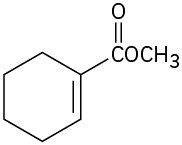
A chemical structure of 1-cyclohexen-1-ylmethanol
12.8 Infrared of Some Common — illustration 15 John McMurry, Organic Chemistry, OpenStax. · CC BY-NC-SA 4.0 · Original image ↗ · Access for free ↗
A chemical structure of a methyl ester with a five-carbon chain off the carbonyl. There is a terminal alkyne and a methyl on the beta C.
12.8 Infrared of Some Common — illustration 16 John McMurry, Organic Chemistry, OpenStax. · CC BY-NC-SA 4.0 · Original image ↗ · Access for free ↗

Strategy

Identify the in each , and then check Table 12.1 to see where those groups absorb.

(a) Absorptions: 3400 to 3650 –1 (O–H), 3020 to 3100 –1 (=C–H), 1640 to 1680 –1 (C═C). This has an alcohol O–H group and an alkene double bond.

(b) Absorptions: 3300 –1 (≡C–H), 2100 to 2260 –1 (C≡C), 1735 –1 (C═O). This has a terminal alkyne triple bond and a saturated ester .

Worked Example 12.6

Identifying from an

The of an unknown is shown in Figure 12.28. What does the contain?

An infrared spectrum of an unknown compound. Significant absorption bands are at 700,1000 and 1700 centimeter inverse.
Figure 12.28 — 12.8 Infrared of Some Common

Figure 12.28 IR spectrum for Worked Example 12.6 .

John McMurry, Organic Chemistry, OpenStax. · CC BY-NC-SA 4.0 · Original image ↗ · Access for free ↗
Strategy

All have many absorptions, but those useful for identifying specific are usually found in the region from 1500 –1 to 3300 –1. Pay particular attention to the region (1670 to 1780 –1), the region (1660 to 2000 –1), the triple-bond region (2000 to 2500 –1), and the C–H region (2500 to 3500 –1).

The shows an intense absorption at 1725 –1 due to a (perhaps an aldehyde, –CHO), a series of weak absorptions from 1800 to 2000 –1 characteristic of , and a C–H absorption near 3030 –1, also characteristic of . In , the is phenylacetaldehyde.
Chemical structure structure of phenylacetaldehyde.
12.8 Infrared of Some Common — illustration 18 John McMurry, Organic Chemistry, OpenStax. · CC BY-NC-SA 4.0 · Original image ↗ · Access for free ↗
Problem 12-9

The of phenylacetylene is shown in Figure 12.29. What absorption bands can you identify?

An infrared spectrum with xignificant absorption bands near 3300 and 1500 inverse centimeters, and in the fingerprint region.
Figure 12.29 — 12.8 Infrared of Some Common

Figure 12.29 The IR spectrum of phenylacetylene, Problem 12-9.

John McMurry, Organic Chemistry, OpenStax. · CC BY-NC-SA 4.0 · Original image ↗ · Access for free ↗
Problem 12-10
Where might the following have absorptions?
(a)
(b)
(c)
Problem 12-11

Where might the following have absorptions?

13.6 Spin–Spin Splitting in 1 H NMR Spectra

OpenStax · 13.6 Spin–Spin Splitting in 1 H NMR Spectra ↗
Instructional page including learning objectives, examples, figures, source tables and questions. Site navigation and embedded media players are omitted; source media links remain available.

13.6 • in 1H

In the 1H we’ve seen thus far, each different kind of in a has given rise to a single . It often happens, though, that the absorption of a splits into multiple , called a multiplet. For example, in the 1H of bromoethane shown in Figure 13.8, the –CH2Br appear as four (a quartet) centered at 3.42 δ and the –CH3 appear as three (a triplet) centered at 1.68 δ.

1 H N M R spectrum of bromoethane shows singlet peak at 0 (T M S), triplet peak at 1.8, and quartet peak at 3.4 parts per million.
Figure 13.8 — 13.6 in 1 H

Figure 13.8 The 1 H NMR spectrum of bromoethane, CH 3 CH 2 Br. The –CH 2 Br protons appear as a quartet at 3.42 δ , and the –CH 3 protons appear as a triplet at 1.68 δ .

John McMurry, Organic Chemistry, OpenStax. · CC BY-NC-SA 4.0 · Original image ↗ · Access for free ↗


Called , multiple absorptions of a are caused by the interaction, or coupling, of the spins of nearby . In other words, the tiny produced by one affects the felt by a neighboring . Look at the –CH3 in bromoethane, for example. The three equivalent –CH3 are neighbored by two other magnetic —the two on the adjacent –CH2Br group. Each of the neighboring –CH2Br has its own spin, which can align either with or against the applied field, producing a tiny effect that is felt by the –CH3 .

There are three ways in which the spins of the two –CH2Br can align, as shown in Figure 13.9. If both spins align with the applied field, the total effective field felt by the neighboring –CH3 is slightly larger than it would be otherwise. Consequently, the applied field necessary to cause is slightly reduced. Alternatively, if one of the –CH2Br spins aligns with the field and one aligns against the field, there is no effect on the neighboring –CH3 . (This arrangement can occur in two ways, depending on which of the two spins aligns which way.) Finally, if both –CH2Br spins align against the applied field, the effective field felt by the –CH3 is slightly smaller than it would be otherwise, and the applied field needed for is slightly increased.

C H 2 B r splits into four branches and C H 3 splits into three branches, each with arrows labeled B proton and J values equals 7 Hertz.
Figure 13.9 — 13.6 in 1 H

Figure 13.9 The origin of spin–spin splitting in bromoethane. The nuclear spins of neighboring protons, indicated by horizontal arrows, align either with or against the applied field, causing the splitting of absorptions into multiplets.

John McMurry, Organic Chemistry, OpenStax. · CC BY-NC-SA 4.0 · Original image ↗ · Access for free ↗

Any given has only one of the three possible alignments of –CH2Br spins, but in a large collection of , all three spin states are represented in a 1 : 2 : 1 statistical ratio. We therefore find that the neighboring –CH3 come into at three slightly different values of the applied field, and we see a 1 : 2 : 1 triplet in the . One is a little above where it would be without coupling, one is at the same place it would be without coupling, and the third is a little below where it would be without coupling.

In the same way that the –CH3 absorption of bromoethane is split into a triplet, the –CH2Br absorption is split into a quartet. The three spins of the neighboring –CH3 can align in four possible combinations: all three with the applied field, two with and one against (three ways), one with and two against (three ways), or all three against. Thus, four are produced for the –CH2Br in a 1 : 3 : 3 : 1 ratio.

As a general rule, called the n + 1 rule, that have n equivalent neighboring show n + 1 in their . For example, the of 2-bromopropane in Figure 13.10 shows a doublet at 1.71 δ and a seven-line multiplet, or septet, at 4.28 δ. The septet is caused by splitting of the –CHBr– signal by six equivalent neighboring on the two (n = 6 leads to 6 + 1 = 7 ). The doublet is due to signal splitting of the six equivalent by the single –CHBr– (n = 1 leads to 2 ). Integration confirms the expected 6 : 1 ratio.

1 H N M R spectrum of 2-bromopropane shows singlet peak at 0 (T M S), doublet peak at 1.8, and septet peak at 4.2 parts per million.
Figure 13.10 — 13.6 in 1 H

Figure 13.10 The 1 H NMR spectrum of 2-bromopropane. The –CH 3 proton signal at 1.71 δ is split into a doublet, and the –CHBr– proton signal at 4.28 δ is split into a septet. Note that the distance between peaks is the same in both multiplets. Note also that the two outer peaks of the septet are small enough to be nearly missed.

John McMurry, Organic Chemistry, OpenStax. · CC BY-NC-SA 4.0 · Original image ↗ · Access for free ↗
The distance between in a multiplet is called the coupling constant and is denoted J. Coupling constants are measured in hertz and generally fall in the range 0 to 18 Hz. The exact value of the coupling constant between two neighboring depends on the geometry of the , but a typical value for an open-chain alkane is J = 6 to 8 Hz. The same coupling constant is shared by both groups of hydrogens whose spins are coupled and is independent of spectrometer field strength. In bromoethane, for instance, the –CH2Br are coupled to the  –CH3 and appear as a quartet with J = 7 Hz. The –CH3 appear as a triplet with the same J = 7 Hz coupling constant.

Because coupling is a reciprocal interaction between two adjacent groups of , it’s sometimes possible to tell which multiplets in a complex are related to each other. If two multiplets have the same coupling constant, they are probably related, and the causing those multiplets are therefore adjacent in the .

The most commonly observed coupling patterns and the relative of lines in their multiplets are listed in Table 13.4. Note that it’s not possible for a given to have five equivalent neighboring . (Why not?) A six-line multiplet, or sextet, is therefore found only when a has five nonequivalent neighboring that coincidentally happen to be coupled with an identical coupling constant J.

Table 13.4 Some Common Spin Multiplicities
Number of equivalent adjacent Multiplet Ratio of
0 Singlet 1
1 Doublet 1 : 1
2 Triplet 1 : 2 : 1
3 Quartet 1 : 3 : 3 : 1
4 Quintet 1 : 4 : 6 : 4 : 1
6 Septet 1 : 6 : 15 : 20 : 15 : 6 : 1

in 1H can be summarized by three rules.

RULE 1
Chemically equivalent don’t show . Equivalent may be on the same carbon or on different carbons, but their signals don’t split.

A structure of chloromethane with text indicating all protons are chemically equivalent, no splitting. A structure of 1,2-dichloroethane with text indicating all protons are chemically equivalent, no splitting.
13.6 in 1 H — illustration 4 John McMurry, Organic Chemistry, OpenStax. · CC BY-NC-SA 4.0 · Original image ↗ · Access for free ↗
RULE 2
The signal of a with n equivalent neighboring is split into a multiplet of n + 1 with coupling constant J. that are farther than two carbon apart don’t usually couple, although they sometimes show weak coupling when they are separated by a π bond.

First structure labeled splitting observed shows adjacent carbon atoms, each bonded to hydrogen. Second structure labeled splitting not usually observed has 3-carbon chain, hydrogens on non-adjacent carbons.
13.6 in 1 H — illustration 5 John McMurry, Organic Chemistry, OpenStax. · CC BY-NC-SA 4.0 · Original image ↗ · Access for free ↗
RULE 3
Two groups of coupled to each other have the same coupling constant, J.

The of para-methoxypropiophenone in Figure 13.11 further illustrates these three rules. The downfield absorptions at 6.91 and 7.93 δ are due to the four -ring . There are two kinds of , each of which gives a signal that is split into a doublet by its neighbor. The –OCH3 signal is unsplit and appears as a sharp singlet at 3.84 δ. The –CH2– next to the appear at 2.93 δ in the region expected for on carbon next to an unsaturated center, and their signal is split into a quartet by coupling with the of the neighboring . The appear as a triplet at 1.20 δ in the usual upfield region.

The H N M R spectrum of para-methoxypropiophenone shows peaks at 0 (T M S), 1.2 (triplet, methyl), 2.9 (quartet, methylene), 3.9 (singlet, methoxy), 6.9 and 7.9 (both doublets, benzene).
Figure 13.11 — 13.6 in 1 H

Figure 13.11 The 1 H NMR spectrum of para -methoxypropiophenone.

John McMurry, Organic Chemistry, OpenStax. · CC BY-NC-SA 4.0 · Original image ↗ · Access for free ↗
Worked Example 13.2

Assigning a Chemical Structure from a 1H

Propose a structure for a , C5H12O, that fits the following 1H data: 0.92 δ (3 H, triplet, J = 7 Hz), 1.20 δ (6 H, singlet), 1.50 δ (2 H, quartet, J = 7 Hz), 1.64 δ (1 H, broad singlet).

Strategy

It’s best to begin solving structural problems by calculating a ’s degree of unsaturation (we’ll see this again in Worked Example 13.4). In the present instance, a formula of C5H12O corresponds to a saturated, open-chain , either an alcohol or an ether.

To interpret the information, let’s look at each absorption individually. The three- absorption at 0.92 δ is due to a in an alkane-like environment, and the triplet-splitting pattern implies that the CH3 is next to a CH2. Thus, our contains an ethyl group, CH3CH2–. The six- singlet at 1.20 δ is due to two equivalent alkane-like attached to a carbon with no hydrogens, (CH3)2C, and the two- quartet at 1.50 δ is due to the CH2 of the ethyl group. All 5 carbons and 11 of the 12 hydrogens in the are now accounted for. The remaining hydrogen, which appears as a broad one- singlet at 1.64 δ, is probably due to an OH group, since there is no other way to account for it. Putting the pieces together gives the structure: 2--2-butanol.

Structure of 2-methyl-2-butanol with shifts for hydrogens on: C 1 and methyl (1.20), O H (1.64), C 3 (1.50), and C 4 (0.92).
13.6 in 1 H — illustration 7 John McMurry, Organic Chemistry, OpenStax. · CC BY-NC-SA 4.0 · Original image ↗ · Access for free ↗
Problem 13-9
Predict the splitting patterns you would expect for each in the following :
(a)
(b)
(c)
(d)
(e)
(f)
Problem 13-10
Draw structures for that meet the following descriptions:
(a)
C2H6O; one singlet
(b)
C3H7Cl; one doublet and one septet
(c)
C4H8Cl2O; two triplets
(d)
C4H8O2; one singlet, one triplet, and one quartet
Problem 13-11

The integrated 1H of a of formula C4H10O is shown in Figure 13.12. Propose a structure.

The 1 H N M R spectrum of a compound has peaks at 0 (T M S), 1.2 (triplet), and 3.5 (quartet).
Figure 13.12 — 13.6 in 1 H

Figure 13.12 An integrated 1 H NMR spectrum for Problem 11.

John McMurry, Organic Chemistry, OpenStax. · CC BY-NC-SA 4.0 · Original image ↗ · Access for free ↗

14.8 Interpreting Ultraviolet Spectra: The Effect of Conjugation

OpenStax · 14.8 Interpreting Ultraviolet Spectra: The Effect of Conjugation ↗

Instructional page including learning objectives, examples, figures, source tables and questions. Site navigation and embedded media players are omitted; source media links remain available.

14.8 • Interpreting : The Effect of

The necessary to effect the π → π* transition in a conjugated depends on the energy gap between HOMO and LUMO, which in turn depends on the nature of the . Thus, by measuring the of an unknown, we can derive structural information about the nature of any conjugated π system present in a .

One of the most important factors affecting the of absorption by a is the extent of . Molecular calculations show that the energy difference between HOMO and LUMO decreases as the extent of increases. Thus, 1,3-butadiene absorbs at λmax = 217 nm, 1,3,5-hexatriene absorbs at λmax = 258 nm, and 1,3,5,7-octatetraene absorbs at λmax = 290 nm. (Remember: longer means lower energy.)

Other kinds of , such as conjugated enones and rings, also have characteristic absorptions that are useful in structure . The absorption maxima of some representative conjugated are given in Table 14.2.


Table 14.2 Absorptions of Some Conjugated
Name Structure λmax (nm)
2--1,3-butadiene 220
1,3-Cyclohexadiene 256
1,3,5-Hexatriene H2C=CH―CH=CH–CH=CH2 258
1,3,5,7-Octatetraene H2C=CH–CH=CH–CH=CH–CH=CH2 290
3-Buten-2-one 219
254
Problem 14-15

Which of the following would you expect to show absorptions in the 200 to 400 nm range?

(a)   (b)   (c)   (d)  (e) 
(f)