State the sign convention and distinguish real rays meeting from backward extensions meeting.
Reflection, refraction, and dispersion
The angle of reflection equals the angle of incidence, measured from the normal. Snell’s law is n₁ sin θ₁ = n₂ sin θ₂. Entering higher index bends a ray toward the normal. Total internal reflection requires travel from higher to lower refractive index and incidence above θc, where sin θc = nlow/nhigh. It cannot occur for incidence from the lower-index side. Dispersion means refractive index depends on wavelength; a prism separates colors because they refract differently. Frequency is unchanged at the stationary interface.
For paraxial thin lenses and spherical mirrors, 1/f = 1/do + 1/di and magnification m = −di/do. In a common real-is-positive convention, a real object has do > 0, a real image di > 0, and converging elements f > 0. A negative di indicates a virtual image; negative magnification indicates inversion. A plane mirror gives a virtual upright image at the same distance behind the mirror as the object is in front. Ray rules differ for reflection and transmission, so sketch rays before interpreting signs.
Optical power is P = 1/f with f in meters, measured in diopters. Thin lenses in contact approximately add powers; separated lenses must be treated sequentially. The first image becomes the second lens’s object, which may be virtual depending on its position relative to incoming rays. A converging lens makes a real inverted image when a real object lies beyond its focal point, and a virtual upright enlarged image when the object lies inside. A diverging lens produces a virtual upright diminished image from an ordinary real object.
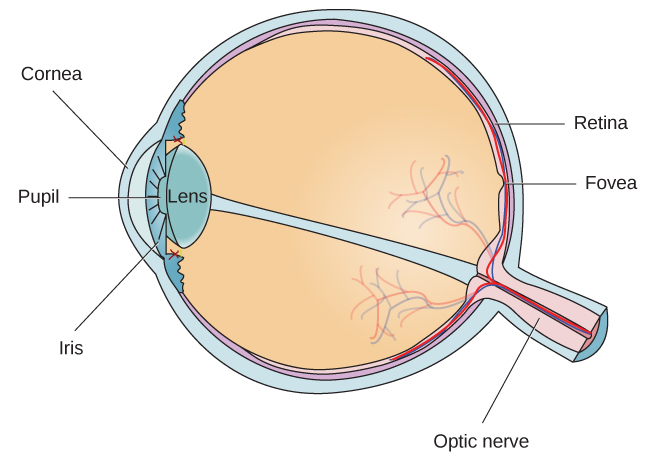
The eye focuses light on the retina; accommodation increases lens power for nearer objects. Myopia focuses distant light before the retina and is corrected with a diverging lens. Hyperopia requires additional converging power for near focus. Microscopes combine an objective’s enlarged real image with an eyepiece’s angular magnification; telescopes enlarge apparent angular separation of distant objects. Spherical aberration occurs because off-axis-height rays need not share a focus; chromatic aberration arises from wavelength-dependent refraction. Diffraction also limits resolution even with otherwise perfect optics.
Try before revealing the answer. A converging thin lens has focal length 10 cm and an object 30 cm away. Find image distance, orientation, magnification, and lens power.
Worked answer
1/di = 1/10 − 1/30 = 1/15 cm⁻¹, so di = +15 cm: a real image. m = −15/30 = −0.5, meaning inverted and half the object’s height. Power uses meters: P = 1/0.10 = +10 diopters.
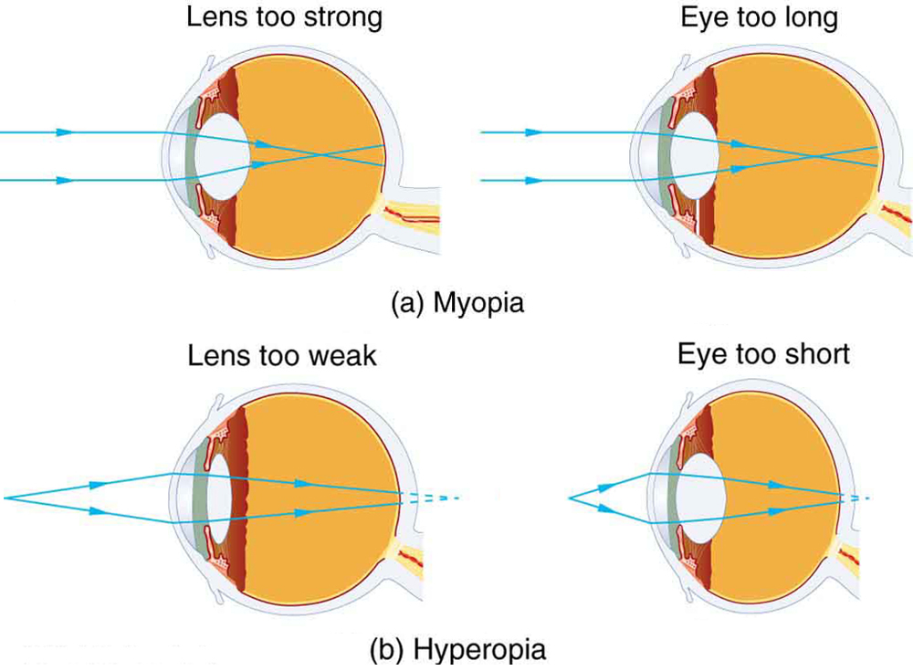
Figure 26.5 (a) The nearsighted (myopic) eye converges rays from a distant object in front of the retina; thus, they are diverging when they strike the retina, producing a blurry image. This can be caused by the lens of the eye being too powerful or the length of the eye being too great. (b) The farsighted (hyperopic) eye is unable to converge the rays from a close object by the time they strike the retina, producing blurry close vision. This can be caused by insufficient power in the lens or by the eye being too short.
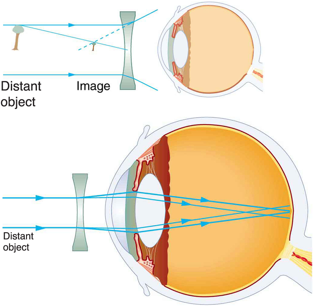
Figure 26.6 Correction of nearsightedness requires a diverging lens that compensates for the overconvergence by the eye. The diverging lens produces an image closer to the eye than the object, so that the nearsighted person can see it clearly.
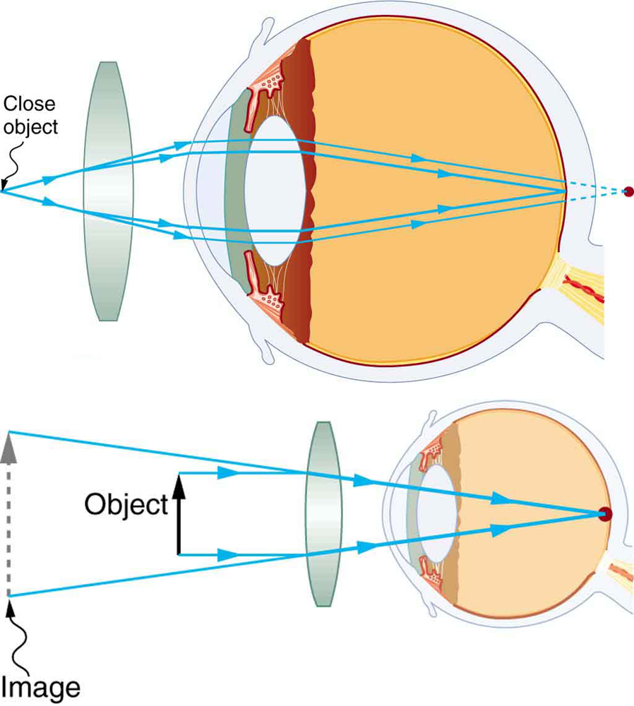
Figure 26.7 Correction of farsightedness uses a converging lens that compensates for the under convergence by the eye. The converging lens produces an image farther from the eye than the object, so that the farsighted person can see it clearly.
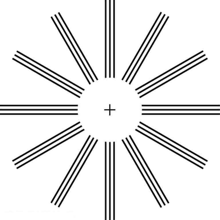
Figure 26.8 This chart can detect astigmatism, unevenness in the focus of the eye. Check each of your eyes separately by looking at the center cross (without spectacles if you wear them). If lines along some axes appear darker or clearer than others, you have an astigmatism.
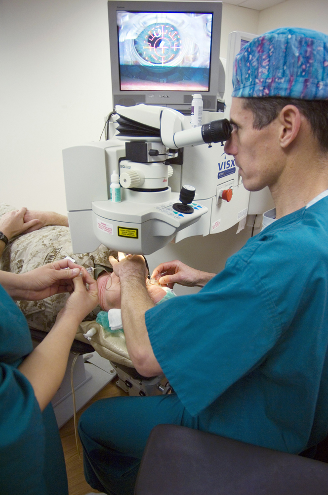
Figure 26.9 Laser vision correction is being performed using the LASIK procedure. Reshaping of the cornea by laser ablation is based on a careful assessment of the patient’s vision and is computer controlled. The upper corneal layer is temporarily peeled back and minimally disturbed in LASIK, providing for more rapid and less painful healing of the less sensitive tissues below. (credit: U.S. Navy photo by Mass Communication Specialist 1st Class Brien Aho)
Paul Peter Urone and Roger Hinrichs, College Physics 2E, OpenStax. (credit: U.S. Navy photo by Mass Communication Specialist 1st Class Brien Aho) · CC BY-NC-SA 4.0
· Original image ↗ · Access for free ↗
A nitrogen-containing functional group. A free amino acid’s amino group is typically protonated (NH₃⁺) near physiological pH; proline’s ring nitrogen is an exception to this formula.
Figure 15.4 This figure shows the genetic code for translating each nucleotide triplet in mRNA into an amino acid or a termination signal in a protein. (credit: modification of work by NIH)
An electrical property that can be positive or negative. Opposite charges attract and like charges repel; net charge is the sum of all charges on a particle.
Figure 4.18 - Kinetics of an enzyme obeying Michaelis-Menten kinetics. Image by Aleia Kim
Kevin Ahern, Indira Rajagopal, and Taralyn Tan, Biochemistry Free For All, Oregon State University, via Biology LibreTexts. Image by Aleia Kim · CC BY-NC-SA 4.0
· Original image ↗ · Access for free ↗
Equilibrium
A state where opposing processes occur at equal rates, so macroscopic concentrations or other properties remain constant.
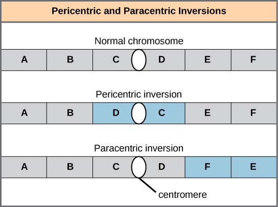
Figure 13.11 Pericentric inversions include the centromere, and paracentric inversions do not. A pericentric inversion can change the chromosome arms' relative lengths. A paracentric inversion cannot.
The exponent needed to obtain a number from a chosen fixed number. For decimal logarithms, log₁₀(100) = 2 because 10² = 100; pH uses the negative decimal logarithm of hydrogen ion activity.
A thin boundary or sheet. Cell membranes are lipid bilayers containing proteins; blotting membranes are synthetic supports that hold proteins or nucleic acids for detection.
Figure 15.11 Eukaryotic pre-mRNA processing. In addition to 5’ Cap and 3’ Poly-A Tail addition, introns must be precisely removed and exons joined to generate a functional mRNA. Nucleotides upstream (towards the 5’cap) of the translation START codon are part of the 5’ untranslated region (5’ UTR). Nucleotides downstream (towards 3’end) of the STOP codon form the 3’ UTR. Both 5’ and 3’ UTRs are important for regulating translation initiation and mRNA stability. Credit: Rao, A., Ryan, K. Fletcher, S. and Tag, A. Department of Biology, Texas A&M University.
Rao, A., Ryan, K. Fletcher, S. and Tag, A. Department of Biology, Texas A&M University.; Mary Ann Clark, Matthew Douglas, and Jung Choi, Biology 2e, OpenStax. · CC BY-NC-SA 4.0
· Original image ↗ · Access for free ↗
Figure 12.19 Reaction diagrams for an endothermic process in the absence (red curve) and presence (blue curve) of a catalyst. The catalyzed pathway involves a two-step mechanism (note the presence of two transition states) and an intermediate species (represented by the valley between the two transitions states).
Figure 7.45 - Non-homologous end joining (left) versus homologous recombination (right) - Wikipedia
Wikipedia, as credited by Kevin Ahern, Indira Rajagopal, and Taralyn Tan, Biochemistry Free For All, Oregon State University, via Biology LibreTexts. · Source attribution
· Original image ↗ · Access for free ↗
Snell’s law
Relationship n₁ sin θ₁ = n₂ sin θ₂ for refraction at a boundary.
The amount of a substance that can dissolve in a solvent under specified conditions. Soluble means able to dissolve; insoluble means only very slightly soluble in that solvent.
Figure 15.7 Messenger RNA is a copy of protein-coding information in the coding strand of DNA, with the substitution of U in the RNA for T in the coding sequence. However, new RNA nucleotides base pair with the nucleotides of the template strand. RNA is synthesized in its 5'-3' direction, using the enzyme RNA polymerase. As the template is read, the DNA unwinds ahead of the polymerase and then rewinds behind it.
A fleeting, high-energy arrangement at a reaction barrier. It is an energy maximum along the reaction path, rather than a stable reaction intermediate.
Figure 12.19 Reaction diagrams for an endothermic process in the absence (red curve) and presence (blue curve) of a catalyst. The catalyzed pathway involves a two-step mechanism (note the presence of two transition states) and an intermediate species (represented by the valley between the two transitions states).
Figure 15.18 Translation begins when an initiator tRNA anticodon recognizes a start codon on mRNA bound to a small ribosomal subunit. The large ribosomal subunit joins the small subunit, and a second tRNA is recruited. As the mRNA moves relative to the ribosome, successive tRNAs move through the ribosome and the polypeptide chain is formed. Entry of a release factor into the A site terminates translation and the components dissociate.
Figure 4.21 - Kcat (turnover number) values for several enzymes. Image by Aleia Kim
Kevin Ahern, Indira Rajagopal, and Taralyn Tan, Biochemistry Free For All, Oregon State University, via Biology LibreTexts. Image by Aleia Kim · CC BY-NC-SA 4.0
· Original image ↗ · Access for free ↗
No matching terms. Try a different name or abbreviation.
College Physics 2E 25 — Section Summary
OpenStax · College Physics 2E 25 — Section Summary ↗ Instructional page including learning objectives, examples, figures, source tables and questions. Site navigation and embedded media players are omitted; source media links remain available.
A straight line that originates at some point is called a ray.
The part of optics dealing with the ray aspect of light is called geometric optics.
Light can travel in three ways from a source to another location: (1) directly from the source through empty space; (2) through various media; (3) after being reflected from a mirror.
The incident angle that produces an angle of refraction of is called critical angle.
Total internal reflection is a phenomenon that occurs at the boundary between two mediums, such that if the incident angle in the first medium is greater than the critical angle, then all the light is reflected back into that medium.
Fiber optics involves the transmission of light down fibers of plastic or glass, applying the principle of total internal reflection.
Endoscopes are used to explore the body through various orifices or minor incisions, based on the transmission of light through optical fibers.
Cladding prevents light from being transmitted between fibers in a bundle.
Light rays entering a converging lens parallel to its axis cross one another at a single point on the opposite side.
For a converging lens, the focal point is the point at which converging light rays cross; for a diverging lens, the focal point is the point from which diverging light rays appear to originate.
The distance from the center of the lens to its focal point is called the focal length .
A lens that causes the light rays to bend away from its axis is called a diverging lens.
Ray tracing is the technique of graphically determining the paths that light rays take.
The image in which light rays from one point on the object actually cross at the location of the image and can be projected onto a screen, a piece of film, or the retina of an eye is called a real image.
Thin lens equations are and (magnification).
The distance of the image from the center of the lens is called image distance.
An image that is on the same side of the lens as the object and cannot be projected on a screen is called a virtual image.
The characteristics of an image formed by a flat mirror are: (a) The image and object are the same distance from the mirror, (b) The image is a virtual image, and (c) The image is situated behind the mirror.
Image length is half the radius of curvature.
A convex mirror is a diverging mirror and forms only one type of image, namely a virtual image.
26.2 Vision Correction
OpenStax · 26.2 Vision Correction ↗ Instructional page including learning objectives, examples, figures, source tables and questions. Site navigation and embedded media players are omitted; source media links remain available.
Learning Objectives
By the end of this section, you will be able to:
Identify and discuss common vision defects.
Explain nearsightedness and farsightedness corrections.
Explain laser vision correction.
The need for some type of vision correction is very common. Common vision defects are easy to understand, and some are simple to correct. Figure 26.5 illustrates two common vision defects. Nearsightedness, or myopia, is the inability to see distant objects clearly while close objects are clear. The eye overconverges the nearly parallel rays from a distant object, and the rays cross in front of the retina. More divergent rays from a close object are converged on the retina for a clear image. The distance to the farthest object that can be seen clearly is called the far point of the eye (normally infinity). Farsightedness, or hyperopia, is the inability to see close objects clearly while distant objects may be clear. A farsighted eye does not converge sufficient rays from a close object to make the rays meet on the retina. Less diverging rays from a distant object can be converged for a clear image. The distance to the closest object that can be seen clearly is called the near point of the eye (normally 25 cm).
Figure 26.5 — 26.2 Vision Correction
Figure 26.5 (a) The nearsighted (myopic) eye converges rays from a distant object in front of the retina; thus, they are diverging when they strike the retina, producing a blurry image. This can be caused by the lens of the eye being too powerful or the length of the eye being too great. (b) The farsighted (hyperopic) eye is unable to converge the rays from a close object by the time they strike the retina, producing blurry close vision. This can be caused by insufficient power in the lens or by the eye being too short.
Since the nearsighted eye over converges light rays, the correction for nearsightedness is to place a diverging spectacle lens in front of the eye. This reduces the power of an eye that is too powerful. Another way of thinking about this is that a diverging spectacle lens produces a case 3 image, which is closer to the eye than the object (see Figure 26.6). To determine the spectacle power needed for correction, you must know the person’s far point—that is, you must know the greatest distance at which the person can see clearly. Then the image produced by a spectacle lens must be at this distance or closer for the nearsighted person to be able to see it clearly. It is worth noting that wearing glasses does not change the eye in any way. The eyeglass lens is simply used to create an image of the object at a distance where the nearsighted person can see it clearly. Whereas someone not wearing glasses can see clearly objects that fall between their near point and their far point, someone wearing glasses can see images that fall between their near point and their far point.
Figure 26.6 — 26.2 Vision Correction
Figure 26.6 Correction of nearsightedness requires a diverging lens that compensates for the overconvergence by the eye. The diverging lens produces an image closer to the eye than the object, so that the nearsighted person can see it clearly.
What power of spectacle lens is needed to correct the vision of a nearsighted person whose far point is 30.0 cm? Assume the spectacle (corrective) lens is held 1.50 cm away from the eye by eyeglass frames.
Strategy
You want this nearsighted person to be able to see very distant objects clearly. That means the spectacle lens must produce an image 30.0 cm from the eye for an object very far away. An image 30.0 cm from the eye will be 28.5 cm to the left of the spectacle lens (see Figure 26.6). Therefore, we must get when . The image distance is negative, because it is on the same side of the spectacle as the object.
Since and are known, the power of the spectacle lens can be found using as written earlier:
26.9
Since , we obtain:
26.10
Discussion
The negative power indicates a diverging (or concave) lens, as expected. The spectacle produces a case 3 image closer to the eye, where the person can see it. If you examine eyeglasses for nearsighted people, you will find the lenses are thinnest in the center. Additionally, if you examine a prescription for eyeglasses for nearsighted people, you will find that the prescribed power is negative and given in units of diopters.
Since the farsighted eye under converges light rays, the correction for farsightedness is to place a converging spectacle lens in front of the eye. This increases the power of an eye that is too weak. Another way of thinking about this is that a converging spectacle lens produces a case 2 image, which is farther from the eye than the object (see Figure 26.7). To determine the spectacle power needed for correction, you must know the person’s near point—that is, you must know the smallest distance at which the person can see clearly. Then the image produced by a spectacle lens must be at this distance or farther for the farsighted person to be able to see it clearly.
Figure 26.7 — 26.2 Vision Correction
Figure 26.7 Correction of farsightedness uses a converging lens that compensates for the under convergence by the eye. The converging lens produces an image farther from the eye than the object, so that the farsighted person can see it clearly.
What power of spectacle lens is needed to allow a farsighted person, whose near point is 1.00 m, to see an object clearly that is 25.0 cm away? Assume the spectacle (corrective) lens is held 1.50 cm away from the eye by eyeglass frames.
Strategy
When an object is held 25.0 cm from the person’s eyes, the spectacle lens must produce an image 1.00 m away (the near point). An image 1.00 m from the eye will be 98.5 cm to the left of the spectacle lens because the spectacle lens is 1.50 cm from the eye (see Figure 26.7). Therefore, . The image distance is negative, because it is on the same side of the spectacle as the object. The object is 23.5 cm to the left of the spectacle, so that .
Since and are known, the power of the spectacle lens can be found using :
26.11
Discussion
The positive power indicates a converging (convex) lens, as expected. The convex spectacle produces a case 2 image farther from the eye, where the person can see it. If you examine eyeglasses of farsighted people, you will find the lenses to be thickest in the center. In addition, a prescription of eyeglasses for farsighted people has a prescribed power that is positive.
Another common vision defect is astigmatism, an unevenness or asymmetry in the focus of the eye. For example, rays passing through a vertical region of the eye may focus closer than rays passing through a horizontal region, resulting in the image appearing elongated. This is mostly due to irregularities in the shape of the cornea but can also be due to lens irregularities or unevenness in the retina. Because of these irregularities, different parts of the lens system produce images at different locations. The eye-brain system can compensate for some of these irregularities, but they generally manifest themselves as less distinct vision or sharper images along certain axes. Figure 26.8 shows a chart used to detect astigmatism. Astigmatism can be at least partially corrected with a spectacle having the opposite irregularity of the eye. If an eyeglass prescription has a cylindrical correction, it is there to correct astigmatism. The normal corrections for short- or farsightedness are spherical corrections, uniform along all axes.
Figure 26.8 — 26.2 Vision Correction
Figure 26.8 This chart can detect astigmatism, unevenness in the focus of the eye. Check each of your eyes separately by looking at the center cross (without spectacles if you wear them). If lines along some axes appear darker or clearer than others, you have an astigmatism.
Contact lenses have advantages over glasses beyond their cosmetic aspects. One problem with glasses is that as the eye moves, it is not at a fixed distance from the spectacle lens. Contacts rest on and move with the eye, eliminating this problem. Because contacts cover a significant portion of the cornea, they provide superior peripheral vision compared with eyeglasses. Contacts also correct some corneal astigmatism caused by surface irregularities. The tear layer between the smooth contact and the cornea fills in the irregularities. Since the index of refraction of the tear layer and the cornea are very similar, you now have a regular optical surface in place of an irregular one. If the curvature of a contact lens is not the same as the cornea (as may be necessary with some individuals to obtain a comfortable fit), the tear layer between the contact and cornea acts as a lens. If the tear layer is thinner in the center than at the edges, it has a negative power, for example. Skilled optometrists will adjust the power of the contact to compensate.
Other advances in vision correction demonstrate the interconnectedness and value of scientific research. In the 1980s, Donna Strickland and Gérard Mourou worked on ways to make small but powerful lasers. Up until that time, powerful lasers had to be quite large in order to function properly. Essentially, the intensity of the beam itself would modify the instrument’s ability to function and create too much heat to be practical. Strickland and Mourou used ultrashort laser pulses passed over a grating that modified the beam but retained its power. Chirped pulse amplification, as it became known, has been used to develop most of the highest-powered lasers in the world, but also some of the smallest and most common. Decades after their initial discovery, Strickland and Mourou were awarded the Nobel Prize for Physics (with Strickland becoming the third woman to receive the award) partly due to CPA’s pivotal role in the increasingly common practice of laser vision correction—an application neither planned during their initial research.
Laser vision correction has progressed rapidly in the last few years. It is the latest and by far the most successful in a series of procedures that correct vision by reshaping the cornea. As noted at the beginning of this section, the cornea accounts for about two-thirds of the power of the eye. Thus, small adjustments of its curvature have the same effect as putting a lens in front of the eye. To a reasonable approximation, the power of multiple lenses placed close together equals the sum of their powers. For example, a concave spectacle lens (for nearsightedness) having has the same effect on vision as reducing the power of the eye itself by 3.00 D. So to correct the eye for nearsightedness, the cornea is flattened to reduce its power. Similarly, to correct for farsightedness, the curvature of the cornea is enhanced to increase the power of the eye—the same effect as the positive power spectacle lens used for farsightedness. Laser vision correction uses high intensity electromagnetic radiation to ablate (to remove material from the surface) and reshape the corneal surfaces.
Today, the most commonly used laser vision correction procedure is Laser in situ Keratomileusis (LASIK). The top layer of the cornea is surgically peeled back and the underlying tissue ablated by multiple bursts of finely controlled ultraviolet radiation produced by an excimer laser. Lasers are used because they not only produce well-focused intense light, but they also emit very pure wavelength electromagnetic radiation that can be controlled more accurately than mixed wavelength light. The 193 nm wavelengthUV commonly used is extremely and strongly absorbed by corneal tissue, allowing precise evaporation of very thin layers. A computer controlled program applies more bursts, usually at a rate of 10 per second, to the areas that require deeper removal. Typically a spot less than 1 mm in diameter and about in thickness is removed by each burst. Nearsightedness, farsightedness, and astigmatism can be corrected with an accuracy that produces normal distant vision in more than 90% of the patients, in many cases right away. The corneal flap is replaced; healing takes place rapidly and is nearly painless. More than 1 million Americans per year undergo LASIK (see Figure 26.9).
Figure 26.9 — 26.2 Vision Correction
Figure 26.9 Laser vision correction is being performed using the LASIK procedure. Reshaping of the cornea by laser ablation is based on a careful assessment of the patient’s vision and is computer controlled. The upper corneal layer is temporarily peeled back and minimally disturbed in LASIK, providing for more rapid and less painful healing of the less sensitive tissues below. (credit: U.S. Navy photo by Mass Communication Specialist 1st Class Brien Aho)
Paul Peter Urone and Roger Hinrichs, College Physics 2E, OpenStax. (credit: U.S. Navy photo by Mass Communication Specialist 1st Class Brien Aho) · CC BY-NC-SA 4.0
· Original image ↗ · Access for free ↗