Sound is a mechanical disturbance, usually longitudinal pressure oscillations in gases and liquids. It cannot propagate through vacuum. Frequency determines pitch; the medium and temperature determine speed. Sound generally travels faster in less compressible, stiffer media, although density also matters: v = √(B/ρ) in a fluid of bulk modulus B. In air near room temperature, v is about 343 m/s. The wavelength is v/f. Crossing a stationary interface preserves frequency while speed and wavelength change.
Intensity I is power per area, in W/m². For an isotropic point source without absorption, I = P/(4πr²); doubling distance reduces intensity fourfold. Sound level β = 10 log₁₀(I/I₀), with I₀ = 10⁻¹² W/m² for the conventional air reference. A 10 dB increase multiplies intensity by ten; a 3 dB increase approximately doubles it. Intensity is proportional to squared pressure amplitude in a given medium. Damping decreases amplitude and energy; it need not change the source frequency.
For sound in a stationary medium, fobserved = fsource(v ± vobserver)/(v ∓ vsource), choosing signs so approaching motion raises observed frequency. Source and observer speeds are relative to the medium. A moving source changes wavefront spacing; a moving observer changes encounter rate. A reflected ultrasound wave is shifted twice, first at the moving reflector and then on return. For reflector speed u much less than sound speed, Δf ≈ 2f₀u cos θ/v. Flow perpendicular to the beam gives little Doppler shift.
A string fixed at both ends and an ideal open–open pipe have fn = nv/(2L), n = 1,2,3… . An open–closed pipe supports odd harmonics fn = nv/(4L), n = 1,3,5…; its closed end is a displacementnode but pressure antinode. Ultrasound has frequency above ordinary human hearing, conventionally 20 kHz. Echo time gives depth d = vt/2. Higher frequency shortens wavelength and can improve resolution but often increases attenuation. A source moving faster than sound produces a shock front from accumulated wavefronts.
Try before revealing the answer. An echo returns 40 µs after an ultrasound pulse. Assume tissue sound speed 1500 m/s. Find depth. Separately, by how much does sound level fall when distance from an ideal point source doubles?
Worked answer
Round-trip distance = 1500 × 40 × 10⁻⁶ = 0.060 m, so depth is 0.030 m = 3 cm. Doubling distance makes intensity one quarter; Δβ = 10 log₁₀(1/4) ≈ −6 dB.
Figure 17.40 Ultrasound is used in medicine to painlessly and noninvasively monitor patient health and diagnose a wide range of disorders. (credit: abbybatchelder, Flickr)
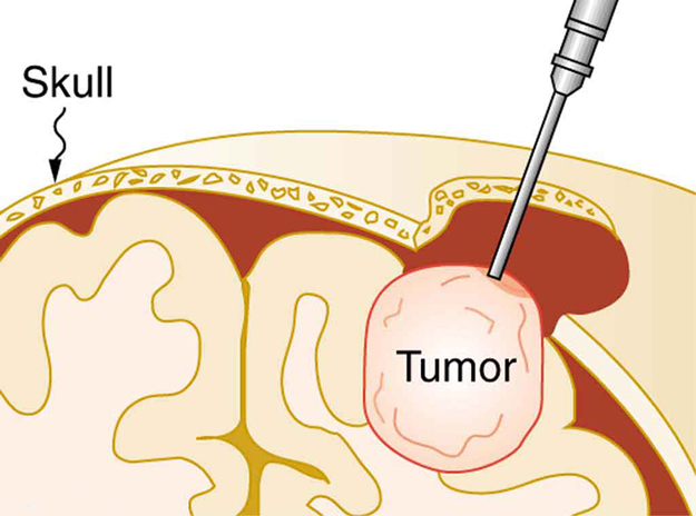
Figure 17.41 The tip of this small probe oscillates at 23 kHz with such a large amplitude that it pulverizes tissue on contact. The debris is then aspirated. The speed of the tip may exceed the speed of sound in tissue, thus creating shock waves and cavitation, rather than a smooth simple harmonic oscillator–type wave.
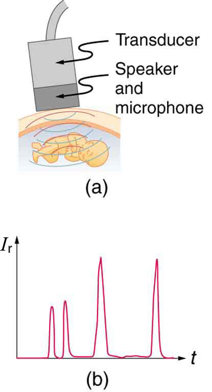
Figure 17.42 (a) An ultrasound speaker doubles as a microphone. Brief bleeps are broadcast, and echoes are recorded from various depths. (b) Graph of echo intensity versus time. The time for echoes to return is directly proportional to the distance of the reflector, yielding this information noninvasively.
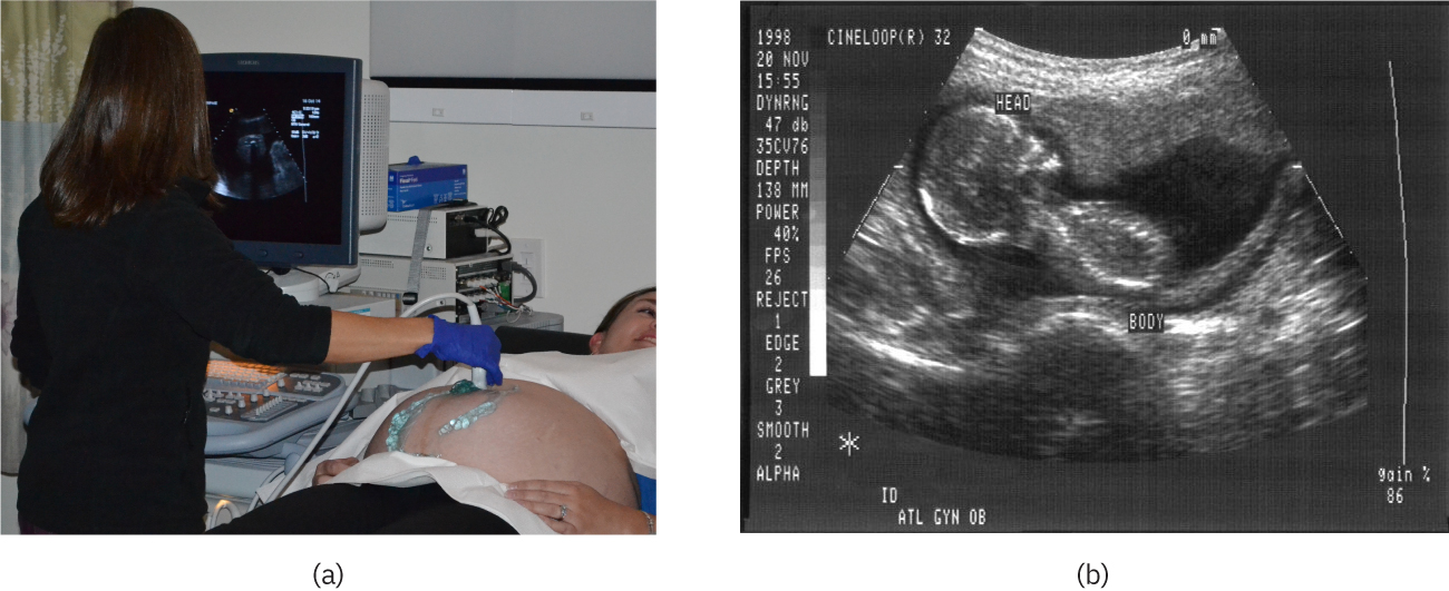
Figure 17.43 (a) An ultrasonic image is produced by sweeping the ultrasonic beam across the area of interest, in this case the person's abdomen. Data are recorded and analyzed in a computer, providing a two-dimensional image (credit: COD Newsroom, Flickr). (b) Ultrasound image of 12-week-old fetus. (credit: Public Health Image Library, CDC)
Paul Peter Urone and Roger Hinrichs, College Physics 2E, OpenStax. (credit: COD Newsroom, Flickr). (b) Ultrasound image of 12-week-old fetus. (credit: Public Health Image Library, CDC) · CC BY-NC-SA 4.0
· Original image ↗ · Access for free ↗Figure 17.44 — 17.7 Ultrasound
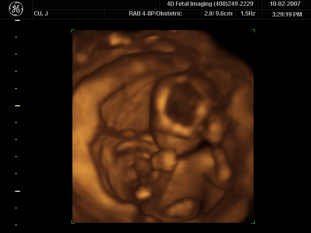
Figure 17.44 A 3D ultrasound image of a fetus. (credit: Jennie Cu, Wikimedia Commons)
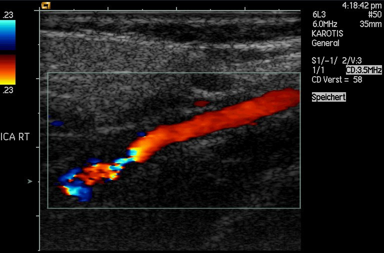
Figure 17.45 This Doppler-shifted ultrasonic image of a partially occluded artery uses color to indicate velocity. The highest velocities are in red, while the lowest are blue. The blood must move faster through the constriction to carry the same flow. (credit: Arning C, Grzyska U, Wikimedia Commons)
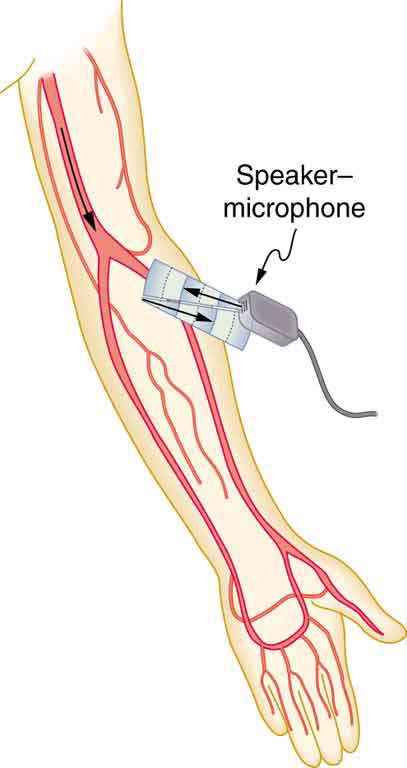
Figure 17.46 Ultrasound is partly reflected by blood cells and plasma back toward the speaker-microphone. Because the cells are moving, two Doppler shifts are produced—one for blood as a moving observer, and the other for the reflected sound coming from a moving source. The magnitude of the shift is directly proportional to blood velocity.
Table 17.5 The Ultrasound Properties of Various Media, Including Soft Tissue Found in the Body. Paul Peter Urone and Roger Hinrichs, College Physics 2E, OpenStax.
A nitrogen-containing functional group. A free amino acid’s amino group is typically protonated (NH₃⁺) near physiological pH; proline’s ring nitrogen is an exception to this formula.
Figure 10.15 Cell Division and Apoptosis. In an adult organism, normal cell division is balanced by apoptosis (programmed cell death) to maintain a constant cell number in homeostasis. Either an increase in cell division or a decrease in apoptosis leads an increase in the number of cells and tumor formation. Credit: Rao, A. and Ryan, K. Department of Biology, Texas A&M University.
Rao, A. and Ryan, K. Department of Biology, Texas A&M University. Mary Ann Clark, Matthew Douglas, and Jung Choi, Biology 2e, OpenStax. · CC BY-NC-SA 4.0
· Original image ↗ · Access for free ↗
Figure 15.4 This figure shows the genetic code for translating each nucleotide triplet in mRNA into an amino acid or a termination signal in a protein. (credit: modification of work by NIH)
An electrical property that can be positive or negative. Opposite charges attract and like charges repel; net charge is the sum of all charges on a particle.
Figure 15.10 A generalized promoter of a gene transcribed by RNA polymerase II is shown. Transcription factors recognize the promoter. RNA polymerase II then binds and forms the transcription initiation complex.
The exponent needed to obtain a number from a chosen fixed number. For decimal logarithms, log₁₀(100) = 2 because 10² = 100; pH uses the negative decimal logarithm of hydrogen ion activity.
A thin boundary or sheet. Cell membranes are lipid bilayers containing proteins; blotting membranes are synthetic supports that hold proteins or nucleic acids for detection.
Figure 15.11 Eukaryotic pre-mRNA processing. In addition to 5’ Cap and 3’ Poly-A Tail addition, introns must be precisely removed and exons joined to generate a functional mRNA. Nucleotides upstream (towards the 5’cap) of the translation START codon are part of the 5’ untranslated region (5’ UTR). Nucleotides downstream (towards 3’end) of the STOP codon form the 3’ UTR. Both 5’ and 3’ UTRs are important for regulating translation initiation and mRNA stability. Credit: Rao, A., Ryan, K. Fletcher, S. and Tag, A. Department of Biology, Texas A&M University.
Rao, A., Ryan, K. Fletcher, S. and Tag, A. Department of Biology, Texas A&M University.; Mary Ann Clark, Matthew Douglas, and Jung Choi, Biology 2e, OpenStax. · CC BY-NC-SA 4.0
· Original image ↗ · Access for free ↗
Figure 12.19 Reaction diagrams for an endothermic process in the absence (red curve) and presence (blue curve) of a catalyst. The catalyzed pathway involves a two-step mechanism (note the presence of two transition states) and an intermediate species (represented by the valley between the two transitions states).
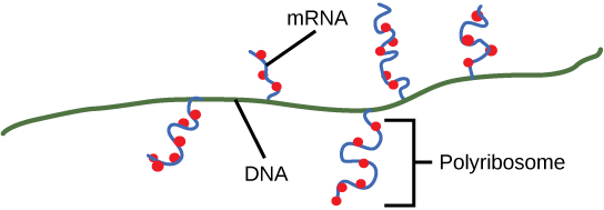
Figure 15.9 Multiple polymerases can transcribe a single bacterial gene while numerous ribosomes concurrently translate the mRNA transcripts into polypeptides. In this way, a specific protein can rapidly reach a high concentration in the bacterial cell.
Figure 7.45 - Non-homologous end joining (left) versus homologous recombination (right) - Wikipedia
Wikipedia, as credited by Kevin Ahern, Indira Rajagopal, and Taralyn Tan, Biochemistry Free For All, Oregon State University, via Biology LibreTexts. · Source attribution
· Original image ↗ · Access for free ↗
The amount of a substance that can dissolve in a solvent under specified conditions. Soluble means able to dissolve; insoluble means only very slightly soluble in that solvent.
Figure 15.7 Messenger RNA is a copy of protein-coding information in the coding strand of DNA, with the substitution of U in the RNA for T in the coding sequence. However, new RNA nucleotides base pair with the nucleotides of the template strand. RNA is synthesized in its 5'-3' direction, using the enzyme RNA polymerase. As the template is read, the DNA unwinds ahead of the polymerase and then rewinds behind it.
A fleeting, high-energy arrangement at a reaction barrier. It is an energy maximum along the reaction path, rather than a stable reaction intermediate.
Figure 12.19 Reaction diagrams for an endothermic process in the absence (red curve) and presence (blue curve) of a catalyst. The catalyzed pathway involves a two-step mechanism (note the presence of two transition states) and an intermediate species (represented by the valley between the two transitions states).
Figure 15.18 Translation begins when an initiator tRNA anticodon recognizes a start codon on mRNA bound to a small ribosomal subunit. The large ribosomal subunit joins the small subunit, and a second tRNA is recruited. As the mRNA moves relative to the ribosome, successive tRNAs move through the ribosome and the polypeptide chain is formed. Entry of a release factor into the A site terminates translation and the components dissociate.
Figure 10.15 Cell Division and Apoptosis. In an adult organism, normal cell division is balanced by apoptosis (programmed cell death) to maintain a constant cell number in homeostasis. Either an increase in cell division or a decrease in apoptosis leads an increase in the number of cells and tumor formation. Credit: Rao, A. and Ryan, K. Department of Biology, Texas A&M University.
Rao, A. and Ryan, K. Department of Biology, Texas A&M University. Mary Ann Clark, Matthew Douglas, and Jung Choi, Biology 2e, OpenStax. · CC BY-NC-SA 4.0
· Original image ↗ · Access for free ↗
ultrasound
Sound at frequencies above the usual human audible range, conventionally above 20 kHz.
No matching terms. Try a different name or abbreviation.
College Physics 2E 17 — Section Summary
OpenStax · College Physics 2E 17 — Section Summary ↗ Instructional page including learning objectives, examples, figures, source tables and questions. Site navigation and embedded media players are omitted; source media links remain available.
The Doppler effect is an alteration in the observed frequency of a sound due to motion of either the source or the observer.
The actual change in frequency is called the Doppler shift.
A sonic boom is constructive interference of sound created by an object moving faster than sound.
A sonic boom is a type of bow wake created when any wave source moves faster than the wave propagation speed.
For a stationary observer and a moving source, the observed frequency is:
where is the frequency of the source, is the speed of the source, and is the speed of sound. The minus sign is used for motion toward the observer and the plus sign for motion away.
For a stationary source and moving observer, the observed frequency is:
Sound interference and resonance have the same properties as defined for all waves.
In air columns, the lowest-frequencyresonance is called the fundamental, whereas all higher resonant frequencies are called overtones. Collectively, they are called harmonics.
The resonant frequencies of a tube closed at one end are:
is the fundamental and is the length of the tube.
The resonant frequencies of a tube open at both ends are:
is the density of a medium through which the sound travels and is the speed of sound through that medium.
The intensity reflection coefficient , a measure of the ratio of the intensity of the wave reflected off a boundary between two media relative to the intensity of the incident wave, is given by
The intensity reflection coefficient is a unitless quantity.
17.7 Ultrasound
OpenStax · 17.7 Ultrasound ↗ Instructional page including learning objectives, examples, figures, source tables and questions. Site navigation and embedded media players are omitted; source media links remain available.
Learning Objectives
By the end of this section, you will be able to:
Define acoustic impedance and intensity reflection coefficient.
Describe medical and other uses of ultrasound technology.
Calculate acoustic impedance using density values and the speed of ultrasound.
Calculate the velocity of a moving object using Doppler-shifted ultrasound.
Figure 17.40 Ultrasound is used in medicine to painlessly and noninvasively monitor patient health and diagnose a wide range of disorders. (credit: abbybatchelder, Flickr)
Any sound with a frequency above 20,000 Hz (or 20 kHz)—that is, above the highest audible frequency—is defined to be ultrasound. In practice, it is possible to create ultrasoundfrequencies up to more than a gigahertz. (Higher frequencies are difficult to create; furthermore, they propagate poorly because they are very strongly absorbed.) Ultrasound has a tremendous number of applications, which range from burglar alarms to use in cleaning delicate objects to the guidance systems of bats. We begin our discussion of ultrasound with some of its applications in medicine, in which it is used extensively both for diagnosis and for therapy.
The characteristics of ultrasound, such as frequency and intensity, are wave properties common to all types of waves. Ultrasound also has a wavelength that limits the fineness of detail it can detect. This characteristic is true of all waves. We can never observe details significantly smaller than the wavelength of our probe; for example, we will never see individual atoms with visible light, because the atoms are so small compared with the wavelength of light.
Ultrasound, like any wave, carries energy that can be absorbed by the medium carrying it, producing effects that vary with intensity. When focused to intensities of to , ultrasound can be used to shatter gallstones or pulverize cancerous tissue in surgical procedures. (See Figure 17.41.) Intensities this great can damage individual cells, variously causing their protoplasm to stream inside them, altering their permeability, or rupturing their walls through cavitation. Cavitation is the creation of vapor cavities in a fluid—the longitudinal vibrations in ultrasound alternatively compress and expand the medium, and at sufficient amplitudes the expansion separates molecules. Most cavitation damage is done when the cavities collapse, producing even greater shock pressures.
Figure 17.41 The tip of this small probe oscillates at 23 kHz with such a large amplitude that it pulverizes tissue on contact. The debris is then aspirated. The speed of the tip may exceed the speed of sound in tissue, thus creating shock waves and cavitation, rather than a smooth simple harmonic oscillator–type wave.
Most of the energy carried by high-intensityultrasound in tissue is converted to thermal energy. In fact, intensities of to are commonly used for deep-heat treatments called ultrasound diathermy. Frequencies of 0.8 to 1 MHz are typical. In both athletics and physical therapy, ultrasound diathermy is most often applied to injured or overworked muscles to relieve pain and improve flexibility. Skill is needed by the therapist to avoid “bone burns” and other tissue damage caused by overheating and cavitation, sometimes made worse by reflection and focusing of the ultrasound by joint and bone tissue.
In some instances, you may encounter a different decibel scale, called the sound pressure level, when ultrasound travels in water or in human and other biological tissues. We shall not use the scale here, but it is notable that numbers for sound pressure levels range 60 to 70 dB higher than you would quote for , the sound intensity level used in this text. Should you encounter a sound pressure level of 220 decibels, then, it is not an astronomically high intensity, but equivalent to about 155 dB—high enough to destroy tissue, but not as unreasonably high as it might seem at first.
When used for imaging, ultrasonic waves are emitted from a transducer, a crystal exhibiting the piezoelectric effect (the expansion and contraction of a substance when a voltage is applied across it, causing a vibration of the crystal). These high-frequency vibrations are transmitted into any tissue in contact with the transducer. Similarly, if a pressure is applied to the crystal (in the form of a wave reflected off tissue layers), a voltage is produced which can be recorded. The crystal therefore acts as both a transmitter and a receiver of sound. Ultrasound is also partially absorbed by tissue on its path, both on its journey away from the transducer and on its return journey. From the time between when the original signal is sent and when the reflections from various boundaries between media are received, (as well as a measure of the intensity loss of the signal), the nature and position of each boundary between tissues and organs may be deduced.
Reflections at boundaries between two different media occur because of differences in a characteristic known as the acoustic impedance of each substance. Impedance is defined as
17.38
where is the density of the medium (in ) and is the speed of sound through the medium (in m/s). The units for are therefore .
Table 17.5 shows the density and speed of sound through various media (including various soft tissues) and the associated acoustic impedances. Note that the acoustic impedances for soft tissue do not vary much but that there is a big difference between the acoustic impedance of soft tissue and air and also between soft tissue and bone.
Table
17.5
The Ultrasound Properties of Various Media, Including Soft Tissue Found in the Body
At the boundary between media of different acoustic impedances, some of the wave energy is reflected and some is transmitted. The greater the difference in acoustic impedance between the two media, the greater the reflection and the smaller the transmission.
The intensity reflection coefficient is defined as the ratio of the intensity of the reflected wave relative to the incident (transmitted) wave. This statement can be written mathematically as
17.39
where and are the acoustic impedances of the two media making up the boundary. A reflection coefficient of zero (corresponding to total transmission and no reflection) occurs when the acoustic impedances of the two media are the same. An impedance “match” (no reflection) provides an efficient coupling of sound energy from one medium to another. The image formed in an ultrasound is made by tracking reflections (as shown in Figure 17.42) and mapping the intensity of the reflected sound waves in a two-dimensional plane.
Example
17.7
Calculate Acoustic Impedance and Intensity Reflection Coefficient: Ultrasound and Fat Tissue
(a) Using the values for density and the speed of ultrasound given in Table 17.5, show that the acoustic impedance of fat tissue is indeed .
(b) Calculate the intensity reflection coefficient of ultrasound when going from fat to muscle tissue.
Strategy for (a)
The acoustic impedance can be calculated using and the values for and found in Table 17.5.
Substitute known values into to find the intensity reflection coefficient:
17.42
Discussion
This result means that only 1.4% of the incident intensity is reflected, with the remaining being transmitted.
The applications of ultrasound in medical diagnostics have produced untold benefits with no known risks. Diagnostic intensities are too low (about ) to cause thermal damage. More significantly, ultrasound has been in use for several decades and detailed follow-up studies do not show evidence of ill effects, quite unlike the case for x-rays.
Figure 17.42 (a) An ultrasound speaker doubles as a microphone. Brief bleeps are broadcast, and echoes are recorded from various depths. (b) Graph of echo intensity versus time. The time for echoes to return is directly proportional to the distance of the reflector, yielding this information noninvasively.
The most common ultrasound applications produce an image like that shown in Figure 17.43. The speaker-microphone broadcasts a directional beam, sweeping the beam across the area of interest. This is accomplished by having multiple ultrasound sources in the probe’s head, which are phased to interfere constructively in a given, adjustable direction. Echoes are measured as a function of position as well as depth. A computer constructs an image that reveals the shape and density of internal structures.
Figure 17.43 (a) An ultrasonic image is produced by sweeping the ultrasonic beam across the area of interest, in this case the person's abdomen. Data are recorded and analyzed in a computer, providing a two-dimensional image (credit: COD Newsroom, Flickr). (b) Ultrasound image of 12-week-old fetus. (credit: Public Health Image Library, CDC)
Paul Peter Urone and Roger Hinrichs, College Physics 2E, OpenStax. (credit: COD Newsroom, Flickr). (b) Ultrasound image of 12-week-old fetus. (credit: Public Health Image Library, CDC) · CC BY-NC-SA 4.0
· Original image ↗ · Access for free ↗
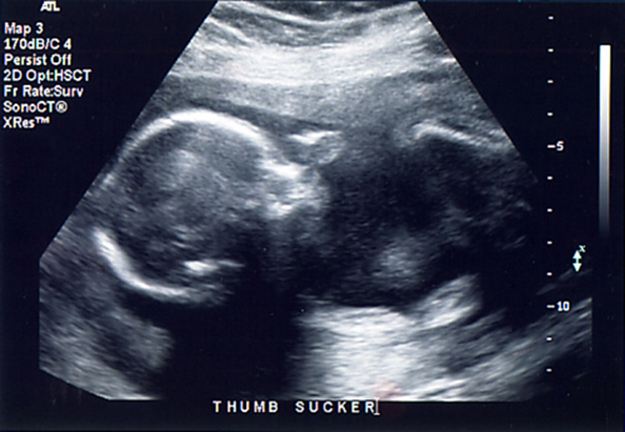
How much detail can ultrasound reveal? The image in Figure 17.43 is typical of low-cost systems, but that in Figure 17.44 shows the remarkable detail possible with more advanced systems, including 3D imaging. Ultrasound today is commonly used in prenatal care. Such imaging can be used to see if the fetus is developing at a normal rate, and help in the determination of serious problems early in the pregnancy. Ultrasound is also in wide use to image the chambers of the heart and the flow of blood within the beating heart, using the Doppler effect (echocardiology).
Whenever a wave is used as a probe, it is very difficult to detect details smaller than its wavelength . Indeed, current technology cannot do quite this well. Abdominal scans may use a 7-MHz frequency, and the speed of sound in tissue is about 1540 m/s—so the wavelength limit to detail would be . In practice, 1-mm detail is attainable, which is sufficient for many purposes. Higher-frequencyultrasound would allow greater detail, but it does not penetrate as well as lower frequencies do. The accepted rule of thumb is that you can effectively scan to a depth of about into tissue. For 7 MHz, this penetration limit is , which is 0.11 m. Higher frequencies may be employed in smaller organs, such as the eye, but are not practical for looking deep into the body.
In addition to shape information, ultrasonic scans can produce density information superior to that found in X-rays, because the intensity of a reflected sound is related to changes in density. Sound is most strongly reflected at places where density changes are greatest.
Another major use of ultrasound in medical diagnostics is to detect motion and determine velocity through the Doppler shift of an echo, known as Doppler-shifted ultrasound. This technique is used to monitor fetal heartbeat, measure blood velocity, and detect occlusions in blood vessels, for example. (See Figure 17.45.) The magnitude of the Doppler shift in an echo is directly proportional to the velocity of whatever reflects the sound. Because an echo is involved, there is actually a double shift. The first occurs because the reflector (say a fetal heart) is a moving observer and receives a Doppler-shifted frequency. The reflector then acts as a moving source, producing a second Doppler shift.
Figure 17.45 This Doppler-shifted ultrasonic image of a partially occluded artery uses color to indicate velocity. The highest velocities are in red, while the lowest are blue. The blood must move faster through the constriction to carry the same flow. (credit: Arning C, Grzyska U, Wikimedia Commons)
A clever technique is used to measure the Doppler shift in an echo. The frequency of the echoed sound is superimposed on the broadcast frequency, producing beats. The beat frequency is , and so it is directly proportional to the Doppler shift () and hence, the reflector’s velocity. The advantage in this technique is that the Doppler shift is small (because the reflector’s velocity is small), so that great accuracy would be needed to measure the shift directly. But measuring the beat frequency is easy, and it is not affected if the broadcast frequency varies somewhat. Furthermore, the beat frequency is in the audible range and can be amplified for audio feedback to the medical observer.
Uses for Doppler-Shifted Radar
Doppler-shifted radar echoes are used to measure wind velocities in storms as well as aircraft and automobile speeds. The principle is the same as for Doppler-shifted ultrasound. There is evidence that bats and dolphins may also sense the velocity of an object (such as prey) reflecting their ultrasound signals by observing its Doppler shift.
Ultrasound that has a frequency of 2.50 MHz is sent toward blood in an artery that is moving toward the source at 20.0 cm/s, as illustrated in Figure 17.46. Use the speed of sound in human tissue as 1540 m/s. (Assume that the frequency of 2.50 MHz is accurate to seven significant figures.)
Figure 17.46 Ultrasound is partly reflected by blood cells and plasma back toward the speaker-microphone. Because the cells are moving, two Doppler shifts are produced—one for blood as a moving observer, and the other for the reflected sound coming from a moving source. The magnitude of the shift is directly proportional to blood velocity.
The first two questions can be answered using and for the Doppler shift. The last question asks for beat frequency, which is the difference between the original and returning frequencies.
The Doppler shifts are quite small compared with the original frequency of 2.50 MHz. It is far easier to measure the beat frequency than it is to measure the echo frequency with an accuracy great enough to see shifts of a few hundred hertz out of a couple of megahertz. Furthermore, variations in the source frequency do not greatly affect the beat frequency, because both and would increase or decrease. Those changes subtract out in
Industrial, retail, and research applications of ultrasound are common. A few are discussed here. Ultrasonic cleaners have many uses. Jewelry, machined parts, and other objects that have odd shapes and crevices are immersed in a cleaning fluid that is agitated with ultrasound typically about 40 kHz in frequency. The intensity is great enough to cause cavitation, which is responsible for most of the cleansing action. Because cavitation-produced shock pressures are large and well transmitted in a fluid, they reach into small crevices where even a low-surface-tension cleaning fluid might not penetrate.
Sonar is a familiar application of ultrasound. Sonar typically employs ultrasonic frequencies in the range from 30.0 to 100 kHz. Bats, dolphins, submarines, and even some birds use ultrasonic sonar. Echoes are analyzed to give distance and size information both for guidance and finding prey. In most sonar applications, the sound reflects quite well because the objects of interest have significantly different density than the medium in which they travel. When the Doppler shift is observed, velocity information can also be obtained. Submarine sonar can be used to obtain such information, and there is evidence that some bats also sense velocity from their echoes.
Similarly, there are a range of relatively inexpensive devices that measure distance by timing ultrasonic echoes. Many cameras, for example, use such information to focus automatically. Some doors open when their ultrasonic ranging devices detect a nearby object, and certain home security lights turn on when their ultrasonic rangers observe motion. Ultrasonic “measuring tapes” also exist to measure such things as room dimensions. Sinks in public restrooms are sometimes automated with ultrasound devices to turn faucets on and off when people wash their hands. These devices reduce the spread of germs and can conserve water.
Ultrasound is used for nondestructive testing in industry and by the military. Because ultrasound reflects well from any large change in density, it can reveal cracks and voids in solids, such as aircraft wings, that are too small to be seen with x-rays. For similar reasons, ultrasound is also good for measuring the thickness of coatings, particularly where there are several layers involved.
Basic research in solid state physics employs ultrasound. Its attenuation is related to a number of physical characteristics, making it a useful probe. Among these characteristics are structural changes such as those found in liquid crystals, the transition of a material to a superconducting phase, as well as density and other properties.
These examples of the uses of ultrasound are meant to whet the appetites of the curious, as well as to illustrate the underlying physics of ultrasound. There are many more applications, as you can easily discover for yourself.
Check Your Understanding
Why is it possible to use ultrasound both to observe a fetus in the womb and also to destroy canceroustumors in the body?
Ultrasound can be used medically at different intensities. Lower intensities do not cause damage and are used for medical imaging. Higher intensities can pulverize and destroy targeted substances in the body, such as tumors.