Nuclear equations conserve charge and nucleon accounting; radioactive loss is probabilistic and exponential.
Composition and binding
Atomic number Z counts protons and defines the element. Mass number A counts protons plus neutrons; isotopes share Z but differ in neutron count. Atomic weight is an abundance-weighted average isotopic mass, not a particular isotope’s integer mass number. Nuclear attraction from the short-range strong interaction competes with proton–proton electrical repulsion. A bound nucleus has less mass than its separated nucleons; binding energy is Δmc². Binding energy per nucleon helps compare stability, but a nucleus’s decay also depends on available products and allowed pathways.
Source: Chemistry 2E 21 — Summary · 12.1 Mass Spectrometry of Small Molecules: Magnetic-Sector Instruments
Track the decay and its timing
Alpha decay lowers A by 4 and Z by 2. Beta-minus decay converts a neutron into a proton, increasing Z by 1 at constant A while emitting an electron and antineutrino. Beta-plus decay or electron capture lowers Z by 1 at constant A. Gamma emission releases excitation energy without changing A or Z. Undecayed nuclei follow N = N₀e⁻λt and activity λN; t½ = ln2/λ. After n half-lives the remaining fraction is 2⁻ⁿ. A plot of ln N against time has slope −λ.
Source: Chemistry 2E 21 — Summary · 12.1 Mass Spectrometry of Small Molecules: Magnetic-Sector Instruments
Mass spectrometry separates ions
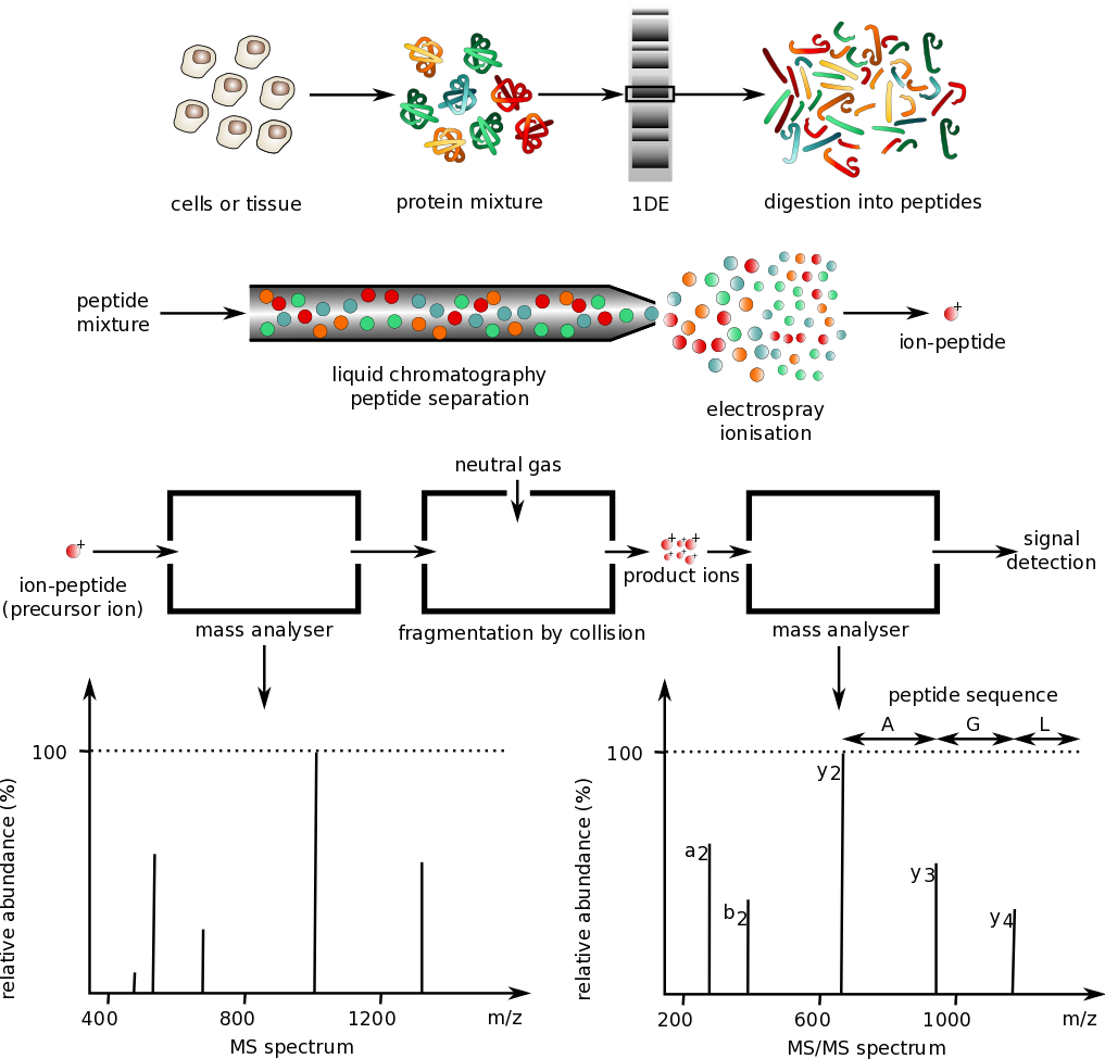
Mass spectrometry ionizes a sample and separates ions by mass-to-charge ratio m/z. Acceleration through a voltage gives kinetic energy qV; a magnetic analyzer bends ions with radius mv/(|q|B), while time-of-flight methods compare travel times after controlled acceleration. The molecular-ion region can constrain molecular mass, but fragmentation and multiple charge states require interpretation. Isotope clusters reveal composition: one chlorine often gives an M:M+2 ratio near 3:1, whereas one bromine gives near 1:1. A peak position is not automatically the neutral molecule’s mass.
Source: Chemistry 2E 21 — Summary · 12.1 Mass Spectrometry of Small Molecules: Magnetic-Sector Instruments
Apply it
Try before revealing the answer. A sample’s activity falls from 800 to 100 counts per minute in 18 hours after background subtraction. Find its half-life. What fraction remains after another 12 hours?
Worked answer
800 → 400 → 200 → 100 is three half-lives, so t½ = 18/3 = 6 hours. Another 12 hours is two more half-lives, leaving 25 counts/min and 1/32 of the original activity. Activity tracks remaining nuclei for a single isotope without additional production.
Source: Chemistry 2E 21 — Summary · 12.1 Mass Spectrometry of Small Molecules: Magnetic-Sector Instruments
12.1 Mass Spectrometry of Small Molecules: Magnetic-Sector Instruments — figures and tables
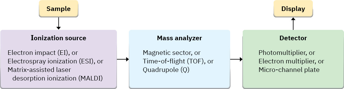
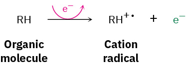
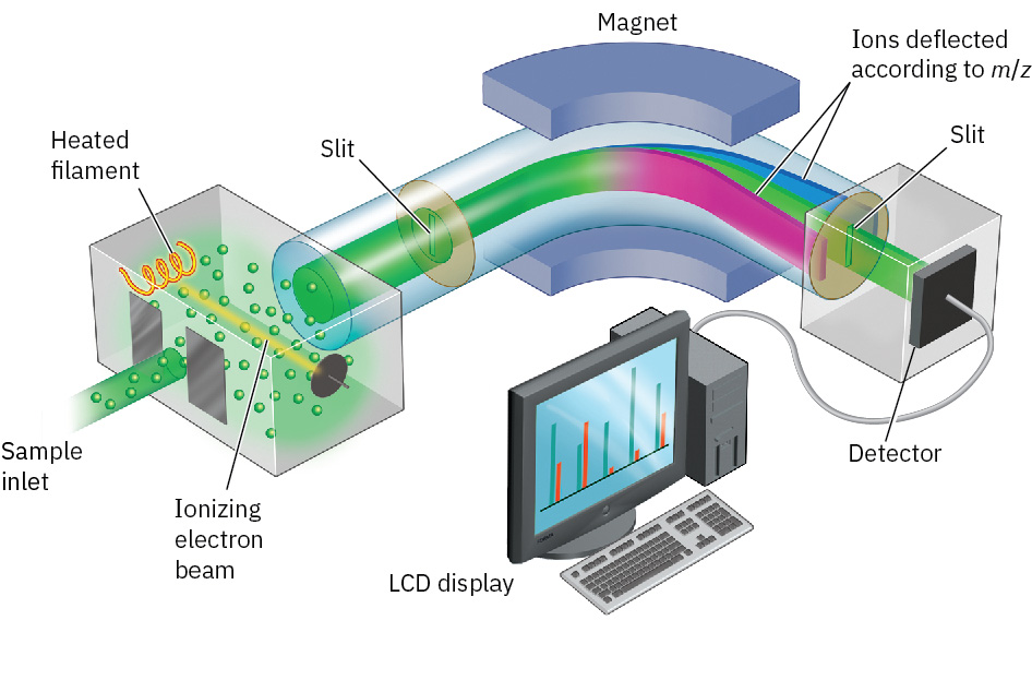
Figure 12.2 Representation of an electron-ionization, magnetic-sector mass spectrometer. Molecules are ionized by collision with high-energy electrons, causing some of the molecules to fragment. Passage of the charged fragments through a magnetic field then sorts them according to their mass.
John McMurry, Organic Chemistry, OpenStax. · CC BY-NC-SA 4.0 · Original image ↗ · Access for free ↗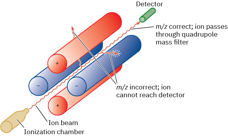
Figure 12.3 Representation of a quadrupole mass analyzer. Only ions of a certain m/z will reach the detector; other ions will collide with the rods.
John McMurry, Organic Chemistry, OpenStax. · CC BY-NC-SA 4.0 · Original image ↗ · Access for free ↗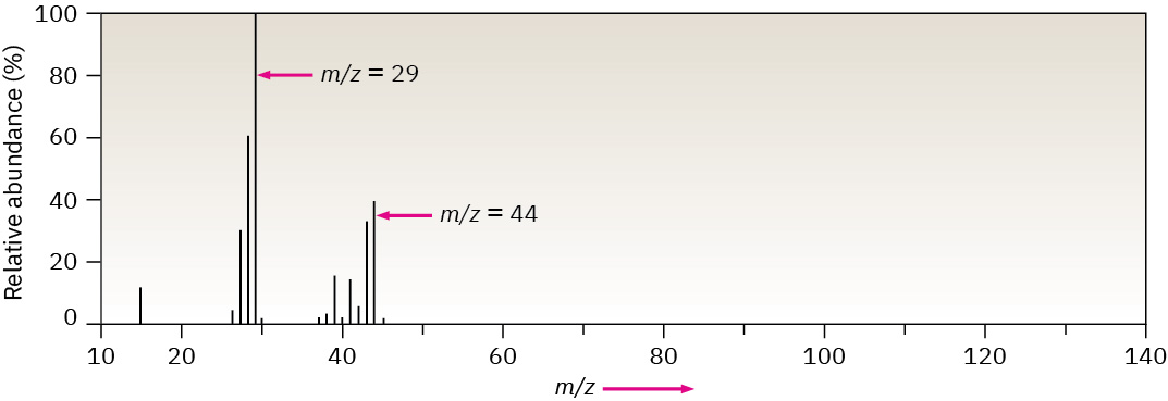
Figure 12.4 Mass spectrum of propane (C 3 H 8 ; MW = 44).
John McMurry, Organic Chemistry, OpenStax. · CC BY-NC-SA 4.0 · Original image ↗ · Access for free ↗Source: 12.1 Mass Spectrometry of Small Molecules: Magnetic-Sector Instruments