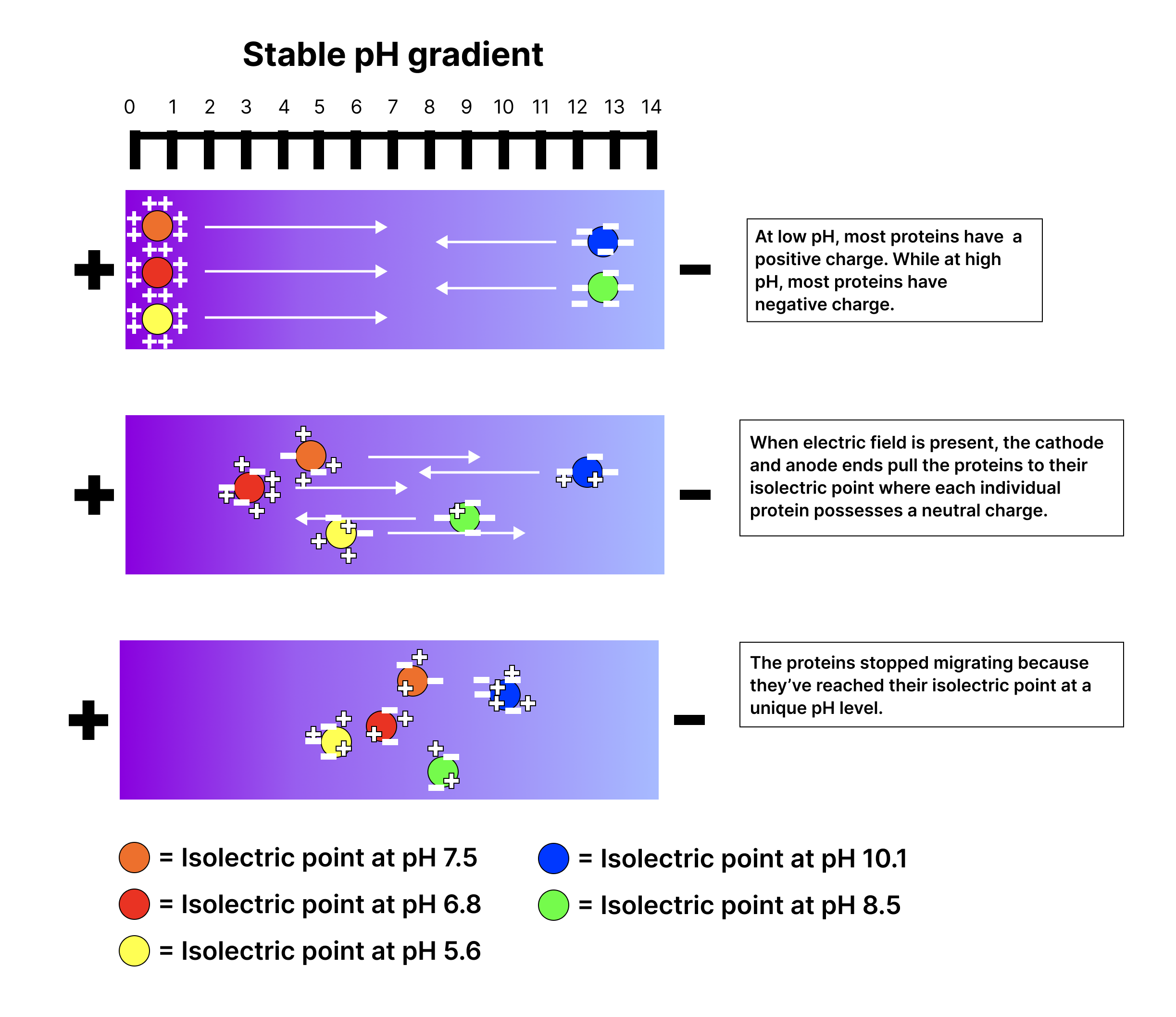
Foundation 4 · 4C · Electricity, magnetism, and electrochemistry

Electrical signaling in nerve cells

Sources ↗

accelerates passive spread between nodes; voltage-gated channels regenerate the at nodes.

changes the cable properties

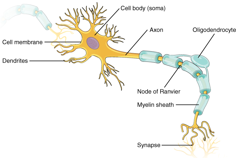
The ’s barrier supplies while channels supply conductance. adds insulation, increasing and reducing per axonal length. Local current therefore spreads farther and the next exposed region more rapidly. Voltage-gated sodium channels concentrated at nodes of Ranvier regenerate , producing . Schwann cells form peripheral , typically one internode per cell; myelinate central axons. Demyelination increases current leakage and can slow or block propagation. The signal is regenerated locally; individual sodium do not travel from one end of the to the other.

Source: 20.7 Nerve Conduction–Electrocardiograms

Apply it

Try before revealing the answer. Two axons have equal diameter, channel properties, and temperature. One loses . Predict changes in leakage, , and propagation.

Worked answer

Loss of insulation lowers and raises effective . More local current leaks across the and more is needed to change voltage, making threshold at nodes harder to reach. slows or fails even though an individual intact node can still generate an .

Source: 20.7 Nerve Conduction–Electrocardiograms

20.7 Nerve –Electrocardiograms — figures and tables

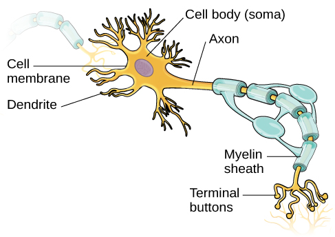
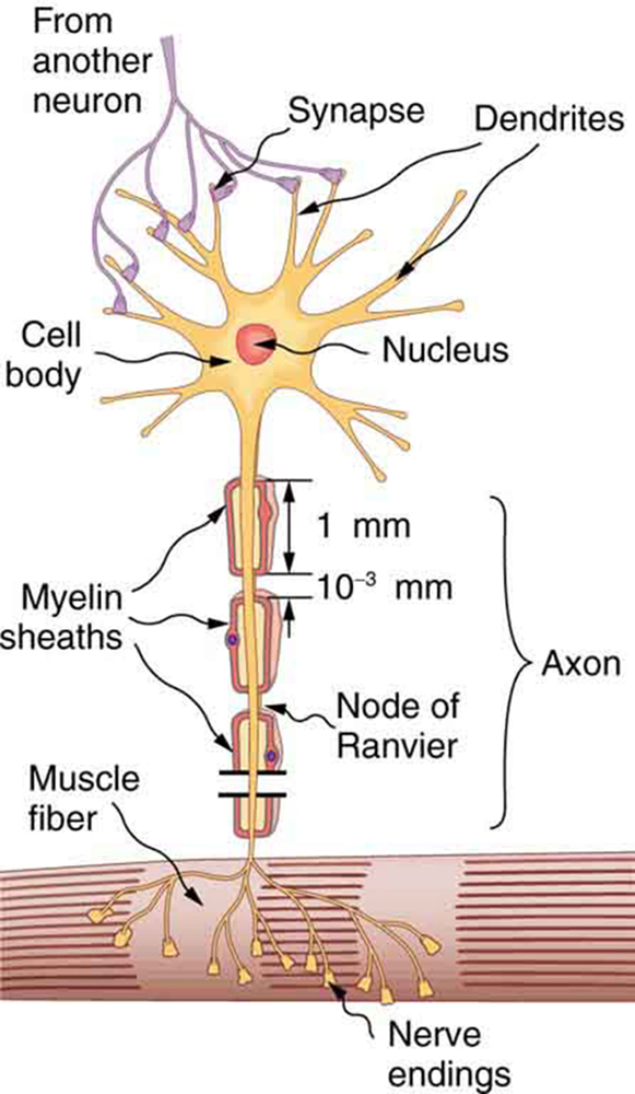
The figure describes a neuron. The neuron has a cell body with a nucleus at the center represented by a circle. The cell body is surrounded by many thin, branching projections called dendrites, represented by ribbon-like structures. The ends of some of these dendrites are shown connected to the ends of dendrites from another neuron at junctions called synapses. The cell body of the neuron also has a long projection called an axon, represented as a vertical tube reaching downward and ending with thin projections inside a muscle fiber, represented by a tubular structure. The ends of the axon are called nerve endings. The axon is covered with myelin sheaths, each of which is one millimeter in length. The myelin sheaths are separated by gaps, called nodes of Ranvier, each of length zero point zero zero one millimeter.
Figure 20.24 — 20.7 Nerve –Electrocardiograms

Figure 20.24 A neuron with its dendrites and long axon. Signals in the form of electric currents reach the cell body through dendrites and across synapses, stimulating the neuron to generate its own signal sent down the axon. The number of interconnections can be far greater than shown here.

Paul Peter Urone and Roger Hinrichs, College Physics 2E, OpenStax. · CC BY-NC-SA 4.0 · Original image ↗ · Access for free ↗
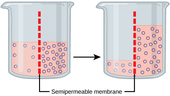
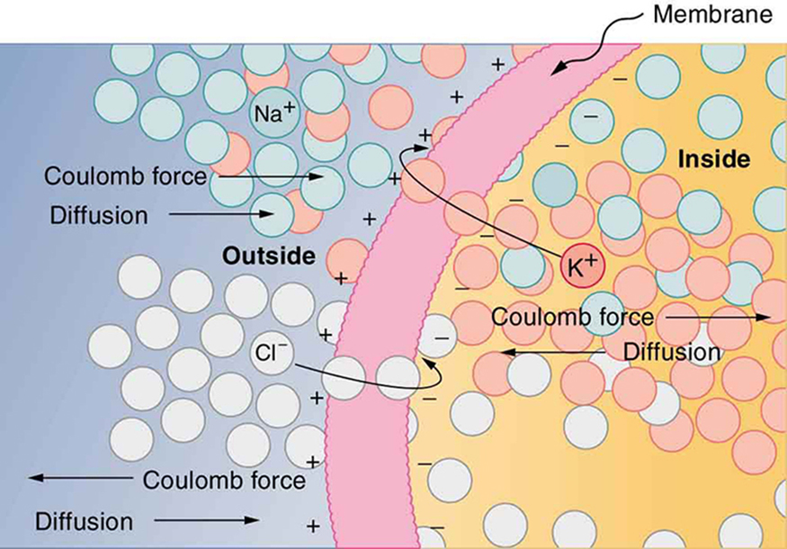
The semipermeable membrane of a cell is shown, with different concentrations of potassium cations, sodium cations, and chloride anions inside and outside the cell. The ions are represented by small, colored circles. In its resting state, the cell membrane is permeable to potassium and chloride ions, but it is impermeable to sodium ions. By diffusion, potassium cations travel out of the cell, going through the cell membrane and forming a layer of positive charge on the outer surface of the membrane. By diffusion, chloride anions go into the cell, going through the cell membrane and forming a layer of negative charge on the inner surface of the membrane. As a result, a voltage is set up across the cell membrane. The Coulomb force prevents all the ions from crossing the membrane.
Figure 20.25 — 20.7 Nerve –Electrocardiograms

Figure 20.25 The semipermeable membrane of a cell has different concentrations of ions inside and out. Diffusion moves the K + K + and Cl - Cl - ions in the direction shown, until the Coulomb force halts further transfer. This results in a layer of positive charge on the outside, a layer of negative charge on the inside, and thus a voltage across the cell membrane. The membrane is normally impermeable to Na + Na + .

Paul Peter Urone and Roger Hinrichs, College Physics 2E, OpenStax. · CC BY-NC-SA 4.0 · Original image ↗ · Access for free ↗
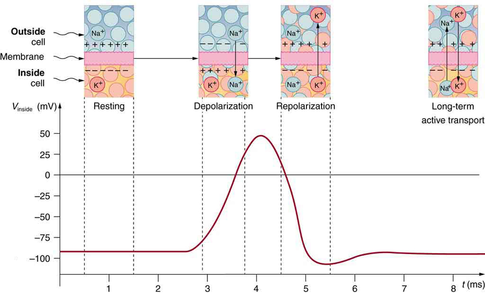
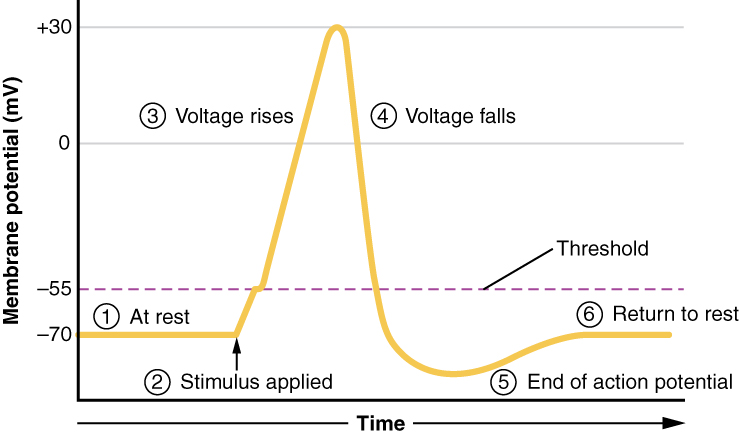
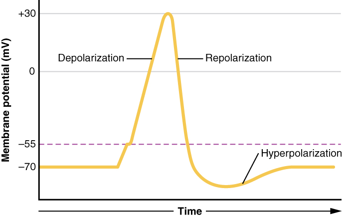
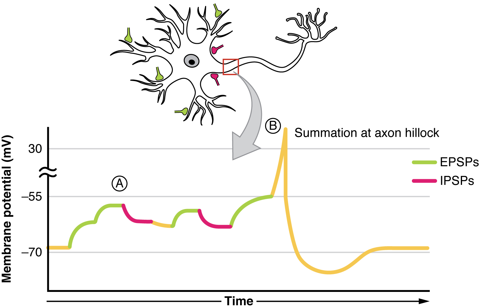
This is a graphical representation of a pulse of voltage, or action potential, inside a nerve cell. The voltage in millivolts is plotted along the vertical axis and the time in milliseconds is plotted along the horizontal axis. Initially, between zero and about two point eight milliseconds, the voltage is a constant at about minus ninety millivolts, corresponding to the resting state. Above this section of the graph, a window shows a small cross-section of the cell membrane, with a positively charged outer surface, a negatively charged inner surface, and no ions moving across the membrane. Between two point eight and four point two milliseconds, the voltage increases to a peak of fifty millivolts, corresponding to depolarization of the membrane. A window above this section shows sodium cations crossing the membrane, from outside to inside the cell, so that the membrane’s inner surface acquires a positive charge and its outer surface has a negative charge. Between about four point two and about five point five milliseconds, the voltage drops to a low of about minus one hundred and ten millivolts, corresponding to repolarization of the membrane. A window above this section shows potassium cations crossing the membrane, from inside to outside the cell, so that the membrane’s outer surface again acquires a positive charge and its inner surface has a negative charge. After that, the voltage rises slightly, going back to a constant of about minus ninety millivolts, corresponding to the resting state. This movement of sodium and potassium ions across the membrane is called active transport, and long-term active transport is shown in a window above the final part of the curve.
Figure 20.26 — 20.7 Nerve –Electrocardiograms

Figure 20.26 An action potential is the pulse of voltage inside a nerve cell graphed here. It is caused by movements of ions across the cell membrane as shown. Depolarization occurs when a stimulus makes the membrane permeable to Na + Na + ions. Repolarization follows as the membrane again becomes impermeable to Na + , Na + , and K + K + moves from high to low concentration. In the long term, active transport slowly maintains the concentration differences, but the cell may fire hundreds of times in rapid succession without seriously depleting them.

Paul Peter Urone and Roger Hinrichs, College Physics 2E, OpenStax. · CC BY-NC-SA 4.0 · Original image ↗ · Access for free ↗
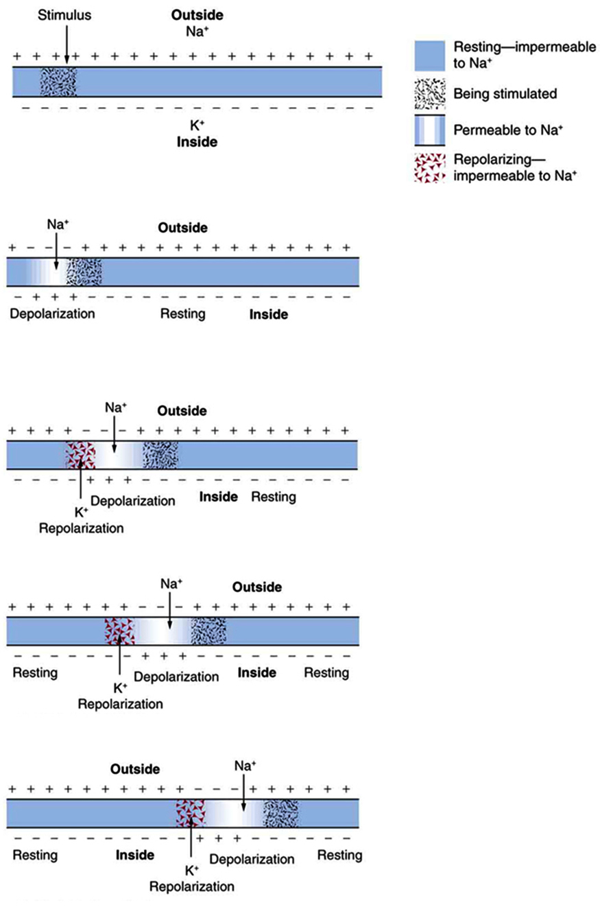
The figure describes the propagation of an action potential, or voltage pulse, along a cell membrane. The cell membrane, represented by a horizontal, blue strip, is shown in five stages, with the electrical signal moving along its length from left to right. Initially, the membrane is in the resting state, with a uniform distribution of positive charges along the outer surface and negative charges along the inner surface. A sodium cation is shown outside the cell, and a potassium cation is shown inside the cell. A small part of the membrane near the left end receives a stimulus, making that part permeable to sodium ions. In the second stage, sodium ions cross the membrane in that area, represented by a white opening in the membrane. The charge distribution in that section of the membrane is reversed; this process is called depolarization. At the same time, an adjacent part of the membrane is stimulated. In the third stage, the depolarized area undergoes repolarization, with potassium ions crossing the membrane from inside to outside the cell. Repolarization is represented by a box containing tiny triangles. At the same time, sodium ions enter the cell through the adjacent area that was stimulated in the second stage. As the cycle is repeated, the electrical signal moves along the membrane, from left to right.
Figure 20.27 — 20.7 Nerve –Electrocardiograms

Figure 20.27 A nerve impulse is the propagation of an action potential along a cell membrane. A stimulus causes an action potential at one location, which changes the permeability of the adjacent membrane, causing an action potential there. This in turn affects the membrane further down, so that the action potential moves slowly (in electrical terms) along the cell membrane. Although the impulse is due to Na + Na + and K + K + going across the membrane, it is equivalent to a wave of charge moving along the outside and inside of the membrane.

Paul Peter Urone and Roger Hinrichs, College Physics 2E, OpenStax. · CC BY-NC-SA 4.0 · Original image ↗ · Access for free ↗
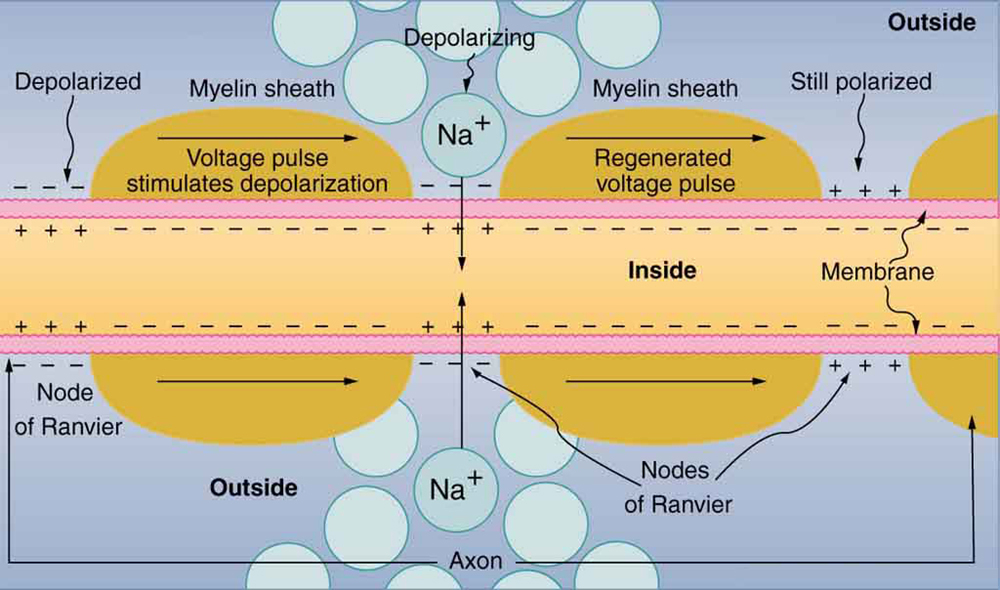
The figure describes the propagation of a nerve impulse, or voltage pulse, down a myelinated axon, from left to right. A cross-section of the axon is shown as a long, horizontally oriented rectangular strip, with a membrane on each side. The axon is covered with myelin sheaths separated by gaps known as nodes of Ranvier. Three gaps are shown. Most of the inner surface of the membrane is negatively charged, and the outer surface is positively charged. The gap on the left is labeled as depolarized, where the charge distribution along the membrane surface is reversed. As the voltage pulse moves from left to right through the first myelinated region, it loses voltage. The gap in the middle, labeled as depolarizing, shows sodium cations crossing the membrane from the outside to the inside of the axon. This regenerates the voltage pulse, which continues to move along the axon. The third gap is labeled as still polarized, because the signal has yet to reach that gap.
Figure 20.28 — 20.7 Nerve –Electrocardiograms

Figure 20.28 Propagation of a nerve impulse down a myelinated axon, from left to right. The signal travels very fast and without energy input in the myelinated regions, but it loses voltage. It is regenerated in the gaps. The signal moves faster than in unmyelinated axons and is insulated from signals in other nerves, limiting cross talk.

Paul Peter Urone and Roger Hinrichs, College Physics 2E, OpenStax. · CC BY-NC-SA 4.0 · Original image ↗ · Access for free ↗
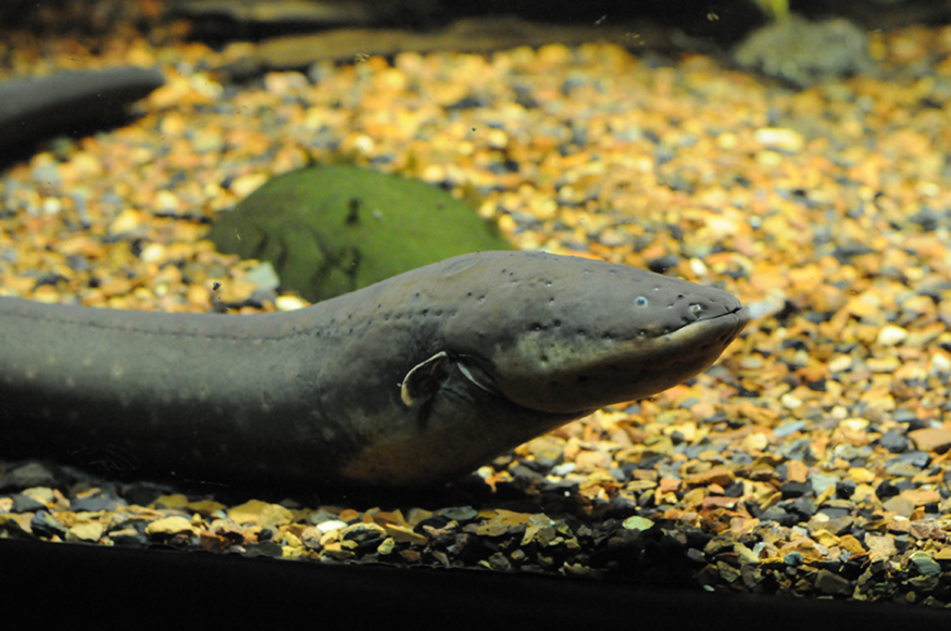
Photograph of an electric eel.
Figure 20.29 — 20.7 Nerve –Electrocardiograms

Figure 20.29 An electric eel flexes its muscles to create a voltage that stuns prey. (credit: chrisbb, Flickr)

Paul Peter Urone and Roger Hinrichs, College Physics 2E, OpenStax. (credit: chrisbb, Flickr) · CC BY-NC-SA 4.0 · Original image ↗ · Access for free ↗
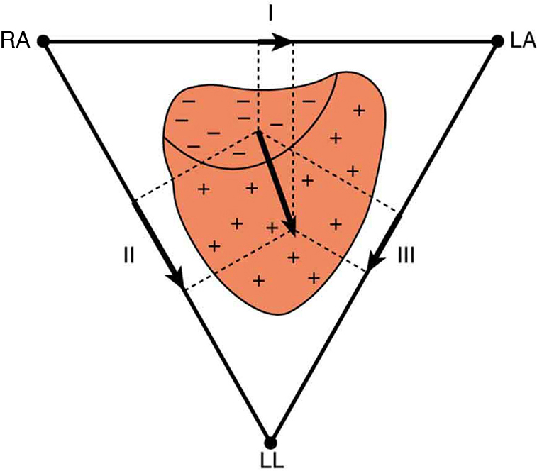
The figure shows that the charge distribution on the outer surface of the heart changes from positive to negative during depolarization. This wave of depolarization, spreading from the upper right toward the lower left of the heart, is represented by a vector pointing in the direction of the wave. The components of this vector are measured by placing electrodes on the patient’s chest. The figure shows three electrodes, labeled R A, L A, and L L, placed to form a triangle around the heart. The electrode R A is close to the right atrium, L A is close to the left atrium, and L L is just below the heart. R A and L A form a pair called lead one, R A and L L form a second pair called lead two, and L A and L L form a third pair called lead three. Each pair of electrodes measures a component of the depolarization vector.
Figure 20.30 — 20.7 Nerve –Electrocardiograms

Figure 20.30 The outer surface of the heart changes from positive to negative during depolarization. This wave of depolarization is spreading from the top of the heart and is represented by a vector pointing in the direction of the wave. This vector is a voltage (potential difference) vector. Three electrodes, labeled RA, LA, and LL, are placed on the patient. Each pair (called leads I, II, and III) measures a component of the depolarization vector and is graphed in an ECG.

Paul Peter Urone and Roger Hinrichs, College Physics 2E, OpenStax. · CC BY-NC-SA 4.0 · Original image ↗ · Access for free ↗
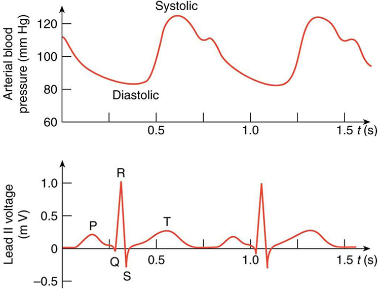
This figure has two graphs, placed one below the other. The lower graph shows an E C G of the lead two potential, and the upper graph shows the corresponding changes in arterial blood pressure. In each case, time is plotted on the horizontal axis, in seconds. The vertical axis of the upper graph shows the arterial blood pressure in millimeters of mercury, and the vertical axis of the lower graph shows the lead two voltage in millivolts. The upper graph is roughly sinusoidal, showing the diastolic or minimum blood pressure at about eighty millimeters of mercury, and the systolic or maximum blood pressure at about one hundred twenty millimeters of mercury. For the lower graph, the main features are labeled P, Q, R, S, and T. The P wave is a smooth curve that rises from zero millivolts to a peak of about zero point two five millivolts and falls to just below zero millivolts when it reaches point Q. From point Q to point R, the voltage rises steeply to about one millivolt, and then drops equally sharply to point S, at negative zero point three millivolts. This is followed by the T wave, which is a smooth curve, broader than the P wave, with a peak of comparable height. All of this is completed in less than seven-tenths of a second, with the voltage returning to zero millivolts. After about one-tenth of a second, the cycle begins again. The systolic blood pressure follows soon after the QRS complex.
Figure 20.31 — 20.7 Nerve –Electrocardiograms

Figure 20.31 A lead II ECG with corresponding arterial blood pressure. The QRS complex is created by the depolarization and contraction of the ventricles and is followed shortly by the maximum or systolic blood pressure. See text for further description.

Paul Peter Urone and Roger Hinrichs, College Physics 2E, OpenStax. · CC BY-NC-SA 4.0 · Original image ↗ · Access for free ↗
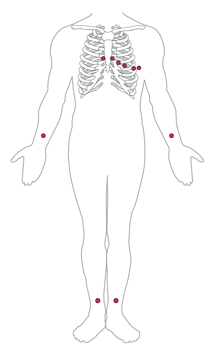
This diagram shows the points where electrodes are placed on the body for an ECG. Dots indicate the placement locations. There are six dots on on the chest and left side of the abdomen. There are dots on each wrist and just above each ankle.
Figure 20.32 — 20.7 Nerve –Electrocardiograms

Figure 20.32 In a 12-lead ECG, six electrodes are placed on the chest, and four electrodes are placed on the limbs.

Paul Peter Urone and Roger Hinrichs, College Physics 2E, OpenStax. · CC BY-NC-SA 4.0 · Original image ↗ · Access for free ↗

Source: 20.7 Nerve Conduction–Electrocardiograms

141 terms, alphabetically ordered

Acid · ·

In the Brønsted–Lowry model, a substance that can donate a . Acidic have a higher activity than water.

Read explanation · OpenStax ↗
action potential

A regenerative -voltage change produced by voltage-dependent -channel activity.

Read explanation · OpenStax ↗
Activation energy ·

The energy barrier that must overcome to reach a and undergo .

Read explanation · OpenStax ↗
Adenosine triphosphate

An energy-carrying ; its powers many cellular processes, including cycles.

Read explanation · OpenStax ↗
Alpha carbon · ·

The central carbon next to the carboxyl carbon. In a standard it binds an , hydrogen, and .

Read explanation · OpenStax ↗
Amide · · ·

A group containing a carbon bonded to nitrogen. are amide linkages.

Read explanation · OpenStax ↗
Amino acid

A building block containing amino and and a characteristic .

Read explanation · OpenStax ↗
Amino acid residue · ·

The part of an incorporated into a chain. Residue can also describe another building block within a larger .

Read explanation · OpenStax ↗
Amino group · · ·

A nitrogen-containing . A free ’s amino group is typically (NH₃⁺) near physiological ; ’s ring nitrogen is an exception to this formula.

Read explanation · OpenStax ↗
ammeter

Current-measuring instrument placed in series with low internal .

Read explanation · OpenStax ↗
Amphipathic

Having both a region and a region, as in a or .

Read explanation · OpenStax ↗
Analytical standard ·

A material with a known identity or amount used to compare or calibrate measurements.

Read explanation · Agilent ↗
Anode

The positive in an apparatus; negatively migrate toward it.

Read explanation · Thermo Fisher ↗
Aqueous solution ·

A in which water is the .

Read explanation · OpenStax ↗
Atom

The smallest unit of an element that retains its chemical identity; it has a surrounded by .

Read explanation · OpenStax ↗
atrium

A cardiac receiving chamber of a ventricle.

Read explanation · OpenStax ↗
axon

Neuronal process that usually conducts toward terminals.

Read explanation · OpenStax ↗
Biomolecule

A made by or involved in living systems, including , , , and .

Read explanation · OpenStax ↗
Blotting ·

Transfer of separated or to a so a specific target can be detected.

Read explanation · Thermo Fisher ↗
Brønsted–Lowry base · · · · · · · · · · ·

In the Brønsted–Lowry model, a substance that can accept a . Basic solutions have lower activity than water.

Read explanation · OpenStax ↗
Calibration

Establishing the relation between a measured signal and known values or .

Read explanation · Agilent ↗
capacitance

stored per potential difference, measured in farads.

Read explanation · OpenStax ↗
Carbohydrate

A class of including sugars and their , used for energy storage, structure, and recognition.

Read explanation · OpenStax ↗
Carbonyl group · ·

A carbon double-bonded to oxygen, C=O; present in and many other .

Read explanation · OpenStax ↗
Carboxyl group · ·

An , COOH. In free it is usually to COO⁻ near physiological .

Read explanation · OpenStax ↗
Catalyst · ·

A substance that speeds a by providing a pathway with lower and is regenerated rather than consumed overall.

Read explanation · OpenStax ↗
Cathode

The negative in an apparatus; positively migrate toward it.

Read explanation · Thermo Fisher ↗
central nervous system

Brain and spinal cord.

Read explanation · OpenStax ↗
Chemical reaction ·

A process that rearranges and chemical bonds, converting into .

Read explanation · OpenStax ↗
Chromatin

The complex of and associated , including , in .

Read explanation · OpenStax ↗
Chromosome ·

An organized with associated that carries . A replicated chromosome has two .

Read explanation · OpenStax ↗
Codon

A sequence of three that specifies an or a stop signal.

Read explanation · OpenStax ↗
Concentration

The amount of a substance in a specified volume or mass of mixture.

Read explanation · OpenStax ↗
conduction

Thermal energy transfer through microscopic interactions without bulk matter flow.

Read explanation · OpenStax ↗
conductor

Material with that can move readily through it.

Read explanation · OpenStax ↗
Covalent bond · ·

A bond in which share pairs. and are covalent.

Read explanation · OpenStax ↗
Cysteine ·

An with a . Two cysteines can form a .

Read explanation · OpenStax ↗
dendrite

Branching neuronal process receiving many synaptic inputs.

Read explanation · OpenStax ↗
Deoxyribonucleic acid

The that stores genetic information used to specify and sequences.

Read explanation · OpenStax ↗
depolarization

A shift in voltage toward a less negative value.

Read explanation · OpenStax ↗
Deprotonation

Removal of a from a or group; this changes its by minus one.

Read explanation · OpenStax ↗
Detergent

An substance that can associate with both water and material; detergents can disrupt or interactions.

Read explanation · Thermo Fisher ↗
Diffusion

Net spreading of particles down a due to random molecular motion.

Read explanation · OpenStax ↗
Disulfide bond · · ·

A covalent sulfur–sulfur bond formed by of two , which can stabilize structure.

Read explanation · NCBI Bookshelf ↗
DNA replication

Copying so genetic information can be passed to daughter cells. Each guides a complementary new strand.

Read explanation · OpenStax ↗
Downstream

Later along a directed sequence or process; in , toward positions following a reference site in the direction of synthesis.

Read explanation · OpenStax ↗
Electric charge · · ·

An electrical property that can be positive or negative. Opposite charges attract and like charges repel; net charge is the sum of all charges on a particle.

Read explanation · OpenStax ↗
Electric field

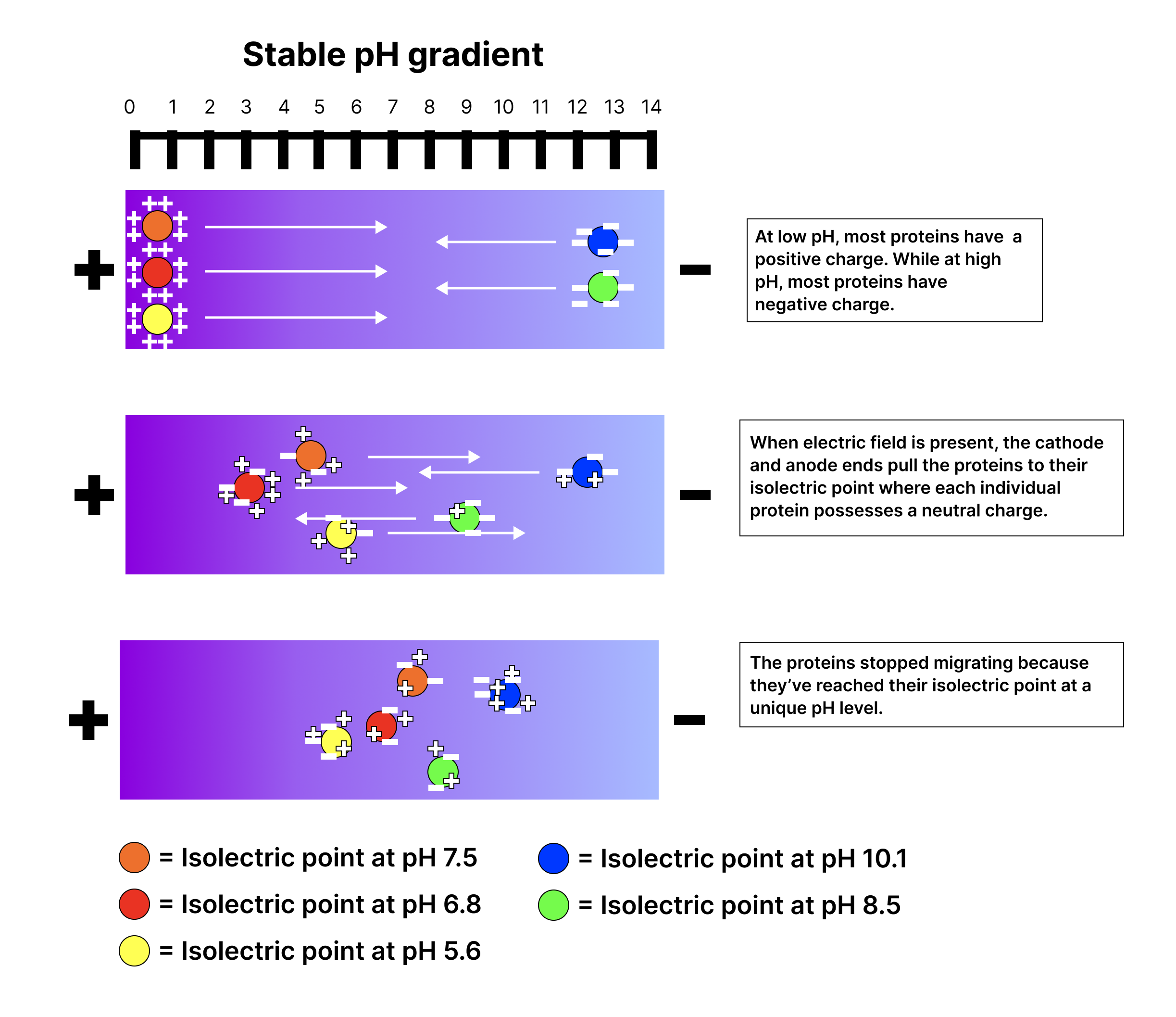
A quantity describing the electrical force per unit positive . An applied field drives through a gel during .

Read explanation · Thermo Fisher ↗
electrical resistance ·

Voltage across a component divided by current through it under specified conditions.

Read explanation · OpenStax ↗
Electrode

A that connects an electrical circuit to a or other medium; the and are electrodes.

Read explanation · Thermo Fisher ↗
Electron

A negatively subatomic particle. Electron sharing and transfer determine much of chemical bonding.

Read explanation · OpenStax ↗
Electrophoresis

Movement of in an , used to separate through a support such as a gel.

Read explanation · Thermo Fisher ↗
Eukaryote ·

An organism whose cells have a -enclosed ; animals, plants, fungi, and protists are eukaryotes.

Read explanation · OpenStax ↗
FACT complex ·

A -associated complex that helps machinery traverse and supports reorganization.

Read explanation · OpenStax ↗
Fraction · ·

A portion of a sample collected during a separation, such as the fluid leaving a column during a time interval.

Read explanation · Waters ↗
Functional group

A group of whose chemical behavior gives a characteristic and properties.

Read explanation · OpenStax ↗
Gene

A sequence that contributes to a functional or , together with the information needed to express it.

Read explanation · OpenStax ↗
Gene expression

Production of a functional from a , including and, for -coding , .

Read explanation · NCBI Bookshelf ↗
Gradient

A change in a quantity, such as , , or density, across a distance or over time.

Read explanation · Thermo Fisher ↗
Histone

A -associated rich in . Core histones form the around which nucleosomal wraps.

Read explanation · OpenStax ↗
Hydrogen ion · ·

A positively hydrogen species. H⁺ denotes a , which is hydrated in water; its activity determines .

Read explanation · OpenStax ↗
Hydrolysis

Breaking a chemical bond by adding water. hydrolysis separates .

Read explanation · OpenStax ↗
Hydrophilic

Interacting favorably with water, typically because a group is or .

Read explanation · OpenStax ↗
Hydrophobic

Interacting poorly with water. often gather in a ’s interior.

Read explanation · OpenStax ↗
Hydroxide ion ·

The negatively OH⁻. Basic contain a higher hydroxide ion activity than water at the same temperature.

Read explanation · OpenStax ↗
infrared spectroscopy ·

Method probing vibrational transitions through absorption of infrared light.

Read explanation · OpenStax ↗
insulator

Material in which cannot move freely over macroscopic distances.

Read explanation · OpenStax ↗
Ion · ·

An or with a net because its numbers of and do not balance.

Read explanation · OpenStax ↗
laminar flow

Smooth fluid flow in organized layers.

Read explanation · OpenStax ↗
linkage

A tendency of nearby on the same to be inherited together.

Read explanation · OpenStax ↗
Lipid

A broad class of largely or , including fats, , and steroids.

Read explanation · OpenStax ↗
locus

The specific position of a or other feature.

Read explanation · OpenStax ↗
Logarithm · · ·

The exponent needed to obtain a number from a chosen fixed number. For decimal logarithms, log₁₀(100) = 2 because 10² = 100; uses the negative decimal logarithm of activity.

Read explanation · OpenStax ↗
Mass spectrometry ·

Analysis of according to their mass-to- ratios, used to identify and or .

Read explanation · Thermo Fisher ↗
mechanical power

Rate of energy transfer, measured in watts.

Read explanation · OpenStax ↗
mechanical work

Energy transferred by a force acting through ; the force component parallel to motion contributes.

Read explanation · OpenStax ↗
Membrane · · · ·

A thin boundary or sheet. Cell membranes are bilayers containing ; membranes are synthetic supports that hold or for detection.

Read explanation · OpenStax ↗
membrane capacitance

Ability of a to store separated across its barrier.

Read explanation · OpenStax ↗
Messenger RNA ·

An that carries coding information used by to synthesize a .

Read explanation · OpenStax ↗
Molecular chaperone · ·

A that assists other in folding or prevents , without specifying their sequence.

Read explanation · NCBI Bookshelf ↗
Molecule

Two or more joined by . A molecule may be electrically or carry a .

Read explanation · OpenStax ↗
Myelin

A -rich insulating sheath around many nerve axons that supports efficient signal .

Read explanation · NHGRI / NIH ↗
neuron

An electrically excitable cell specialized for receiving and transmitting information.

Read explanation · OpenStax ↗
Neutral

Having no net . For a , neutral instead means equal and activities; its is about 7 at 25 °C.

Read explanation · OpenStax ↗
Nitrogenous base · · ·

A nitrogen-containing ring component of ; its pairing interactions help specify sequences.

Read explanation · OpenStax ↗
node of Ranvier

Gap between segments containing a high density of voltage-gated channels.

Read explanation · OpenStax ↗
Nonpolar ·

Lacking a substantial separation of . Nonpolar groups generally interact poorly with water.

Read explanation · OpenStax ↗
Nucleic acid

A of , such as or , used in information storage, expression, and other cellular functions.

Read explanation · OpenStax ↗
Nucleosome

The basic unit of : wrapped around a .

Read explanation · OpenStax ↗
Nucleotide

A joined to a sugar and one or more .

Read explanation · OpenStax ↗
Nucleus ·

A -enclosed compartment containing most of a cell’s .

Read explanation · OpenStax ↗
Octamer

A complex made of eight . A core contains eight .

Read explanation · OpenStax ↗
oligodendrocyte

A central glial cell that can form segments on multiple axons.

Read explanation · OpenStax ↗
osmosis

Net water movement across a selectively permeable in response to water chemical-potential differences.

Read explanation · OpenStax ↗
Oxidation ·

Loss of or an increase in oxidation state. Oxidizing two can form a .

Read explanation · OpenStax ↗
Peptide

A chain of joined by ; the term commonly describes chains shorter than a typical .

Read explanation · OpenStax ↗
Peptide bond ·

A covalent between the carboxyl carbon of one and the amino nitrogen of the next.

Read explanation · OpenStax ↗
peripheral nervous system

Nerves and ganglia outside the brain and spinal cord.

Read explanation · OpenStax ↗
pH

A measure of , defined as the negative of activity. Lower pH means greater ; pH influences .

Read explanation · OpenStax ↗
Phosphate group ·

A phosphorus-containing group bonded to oxygens; phosphate groups occur in , , and modified .

Read explanation · OpenStax ↗
Phospholipid

An with a -containing region and tails; a major component of cellular .

Read explanation · OpenStax ↗
physical displacement ·

Signed or difference between final and initial position.

Read explanation · OpenStax ↗
Poiseuille’s law

Laminar tube-flow relationship in which flow is proportional to pressure difference and the fourth of radius.

Read explanation · OpenStax ↗
Polar

Having an uneven distribution of . Polar groups often interact favorably with water.

Read explanation · OpenStax ↗
Polymer

A large made from many linked molecular building blocks.

Read explanation · OpenStax ↗
Polypeptide

A chain of connected by . A functional can contain one or several such chains.

Read explanation · OpenStax ↗
Proline ·

An whose loops back to the nitrogen, restricting rotation and often creating bends.

Read explanation · OpenStax ↗
Protein

One or more folded chains that perform biological roles such as , transport, signaling, or structural support.

Read explanation · OpenStax ↗
Protein aggregation · ·

Association of into larger clusters, often when exposed surfaces of unfolded stick together.

Read explanation · NCBI Bookshelf ↗
Protein backbone ·

The repeating nitrogen–alpha-carbon– chain of a , excluding its variable .

Read explanation · OpenStax ↗
Proton

A positively subatomic particle. In – chemistry, donating a proton means transferring a .

Read explanation · OpenStax ↗
Protonation

Addition of a to an , , or ; this changes its by plus one.

Read explanation · OpenStax ↗
Quantification · ·

Estimating the amount or of a substance from measured signals and suitable or .

Read explanation · Waters ↗
R group · ·

The variable side chain attached to an ’s . It determines identity, , , and chemical behavior.

Read explanation · OpenStax ↗
Reactant

A starting substance transformed during a .

Read explanation · OpenStax ↗
Reaction intermediate · ·

A species formed in one step and consumed in another. It occupies a local energy minimum between .

Read explanation · OpenStax ↗
Reaction product ·

A substance formed by a .

Read explanation · OpenStax ↗
reaction quotient

-to- activity ratio with stoichiometric exponents at the current composition.

Read explanation · OpenStax ↗
repolarization

Return of voltage toward its resting value after .

Read explanation · OpenStax ↗
Ribonucleic acid ·

A with roles in , regulation, and ; carries -coding information.

Read explanation · OpenStax ↗
Ribosome ·

A complex of and that translates into a .

Read explanation · OpenStax ↗
saltatory conduction

Regeneration of at separated nodes along a myelinated .

Read explanation · OpenStax ↗
Schwann cell

glial cell that can form a segment around an .

Read explanation · OpenStax ↗
Sister chromatid

One of the two copies of a produced by . An intact sister chromatid can supply a template for accurate repair.

Read explanation · Biology LibreTexts / Oregon State University ↗
sodium–potassium ATPase ·

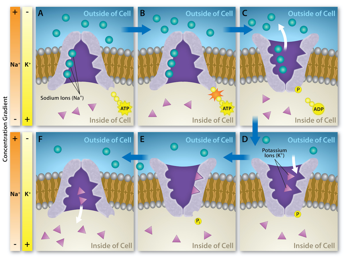
The -driven pump exporting three Na+ and importing two K+ per cycle.

Read explanation · OpenStax ↗
Solubility ·

The amount of a substance that can dissolve in a under specified conditions. Soluble means able to dissolve; insoluble means only very slightly soluble in that .

Read explanation · OpenStax ↗
Solute

A dissolved component of a .

Read explanation · OpenStax ↗
Solution

A homogeneous mixture in which are dissolved in a .

Read explanation · OpenStax ↗
Solvent

The component of a that dissolves other substances, usually the component present in the greatest amount.

Read explanation · OpenStax ↗
spatial vector ·

Quantity with both magnitude and direction, combined by adding components.

Read explanation · OpenStax ↗
Subunit ·

An individual chain within a containing multiple chains.

Read explanation · OpenStax ↗
synapse

A specialized connection through which a cell communicates with another electrically or chemically.

Read explanation · OpenStax ↗
Template strand · · ·

A preexisting strand whose sequence guides complementary nucleic- synthesis.

Read explanation · OpenStax ↗
Thiol group · · · ·

A sulfur bonded to hydrogen, –SH. thiols can form through .

Read explanation · OpenStax ↗
Transcription

Synthesis of using a template.

Read explanation · OpenStax ↗
Transition state

A fleeting, high-energy arrangement at a barrier. It is an energy maximum along the path, rather than a stable .

Read explanation · OpenStax ↗
Translation ·

Synthesis of a on a using the sequence of in .

Read explanation · OpenStax ↗
Upstream

Toward positions preceding a reference site in the direction opposite ; positions before +1 have negative numbering.

Read explanation · OpenStax ↗
viscosity

of a fluid to shear flow, measured in Pa·s.

Read explanation · OpenStax ↗
voltmeter

Potential-difference instrument placed in parallel with high internal .

Read explanation · OpenStax ↗
wealth

Stock of assets minus liabilities at a point in time.

Read explanation · OpenStax ↗

20.7 Nerve Conduction–Electrocardiograms

OpenStax · 20.7 Nerve Conduction–Electrocardiograms ↗
Instructional page including learning objectives, examples, figures, source tables and questions. Site navigation and embedded media players are omitted; source media links remain available.

Learning Objectives

By the end of this section, you will be able to:

  • Explain the process by which electric signals are transmitted along a .
  • Explain the effects sheaths have on signal propagation.
  • Explain what the features of an ECG signal indicate.

Nerve

Electric currents in the vastly complex system of billions of nerves in our body allow us to sense the world, control parts of our body, and think. These are representative of the three major functions of nerves. First, nerves carry messages from our sensory organs and others to the , consisting of the brain and spinal cord. Second, nerves carry messages from the to muscles and other organs. Third, nerves transmit and process signals within the . The sheer number of nerve cells and the incredibly greater number of connections between them makes this system the subtle wonder that it is. Nerve is a general term for electrical signals carried by nerve cells. It is one aspect of bioelectricity, or electrical effects in and created by biological systems.

Nerve cells, properly called , look different from other cells—they have tendrils, some of them many centimeters long, connecting them with other cells. (See Figure 20.24.) Signals arrive at the cell body across or through , stimulating the to generate its own signal, sent along its long to other nerve or muscle cells. Signals may arrive from many other locations and be transmitted to yet others, conditioning the by use, giving the system its complexity and its ability to learn.

The figure describes a neuron. The neuron has a cell body with a nucleus at the center represented by a circle. The cell body is surrounded by many thin, branching projections called dendrites, represented by ribbon-like structures. The ends of some of these dendrites are shown connected to the ends of dendrites from another neuron at junctions called synapses. The cell body of the neuron also has a long projection called an axon, represented as a vertical tube reaching downward and ending with thin projections inside a muscle fiber, represented by a tubular structure. The ends of the axon are called nerve endings. The axon is covered with myelin sheaths, each of which is one millimeter in length. The myelin sheaths are separated by gaps, called nodes of Ranvier, each of length zero point zero zero one millimeter.
Figure 20.24 — 20.7 Nerve –Electrocardiograms

Figure 20.24 A neuron with its dendrites and long axon. Signals in the form of electric currents reach the cell body through dendrites and across synapses, stimulating the neuron to generate its own signal sent down the axon. The number of interconnections can be far greater than shown here.

Paul Peter Urone and Roger Hinrichs, College Physics 2E, OpenStax. · CC BY-NC-SA 4.0 · Original image ↗ · Access for free ↗

The method by which these electric currents are generated and transmitted is more complex than the simple movement of free in a , but it can be understood with principles already discussed in this text. The most important of these are the Coulomb force and .

Figure 20.25 illustrates how a voltage (potential difference) is created across the of a in its resting state. This thin separates electrically fluids having differing of , the most important varieties being Na+, K+, and Cl- (these are sodium, potassium, and chlorine with single plus or minus as indicated). As discussed in Molecular Transport Phenomena: Diffusion, Osmosis, and Related Processes, free will diffuse from a region of high to one of low . But the is semipermeable, meaning that some may cross it while others cannot. In its resting state, the is permeable to K+ and Cl-, and impermeable to Na+. of K+ and Cl- thus creates the layers of positive and negative on the outside and inside of the . The Coulomb force prevents the from diffusing across in their entirety. Once the layer has built up, the repulsion of like prevents more from moving across, and the attraction of unlike prevents more from leaving either side. The result is two layers of right on the , with being balanced by the Coulomb force. A tiny of the move across and the fluids remain (other are present), while a separation of and a voltage have been created across the .

The semipermeable membrane of a cell is shown, with different concentrations of potassium cations, sodium cations, and chloride anions inside and outside the cell. The ions are represented by small, colored circles. In its resting state, the cell membrane is permeable to potassium and chloride ions, but it is impermeable to sodium ions. By diffusion, potassium cations travel out of the cell, going through the cell membrane and forming a layer of positive charge on the outer surface of the membrane. By diffusion, chloride anions go into the cell, going through the cell membrane and forming a layer of negative charge on the inner surface of the membrane. As a result, a voltage is set up across the cell membrane. The Coulomb force prevents all the ions from crossing the membrane.
Figure 20.25 — 20.7 Nerve –Electrocardiograms

Figure 20.25 The semipermeable membrane of a cell has different concentrations of ions inside and out. Diffusion moves the K + K + and Cl - Cl - ions in the direction shown, until the Coulomb force halts further transfer. This results in a layer of positive charge on the outside, a layer of negative charge on the inside, and thus a voltage across the cell membrane. The membrane is normally impermeable to Na + Na + .

Paul Peter Urone and Roger Hinrichs, College Physics 2E, OpenStax. · CC BY-NC-SA 4.0 · Original image ↗ · Access for free ↗
This is a graphical representation of a pulse of voltage, or action potential, inside a nerve cell. The voltage in millivolts is plotted along the vertical axis and the time in milliseconds is plotted along the horizontal axis. Initially, between zero and about two point eight milliseconds, the voltage is a constant at about minus ninety millivolts, corresponding to the resting state. Above this section of the graph, a window shows a small cross-section of the cell membrane, with a positively charged outer surface, a negatively charged inner surface, and no ions moving across the membrane. Between two point eight and four point two milliseconds, the voltage increases to a peak of fifty millivolts, corresponding to depolarization of the membrane. A window above this section shows sodium cations crossing the membrane, from outside to inside the cell, so that the membrane’s inner surface acquires a positive charge and its outer surface has a negative charge. Between about four point two and about five point five milliseconds, the voltage drops to a low of about minus one hundred and ten millivolts, corresponding to repolarization of the membrane. A window above this section shows potassium cations crossing the membrane, from inside to outside the cell, so that the membrane’s outer surface again acquires a positive charge and its inner surface has a negative charge. After that, the voltage rises slightly, going back to a constant of about minus ninety millivolts, corresponding to the resting state. This movement of sodium and potassium ions across the membrane is called active transport, and long-term active transport is shown in a window above the final part of the curve.
Figure 20.26 — 20.7 Nerve –Electrocardiograms

Figure 20.26 An action potential is the pulse of voltage inside a nerve cell graphed here. It is caused by movements of ions across the cell membrane as shown. Depolarization occurs when a stimulus makes the membrane permeable to Na + Na + ions. Repolarization follows as the membrane again becomes impermeable to Na + , Na + , and K + K + moves from high to low concentration. In the long term, active transport slowly maintains the concentration differences, but the cell may fire hundreds of times in rapid succession without seriously depleting them.

Paul Peter Urone and Roger Hinrichs, College Physics 2E, OpenStax. · CC BY-NC-SA 4.0 · Original image ↗ · Access for free ↗

The separation of creates a potential difference of 70 to 90 mV across the . While this is a small voltage, the resulting (E=V/d) across the only 8-nm-thick is immense (on the order of 11 MV/m!) and has fundamental effects on its structure and permeability. Now, if the exterior of a is taken to be at 0 V, then the interior has a resting potential of about –90 mV. Such voltages are created across the of almost all types of animal cells but are largest in nerve and muscle cells. In , fully 25% of the energy used by cells goes toward creating and maintaining these potentials.

Electric currents along the are created by any stimulus that changes the ’s permeability. The thus temporarily becomes permeable to Na+, which then rushes in, driven both by and the Coulomb force. This inrush of Na+ first neutralizes the inside , or depolarizes it, and then makes it slightly positive. The causes the to again become impermeable to Na+, and the movement of K+ quickly returns the cell to its resting potential, or repolarizes it. This sequence of events results in a voltage pulse, called the . (See Figure 20.26.) Only small of the move, so that the cell can fire many hundreds of times without depleting the excess of Na+ and K+. Eventually, the cell must replenish these to maintain the differences that create bioelectricity. This is an example of active transport, wherein cell energy is used to move across against and the Coulomb force.

The is a voltage pulse at one location on a . How does it get transmitted along the , and in particular down an , as a nerve impulse? The answer is that the changing voltage and affect the permeability of the adjacent , so that the same process takes place there. The adjacent depolarizes, affecting the further down, and so on, as illustrated in Figure 20.27. Thus the stimulated at one location triggers a nerve impulse that moves slowly (about 1 m/s) along the .

The figure describes the propagation of an action potential, or voltage pulse, along a cell membrane. The cell membrane, represented by a horizontal, blue strip, is shown in five stages, with the electrical signal moving along its length from left to right. Initially, the membrane is in the resting state, with a uniform distribution of positive charges along the outer surface and negative charges along the inner surface. A sodium cation is shown outside the cell, and a potassium cation is shown inside the cell. A small part of the membrane near the left end receives a stimulus, making that part permeable to sodium ions. In the second stage, sodium ions cross the membrane in that area, represented by a white opening in the membrane. The charge distribution in that section of the membrane is reversed; this process is called depolarization. At the same time, an adjacent part of the membrane is stimulated. In the third stage, the depolarized area undergoes repolarization, with potassium ions crossing the membrane from inside to outside the cell. Repolarization is represented by a box containing tiny triangles. At the same time, sodium ions enter the cell through the adjacent area that was stimulated in the second stage. As the cycle is repeated, the electrical signal moves along the membrane, from left to right.
Figure 20.27 — 20.7 Nerve –Electrocardiograms

Figure 20.27 A nerve impulse is the propagation of an action potential along a cell membrane. A stimulus causes an action potential at one location, which changes the permeability of the adjacent membrane, causing an action potential there. This in turn affects the membrane further down, so that the action potential moves slowly (in electrical terms) along the cell membrane. Although the impulse is due to Na + Na + and K + K + going across the membrane, it is equivalent to a wave of charge moving along the outside and inside of the membrane.

Paul Peter Urone and Roger Hinrichs, College Physics 2E, OpenStax. · CC BY-NC-SA 4.0 · Original image ↗ · Access for free ↗

Some axons, like that in Figure 20.24, are sheathed with , consisting of fat-containing cells. Figure 20.28 shows an enlarged view of an having sheaths characteristically separated by unmyelinated gaps (called nodes of Ranvier). This arrangement gives the a number of interesting properties. Since is an , it prevents signals from jumping between adjacent nerves (cross talk). Additionally, the myelinated regions transmit electrical signals at a very high speed, as an ordinary or resistor would. There is no in the myelinated regions, so that no cell energy is used in them. There is an signal loss in the , but the signal is regenerated in the gaps, where the voltage pulse triggers the at full voltage. So a myelinated transmits a nerve impulse faster, with less energy consumption, and is better protected from cross talk than an unmyelinated one. Not all axons are myelinated, so that cross talk and slow signal transmission are a characteristic of the normal operation of these axons, another variable in the nervous system.

The degeneration or destruction of the sheaths that surround the nerve fibers impairs signal transmission and can lead to numerous neurological effects. One of the most prominent of these diseases comes from the body’s own immune system attacking the in the —multiple sclerosis. symptoms include fatigue, vision problems, weakness of arms and legs, loss of balance, and tingling or numbness in one’s extremities (neuropathy). It is more apt to strike younger adults, especially females. Causes might come from infection, environmental or geographic affects, or genetics. At the moment there is no known cure for .

Most animal cells can fire or create their own . Muscle cells contract when they fire and are often induced to do so by a nerve impulse. In , nerve and muscle cells are physiologically similar, and there are even hybrid cells, such as in the heart, that have characteristics of both nerves and muscles. Some animals, like the infamous electric eel (see Figure 20.29), use muscles ganged so that their voltages add in order to create a shock great enough to stun prey.

The figure describes the propagation of a nerve impulse, or voltage pulse, down a myelinated axon, from left to right. A cross-section of the axon is shown as a long, horizontally oriented rectangular strip, with a membrane on each side. The axon is covered with myelin sheaths separated by gaps known as nodes of Ranvier. Three gaps are shown. Most of the inner surface of the membrane is negatively charged, and the outer surface is positively charged. The gap on the left is labeled as depolarized, where the charge distribution along the membrane surface is reversed. As the voltage pulse moves from left to right through the first myelinated region, it loses voltage. The gap in the middle, labeled as depolarizing, shows sodium cations crossing the membrane from the outside to the inside of the axon. This regenerates the voltage pulse, which continues to move along the axon. The third gap is labeled as still polarized, because the signal has yet to reach that gap.
Figure 20.28 — 20.7 Nerve –Electrocardiograms

Figure 20.28 Propagation of a nerve impulse down a myelinated axon, from left to right. The signal travels very fast and without energy input in the myelinated regions, but it loses voltage. It is regenerated in the gaps. The signal moves faster than in unmyelinated axons and is insulated from signals in other nerves, limiting cross talk.

Paul Peter Urone and Roger Hinrichs, College Physics 2E, OpenStax. · CC BY-NC-SA 4.0 · Original image ↗ · Access for free ↗
Photograph of an electric eel.
Figure 20.29 — 20.7 Nerve –Electrocardiograms

Figure 20.29 An electric eel flexes its muscles to create a voltage that stuns prey. (credit: chrisbb, Flickr)

Paul Peter Urone and Roger Hinrichs, College Physics 2E, OpenStax. (credit: chrisbb, Flickr) · CC BY-NC-SA 4.0 · Original image ↗ · Access for free ↗

Electrocardiograms

Just as nerve impulses are transmitted by and of adjacent , the that causes muscle contraction can also stimulate adjacent muscle cells to depolarize (fire) and contract. Thus, a wave can be sent across the heart, coordinating its rhythmic contractions and enabling it to perform its vital function of propelling blood through the circulatory system. Figure 20.30 is a simplified graphic of a wave spreading across the heart from the sinoatrial (SA) node, the heart’s natural pacemaker.

The figure shows that the charge distribution on the outer surface of the heart changes from positive to negative during depolarization. This wave of depolarization, spreading from the upper right toward the lower left of the heart, is represented by a vector pointing in the direction of the wave. The components of this vector are measured by placing electrodes on the patient’s chest. The figure shows three electrodes, labeled R A, L A, and L L, placed to form a triangle around the heart. The electrode R A is close to the right atrium, L A is close to the left atrium, and L L is just below the heart. R A and L A form a pair called lead one, R A and L L form a second pair called lead two, and L A and L L form a third pair called lead three. Each pair of electrodes measures a component of the depolarization vector.
Figure 20.30 — 20.7 Nerve –Electrocardiograms

Figure 20.30 The outer surface of the heart changes from positive to negative during depolarization. This wave of depolarization is spreading from the top of the heart and is represented by a vector pointing in the direction of the wave. This vector is a voltage (potential difference) vector. Three electrodes, labeled RA, LA, and LL, are placed on the patient. Each pair (called leads I, II, and III) measures a component of the depolarization vector and is graphed in an ECG.

Paul Peter Urone and Roger Hinrichs, College Physics 2E, OpenStax. · CC BY-NC-SA 4.0 · Original image ↗ · Access for free ↗

An electrocardiogram (ECG) is a record of the voltages created by the wave of and subsequent in the heart. (They are also abbreviated EKG.) Voltages between pairs of placed on the chest are components of the voltage wave on the heart. Standard ECGs have 12 or more , but only three are shown in Figure 20.30 for clarity. Decades ago, three- ECGs were performed by placing on the left and right arms and the left leg. The voltage between the right arm and the left leg is called the lead II potential and is the most often graphed. We shall examine the lead II potential as an indicator of heart-muscle function and see that it is coordinated with arterial blood pressure as well.

Heart function and its four-chamber action are explored in Viscosity and Laminar Flow; Poiseuille’s Law. Basically, the right and left receive blood from the body and lungs, respectively, and pump the blood into the ventricles. The right and left ventricles, in turn, pump blood through the lungs and the rest of the body, respectively. of the heart muscle causes it to contract. After contraction it is repolarized to ready it for the next beat. The ECG measures components of and of the heart muscle and can yield significant information on the functioning and malfunctioning of the heart.

Figure 20.31 shows an ECG of the lead II potential and a graph of the corresponding arterial blood pressure. The major features are labeled P, , R, S, and T. The P wave is generated by the and contraction of the as they pump blood into the ventricles. The QRS complex is created by the of the ventricles as they pump blood to the lungs and body. Since the shape of the heart and the path of the wave are not simple, the QRS complex has this typical shape and time span. The lead II QRS signal also masks the of the , which occur at the same time. Finally, the T wave is generated by the of the ventricles and is followed by the next P wave in the next heartbeat. Arterial blood pressure varies with each part of the heartbeat, with systolic (maximum) pressure occurring closely after the QRS complex, which signals contraction of the ventricles.

This figure has two graphs, placed one below the other. The lower graph shows an E C G of the lead two potential, and the upper graph shows the corresponding changes in arterial blood pressure. In each case, time is plotted on the horizontal axis, in seconds. The vertical axis of the upper graph shows the arterial blood pressure in millimeters of mercury, and the vertical axis of the lower graph shows the lead two voltage in millivolts. The upper graph is roughly sinusoidal, showing the diastolic or minimum blood pressure at about eighty millimeters of mercury, and the systolic or maximum blood pressure at about one hundred twenty millimeters of mercury. For the lower graph, the main features are labeled P, Q, R, S, and T. The P wave is a smooth curve that rises from zero millivolts to a peak of about zero point two five millivolts and falls to just below zero millivolts when it reaches point Q. From point Q to point R, the voltage rises steeply to about one millivolt, and then drops equally sharply to point S, at negative zero point three millivolts. This is followed by the T wave, which is a smooth curve, broader than the P wave, with a peak of comparable height. All of this is completed in less than seven-tenths of a second, with the voltage returning to zero millivolts. After about one-tenth of a second, the cycle begins again. The systolic blood pressure follows soon after the QRS complex.
Figure 20.31 — 20.7 Nerve –Electrocardiograms

Figure 20.31 A lead II ECG with corresponding arterial blood pressure. The QRS complex is created by the depolarization and contraction of the ventricles and is followed shortly by the maximum or systolic blood pressure. See text for further description.

Paul Peter Urone and Roger Hinrichs, College Physics 2E, OpenStax. · CC BY-NC-SA 4.0 · Original image ↗ · Access for free ↗

Taken together, the 12 leads of a state-of-the-art ECG can yield a of information about the heart. For example, regions of damaged heart tissue, called infarcts, reflect electrical waves and are apparent in one or more lead potentials. Subtle changes due to slight or gradual damage to the heart are most readily detected by comparing a recent ECG to an older one. This is particularly the case since individual heart shape, size, and orientation can cause variations in ECGs from one individual to another. ECG technology has advanced to the point where a portable ECG monitor can be incorporated into wearable devices and other small objects. See Figure 20.32.
This diagram shows the points where electrodes are placed on the body for an ECG. Dots indicate the placement locations. There are six dots on on the chest and left side of the abdomen. There are dots on each wrist and just above each ankle.

Figure 20.32 — 20.7 Nerve –Electrocardiograms

Figure 20.32 In a 12-lead ECG, six electrodes are placed on the chest, and four electrodes are placed on the limbs.

Paul Peter Urone and Roger Hinrichs, College Physics 2E, OpenStax. · CC BY-NC-SA 4.0 · Original image ↗ · Access for free ↗

PhET Explorations

Construct the circuit shown and use the simulation’s and to measure the current in the circuit and the voltage across each component.