Bacteria inherit DNA during division and can also acquire DNA horizontally; distinguish the vehicle of transfer from the later fate of the DNA.
Mobile DNA and inheritance
Plasmids are usually independently replicating extrachromosomal DNA molecules with their own origins; they may carry resistance, metabolic, or transfer genes. Copy number and partitioning affect inheritance during cell division. Some plasmids integrate into the chromosome; an F factor integrated in this way can make an Hfr donor capable of transferring chromosomal genes.
Transposable elements move between DNA locations through encoded mobility machinery, sometimes by a copy-and-paste route and sometimes by excision and insertion. They can disrupt genes, change expression, or carry resistance determinants between a chromosome and plasmid. A transposon is not automatically an independently replicating plasmid, and mobility within a cell is distinct from transfer between cells.
Transformation, conjugation, and transduction
Transformation is uptake of extracellular DNA by a competent cell, followed by maintenance or recombination. Conjugation transfers DNA through direct cell contact using a transfer apparatus; the F plasmid is a classic example. In Hfr crosses, transfer may begin in an integrated F sequence and proceed into neighboring chromosomal genes, with interrupted mating revealing gene order.
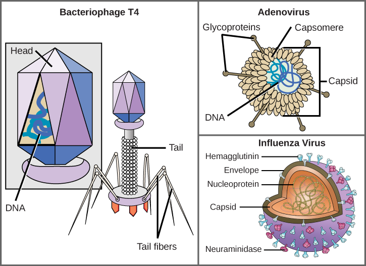
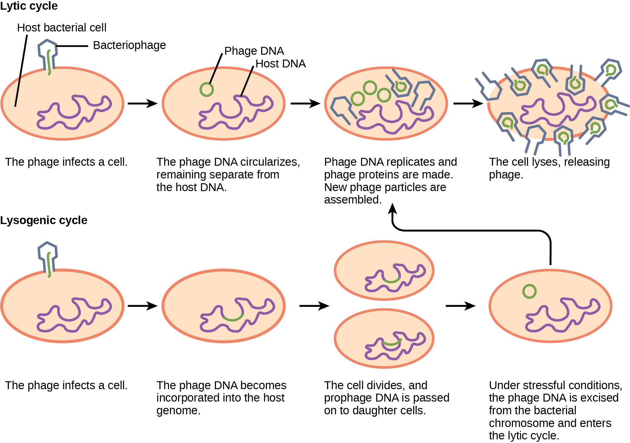
Transduction uses a bacteriophage as the carrier. Generalized transduction can package different bacterial DNA fragments during lytic growth; specialized transduction transfers genes near a prophage integration site after faulty excision. DNase can destroy exposed transforming DNA but does not readily reach DNA protected inside an intact phage particle or a conjugative transfer junction.
Practice: apply the mechanism
Question. Resistance transfers between two bacterial cultures only when cells contact one another; adding extracellular DNase does not stop it. Which mechanism is most consistent, and what additional control distinguishes contact from phage transfer?
Show reasoning
Conjugation is most consistent with contact dependence and DNase resistance. Separate donor and recipient cells with a barrier that blocks cells while allowing medium and suitable phage particles to pass, and test cell-free donor filtrate. Transfer through filtrate would support a phage-mediated alternative. DNase resistance alone cannot distinguish conjugation from transduction.
22.2 Structure of Prokaryotes: Bacteria and Archaea — figures and tables
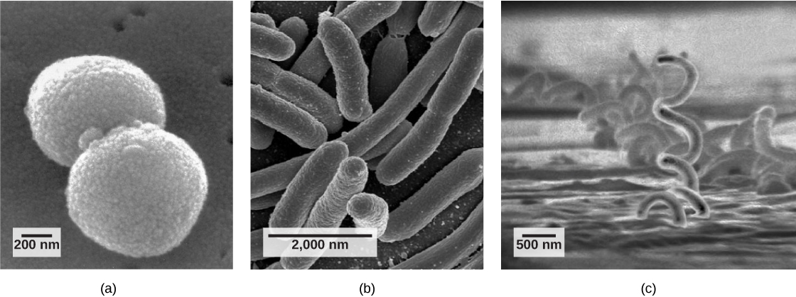
Figure 22.9 Common prokaryotic cell types. Prokaryotes fall into three basic categories based on their shape, visualized here using scanning electron microscopy: (a) cocci, or spherical (a pair is shown); (b) bacilli, or rod-shaped; and (c) spirilli, or spiral-shaped. (credit a: modification of work by Janice Haney Carr, Dr. Richard Facklam, CDC; credit c: modification of work by Dr. David Cox; scale-bar data from Matt Russell)
Mary Ann Clark, Matthew Douglas, and Jung Choi, Biology 2E, OpenStax. (credit a: modification of work by Janice Haney Carr, Dr. Richard Facklam, CDC; credit c: modification of work by Dr. David Cox; scale-bar data from Matt Russell) · CC BY-NC-SA 4.0 · Original image ↗ · Access for free ↗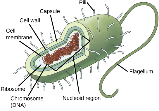
Figure 22.10 The features of a typical prokaryotic cell. Flagella, capsules, and pili are not found in all prokaryotes.
Mary Ann Clark, Matthew Douglas, and Jung Choi, Biology 2E, OpenStax. · CC BY-NC-SA 4.0 · Original image ↗ · Access for free ↗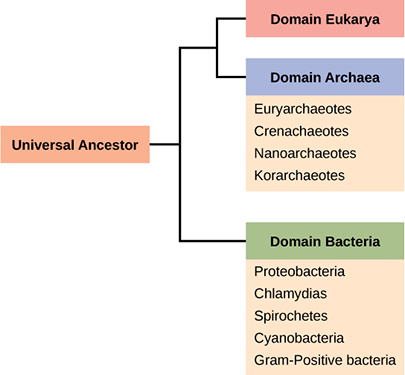
Figure 22.11 The three domains of living organisms. Bacteria and Archaea are both prokaryotes but differ enough to be placed in separate domains. An ancestor of modern Archaea is believed to have given rise to Eukarya, the third domain of life. Major groups of Archaea and Bacteria are shown.
Mary Ann Clark, Matthew Douglas, and Jung Choi, Biology 2E, OpenStax. · CC BY-NC-SA 4.0 · Original image ↗ · Access for free ↗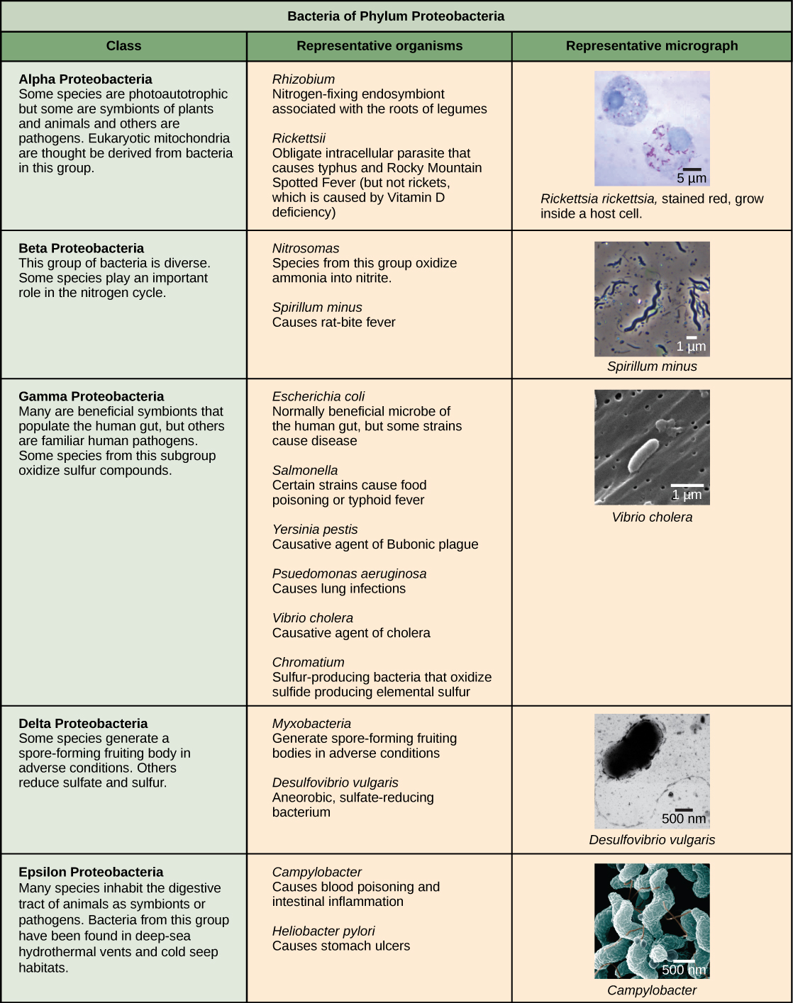
Figure 22.12 The Proteobacteria. Phylum Proteobacteria is one of up to 52 bacteria phyla. Proteobacteria is further subdivided into five classes, Alpha through Epsilon. (credit “Rickettsia rickettsii”: modification of work by CDC; credit “Spirillum minus”: modification of work by Wolframm Adlassnig; credit “Vibrio cholera”: modification of work by Janice Haney Carr, CDC; credit “Desulfovibrio vulgaris”: modification of work by Graham Bradley; credit “Campylobacter”: modification of work by De Wood, Pooley, USDA, ARS, EMU; scale-bar data from Matt Russell)
Mary Ann Clark, Matthew Douglas, and Jung Choi, Biology 2E, OpenStax. (credit “Rickettsia rickettsii”: modification of work by CDC; credit “Spirillum minus”: modification of work by Wolframm Adlassnig; credit “Vibrio cholera”: modification of work by Janice Haney Carr, CDC; credit “Desulfovibrio vulgaris”: modification of work by Graham Bradley; credit “Campylobacter”: modification of work by De Wood, Pooley, USDA, ARS, EMU; scale-bar data from Matt Russell) · CC BY-NC-SA 4.0 · Original image ↗ · Access for free ↗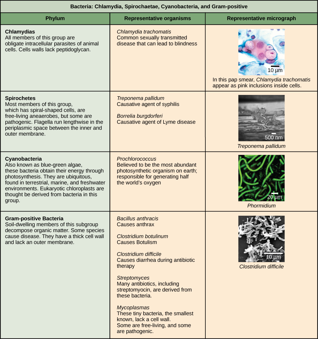
Figure 22.13 Other bacterial phyla. Chlamydia, Spirochetes, Cyanobacteria, and Gram-positive bacteria are described in this table. Note that bacterial shape is not phylum-dependent; bacteria within a phylum may be cocci, rod-shaped, or spiral. (credit “Chlamydia trachomatis” : modification of work by Dr. Lance Liotta Laboratory, NCI; credit “Treponema pallidum” : modification of work by Dr. David Cox, CDC; credit “Phormidium” : modification of work by USGS; credit “Clostridium difficile” : modification of work by Lois S. Wiggs, CDC; scale-bar data from Matt Russell)
Mary Ann Clark, Matthew Douglas, and Jung Choi, Biology 2E, OpenStax. (credit “Chlamydia trachomatis” : modification of work by Dr. Lance Liotta Laboratory, NCI; credit “Treponema pallidum” : modification of work by Dr. David Cox, CDC; credit “Phormidium” : modification of work by USGS; credit “Clostridium difficile” : modification of work by Lois S. Wiggs, CDC; scale-bar data from Matt Russell) · CC BY-NC-SA 4.0 · Original image ↗ · Access for free ↗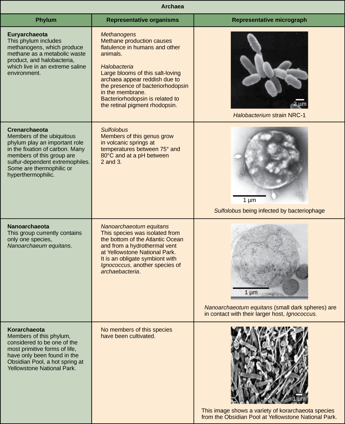
Figure 22.14 Archaeal phyla. Archaea are separated into four phyla: the Korarchaeota, Euryarchaeota, Crenarchaeota, and Nanoarchaeota. (credit “Halobacterium” : modification of work by NASA; credit “Nanoarchaeotum equitans” : modification of work by Karl O. Stetter; credit “Korarchaeota”: modification of work by Office of Science of the U.S. Dept. of Energy; scale-bar data from Matt Russell)
Mary Ann Clark, Matthew Douglas, and Jung Choi, Biology 2E, OpenStax. (credit “Halobacterium” : modification of work by NASA; credit “Nanoarchaeotum equitans” : modification of work by Karl O. Stetter; credit “Korarchaeota”: modification of work by Office of Science of the U.S. Dept. of Energy; scale-bar data from Matt Russell) · CC BY-NC-SA 4.0 · Original image ↗ · Access for free ↗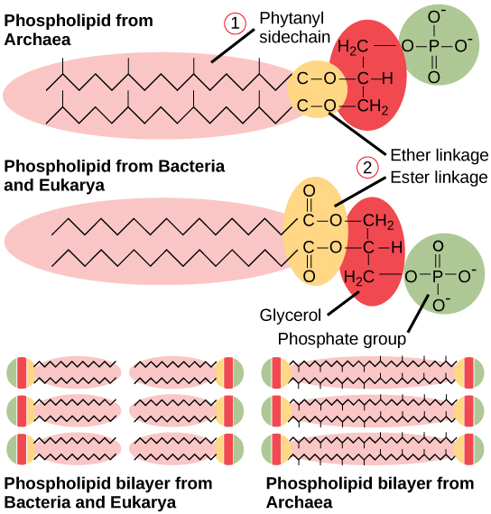
Figure 22.15 Bacterial and archaeal phospholipids. Archaeal phospholipids differ from those found in Bacteria and Eukarya in two ways. First, they have branched phytanyl sidechains instead of linear ones. Second, an ether bond instead of an ester bond connects the lipid to the glycerol.
Mary Ann Clark, Matthew Douglas, and Jung Choi, Biology 2E, OpenStax. · CC BY-NC-SA 4.0 · Original image ↗ · Access for free ↗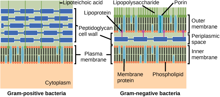
Figure 22.16 Cell walls in Gram-positive and Gram-negative bacteria. Bacteria are divided into two major groups: Gram positive and Gram negative. Both groups have a cell wall composed of peptidoglycan: in Gram-positive bacteria, the wall is thick, whereas in Gram-negative bacteria, the wall is thin. In Gram-negative bacteria, the cell wall is surrounded by an outer membrane that contains lipopolysaccharides and lipoproteins. Porins are proteins in this cell membrane that allow substances to pass through the outer membrane of Gram-negative bacteria. In Gram-positive bacteria, lipoteichoic acid anchors the cell wall to the cell membrane. (credit: modification of work by "Franciscosp2"/Wikimedia Commons)
Mary Ann Clark, Matthew Douglas, and Jung Choi, Biology 2E, OpenStax. (credit: modification of work by "Franciscosp2"/Wikimedia Commons) · CC BY-NC-SA 4.0 · Original image ↗ · Access for free ↗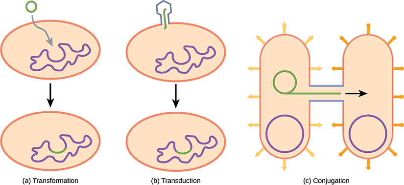
Figure 22.17 Gene transfer mechanisms in prokaryotes. There are three mechanisms by which prokaryotes can exchange DNA. In (a) transformation, the cell takes up prokaryotic DNA directly from the environment. The DNA may remain separate as plasmid DNA or be incorporated into the host genome. In (b) transduction, a bacteriophage injects DNA into the cell that contains a small fragment of DNA from a different prokaryote. In (c) conjugation, DNA is transferred from one cell to another via a mating bridge, or pilus, that connects the two cells after the sex pilus draws the two bacteria close enough to form the bridge.
Mary Ann Clark, Matthew Douglas, and Jung Choi, Biology 2E, OpenStax. · CC BY-NC-SA 4.0 · Original image ↗ · Access for free ↗| Structural Characteristic | Bacteria | Archaea |
|---|---|---|
| Cell type | Prokaryotic | Prokaryotic |
| Cell morphology | Variable | Variable |
| Cell wall | Contains peptidoglycan | Does not contain peptidoglycan |
| Cell membrane type | Lipid bilayer | Lipid bilayer or lipid monolayer |
| Plasma membrane lipids | Fatty acids-glycerol ester | Phytanyl-glycerol ethers |
| Chromosome | Typically circular | Typically circular |
| Replication origins | Single | Multiple |
| RNA polymerase | Single | Multiple |
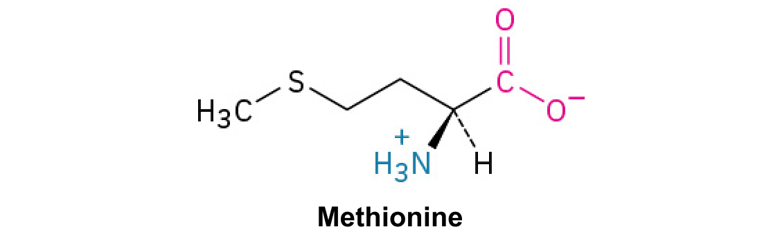
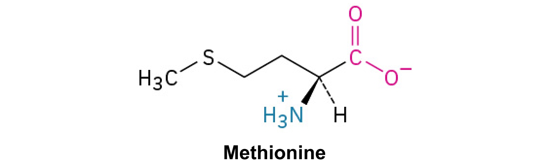
| Initiator tRNA | Formyl-methionine | Methionine |
| Streptomycin inhibition | Sensitive | Resistant |
| Calvin cycle | Yes | No |
Table 22.2. Mary Ann Clark, Matthew Douglas, and Jung Choi, Biology 2E, OpenStax.