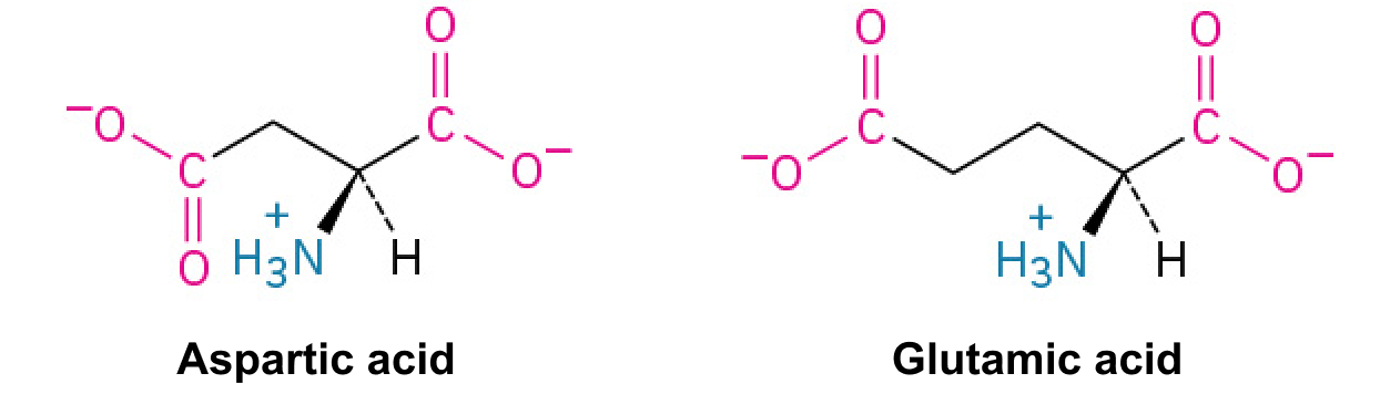
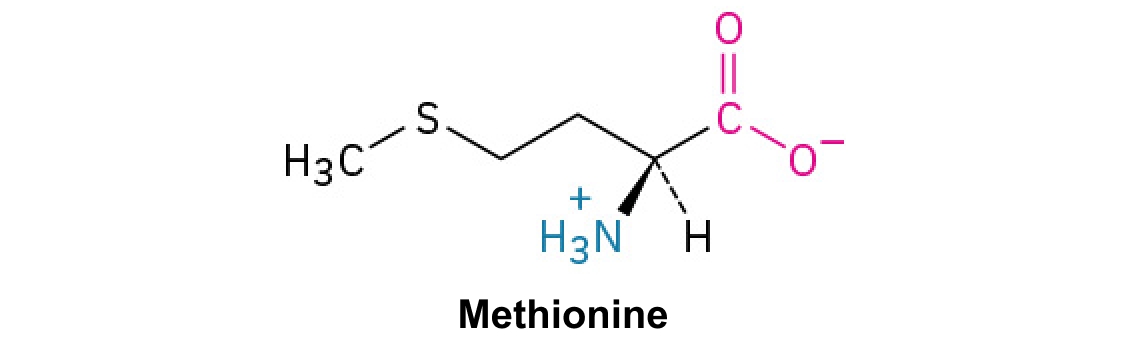
Foundation 2 · 2B · Prokaryotes and viruses

Cell theory and microbial structure

Sources ↗

Cells are the units of cellular life; , , and share core chemistry but differ in organization and surface structures.

What cell theory claims and how evidence supports it

Cell theory states that cellular organisms consist of cells, that the cell is the basic unit of biological organization, and that new cells arise from pre-existing cells. Microscopy established recurring cellular structure in tissues; observation of division connected reproduction to existing cells. Experiments excluding environmental contamination undermined routine spontaneous-generation claims.

All cells possess a plasma , , hereditary information, and , although specialized mature cells can lose some structures. Viruses are acellular and depend on cells for reproduction, so they do not overturn the theory by dividing independently. Cell theory describes modern cellular continuity; it does not by itself explain the first origin of life.

Source: 22.2 Structure of Prokaryotes: Bacteria and Archaea · 4.3 Eukaryotic Cells

, , and

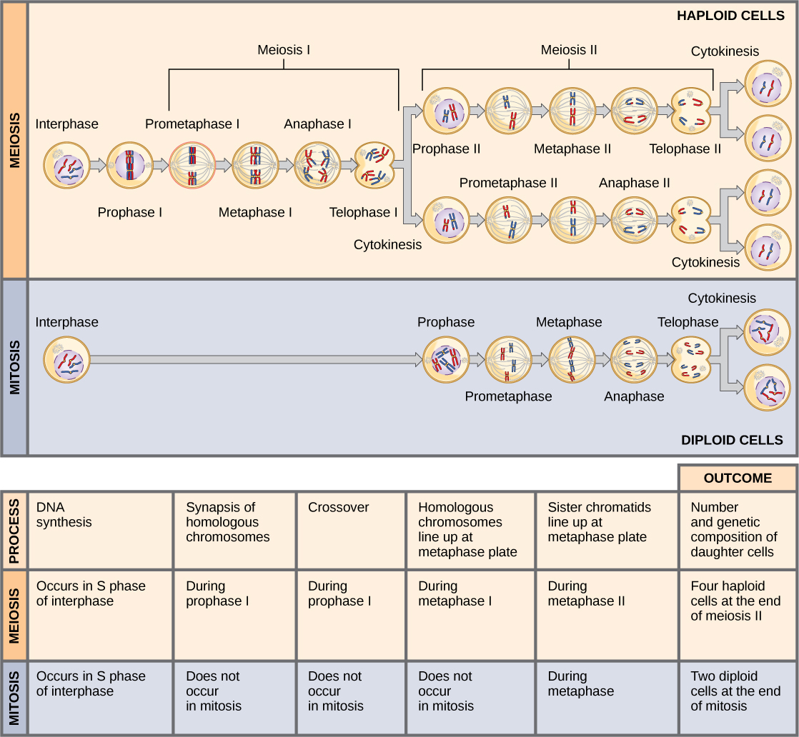
and lack a -enclosed and usually divide by ; their occupies a . have a and -bound and commonly segregate by . are generally 70S, while are 80S; and retain ancestry.

walls commonly contain . lack true and often have ether-linked ; their information-processing machinery shares some features with . Not all are extremophiles, and not all are . Size is useful context, not an absolute classification rule.

Source: 22.2 Structure of Prokaryotes: Bacteria and Archaea · 4.3 Eukaryotic Cells

Shapes, walls, and rotary

are spherical, rod-shaped, and spirilla or spirochetes helical; arrangement after division can further distinguish groups. have a thick layer, while have a thinner layer plus an outer containing . Wall structure affects staining, permeability, and antibiotic targets. A capsule can aid adhesion or protection but is distinct from the cell wall.

A consists of a filament, hook, and basal motor. An , usually motive force and sometimes sodium, drives rotation rather than -powered bending. Rotation and coordination of alter swimming paths. have roles in or genetic exchange rather than the same rotary propulsion mechanism.

Source: 22.2 Structure of Prokaryotes: Bacteria and Archaea · 4.3 Eukaryotic Cells

Practice: apply the mechanism

Question. An antimicrobial blocks cross-linking. Why might it damage a growing more directly than a human cell, and why does this not mean every microorganism must be sensitive?

Show reasoning

Human cells lack walls, so the molecular target is selective for susceptible . Growing actively build and remodel walls and can lose osmotic protection when cross-linking fails. lack true , some lack conventional walls, and resistance or limited drug entry can also prevent sensitivity.

Source: 22.2 Structure of Prokaryotes: Bacteria and Archaea · 4.3 Eukaryotic Cells

22.2 Structure of : and — figures and tables

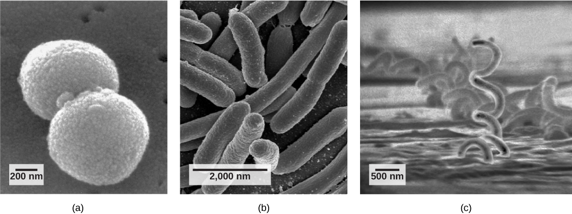
Part a: The micrograph shows ball-shaped cocci about 800 nanometers across. Part b: The micrograph shows elongated, oval-shaped bacilli about 2000 nanometers long. Part c: The micrograph shows corkscrew-shaped spirilli that are quite long and 500 nanometers in diameter.
Figure 22.9 — 22.2 Structure of : and

Figure 22.9 Common prokaryotic cell types. Prokaryotes fall into three basic categories based on their shape, visualized here using scanning electron microscopy: (a) cocci, or spherical (a pair is shown); (b) bacilli, or rod-shaped; and (c) spirilli, or spiral-shaped. (credit a: modification of work by Janice Haney Carr, Dr. Richard Facklam, CDC; credit c: modification of work by Dr. David Cox; scale-bar data from Matt Russell)

Mary Ann Clark, Matthew Douglas, and Jung Choi, Biology 2E, OpenStax. (credit a: modification of work by Janice Haney Carr, Dr. Richard Facklam, CDC; credit c: modification of work by Dr. David Cox; scale-bar data from Matt Russell) · CC BY-NC-SA 4.0 · Original image ↗ · Access for free ↗
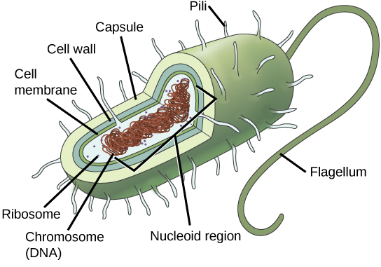
In this illustration, the prokaryotic cell is rod shaped. The circular chromosome is concentrated in a region called the nucleoid. The fluid inside the cell is called the cytoplasm. Ribosomes, depicted as small circles, float in the cytoplasm. The cytoplasm is encased by a plasma membrane, which in turn is encased by a cell wall. A capsule surrounds the cell wall. The bacterium depicted has a flagellum protruding from one narrow end. Pili are small protrusions that project from the capsule all over the bacterium, like hair.
Figure 22.10 — 22.2 Structure of : and

Figure 22.10 The features of a typical prokaryotic cell. Flagella, capsules, and pili are not found in all prokaryotes.

Mary Ann Clark, Matthew Douglas, and Jung Choi, Biology 2E, OpenStax. · CC BY-NC-SA 4.0 · Original image ↗ · Access for free ↗
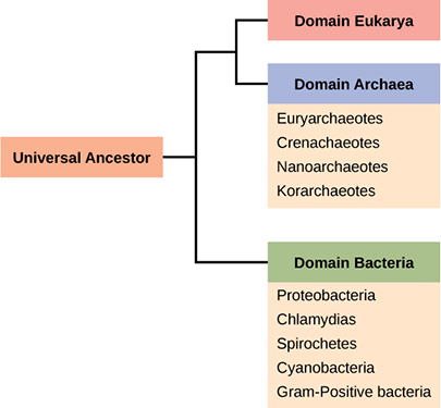
The trunk of the phylogenetic tree is a universal ancestor. The tree forms two branches. One branch leads to the domain bacteria, which includes the phyla proteobacteria, chlamydias, spirochetes, cyanobacteria, and Gram-positive bacteria. The other branch branches again, into the eukarya and archaea domains. Domain archaea includes the phyla euryarchaeotes, crenarchaeotes, nanoarchaeotes, and korarchaeotes.
Figure 22.11 — 22.2 Structure of : and

Figure 22.11 The three domains of living organisms. Bacteria and Archaea are both prokaryotes but differ enough to be placed in separate domains. An ancestor of modern Archaea is believed to have given rise to Eukarya, the third domain of life. Major groups of Archaea and Bacteria are shown.

Mary Ann Clark, Matthew Douglas, and Jung Choi, Biology 2E, OpenStax. · CC BY-NC-SA 4.0 · Original image ↗ · Access for free ↗
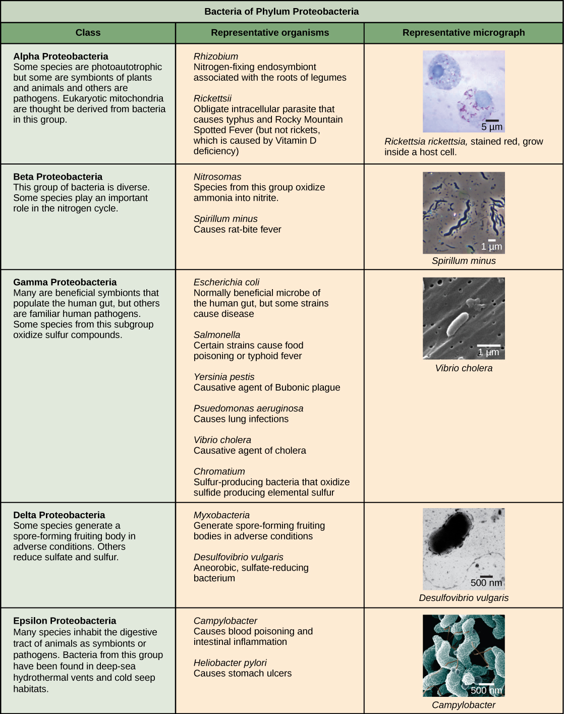
Characteristics of the five classes of bacterial phylum proteobacteria are described. The classes are alpha, beta, gamma, delta and epsilon. Most species of Alpha Proteobacteria are photoautotrophic but some are symbionts of plants and animals, and others are pathogens. Eukaryotic mitochondria are thought to be derived from bacteria in this group. Representative organisms include Rhizobium, a nitrogen-fixing endosymbiont associated with the roots of legumes, and Rickettsia, an obligate intracellular parasite that causes typhus and Rocky Mountain Spotted Fever (but not rickets, which is caused by Vitamin D deficiency). A micrograph shows rod-shaped Rickettsia rickettsii inside a much larger eukaryotic cell. Beta Proteobacteria is a diverse group of bacteria. Some species play an important role in the nitrogen cycle. Representative organisms include Nitrosomas, which oxidize ammonia into nitrite, and Spirillum minus, which causes rat bite fever. A micrograph of spiral-shaped Spirillum minus is shown. Gamma Proteobacteria include many beneficial symbionts that populate the human gut, as well as familiar human pathogens. Some species from this subgroup oxidize sulfur compounds. Representative organisms include Escherichia coli, normally a beneficial microbe of the human gut, but some strains cause disease; Salmonella, certain strains of which cause food poisoning or typhoid fever; Yersinia pestis, the causative agent of Bubonic plague; Psuedomonas aeruganosa, which causes lung infections; Vibrio cholera, the causative agent of cholera; and Chromatium, sulfur-producing bacteria that oxidize sulfur, producing H2S. Micrograph shows rod-shaped Vibrio cholera, which are about 1 micron long. Some species of Delta Proteobacteria generate a spore-forming fruiting body in adverse conditions. Others reduce sulfate and sulfur. Representative organisms include Myxobacteria, which generate spore-forming fruiting bodies in adverse conditions, and Desulfovibrio vulgaris, an aneorobic, sulfate-reducing bacterium. Micrograph shows a bent rod-shaped Desulfovibrio vulgaris bacterium with a long flagellum. Epsilon Proteobacteria includes many species that inhabit the digestive tract of animals as symbionts or pathogens. Bacteria from this group have been found in deep-sea hydrothermal vents and cold seep habitats. Representative organisms include Campylobacter, which causes blood poisoning and intestinal inflammation, and Heliobacter pylori, which causes stomach ulcers. Micrograph shows short spiral, or s shaped, Campylobacter bacteria.
Figure 22.12 — 22.2 Structure of : and

Figure 22.12 The Proteobacteria. Phylum Proteobacteria is one of up to 52 bacteria phyla. Proteobacteria is further subdivided into five classes, Alpha through Epsilon. (credit “Rickettsia rickettsii”: modification of work by CDC; credit “Spirillum minus”: modification of work by Wolframm Adlassnig; credit “Vibrio cholera”: modification of work by Janice Haney Carr, CDC; credit “Desulfovibrio vulgaris”: modification of work by Graham Bradley; credit “Campylobacter”: modification of work by De Wood, Pooley, USDA, ARS, EMU; scale-bar data from Matt Russell)

Mary Ann Clark, Matthew Douglas, and Jung Choi, Biology 2E, OpenStax. (credit “Rickettsia rickettsii”: modification of work by CDC; credit “Spirillum minus”: modification of work by Wolframm Adlassnig; credit “Vibrio cholera”: modification of work by Janice Haney Carr, CDC; credit “Desulfovibrio vulgaris”: modification of work by Graham Bradley; credit “Campylobacter”: modification of work by De Wood, Pooley, USDA, ARS, EMU; scale-bar data from Matt Russell) · CC BY-NC-SA 4.0 · Original image ↗ · Access for free ↗
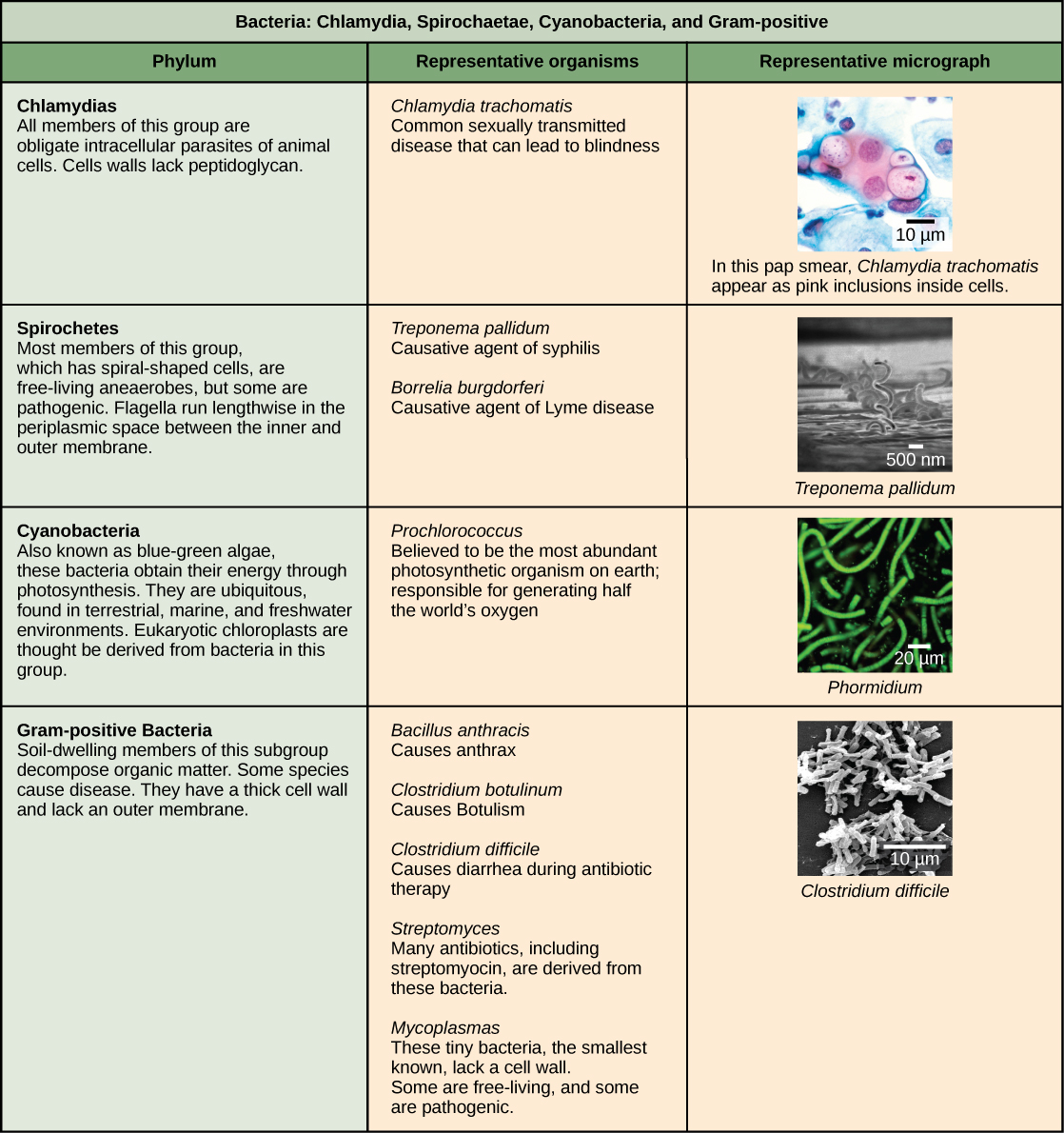
This table describes the other four bacterial phyla classes. The first phylum described is chlamydias. All members of this group are obligate intracellular parasites of animal cells. Cells walls lack peptidoglycan. Micrograph shows a pap smear of cells infected with Chlamydia trachomatis. Chlamydia infection is the most common sexually transmitted disease and can lead to blindness. All members of the phylum Spirochetes have spiral-shaped cells. Most are free-living anaerobes, but some are pathogenic. Flagella run lengthwise in the periplasmic space between the inner and outer membrane. Representative species include Treponema pallidum, the causative agent of syphilis and Borrelia burgdorferi, the causative agent of Lyme disease Micrograph shows corkscrew-shaped Trepanema pallidum, about 1 micron across. Bacteria in the phylum Cyanobacteria, also known as blue-green algae, obtain their energy through photosynthesis. They are ubiquitous, found in terrestrial, marine, and freshwater environments. Eukaryotic chloroplasts are thought to be derived from bacteria in this group. The cyanobacterium Prochlorococcus is believed to be the most abundant photosynthetic organism on earth, responsible for generating half the world’s oxygen. Micrograph shows a long, thin rod-shaped species called Phormidium. Gram-positive Bacteria have a thick cell wall and lack an outer membrane. Soil-dwelling members of this subgroup decompose organic matter. Some species cause disease. Representative species include Bacillus anthracis, which causes anthrax; Clostridium botulinum, which causes botulism; Clostridium difficile, which causes diarrhea during antibiotic therapy; Streptomyces, from which many antibiotics, including streptomyocin, are derived; and Mycoplasmas, the smallest known bacteria, which lack a cell wall. Some are free-living, and some are pathogenic. Micrograph shows Clostridium difficile, which are rod-shaped and about 3 microns long.
Figure 22.13 — 22.2 Structure of : and

Figure 22.13 Other bacterial phyla. Chlamydia, Spirochetes, Cyanobacteria, and Gram-positive bacteria are described in this table. Note that bacterial shape is not phylum-dependent; bacteria within a phylum may be cocci, rod-shaped, or spiral. (credit “Chlamydia trachomatis” : modification of work by Dr. Lance Liotta Laboratory, NCI; credit “Treponema pallidum” : modification of work by Dr. David Cox, CDC; credit “Phormidium” : modification of work by USGS; credit “Clostridium difficile” : modification of work by Lois S. Wiggs, CDC; scale-bar data from Matt Russell)

Mary Ann Clark, Matthew Douglas, and Jung Choi, Biology 2E, OpenStax. (credit “Chlamydia trachomatis” : modification of work by Dr. Lance Liotta Laboratory, NCI; credit “Treponema pallidum” : modification of work by Dr. David Cox, CDC; credit “Phormidium” : modification of work by USGS; credit “Clostridium difficile” : modification of work by Lois S. Wiggs, CDC; scale-bar data from Matt Russell) · CC BY-NC-SA 4.0 · Original image ↗ · Access for free ↗
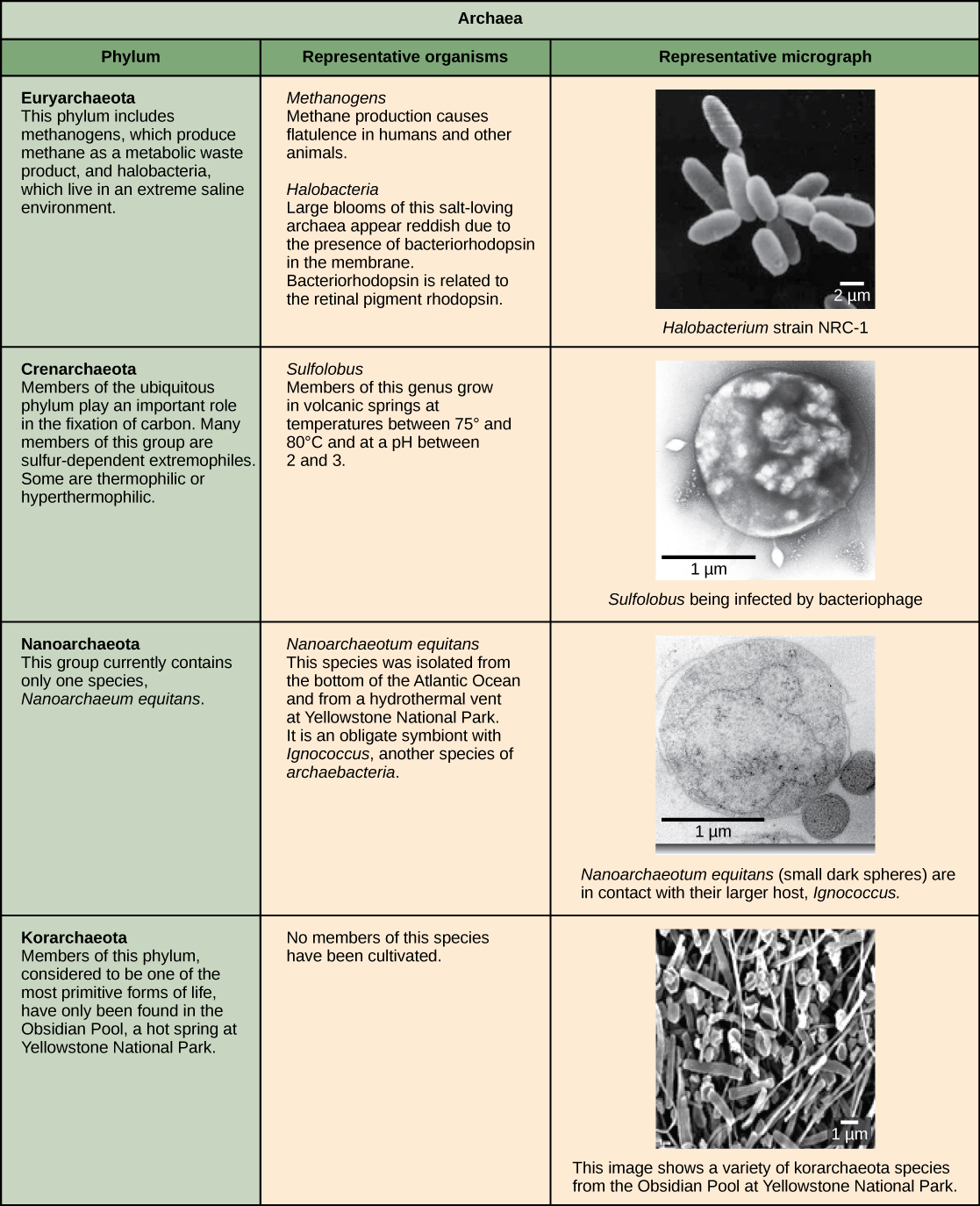
Characteristics of the four phyla of archaea are described. Euryarchaeotes includes methanogens, which produce methane as a metabolic waste product, and halobacteria, which live in an extreme saline environment. Methanogens cause flatulence in humans and other animals. Halobacteria can grow in large blooms that appear reddish, due to the presence of bacteriorhodopsin in the membrane. Bacteriorhodopsin is related to the retinal pigment rhodopsin. Micrograph shows rod shaped Halobacterium. Members of the ubiquitous Crenarchaeotes phylum play an important role in the fixation of carbon. Many members of this group are sulfur dependent extremophiles. Some are thermophilic or hyperthermophilic. Micrograph shows cocci shaped Sulfolobus, a genus which grows in volcanic springs at temperatures between 75 degrees and 80 degrees Celsius and at a lower case p upper case H between 2 and 3. The phylum Nanoarchaeotes currently contains only one species, Nanoarchaeum equitans, which has been isolated from the bottom of the Atlantic Ocean, and from the a hydrothermal vent at Yellowstone National Park. It is an obligate symbiont with Ignicoccus, another species of archaebacteria. Micrograph shows two small, round N. equitans cells attached to a larger Ignicoccus cell. Korarchaeotes are considered to be one of the most primitive forms of life and so far have only been found in the Obsidian Pool, a hot spring at Yellowstone National Park. Micrograph shows a variety of specimens from this group which vary in shape.
Figure 22.14 — 22.2 Structure of : and

Figure 22.14 Archaeal phyla. Archaea are separated into four phyla: the Korarchaeota, Euryarchaeota, Crenarchaeota, and Nanoarchaeota. (credit “Halobacterium” : modification of work by NASA; credit “Nanoarchaeotum equitans” : modification of work by Karl O. Stetter; credit “Korarchaeota”: modification of work by Office of Science of the U.S. Dept. of Energy; scale-bar data from Matt Russell)

Mary Ann Clark, Matthew Douglas, and Jung Choi, Biology 2E, OpenStax. (credit “Halobacterium” : modification of work by NASA; credit “Nanoarchaeotum equitans” : modification of work by Karl O. Stetter; credit “Korarchaeota”: modification of work by Office of Science of the U.S. Dept. of Energy; scale-bar data from Matt Russell) · CC BY-NC-SA 4.0 · Original image ↗ · Access for free ↗
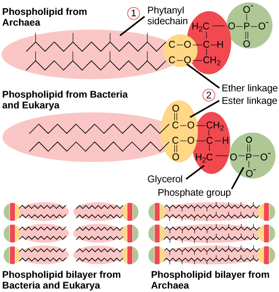
This illustration compares phospholipids from Bacteria and Eukarya to those from Archaea. In Bacteria and Eukarya, fatty acids are attached to glycerol by an ester linkage, while in Archaea, isoprene chains are linked to glycerol by an ether linkage. In the ester linkage, the first carbon in the fatty acid chain has an oxygen double-bonded to it, whereas in the ether linkage, it does not. In Archaea, the isoprene chains have methyl groups branching off from them, whereas such branches are absent in Bacteria and Eukarya. Both types of phospholipids result in similar lipid bilayers.
Figure 22.15 — 22.2 Structure of : and

Figure 22.15 Bacterial and archaeal phospholipids. Archaeal phospholipids differ from those found in Bacteria and Eukarya in two ways. First, they have branched phytanyl sidechains instead of linear ones. Second, an ether bond instead of an ester bond connects the lipid to the glycerol.

Mary Ann Clark, Matthew Douglas, and Jung Choi, Biology 2E, OpenStax. · CC BY-NC-SA 4.0 · Original image ↗ · Access for free ↗
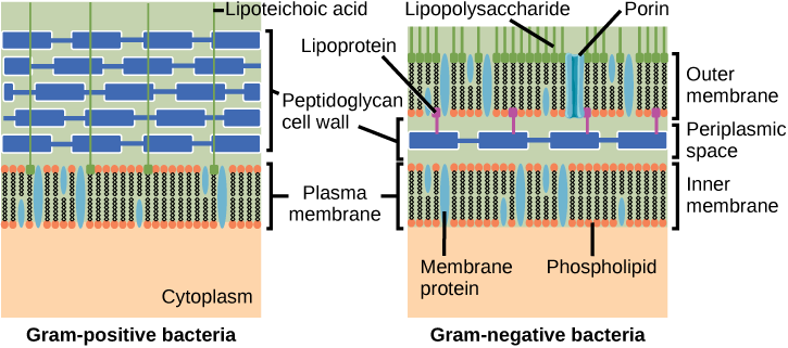
The left illustration shows the cell wall of Gram-positive bacteria. The cell wall is a thick layer of peptidoglycan that exists outside the plasma membrane. A long, thin molecule called lipoteichoic acid anchors the cell wall to the cell membrane. The right illustration shows Gram-negative bacteria. In Gram-negative bacteria, a thin peptidoglycan cell wall is sandwiched between an outer and an inner plasma membrane. The space between the two membranes is called the periplasmic space. Lipoproteins anchor the cell wall to the outer membrane. Lipopolysaccharides protrude from the outer membrane. Porins are proteins in the outer membrane that allow entry of substances.
Figure 22.16 — 22.2 Structure of : and

Figure 22.16 Cell walls in Gram-positive and Gram-negative bacteria. Bacteria are divided into two major groups: Gram positive and Gram negative. Both groups have a cell wall composed of peptidoglycan: in Gram-positive bacteria, the wall is thick, whereas in Gram-negative bacteria, the wall is thin. In Gram-negative bacteria, the cell wall is surrounded by an outer membrane that contains lipopolysaccharides and lipoproteins. Porins are proteins in this cell membrane that allow substances to pass through the outer membrane of Gram-negative bacteria. In Gram-positive bacteria, lipoteichoic acid anchors the cell wall to the cell membrane. (credit: modification of work by "Franciscosp2"/Wikimedia Commons)

Mary Ann Clark, Matthew Douglas, and Jung Choi, Biology 2E, OpenStax. (credit: modification of work by "Franciscosp2"/Wikimedia Commons) · CC BY-NC-SA 4.0 · Original image ↗ · Access for free ↗
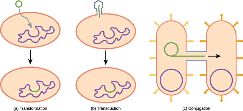
Illustration A shows transformation, where a small, circular piece of DNA is absorbed by a prokaryotic cell. The DNA then gets incorporated in the genome. Illustration B shows transduction, where a bacteriophage injects DNA into a prokaryotic cell. The DNA then gets incorporated in the genome. Illustration C shows conjugation, where two bacteria are connected by a pilus. A small loop of DNA is transferred from one cell to the other via the pilus.
Figure 22.17 — 22.2 Structure of : and

Figure 22.17 Gene transfer mechanisms in prokaryotes. There are three mechanisms by which prokaryotes can exchange DNA. In (a) transformation, the cell takes up prokaryotic DNA directly from the environment. The DNA may remain separate as plasmid DNA or be incorporated into the host genome. In (b) transduction, a bacteriophage injects DNA into the cell that contains a small fragment of DNA from a different prokaryote. In (c) conjugation, DNA is transferred from one cell to another via a mating bridge, or pilus, that connects the two cells after the sex pilus draws the two bacteria close enough to form the bridge.

Mary Ann Clark, Matthew Douglas, and Jung Choi, Biology 2E, OpenStax. · CC BY-NC-SA 4.0 · Original image ↗ · Access for free ↗
Structural Characteristic
Cell type
Cell morphology Variable Variable
Cell wall Contains Does not contain
type bilayer bilayer or monolayer
Plasma - ester Phytanyl- ethers
Typically circular Typically circular
Single Multiple
Single Multiple
Formyl-
Streptomycin inhibition Sensitive Resistant
Calvin cycle Yes No

Table 22.2. Mary Ann Clark, Matthew Douglas, and Jung Choi, Biology 2E, OpenStax.

Source: 22.2 Structure of Prokaryotes: Bacteria and Archaea

4.3 Cells — figures and tables

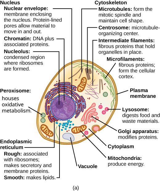
Part a: This illustration shows a typical eukaryotic animal cell, which is egg shaped. The fluid inside the cell is called the cytoplasm, and the cell is surrounded by a cell membrane. The nucleus takes up about one-half the width of the cell. Inside the nucleus is the chromatin, which is composed of DNA and associated proteins. A region of the chromatin is condensed into the nucleolus, a structure where ribosomes are synthesized. The nucleus is encased in a nuclear envelope, which is perforated by protein-lined pores that allow entry of material into the nucleus. The nucleus is surrounded by the rough and smooth endoplasmic reticulum, or ER. The smooth ER is the site of lipid synthesis. The rough ER has embedded ribosomes that give it a bumpy appearance. It synthesizes membrane and secretory proteins. In addition to the ER, many other organelles float inside the cytoplasm. These include the Golgi apparatus, which modifies proteins and lipids synthesized in the ER. The Golgi apparatus is made of layers of flat membranes. Mitochondria, which produce food for the cell, have an outer membrane and a highly folded inner membrane. Other, smaller organelles include peroxisomes that house oxidative metabolism, lysosomes that digest food, and vacuoles. Ribosomes, responsible for protein synthesis, also float freely in the cytoplasm and are depicted as small dots. The last cellular component shown is the cytoskeleton, which has four different types of components: microfilaments, intermediate filaments, microtubules, and centrosomes. Microfilaments are fibrous proteins that line the cell membrane and make up the cellular cortex. Intermediate filaments are fibrous proteins that hold organelles in place. Microtubules form the mitotic spindle and maintain cell shape. Centrosomes are made of two tubular structures at right angles to one another. They form the microtubule-organizing center.
Figure 4.8 — 4.3 Cells

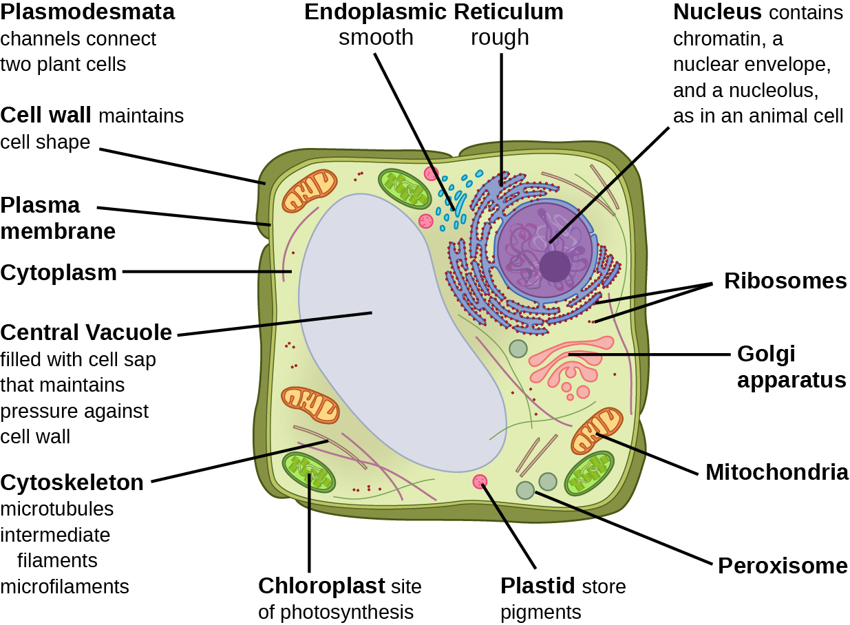
Figure 4.8 These figures show the major organelles and other cell components of (a) a typical animal cell and (b) a typical eukaryotic plant cell. The plant cell has a cell wall, chloroplasts, plastids, and a central vacuole—structures not in animal cells. Most cells do not have lysosomes or centrosomes.

Mary Ann Clark, Matthew Douglas, and Jung Choi, Biology 2E, OpenStax. · CC BY-NC-SA 4.0 · Original image ↗ · Access for free ↗
Part b: This illustration depicts a typical eukaryotic plant cell. The nucleus of a plant cell contains chromatin and a nucleolus, the same as an animal cell. Other structures that the plant cell has in common with the animal cell include rough and smooth endoplasmic reticulum, the Golgi apparatus, mitochondria, peroxisomes, and ribosomes. The fluid inside the plant cell is called the cytoplasm, just as it is in an animal cell. The plant cell has three of the four cytoskeletal components found in animal cells: microtubules, intermediate filaments, and microfilaments. Plant cells do not have centrosomes. Plant cells have four structures not found in animals cells: chloroplasts, plastids, a central vacuole, and a cell wall. Chloroplasts are responsible for photosynthesis; they have an outer membrane, an inner membrane, and stack of membranes inside the inner membrane. The central vacuole is a very large, fluid-filled structure that maintains pressure against the cell wall. Plastids store pigments. The cell wall is outside the cell membrane.
Figure 4.8 — 4.3 Cells

Figure 4.8 These figures show the major organelles and other cell components of (a) a typical animal cell and (b) a typical eukaryotic plant cell. The plant cell has a cell wall, chloroplasts, plastids, and a central vacuole—structures not in animal cells. Most cells do not have lysosomes or centrosomes.

Mary Ann Clark, Matthew Douglas, and Jung Choi, Biology 2E, OpenStax. · CC BY-NC-SA 4.0 · Original image ↗ · Access for free ↗
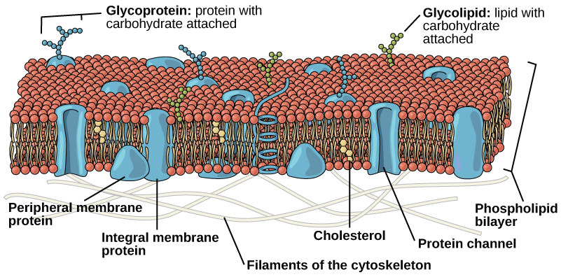
The plasma membrane is composed of a phospholipid bilayer. In the bilayer, the two long hydrophobic tails of phospholipids face toward the center, and the hydrophilic head group faces the exterior. Integral membrane proteins and protein channels span the entire bilayer. Protein channels have a pore in the middle. Peripheral membrane proteins sit on the surface of the phospholipids, and are associated with the phospholipid head groups. On the exterior side of the membrane, carbohydrates are attached to certain proteins and lipids. Filaments of the cytoskeleton line the interior of the membrane.
Figure 4.9 — 4.3 Cells

Figure 4.9 The eukaryotic plasma membrane is a phospholipid bilayer with proteins and cholesterol embedded in it.

Mary Ann Clark, Matthew Douglas, and Jung Choi, Biology 2E, OpenStax. · CC BY-NC-SA 4.0 · Original image ↗ · Access for free ↗
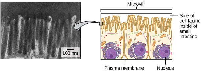
The left part of this figure is a transmission electron micrograph of microvilli, which appear as long, slender stalks extending from the plasma membrane. The right side illustrates cells containing microvilli. The microvilli cover the surface of the cell facing the interior of the small intestine.
Figure 4.10 — 4.3 Cells

Figure 4.10 Microvilli, as they appear on cells lining the small intestine, increase the surface area available for absorption. These microvilli are only on the area of the plasma membrane that faces the cavity from which substances will be absorbed. (credit "micrograph": modification of work by Louisa Howard)

Mary Ann Clark, Matthew Douglas, and Jung Choi, Biology 2E, OpenStax. (credit "micrograph": modification of work by Louisa Howard) · CC BY-NC-SA 4.0 · Original image ↗ · Access for free ↗
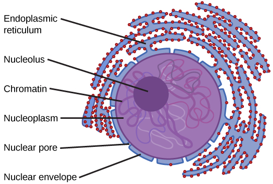
The two-dimensional image depicts the nucleus of a cell as a circular object with two membranes; several gaps appear in the circle, representing nuclear pores. Surrounding the nucleus are membranous sacks representing the endoplasmic reticulum. Inside the nucleus is another circle, approximately ten percent of the total size of the nucleus, representing the nucleolus.
Figure 4.11 — 4.3 Cells

Figure 4.11 The nucleus stores chromatin (DNA plus proteins) in a gel-like substance called the nucleoplasm. The nucleolus is a condensed chromatin region where ribosome synthesis occurs. We call the nucleus' boundary the nuclear envelope. It consists of two phospholipid bilayers: an outer and an inner membrane. The nuclear membrane is continuous with the endoplasmic reticulum. Nuclear pores allow substances to enter and exit the nucleus.

Mary Ann Clark, Matthew Douglas, and Jung Choi, Biology 2E, OpenStax. · CC BY-NC-SA 4.0 · Original image ↗ · Access for free ↗
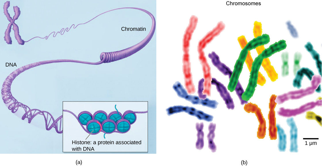
Part a: In this illustration, DNA tightly coiled into two thick cylinders is shown in the upper right. A close-up shows how the DNA is coiled around proteins called histones. Part b: This image shows paired chromosomes.  The chromosomes are shown as a collection of slender tubes.
Figure 4.12 — 4.3 Cells

Figure 4.12 (a) This image shows various levels of chromatin's organization (DNA and protein). (b) This image shows paired chromosomes. (credit b: modification of work by NIH; scale-bar data from Matt Russell)

Mary Ann Clark, Matthew Douglas, and Jung Choi, Biology 2E, OpenStax. (credit b: modification of work by NIH; scale-bar data from Matt Russell) · CC BY-NC-SA 4.0 · Original image ↗ · Access for free ↗
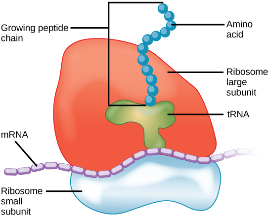
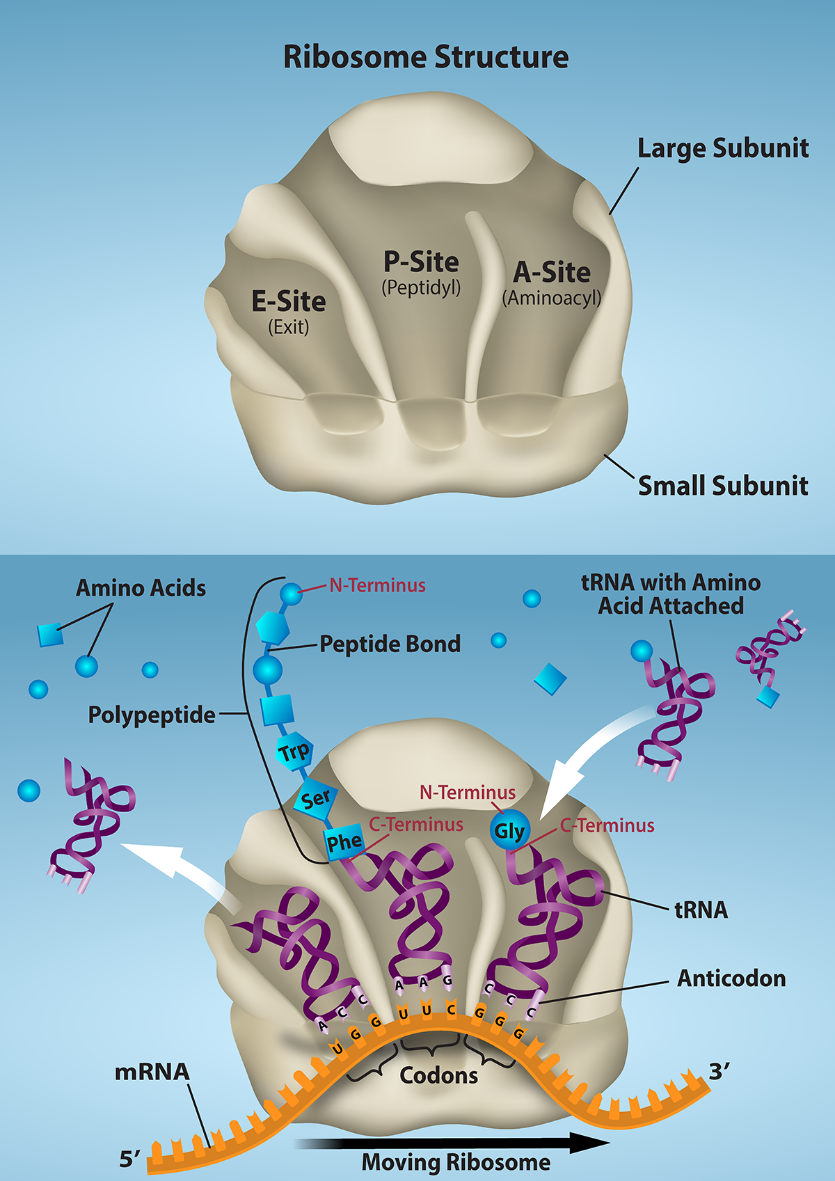
The ribosome consists of a small subunit and a large subunit, which is about three times as big as the small one. The large subunit sits on top of the small one. A chain of m R N A threads between the large and small subunits. A protein chain extends from the top of the large subunit.
Figure 4.13 — 4.3 Cells

Figure 4.13 A large subunit (top) and a small subunit (bottom) comprise ribosomes. During protein synthesis, ribosomes assemble amino acids into proteins.

Mary Ann Clark, Matthew Douglas, and Jung Choi, Biology 2E, OpenStax. · CC BY-NC-SA 4.0 · Original image ↗ · Access for free ↗
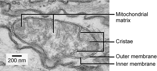
This transmission electron micrograph of a mitochondrion shows an oval outer membrane and an inner membrane with many folds called cristae. Inside the inner membrane is a space called the mitochondrial matrix.
Figure 4.14 — 4.3 Cells

Figure 4.14 This electron micrograph shows a mitochondrion through an electron microscope. This organelle has an outer membrane and an inner membrane. The inner membrane contains folds, called cristae, which increase its surface area. We call the space between the two membranes the intermembrane space, and the space inside the inner membrane the mitochondrial matrix. ATP synthesis takes place on the inner membrane. (credit: modification of work by Matthew Britton; scale-bar data from Matt Russell)

Mary Ann Clark, Matthew Douglas, and Jung Choi, Biology 2E, OpenStax. (credit: modification of work by Matthew Britton; scale-bar data from Matt Russell) · CC BY-NC-SA 4.0 · Original image ↗ · Access for free ↗
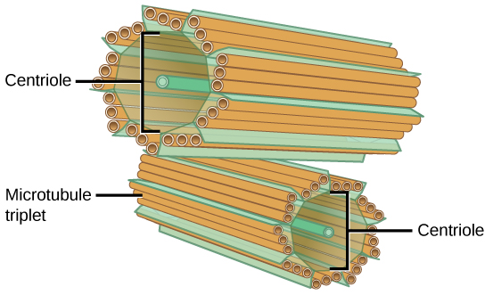
The image depicts two tube-like structures, one on top of the other, at right angles. Each of the tubes is labeled as the centriole. Each tube is composed of smaller tubes grouped in threes; these are labeled 'microtubule triplet.' Each centriole tube is composed of nine triplets arranged to form the wall of the tube.
Figure 4.15 — 4.3 Cells

Figure 4.15 The centrosome consists of two centrioles that lie at right angles to each other. Each centriole is a cylinder comprised of nine triplets of microtubules. Nontubulin proteins (indicated by the green lines) hold the microtubule triplets together.

Mary Ann Clark, Matthew Douglas, and Jung Choi, Biology 2E, OpenStax. · CC BY-NC-SA 4.0 · Original image ↗ · Access for free ↗
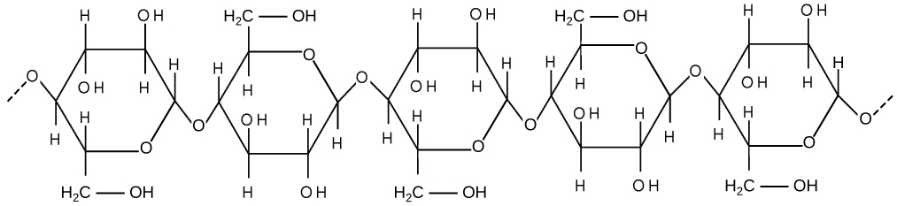
This illustration shows three glucose subunits that are attached together. Dashed lines at each end indicate that many more subunits make up an entire cellulose fiber. Each glucose subunit is a closed ring composed of carbon, hydrogen, and oxygen atoms.
Figure 4.16 — 4.3 Cells

Figure 4.16 Cellulose is a long chain of β-glucose molecules connected by a 1-4 linkage. The dashed lines at each end of the figure indicate a series of many more glucose units. The size of the page makes it impossible to portray an entire cellulose molecule.

Mary Ann Clark, Matthew Douglas, and Jung Choi, Biology 2E, OpenStax. · CC BY-NC-SA 4.0 · Original image ↗ · Access for free ↗
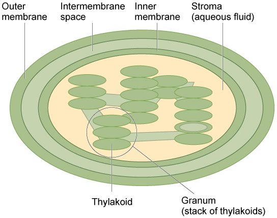
This illustration shows a chloroplast, which has an outer membrane and an inner membrane. The space between the outer and inner membranes is called the intermembrane space. Inside the inner membrane are flat, pancake-like structures called thylakoids. The thylakoids form stacks called grana. The liquid inside the inner membrane is called the stroma, and the space inside the thylakoids is called the thylakoid space.
Figure 4.17 — 4.3 Cells

Figure 4.17 The chloroplast has an outer membrane, an inner membrane, and membrane structures - thylakoids that are stacked into grana. We call the space inside the thylakoid membranes the thylakoid space. The light harvesting reactions take place in the thylakoid membranes, and sugar synthesis takes place in the fluid inside the inner membrane, which we call the stroma. Chloroplasts also have their own genome, which is contained on a single circular chromosome.

Mary Ann Clark, Matthew Douglas, and Jung Choi, Biology 2E, OpenStax. · CC BY-NC-SA 4.0 · Original image ↗ · Access for free ↗

Source: 4.3 Eukaryotic Cells

253 terms, alphabetically ordered

3′ end · ·

The end of a nucleic- strand associated with the sugar’s 3′ position. extends a strand at a free .

Read explanation · OpenStax ↗
3′ hydroxyl group · · ·

The at the sugar’s 3′ carbon that attacks the incoming ’s during strand extension.

Read explanation · OpenStax ↗
A site · ·

The site that accepts most incoming during elongation and at termination.

Read explanation · OpenStax ↗
acceleration

Rate of change of , measured in m/s².

Read explanation · OpenStax ↗
Acid · ·

In the Brønsted–Lowry model, a substance that can donate a . Acidic have a higher activity than water.

Read explanation · OpenStax ↗
Activation energy ·

The energy barrier that must overcome to reach a and undergo .

Read explanation · OpenStax ↗
Active catalytic-site concentration ·

The total of active sites used to calculate from . It equals active when each has one site.

Read explanation · Biology LibreTexts ↗
Active site

The region of an that binds its and carries out .

Read explanation · OpenStax ↗
adaptation

A heritable feature shaped by selection because it improves reproductive success in a relevant environment.

Read explanation · OpenStax ↗
Adenosine triphosphate

An energy-carrying ; its powers many cellular processes, including cycles.

Read explanation · OpenStax ↗
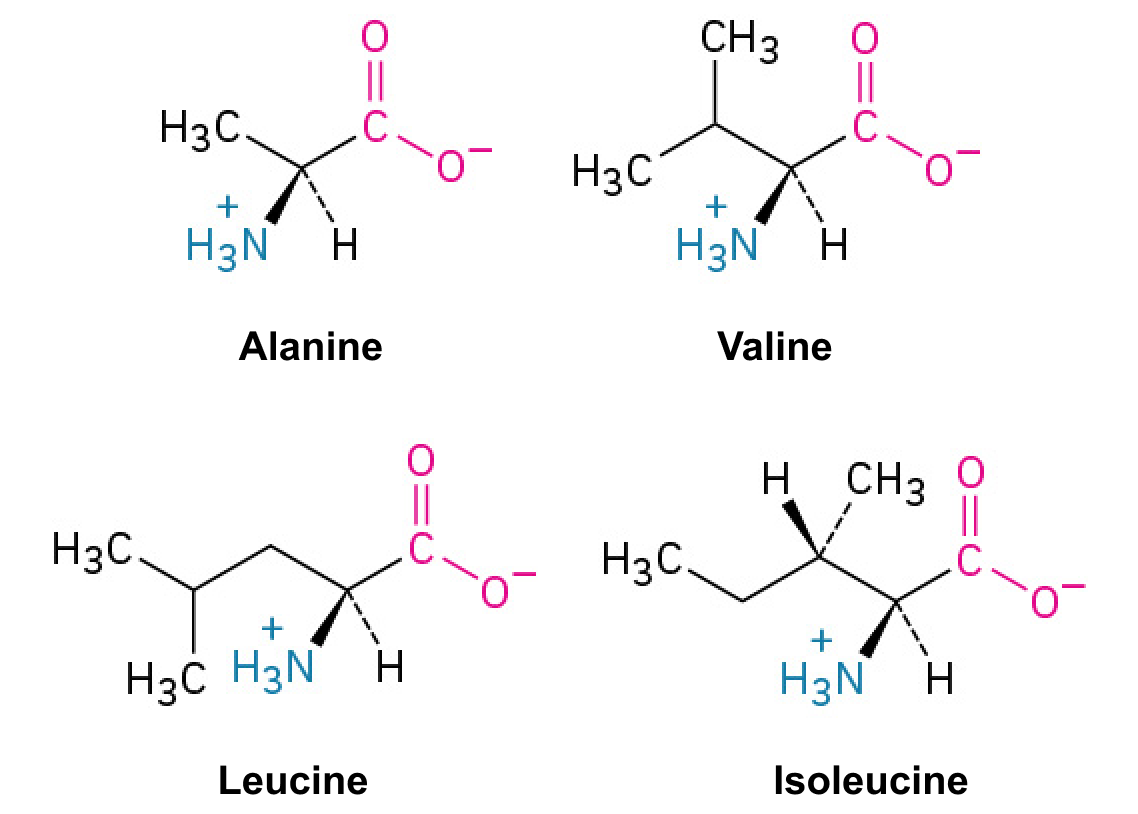
Alanine ·

A with a .

Read explanation · OpenStax ↗
Allele

One version or copy of a at a ; a typical cell carries two copies of an autosomal .

Read explanation · NCBI Bookshelf / Sinauer Associates ↗
Alpha carbon · ·

The central carbon next to the carboxyl carbon. In a standard it binds an , hydrogen, and .

Read explanation · OpenStax ↗
Alpha helix · · · · · ·

A helical stabilized by between a oxygen and an hydrogen four later.

Read explanation · OpenStax ↗
Amide · · ·

A group containing a carbon bonded to nitrogen. are amide linkages.

Read explanation · OpenStax ↗
Amino acid

A building block containing amino and and a characteristic .

Read explanation · OpenStax ↗
Amino acid residue · ·

The part of an incorporated into a chain. Residue can also describe another building block within a larger .

Read explanation · OpenStax ↗
Amino group · · ·

A nitrogen-containing . A free ’s amino group is typically (NH₃⁺) near physiological ; ’s ring nitrogen is an exception to this formula.

Read explanation · OpenStax ↗
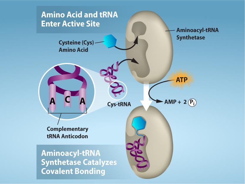
Aminoacyl-tRNA · ·

A covalently linked to an at its , ready to deliver that during .

Read explanation · OpenStax ↗
Amphipathic

Having both a region and a region, as in a or .

Read explanation · OpenStax ↗
Amylose

The largely unbranched component of , made mainly of linked by alpha-1,4 .

Read explanation · OpenStax ↗
Analytical standard ·

A material with a known identity or amount used to compare or calibrate measurements.

Read explanation · Agilent ↗
Anion ·

An with a negative net .

Read explanation · OpenStax ↗
anomeric carbon

The former carbon of a sugar, which becomes the or hemiketal center upon cyclization.

Read explanation · OpenStax ↗
Antibody

An that recognizes a specific region () on an . Antibodies help immune defenses and are used as detection tools in .

Read explanation · OpenStax ↗
Anticodon

A triplet that pairs to an . The anticodon for 5′-AUG-3′ is aligned 3′-UAC-5′, or written 5′-CAU-3′.

Read explanation · NCBI Bookshelf / Garland Science ↗
Antigen

A or molecular structure specifically recognized by an or other adaptive immune .

Read explanation · OpenStax ↗
Antiparallel

Running in opposite directions, as with two strands or neighboring strands in some .

Read explanation · OpenStax ↗
Aqueous solution ·

A in which water is the .

Read explanation · OpenStax ↗
Archaea ·

A of distinct from , with characteristic chemistry and -expression machinery.

Read explanation · OpenStax ↗
Assay

A procedure used to detect a substance or measure its amount, activity, or another property.

Read explanation · Thermo Fisher ↗
Atom

The smallest unit of an element that retains its chemical identity; it has a surrounded by .

Read explanation · OpenStax ↗
attachment

Enduring emotional bond and expectations about another’s availability and responsiveness.

Read explanation · OpenStax ↗
autotroph

An organism using inorganic carbon, usually CO2, as its principal carbon source.

Read explanation · OpenStax ↗
bacillus

A rod-shaped cell.

Read explanation · OpenStax ↗
Bacteria ·

A of single-celled . Bacterial cells lack a -bound and are distinct from .

Read explanation · OpenStax ↗
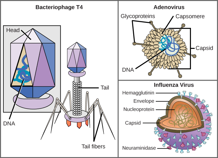
bacteriophage · ·

A virus that infects .

Read explanation · OpenStax ↗
Beta sheet · · · · · ·

A made of aligned segments connected by ; strands may be parallel or .

Read explanation · OpenStax ↗
binary fission

Cell reproduction through , , and division into two daughter cells.

Read explanation · OpenStax ↗
Biomolecule

A made by or involved in living systems, including , , , and .

Read explanation · OpenStax ↗
Blotting ·

Transfer of separated or to a so a specific target can be detected.

Read explanation · Thermo Fisher ↗
Brønsted–Lowry base · · · · · · · · · · ·

In the Brønsted–Lowry model, a substance that can accept a . Basic solutions have lower activity than water.

Read explanation · OpenStax ↗
Buffer ·

A that resists changes when small amounts of or are added, commonly through a weak and its .

Read explanation · OpenStax ↗
Calibration

Establishing the relation between a measured signal and known values or .

Read explanation · Agilent ↗
Carbohydrate

A class of including sugars and their , used for energy storage, structure, and recognition.

Read explanation · OpenStax ↗
Carbonyl group · ·

A carbon double-bonded to oxygen, C=O; present in and many other .

Read explanation · OpenStax ↗
Carboxyl group · ·

An , COOH. In free it is usually to COO⁻ near physiological .

Read explanation · OpenStax ↗
Catalyst · ·

A substance that speeds a by providing a pathway with lower and is regenerated rather than consumed overall.

Read explanation · OpenStax ↗
Cation ·

An with a positive net .

Read explanation · OpenStax ↗
Cellulose

A structural in plant cell walls, with beta-1,4 linkages; also used as a support.

Read explanation · OpenStax ↗
Centrifugation ·

Spinning a sample to accelerate separation of particles according to their behavior.

Read explanation · Beckman Coulter ↗
centriole

A cylindrical structure with nine triplets found in and related basal bodies.

Read explanation · OpenStax ↗
centrosome

A major animal-cell -organizing center, usually containing and surrounding nucleating material.

Read explanation · OpenStax ↗
Chemical compound ·

A substance containing of two or more different chemically combined in a definite composition.

Read explanation · OpenStax ↗
Chemical element

A substance defined by having the same number of , such as carbon or oxygen.

Read explanation · OpenStax ↗
Chemical reaction ·

A process that rearranges and chemical bonds, converting into .

Read explanation · OpenStax ↗
Chloroplast

A photosynthetic in plants and algae with its own and -synthesis machinery.

Read explanation · OpenStax ↗
Chromatin

The complex of and associated , including , in .

Read explanation · OpenStax ↗
Chromatography

Separation of mixture components by their different interactions with a and a moving .

Read explanation · Waters ↗
Chromatography resin ·

The solid or beads in a column, often carrying chemical groups that determine separation behavior.

Read explanation · Cytiva ↗
Chromosome ·

An organized with associated that carries . A replicated chromosome has two .

Read explanation · OpenStax ↗
coccus

A spherical cell.

Read explanation · OpenStax ↗
Codon

A sequence of three that specifies an or a stop signal.

Read explanation · OpenStax ↗
Concentration

The amount of a substance in a specified volume or mass of mixture.

Read explanation · OpenStax ↗
conjugate base

Species formed when an loses one .

Read explanation · OpenStax ↗
conjugation

Overlap of adjacent p orbitals that delocalizes across multiple .

Read explanation · OpenStax ↗
Cooperativity · ·

An interaction in which binding at one site changes binding at other sites. Positive cooperativity can produce a response.

Read explanation · Biology LibreTexts ↗
Copy number ·

The number of copies of a segment in a defined sample or ; it is distinct from or abundance.

Read explanation · NHGRI / NIH ↗
Covalent bond · ·

A bond in which share pairs. and are covalent.

Read explanation · OpenStax ↗
crista

A fold of the inner , increasing its available surface for respiratory machinery.

Read explanation · OpenStax ↗
Cysteine ·

An with a . Two cysteines can form a .

Read explanation · OpenStax ↗
Cytoplasm

Cell contents inside the plasma and outside the , where one is present.

Read explanation · OpenStax ↗
Cytoskeleton

A network of filaments that supports cell shape, organizes components, and enables movement.

Read explanation · OpenStax ↗
Cytosol

The aqueous part of the outside -enclosed .

Read explanation · OpenStax ↗
Density gradient

A fluid whose density varies with position, used to improve separation during .

Read explanation · Beckman Coulter ↗
Deoxyribonucleic acid

The that stores genetic information used to specify and sequences.

Read explanation · OpenStax ↗
Deprotonation

Removal of a from a or group; this changes its by minus one.

Read explanation · OpenStax ↗
Detergent

An substance that can associate with both water and material; detergents can disrupt or interactions.

Read explanation · Thermo Fisher ↗
Diploid

Having two sets; comparisons of must account for and sample composition.

Read explanation · NHGRI / NIH ↗
Disulfide bond · · ·

A covalent sulfur–sulfur bond formed by of two , which can stabilize structure.

Read explanation · NCBI Bookshelf ↗
DNA polymerase ·

An that synthesizes 5′ to 3′ using a template and a with a free .

Read explanation · OpenStax ↗
DNA replication

Copying so genetic information can be passed to daughter cells. Each guides a complementary new strand.

Read explanation · OpenStax ↗
Electric charge · · ·

An electrical property that can be positive or negative. Opposite charges attract and like charges repel; net charge is the sum of all charges on a particle.

Read explanation · OpenStax ↗
Electron

A negatively subatomic particle. Electron sharing and transfer determine much of chemical bonding.

Read explanation · OpenStax ↗
Electronegativity ·

An ’s tendency to attract shared in a chemical bond. Unequal attraction can make a bond .

Read explanation · OpenStax ↗
Endocrine signaling ·

Communication in which enter the circulation and act on distant target cells.

Read explanation · OpenStax ↗
Endomembrane system

The interacting and compartments involved in processes such as modification, transport, and .

Read explanation · OpenStax ↗
Endoplasmic reticulum

An interconnected network continuous with the ; it includes regions involved in processing and synthesis.

Read explanation · OpenStax ↗
Enzyme

A biological that speeds a without being consumed. Most enzymes are .

Read explanation · OpenStax ↗
Enzyme saturation · ·

The high- limit in which sites spend almost all their time processing and the rate approaches .

Read explanation · Biology LibreTexts ↗
Epitope

The particular part of an recognized by an or immune .

Read explanation · OpenStax ↗
Eukaryote ·

An organism whose cells have a -enclosed ; animals, plants, fungi, and protists are eukaryotes.

Read explanation · OpenStax ↗
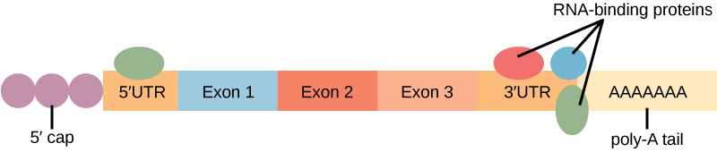
Exon

A segment retained in a mature spliced . In an exon may contain coding sequence, untranslated sequence, or both.

Read explanation · OpenStax ↗
Experimental control ·

A comparison sample or condition used to distinguish the effect being tested from other influences.

Read explanation · Thermo Fisher ↗
FACT complex ·

A -associated complex that helps machinery traverse and supports reorganization.

Read explanation · OpenStax ↗
Fatty acid

A attached to a chain; fatty acids are components of many .

Read explanation · OpenStax ↗
First-order kinetics

A rate dependence proportional to the first power of a . At low , a is approximately first-order in .

Read explanation · Biology LibreTexts ↗
flagellum

A locomotor appendage; forms bend with motors, whereas forms rotate with an -driven motor.

Read explanation · OpenStax ↗
Functional group

A group of whose chemical behavior gives a characteristic and properties.

Read explanation · OpenStax ↗
gamete

A reproductive cell that contributes a during fertilization.

Read explanation · OpenStax ↗
Gene

A sequence that contributes to a functional or , together with the information needed to express it.

Read explanation · OpenStax ↗
Gene expression

Production of a functional from a , including and, for -coding , .

Read explanation · NCBI Bookshelf ↗
Genome

The complete genetic material of an organism or cell.

Read explanation · OpenStax ↗
Glucagon

A pancreatic that helps raise , especially by promoting release from the liver.

Read explanation · OpenStax ↗
Glucose

A six-carbon sugar that is a major cellular energy source and a building block of , , and .

Read explanation · OpenStax ↗
Glutamate · ·

An whose is usually negatively near physiological .

Read explanation · OpenStax ↗
Glycerol

A three-carbon alcohol with three ; used in sample to increase density and in some .

Read explanation · OpenStax ↗
glycogen

A highly branched used for storage in animals.

Read explanation · Biology LibreTexts / Oregon State University ↗
Glycosidic bond

A involving a sugar’s . In a , an N-glycosidic bond links the sugar to the .

Read explanation · Biology LibreTexts / Oregon State University ↗
Golgi apparatus ·

A stack of compartments that modifies and sorts and traveling through the .

Read explanation · OpenStax ↗
Gradient

A change in a quantity, such as , , or density, across a distance or over time.

Read explanation · Thermo Fisher ↗
Gram-negative

Describing with a thin layer and an outer , producing a characteristic Gram-stain response.

Read explanation · OpenStax ↗
Gram-positive

Describing with a thick wall and a characteristic Gram-stain response.

Read explanation · OpenStax ↗
Half-life

The time required for half of a population or amount to remain during decay; a constant half-life is characteristic of decay.

Read explanation · OpenStax ↗
Haploid

Having one set of , as in a human .

Read explanation · OpenStax ↗
hemiacetal

Carbon bonded to both and alkoxy groups, usually formed by alcohol addition to a .

Read explanation · OpenStax ↗
heterotroph

An organism obtaining carbon from organic .

Read explanation · OpenStax ↗
Histone

A -associated rich in . Core histones form the around which nucleosomal wraps.

Read explanation · OpenStax ↗
Hormone

A chemical messenger released by cells that changes the activity of target cells with matching .

Read explanation · OpenStax ↗
Hydrocarbon · ·

A made only of carbon and hydrogen. Hydrocarbon regions are generally .

Read explanation · OpenStax ↗
Hydrogen bond ·

An attraction between a hydrogen bonded to an and another . hydrogen bonds stabilize .

Read explanation · OpenStax ↗
Hydrogen ion · ·

A positively hydrogen species. H⁺ denotes a , which is hydrated in water; its activity determines .

Read explanation · OpenStax ↗
Hydrolysis

Breaking a chemical bond by adding water. hydrolysis separates .

Read explanation · OpenStax ↗
Hydrophilic

Interacting favorably with water, typically because a group is or .

Read explanation · OpenStax ↗
Hydrophobic

Interacting poorly with water. often gather in a ’s interior.

Read explanation · OpenStax ↗
Hydroxide ion ·

The negatively OH⁻. Basic contain a higher hydroxide ion activity than water at the same temperature.

Read explanation · OpenStax ↗
Hydroxyl group ·

An oxygen bonded to hydrogen, –OH. Hydroxyl groups can and often increase .

Read explanation · OpenStax ↗
Hyperbolic curve ·

A curve that approaches an asymptote. The standard rate curve is a rectangular hyperbola approaching .

Read explanation · Biology LibreTexts ↗
Immunoglobulin

An or the -bound form of the same type of -binding .

Read explanation · OpenStax ↗
Initial reaction rate · · · ·

The early rate of formation, measured before substantial depletion or accumulation, usually after the brief binding transient.

Read explanation · Biology LibreTexts ↗
Initiator tRNA ·

The specialized that pairs with a and occupies the during initiation.

Read explanation · OpenStax ↗
Insulin

A from that lowers by promoting uptake and storage and reducing production.

Read explanation · OpenStax ↗
Intron ·

A segment removed from a precursor during . Intronic signals or regulatory sequences can affect .

Read explanation · OpenStax ↗
Ion · ·

An or with a net because its numbers of and do not balance.

Read explanation · OpenStax ↗
Ionizing radiation

Radiation energetic enough to remove from or ; it can damage directly or through reactive chemical species.

Read explanation · Biology LibreTexts / Oregon State University ↗
linkage

A tendency of nearby on the same to be inherited together.

Read explanation · OpenStax ↗
Lipid

A broad class of largely or , including fats, , and steroids.

Read explanation · OpenStax ↗
lipopolysaccharide

A –polysaccharide component of the outer of typical .

Read explanation · OpenStax ↗
locus

The specific position of a or other feature.

Read explanation · OpenStax ↗
Logarithm · · ·

The exponent needed to obtain a number from a chosen fixed number. For decimal logarithms, log₁₀(100) = 2 because 10² = 100; uses the negative decimal logarithm of activity.

Read explanation · OpenStax ↗
Lysosome

An -enclosed compartment that uses hydrolytic to digest and recycle cellular material.

Read explanation · OpenStax ↗
Macromolecule

A large , such as a , , or polysaccharide.

Read explanation · OpenStax ↗
Matrix

A supporting material, such as a gel or the solid support in a column. Its properties influence movement and binding of sample .

Read explanation · Thermo Fisher ↗
Maximum reaction rate · ·

The limiting as becomes saturating. For a fixed under given conditions, Vmax = times .

Read explanation · Biology LibreTexts ↗
Membrane · · · ·

A thin boundary or sheet. Cell membranes are bilayers containing ; membranes are synthetic supports that hold or for detection.

Read explanation · OpenStax ↗
Messenger RNA ·

An that carries coding information used by to synthesize a .

Read explanation · OpenStax ↗
Metabolism

The chemical that build , break them down, and transform energy in a living system.

Read explanation · OpenStax ↗
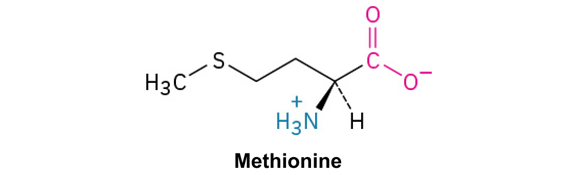
Methionine ·

A with sulfur in a . Unlike , it does not form .

Read explanation · OpenStax ↗
Methyl group ·

A –CH₃ group, consisting of one carbon bonded to three hydrogens.

Read explanation · OpenStax ↗
Michaelis–Menten kinetics · ·

A model with = [S]/(Km + ). Its curve is ; may require a different model.

Read explanation · Biology LibreTexts ↗
Microtubule

A hollow tube built from , supporting cell shape, intracellular transport, and separation.

Read explanation · OpenStax ↗
microvillus

A microscopic cell-surface projection increasing surface area, often for absorption.

Read explanation · OpenStax ↗
Missense mutation ·

A coding-sequence that changes the specified ; its effect depends on the and context.

Read explanation · OpenStax ↗
Mitochondrion ·

A with its own , central to aerobic production and several other processes.

Read explanation · OpenStax ↗
mitosis

division that separates duplicated into daughter , ordinarily preserving .

Read explanation · OpenStax ↗
Mobile phase

The moving fluid that carries a sample through a system.

Read explanation · Waters ↗
Molecular chaperone · ·

A that assists other in folding or prevents , without specifying their sequence.

Read explanation · NCBI Bookshelf ↗
molecular clock

An estimate of evolutionary time based on accumulated sequence changes and an assumed or rate.

Read explanation · OpenStax ↗
Molecule

Two or more joined by . A molecule may be electrically or carry a .

Read explanation · OpenStax ↗
Mutation

A change in a sequence; some mutations alter a ’s sequence.

Read explanation · OpenStax ↗
Neutral

Having no net . For a , neutral instead means equal and activities; its is about 7 at 25 °C.

Read explanation · OpenStax ↗
Nitrogenous base · · ·

A nitrogen-containing ring component of ; its pairing interactions help specify sequences.

Read explanation · OpenStax ↗
Nonpolar ·

Lacking a substantial separation of . Nonpolar groups generally interact poorly with water.

Read explanation · OpenStax ↗
Nonsense mutation ·

A that converts an amino- into a premature , potentially truncating the or destabilizing its .

Read explanation · OpenStax ↗
Nuclear envelope

The double enclosing the and continuous with the .

Read explanation · OpenStax ↗
Nucleic acid

A of , such as or , used in information storage, expression, and other cellular functions.

Read explanation · OpenStax ↗
Nucleoid

The region of a cell containing its , without a surrounding .

Read explanation · OpenStax ↗
Nucleolus

A region where much is and processed and begin assembly.

Read explanation · OpenStax ↗
Nucleosome

The basic unit of : wrapped around a .

Read explanation · OpenStax ↗
Nucleotide

A joined to a sugar and one or more .

Read explanation · OpenStax ↗
Nucleus ·

A -enclosed compartment containing most of a cell’s .

Read explanation · OpenStax ↗
Octamer

A complex made of eight . A core contains eight .

Read explanation · OpenStax ↗
Organelle ·

A specialized structure within a cell that carries out particular functions, such as a or .

Read explanation · OpenStax ↗
Origin of replication · ·

A site where begins; two forks commonly proceed outward from an origin.

Read explanation · OpenStax ↗
osmosis

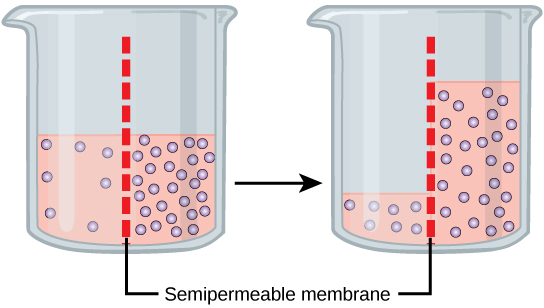
Net water movement across a selectively permeable in response to water chemical-potential differences.

Read explanation · OpenStax ↗
osmotic pressure

The pressure required to prevent net across a under specified conditions.

Read explanation · OpenStax ↗
Oxidation ·

Loss of or an increase in oxidation state. Oxidizing two can form a .

Read explanation · OpenStax ↗
P site ·

The site that holds before and binds the during initiation.

Read explanation · OpenStax ↗
Pancreas

An organ with digestive and functions; its islets contain cells that secrete and .

Read explanation · OpenStax ↗
Pancreatic beta cell ·

An cell in a pancreatic islet that secretes .

Read explanation · OpenStax ↗
Pathogen

An infectious agent capable of causing disease in a host.

Read explanation · OpenStax ↗
Peptide

A chain of joined by ; the term commonly describes chains shorter than a typical .

Read explanation · OpenStax ↗
Peptide bond ·

A covalent between the carboxyl carbon of one and the amino nitrogen of the next.

Read explanation · OpenStax ↗
Peptide hormone

An amino--based chemical messenger released by ; and are examples.

Read explanation · OpenStax ↗
peptidoglycan

A wall of sugar chains cross-linked by short .

Read explanation · OpenStax ↗
Peptidyl transferase · ·

The large-- activity that transfers a growing from to the on .

Read explanation · OpenStax ↗
Peptidyl-tRNA

A carrying the growing through an ester at its .

Read explanation · OpenStax ↗
peroxisome

An supporting oxidative and peroxide .

Read explanation · OpenStax ↗
pH

A measure of , defined as the negative of activity. Lower pH means greater ; pH influences .

Read explanation · OpenStax ↗
Phosphate group ·

A phosphorus-containing group bonded to oxygens; phosphate groups occur in , , and modified .

Read explanation · OpenStax ↗
Phospholipid

An with a -containing region and tails; a major component of cellular .

Read explanation · OpenStax ↗
pilus

A surface appendage used in adhesion or selected transfer processes.

Read explanation · OpenStax ↗
Plasmid

A that replicates separately from a cell’s ; engineered plasmids can carry into experimental cells.

Read explanation · NCBI Bookshelf / Sinauer Associates ↗
ploidy

The number of complete sets in a cell.

Read explanation · OpenStax ↗
Polar

Having an uneven distribution of . Polar groups often interact favorably with water.

Read explanation · OpenStax ↗
Polymer

A large made from many linked molecular building blocks.

Read explanation · OpenStax ↗
Polypeptide

A chain of connected by . A functional can contain one or several such chains.

Read explanation · OpenStax ↗
Porous material · · · ·

A material containing small spaces that can admit of suitable size.

Read explanation · Cytiva ↗
Primer · ·

A short nucleic- strand paired with a template that provides a free for polymerase extension.

Read explanation · OpenStax ↗
Prokaryote ·

An organism whose cells lack a -bound ; and are prokaryotes.

Read explanation · OpenStax ↗
Proline ·

An whose loops back to the nitrogen, restricting rotation and often creating bends.

Read explanation · OpenStax ↗
Protein

One or more folded chains that perform biological roles such as , transport, signaling, or structural support.

Read explanation · OpenStax ↗
Protein aggregation · ·

Association of into larger clusters, often when exposed surfaces of unfolded stick together.

Read explanation · NCBI Bookshelf ↗
Protein backbone ·

The repeating nitrogen–alpha-carbon– chain of a , excluding its variable .

Read explanation · OpenStax ↗
Protein domain · ·

A region of a that forms a recognizable structural or functional unit.

Read explanation · OpenStax ↗
Proton

A positively subatomic particle. In – chemistry, donating a proton means transferring a .

Read explanation · OpenStax ↗
Protonation

Addition of a to an , , or ; this changes its by plus one.

Read explanation · OpenStax ↗
R group · ·

The variable side chain attached to an ’s . It determines identity, , , and chemical behavior.

Read explanation · OpenStax ↗
Reactant

A starting substance transformed during a .

Read explanation · OpenStax ↗
Reaction intermediate · ·

A species formed in one step and consumed in another. It occupies a local energy minimum between .

Read explanation · OpenStax ↗
Reaction product ·

A substance formed by a .

Read explanation · OpenStax ↗
Reaction rate · ·

How quickly are consumed or form, usually expressed as a change per unit time.

Read explanation · OpenStax ↗
Reading frame · ·

The grouping of a sequence into consecutive triplets during . Initiation establishes the frame used by a .

Read explanation · OpenStax ↗
Receptor

A that recognizes a particular signal and helps produce a cellular response.

Read explanation · OpenStax ↗
Release factor ·

A that recognizes a stop signal and promotes of the – to release the .

Read explanation · OpenStax ↗
Ribonucleic acid ·

A with roles in , regulation, and ; carries -coding information.

Read explanation · OpenStax ↗
Ribosomal RNA · ·

An component of that contributes to their structure, decoding, and ; it is not into .

Read explanation · OpenStax ↗
Ribosome ·

A complex of and that translates into a .

Read explanation · OpenStax ↗
RNA polymerase

An that synthesizes 5′ → 3′. Cellular uses a template and can begin without a .

Read explanation · OpenStax ↗
RNA splicing · ·

Removal of and joining of in a precursor .

Read explanation · NCBI Bookshelf / Garland Science ↗
RNA stability

Persistence of an before degradation; greater stability generally means a longer lifetime or under specified conditions.

Read explanation · OpenStax ↗
Salt

An electrically made from and . Dissolved salts can screen or compete with – interactions.

Read explanation · Cytiva ↗
Secondary structure

Local folding, such as and , stabilized mainly by .

Read explanation · OpenStax ↗
Secretory pathway ·

The route through the , , and used to deliver many to the cell surface, outside the cell, or compartments.

Read explanation · OpenStax ↗
Sedimentation ·

Movement of particles through a fluid under gravity or centrifugal .

Read explanation · Beckman Coulter ↗
segregation

Separation of paired into different through segregation.

Read explanation · OpenStax ↗
Sigmoidal curve ·

An S-shaped curve. A sigmoidal rate-versus- curve can indicate rather than simple behavior.

Read explanation · Biology LibreTexts ↗
Sister chromatid

One of the two copies of a produced by . An intact sister chromatid can supply a template for accurate repair.

Read explanation · Biology LibreTexts / Oregon State University ↗
Solubility ·

The amount of a substance that can dissolve in a under specified conditions. Soluble means able to dissolve; insoluble means only very slightly soluble in that .

Read explanation · OpenStax ↗
Solute

A dissolved component of a .

Read explanation · OpenStax ↗
Solution

A homogeneous mixture in which are dissolved in a .

Read explanation · OpenStax ↗
Solvent

The component of a that dissolves other substances, usually the component present in the greatest amount.

Read explanation · OpenStax ↗
Starch

A plant storage made of chains; its two main forms are and amylopectin.

Read explanation · OpenStax ↗
Start codon ·

A recognized during initiation that establishes the ; AUG is the usual example.

Read explanation · OpenStax ↗
Stationary phase

The material that remains fixed in a system.

Read explanation · Waters ↗
Stop codon · · ·

An that recruits a to end : UAA, UAG, or UGA in the standard code.

Read explanation · OpenStax ↗
Substitution · ·

Replacement of one by another. In a coding region it can be , , or .

Read explanation · OpenStax ↗
Substrate

A that an binds and chemically transforms.

Read explanation · OpenStax ↗
Substrate concentration

The amount of per volume. Square brackets denote ; [S] is varied to measure an curve.

Read explanation · Biology LibreTexts ↗
Subunit ·

An individual chain within a containing multiple chains.

Read explanation · OpenStax ↗
Synonymous mutation · · · · ·

A coding-sequence that leaves the specified unchanged. It can still affect , , or .

Read explanation · OpenStax ↗
Template strand · · ·

A preexisting strand whose sequence guides complementary nucleic- synthesis.

Read explanation · OpenStax ↗
Thioether

A sulfur bonded to two carbon groups, as in ’s . It has no hydrogen.

Read explanation · OpenStax ↗
Thiol group · · · ·

A sulfur bonded to hydrogen, –SH. thiols can form through .

Read explanation · OpenStax ↗
Transcription

Synthesis of using a template.

Read explanation · OpenStax ↗
transduction

Transfer of mediated by a .

Read explanation · OpenStax ↗
Transfer RNA ·

An adaptor that carries an and pairs its with a on .

Read explanation · OpenStax ↗
Transition state

A fleeting, high-energy arrangement at a barrier. It is an energy maximum along the path, rather than a stable .

Read explanation · OpenStax ↗
Translation ·

Synthesis of a on a using the sequence of in .

Read explanation · OpenStax ↗
Tubulin

A that forms alpha–beta dimers; these dimers assemble into .

Read explanation · OpenStax ↗
Turnover number · · ·

The maximum number of cycles per per unit time, calculated as /. Units are inverse time, commonly s⁻¹.

Read explanation · IUBMB ↗
velocity

Rate of change of position, including direction.

Read explanation · OpenStax ↗
Vesicle · · ·

A small -enclosed compartment that can transport material between cellular locations.

Read explanation · OpenStax ↗

22.2 Structure of Prokaryotes: Bacteria and Archaea

OpenStax · 22.2 Structure of Prokaryotes: Bacteria and Archaea ↗
Instructional page including learning objectives, examples, figures, source tables and questions. Site navigation and embedded media players are omitted; source media links remain available.

Learning Objectives

By the end of this section, you will be able to do the following:

  • Describe the basic structure of a typical
  • Describe important differences in structure between and

There are many differences between and cells. The name "" suggests that are defined by exclusion—they are not , or organisms whose cells contain a and other internal -bound . However, all cells have four common structures: the plasma , which functions as a barrier for the cell and separates the cell from its environment; the , a complex of organic and inside the cell; a double-stranded , the informational archive of the cell; and , where takes place. come in various shapes, but many fall into three categories: (spherical), (rod-shaped), and spirilli (spiral-shaped) (Figure 22.9).

Part a: The micrograph shows ball-shaped cocci about 800 nanometers across. Part b: The micrograph shows elongated, oval-shaped bacilli about 2000 nanometers long. Part c: The micrograph shows corkscrew-shaped spirilli that are quite long and 500 nanometers in diameter.
Figure 22.9 — 22.2 Structure of : and

Figure 22.9 Common prokaryotic cell types. Prokaryotes fall into three basic categories based on their shape, visualized here using scanning electron microscopy: (a) cocci, or spherical (a pair is shown); (b) bacilli, or rod-shaped; and (c) spirilli, or spiral-shaped. (credit a: modification of work by Janice Haney Carr, Dr. Richard Facklam, CDC; credit c: modification of work by Dr. David Cox; scale-bar data from Matt Russell)

Mary Ann Clark, Matthew Douglas, and Jung Choi, Biology 2E, OpenStax. (credit a: modification of work by Janice Haney Carr, Dr. Richard Facklam, CDC; credit c: modification of work by Dr. David Cox; scale-bar data from Matt Russell) · CC BY-NC-SA 4.0 · Original image ↗ · Access for free ↗

The Cell

Recall that are unicellular organisms that lack -bound or other internal -bound structures (Figure 22.10). Their —usually single—consists of a piece of circular, double-stranded located in an area of the cell called the . Most have a cell wall outside the plasma . The cell wall functions as a protective layer, and it is responsible for the organism’s shape. Some species have a capsule outside the cell wall. The capsule enables the organism to attach to surfaces, protects it from dehydration and attack by phagocytic cells, and makes more resistant to our immune responses. Some species also have (singular, ) used for locomotion, and (singular, ) used for to surfaces including the surfaces of other cells. , which consist of extra- , are also present in many species of and .

In this illustration, the prokaryotic cell is rod shaped. The circular chromosome is concentrated in a region called the nucleoid. The fluid inside the cell is called the cytoplasm. Ribosomes, depicted as small circles, float in the cytoplasm. The cytoplasm is encased by a plasma membrane, which in turn is encased by a cell wall. A capsule surrounds the cell wall. The bacterium depicted has a flagellum protruding from one narrow end. Pili are small protrusions that project from the capsule all over the bacterium, like hair.
Figure 22.10 — 22.2 Structure of : and

Figure 22.10 The features of a typical prokaryotic cell. Flagella, capsules, and pili are not found in all prokaryotes.

Mary Ann Clark, Matthew Douglas, and Jung Choi, Biology 2E, OpenStax. · CC BY-NC-SA 4.0 · Original image ↗ · Access for free ↗

Recall that are divided into two different , and , which together with Eukarya, comprise the three of life (Figure 22.11).

The trunk of the phylogenetic tree is a universal ancestor. The tree forms two branches. One branch leads to the domain bacteria, which includes the phyla proteobacteria, chlamydias, spirochetes, cyanobacteria, and Gram-positive bacteria. The other branch branches again, into the eukarya and archaea domains. Domain archaea includes the phyla euryarchaeotes, crenarchaeotes, nanoarchaeotes, and korarchaeotes.
Figure 22.11 — 22.2 Structure of : and

Figure 22.11 The three domains of living organisms. Bacteria and Archaea are both prokaryotes but differ enough to be placed in separate domains. An ancestor of modern Archaea is believed to have given rise to Eukarya, the third domain of life. Major groups of Archaea and Bacteria are shown.

Mary Ann Clark, Matthew Douglas, and Jung Choi, Biology 2E, OpenStax. · CC BY-NC-SA 4.0 · Original image ↗ · Access for free ↗

Characteristics of phyla are described in Figure 22.12 and Figure 22.13. Major phyla include the Proteobacteria, the Chlamydias, the Spirochaetes, the photosynthetic Cyanobacteria, and the . The Proteobacteria are in turn subdivided into several classes, from the Alpha- to the Epsilon proteobacteria. are thought to be the descendants of alphaproteobacteria, while are derived from cyanobacteria. phyla are described in Figure 22.14.

Characteristics of the five classes of bacterial phylum proteobacteria are described. The classes are alpha, beta, gamma, delta and epsilon. Most species of Alpha Proteobacteria are photoautotrophic but some are symbionts of plants and animals, and others are pathogens. Eukaryotic mitochondria are thought to be derived from bacteria in this group. Representative organisms include Rhizobium, a nitrogen-fixing endosymbiont associated with the roots of legumes, and Rickettsia, an obligate intracellular parasite that causes typhus and Rocky Mountain Spotted Fever (but not rickets, which is caused by Vitamin D deficiency). A micrograph shows rod-shaped Rickettsia rickettsii inside a much larger eukaryotic cell. Beta Proteobacteria is a diverse group of bacteria. Some species play an important role in the nitrogen cycle. Representative organisms include Nitrosomas, which oxidize ammonia into nitrite, and Spirillum minus, which causes rat bite fever. A micrograph of spiral-shaped Spirillum minus is shown. Gamma Proteobacteria include many beneficial symbionts that populate the human gut, as well as familiar human pathogens. Some species from this subgroup oxidize sulfur compounds. Representative organisms include Escherichia coli, normally a beneficial microbe of the human gut, but some strains cause disease; Salmonella, certain strains of which cause food poisoning or typhoid fever; Yersinia pestis, the causative agent of Bubonic plague; Psuedomonas aeruganosa, which causes lung infections; Vibrio cholera, the causative agent of cholera; and Chromatium, sulfur-producing bacteria that oxidize sulfur, producing H2S. Micrograph shows rod-shaped Vibrio cholera, which are about 1 micron long. Some species of Delta Proteobacteria generate a spore-forming fruiting body in adverse conditions. Others reduce sulfate and sulfur. Representative organisms include Myxobacteria, which generate spore-forming fruiting bodies in adverse conditions, and Desulfovibrio vulgaris, an aneorobic, sulfate-reducing bacterium. Micrograph shows a bent rod-shaped Desulfovibrio vulgaris bacterium with a long flagellum. Epsilon Proteobacteria includes many species that inhabit the digestive tract of animals as symbionts or pathogens. Bacteria from this group have been found in deep-sea hydrothermal vents and cold seep habitats. Representative organisms include Campylobacter, which causes blood poisoning and intestinal inflammation, and Heliobacter pylori, which causes stomach ulcers. Micrograph shows short spiral, or s shaped, Campylobacter bacteria.
Figure 22.12 — 22.2 Structure of : and

Figure 22.12 The Proteobacteria. Phylum Proteobacteria is one of up to 52 bacteria phyla. Proteobacteria is further subdivided into five classes, Alpha through Epsilon. (credit “Rickettsia rickettsii”: modification of work by CDC; credit “Spirillum minus”: modification of work by Wolframm Adlassnig; credit “Vibrio cholera”: modification of work by Janice Haney Carr, CDC; credit “Desulfovibrio vulgaris”: modification of work by Graham Bradley; credit “Campylobacter”: modification of work by De Wood, Pooley, USDA, ARS, EMU; scale-bar data from Matt Russell)

Mary Ann Clark, Matthew Douglas, and Jung Choi, Biology 2E, OpenStax. (credit “Rickettsia rickettsii”: modification of work by CDC; credit “Spirillum minus”: modification of work by Wolframm Adlassnig; credit “Vibrio cholera”: modification of work by Janice Haney Carr, CDC; credit “Desulfovibrio vulgaris”: modification of work by Graham Bradley; credit “Campylobacter”: modification of work by De Wood, Pooley, USDA, ARS, EMU; scale-bar data from Matt Russell) · CC BY-NC-SA 4.0 · Original image ↗ · Access for free ↗
This table describes the other four bacterial phyla classes. The first phylum described is chlamydias. All members of this group are obligate intracellular parasites of animal cells. Cells walls lack peptidoglycan. Micrograph shows a pap smear of cells infected with Chlamydia trachomatis. Chlamydia infection is the most common sexually transmitted disease and can lead to blindness. All members of the phylum Spirochetes have spiral-shaped cells. Most are free-living anaerobes, but some are pathogenic. Flagella run lengthwise in the periplasmic space between the inner and outer membrane. Representative species include Treponema pallidum, the causative agent of syphilis and Borrelia burgdorferi, the causative agent of Lyme disease Micrograph shows corkscrew-shaped Trepanema pallidum, about 1 micron across. Bacteria in the phylum Cyanobacteria, also known as blue-green algae, obtain their energy through photosynthesis. They are ubiquitous, found in terrestrial, marine, and freshwater environments. Eukaryotic chloroplasts are thought to be derived from bacteria in this group. The cyanobacterium Prochlorococcus is believed to be the most abundant photosynthetic organism on earth, responsible for generating half the world’s oxygen. Micrograph shows a long, thin rod-shaped species called Phormidium. Gram-positive Bacteria have a thick cell wall and lack an outer membrane. Soil-dwelling members of this subgroup decompose organic matter. Some species cause disease. Representative species include Bacillus anthracis, which causes anthrax; Clostridium botulinum, which causes botulism; Clostridium difficile, which causes diarrhea during antibiotic therapy; Streptomyces, from which many antibiotics, including streptomyocin, are derived; and Mycoplasmas, the smallest known bacteria, which lack a cell wall. Some are free-living, and some are pathogenic. Micrograph shows Clostridium difficile, which are rod-shaped and about 3 microns long.
Figure 22.13 — 22.2 Structure of : and

Figure 22.13 Other bacterial phyla. Chlamydia, Spirochetes, Cyanobacteria, and Gram-positive bacteria are described in this table. Note that bacterial shape is not phylum-dependent; bacteria within a phylum may be cocci, rod-shaped, or spiral. (credit “Chlamydia trachomatis” : modification of work by Dr. Lance Liotta Laboratory, NCI; credit “Treponema pallidum” : modification of work by Dr. David Cox, CDC; credit “Phormidium” : modification of work by USGS; credit “Clostridium difficile” : modification of work by Lois S. Wiggs, CDC; scale-bar data from Matt Russell)

Mary Ann Clark, Matthew Douglas, and Jung Choi, Biology 2E, OpenStax. (credit “Chlamydia trachomatis” : modification of work by Dr. Lance Liotta Laboratory, NCI; credit “Treponema pallidum” : modification of work by Dr. David Cox, CDC; credit “Phormidium” : modification of work by USGS; credit “Clostridium difficile” : modification of work by Lois S. Wiggs, CDC; scale-bar data from Matt Russell) · CC BY-NC-SA 4.0 · Original image ↗ · Access for free ↗
Characteristics of the four phyla of archaea are described. Euryarchaeotes includes methanogens, which produce methane as a metabolic waste product, and halobacteria, which live in an extreme saline environment. Methanogens cause flatulence in humans and other animals. Halobacteria can grow in large blooms that appear reddish, due to the presence of bacteriorhodopsin in the membrane. Bacteriorhodopsin is related to the retinal pigment rhodopsin. Micrograph shows rod shaped Halobacterium. Members of the ubiquitous Crenarchaeotes phylum play an important role in the fixation of carbon. Many members of this group are sulfur dependent extremophiles. Some are thermophilic or hyperthermophilic. Micrograph shows cocci shaped Sulfolobus, a genus which grows in volcanic springs at temperatures between 75 degrees and 80 degrees Celsius and at a lower case p upper case H between 2 and 3. The phylum Nanoarchaeotes currently contains only one species, Nanoarchaeum equitans, which has been isolated from the bottom of the Atlantic Ocean, and from the a hydrothermal vent at Yellowstone National Park. It is an obligate symbiont with Ignicoccus, another species of archaebacteria. Micrograph shows two small, round N. equitans cells attached to a larger Ignicoccus cell. Korarchaeotes are considered to be one of the most primitive forms of life and so far have only been found in the Obsidian Pool, a hot spring at Yellowstone National Park. Micrograph shows a variety of specimens from this group which vary in shape.
Figure 22.14 — 22.2 Structure of : and

Figure 22.14 Archaeal phyla. Archaea are separated into four phyla: the Korarchaeota, Euryarchaeota, Crenarchaeota, and Nanoarchaeota. (credit “Halobacterium” : modification of work by NASA; credit “Nanoarchaeotum equitans” : modification of work by Karl O. Stetter; credit “Korarchaeota”: modification of work by Office of Science of the U.S. Dept. of Energy; scale-bar data from Matt Russell)

Mary Ann Clark, Matthew Douglas, and Jung Choi, Biology 2E, OpenStax. (credit “Halobacterium” : modification of work by NASA; credit “Nanoarchaeotum equitans” : modification of work by Karl O. Stetter; credit “Korarchaeota”: modification of work by Office of Science of the U.S. Dept. of Energy; scale-bar data from Matt Russell) · CC BY-NC-SA 4.0 · Original image ↗ · Access for free ↗

The Plasma of

The plasma is a thin bilayer (6 to 8 nanometers) that completely surrounds the cell and separates the inside from the outside. Its selectively permeable nature keeps , , and other within the cell and prevents them from diffusing into the extracellular environment, while other may move through the . Recall that the general structure of a is a bilayer composed of two layers of . In , isoprene (phytanyl) chains linked to replace the linked to in . Some are monolayers instead of bilayers (Figure 22.15).

This illustration compares phospholipids from Bacteria and Eukarya to those from Archaea. In Bacteria and Eukarya, fatty acids are attached to glycerol by an ester linkage, while in Archaea, isoprene chains are linked to glycerol by an ether linkage. In the ester linkage, the first carbon in the fatty acid chain has an oxygen double-bonded to it, whereas in the ether linkage, it does not. In Archaea, the isoprene chains have methyl groups branching off from them, whereas such branches are absent in Bacteria and Eukarya. Both types of phospholipids result in similar lipid bilayers.
Figure 22.15 — 22.2 Structure of : and

Figure 22.15 Bacterial and archaeal phospholipids. Archaeal phospholipids differ from those found in Bacteria and Eukarya in two ways. First, they have branched phytanyl sidechains instead of linear ones. Second, an ether bond instead of an ester bond connects the lipid to the glycerol.

Mary Ann Clark, Matthew Douglas, and Jung Choi, Biology 2E, OpenStax. · CC BY-NC-SA 4.0 · Original image ↗ · Access for free ↗

The Cell Wall of

The of cells has a high of dissolved . Therefore, the within the cell is relatively high. The cell wall is a protective layer that surrounds some cells and gives them shape and rigidity. It is located outside the and prevents osmotic lysis (bursting due to increasing volume). The chemical composition of the cell wall varies between and , and also varies between species.

cell walls contain , composed of polysaccharide chains that are cross-linked by unusual containing both L- and D- including D- and D-. ( normally have only L-; as a consequence, many of our antibiotics work by mimicking D- and therefore have specific effects on cell-wall development.) There are more than 100 different forms of . S-layer (surface layer) are also present on the outside of cell walls of both and .

are divided into two major groups: Gram positive and Gram negative, based on their to Gram staining. Note that all belong to two phyla (Firmicutes and Actinobacteria); in the other phyla (Proteobacteria, Chlamydias, Spirochetes, Cyanobacteria, and others) are . The Gram staining method is named after its inventor, Danish scientist Hans Christian Gram (1853–1938). The different responses to the staining procedure are ultimately due to cell wall structure. organisms typically lack the outer found in organisms (Figure 22.16). Up to 90 percent of the cell-wall in is composed of , and most of the rest is composed of substances called teichoic . Teichoic may be covalently linked to in the plasma to form lipoteichoic . Lipoteichoic anchor the cell wall to the . have a relatively thin cell wall composed of a few layers of (only 10 percent of the total cell wall), surrounded by an outer envelope containing lipopolysaccharides () and lipoproteins. This outer envelope is sometimes referred to as a second bilayer. The chemistry of this outer envelope is very different, however, from that of the typical bilayer that forms plasma .

Visual Connection

The left illustration shows the cell wall of Gram-positive bacteria. The cell wall is a thick layer of peptidoglycan that exists outside the plasma membrane. A long, thin molecule called lipoteichoic acid anchors the cell wall to the cell membrane. The right illustration shows Gram-negative bacteria. In Gram-negative bacteria, a thin peptidoglycan cell wall is sandwiched between an outer and an inner plasma membrane. The space between the two membranes is called the periplasmic space. Lipoproteins anchor the cell wall to the outer membrane. Lipopolysaccharides protrude from the outer membrane. Porins are proteins in the outer membrane that allow entry of substances.
Figure 22.16 — 22.2 Structure of : and

Figure 22.16 Cell walls in Gram-positive and Gram-negative bacteria. Bacteria are divided into two major groups: Gram positive and Gram negative. Both groups have a cell wall composed of peptidoglycan: in Gram-positive bacteria, the wall is thick, whereas in Gram-negative bacteria, the wall is thin. In Gram-negative bacteria, the cell wall is surrounded by an outer membrane that contains lipopolysaccharides and lipoproteins. Porins are proteins in this cell membrane that allow substances to pass through the outer membrane of Gram-negative bacteria. In Gram-positive bacteria, lipoteichoic acid anchors the cell wall to the cell membrane. (credit: modification of work by "Franciscosp2"/Wikimedia Commons)

Mary Ann Clark, Matthew Douglas, and Jung Choi, Biology 2E, OpenStax. (credit: modification of work by "Franciscosp2"/Wikimedia Commons) · CC BY-NC-SA 4.0 · Original image ↗ · Access for free ↗

Which of the following statements is true?

  1. have a single cell wall anchored to the by lipoteichoic .
  2. Porins allow entry of substances into both and .
  3. The cell wall of is thick, and the cell wall of is thin.
  4. have a cell wall made of , whereas have a cell wall made of lipoteichoic .

Archaean cell walls do not have . There are four different types of archaean cell walls. One type is composed of pseudopeptidoglycan, which is similar to in morphology but contains different sugars in the polysaccharide chain. The other three types of cell walls are composed of polysaccharides, glycoproteins, or pure . Other differences between and are seen in Table 22.2. Note that features related to , and in are similar to those seen in .

Differences and Similarities between and
Structural Characteristic
Cell type
Cell morphology Variable Variable
Cell wall Contains Does not contain
type bilayer bilayer or monolayer
Plasma - ester Phytanyl- ethers
Typically circular Typically circular
Single Multiple
Single Multiple
Formyl-
Streptomycin inhibition Sensitive Resistant
Calvin cycle Yes No
Table 22.2

Reproduction

Reproduction in is asexual and usually takes place by . (Recall that the of a is a single, circular .) do not undergo ; instead, the is replicated and the two resulting copies separate from one another, due to the growth of the cell. The , now enlarged, is pinched inward at its equator and the two resulting cells, which are clones, separate. does not provide an opportunity for genetic recombination or genetic diversity, but can share by three other mechanisms.

In transformation, the takes in shed by other into its environment. If a nonpathogenic takes up for a toxin from a and incorporates the new into its own , it too may become pathogenic. In , , the viruses that infect , may move short pieces of from one to another. results in a recombinant organism. also have viruses that may translocate genetic material from one individual to another. In , is transferred from one to another by means of a , which brings the organisms into contact with one another, and provides a channel for transfer of . The transferred can be in the form of a or as a composite , containing both and . These three processes of exchange are shown in Figure 22.17.

Reproduction can be very rapid: a few minutes for some species. This short generation time coupled with mechanisms of genetic recombination and high rates of result in the rapid evolution of , allowing them to respond to environmental changes (such as the introduction of an antibiotic) very quickly.

Illustration A shows transformation, where a small, circular piece of DNA is absorbed by a prokaryotic cell. The DNA then gets incorporated in the genome. Illustration B shows transduction, where a bacteriophage injects DNA into a prokaryotic cell. The DNA then gets incorporated in the genome. Illustration C shows conjugation, where two bacteria are connected by a pilus. A small loop of DNA is transferred from one cell to the other via the pilus.
Figure 22.17 — 22.2 Structure of : and

Figure 22.17 Gene transfer mechanisms in prokaryotes. There are three mechanisms by which prokaryotes can exchange DNA. In (a) transformation, the cell takes up prokaryotic DNA directly from the environment. The DNA may remain separate as plasmid DNA or be incorporated into the host genome. In (b) transduction, a bacteriophage injects DNA into the cell that contains a small fragment of DNA from a different prokaryote. In (c) conjugation, DNA is transferred from one cell to another via a mating bridge, or pilus, that connects the two cells after the sex pilus draws the two bacteria close enough to form the bridge.

Mary Ann Clark, Matthew Douglas, and Jung Choi, Biology 2E, OpenStax. · CC BY-NC-SA 4.0 · Original image ↗ · Access for free ↗

Evolution Connection

The Evolution of

How do scientists answer questions about the evolution of ? Unlike with animals, artifacts in the fossil record of offer very little information. Fossils of ancient look like tiny bubbles in rock. Some scientists turn to genetics and to the principle of the , which holds that the more recently two species have diverged, the more similar their (and thus ) will be. Conversely, species that diverged long ago will have more that are dissimilar.

Scientists at the NASA Astrobiology Institute and at the European Molecular Biology Laboratory collaborated to analyze the molecular evolution of 32 specific common to 72 species of .2 The model they derived from their data indicates that three important groups of —Actinobacteria, Deinococcus, and Cyanobacteria (collectively called Terrabacteria by the authors)—were the first to colonize land. Actinobacteria are a group of very common that produce branched structures like fungal mycelia, and include species important in decomposition of organic wastes. You will recall that Deinococcus is a genus of that is highly resistant to . It has a thick layer in addition to a second external , so it has features of both and .

Cyanobacteria are photosynthesizers, and were probably responsible for the production of oxygen on the ancient earth. The timelines of divergence suggest that (members of the ) diverged from common ancestral species between 2.5 and 3.2 billion years ago, whereas the diverged earlier: between 3.1 and 4.1 billion years ago. Eukarya later diverged from the archaean line. The work further suggests that stromatolites that formed prior to the advent of cyanobacteria (about 2.6 billion years ago) photosynthesized in an anoxic environment and that because of the modifications of the Terrabacteria for land (resistance to drying and the possession of that protect the organism from excess light), photosynthesis using oxygen may be closely linked to to survive on land.

4.3 Eukaryotic Cells

OpenStax · 4.3 Eukaryotic Cells ↗

Instructional page including learning objectives, examples, figures, source tables and questions. Site navigation and embedded media players are omitted; source media links remain available.

Learning Objectives

By the end of this section, you will be able to do the following:

  • Describe the structure of cells
  • Compare animal cells with plant cells
  • State the role of the plasma
  • Summarize the functions of the major cell

Have you ever heard the phrase “form follows function?” It’s a philosophy that many industries follow. In architecture, this means that buildings should be constructed to support the activities that will be carried out inside them. For example, a skyscraper should include several elevator banks. A hospital should have its emergency room easily accessible.

Our natural world also utilizes the principle of form following function, especially in cell biology, and this will become clear as we explore cells (Figure 4.8). Unlike cells, cells have: 1) a -bound ; 2) numerous -bound such as the , , , , and others; and 3) several, rod-shaped . Because a surrounds cell’s , it has a “true .” The word “” means “little organ,” and, as we already mentioned, have specialized cellular functions, just as your body's organs have specialized functions.

At this point, it should be clear to you that cells have a more complex structure than cells. allow different functions to be compartmentalized in different areas of the cell. Before turning to , let’s first examine two important components of the cell: the plasma and the .

Visual Connection

Part a: This illustration shows a typical eukaryotic animal cell, which is egg shaped. The fluid inside the cell is called the cytoplasm, and the cell is surrounded by a cell membrane. The nucleus takes up about one-half the width of the cell. Inside the nucleus is the chromatin, which is composed of DNA and associated proteins. A region of the chromatin is condensed into the nucleolus, a structure where ribosomes are synthesized. The nucleus is encased in a nuclear envelope, which is perforated by protein-lined pores that allow entry of material into the nucleus. The nucleus is surrounded by the rough and smooth endoplasmic reticulum, or ER. The smooth ER is the site of lipid synthesis. The rough ER has embedded ribosomes that give it a bumpy appearance. It synthesizes membrane and secretory proteins. In addition to the ER, many other organelles float inside the cytoplasm. These include the Golgi apparatus, which modifies proteins and lipids synthesized in the ER. The Golgi apparatus is made of layers of flat membranes. Mitochondria, which produce food for the cell, have an outer membrane and a highly folded inner membrane. Other, smaller organelles include peroxisomes that house oxidative metabolism, lysosomes that digest food, and vacuoles. Ribosomes, responsible for protein synthesis, also float freely in the cytoplasm and are depicted as small dots. The last cellular component shown is the cytoskeleton, which has four different types of components: microfilaments, intermediate filaments, microtubules, and centrosomes. Microfilaments are fibrous proteins that line the cell membrane and make up the cellular cortex. Intermediate filaments are fibrous proteins that hold organelles in place. Microtubules form the mitotic spindle and maintain cell shape. Centrosomes are made of two tubular structures at right angles to one another. They form the microtubule-organizing center.
Figure 4.8 — 4.3 Cells

Figure 4.8 These figures show the major organelles and other cell components of (a) a typical animal cell and (b) a typical eukaryotic plant cell. The plant cell has a cell wall, chloroplasts, plastids, and a central vacuole—structures not in animal cells. Most cells do not have lysosomes or centrosomes.

Mary Ann Clark, Matthew Douglas, and Jung Choi, Biology 2E, OpenStax. · CC BY-NC-SA 4.0 · Original image ↗ · Access for free ↗
Part b: This illustration depicts a typical eukaryotic plant cell. The nucleus of a plant cell contains chromatin and a nucleolus, the same as an animal cell. Other structures that the plant cell has in common with the animal cell include rough and smooth endoplasmic reticulum, the Golgi apparatus, mitochondria, peroxisomes, and ribosomes. The fluid inside the plant cell is called the cytoplasm, just as it is in an animal cell. The plant cell has three of the four cytoskeletal components found in animal cells: microtubules, intermediate filaments, and microfilaments. Plant cells do not have centrosomes. Plant cells have four structures not found in animals cells: chloroplasts, plastids, a central vacuole, and a cell wall. Chloroplasts are responsible for photosynthesis; they have an outer membrane, an inner membrane, and stack of membranes inside the inner membrane. The central vacuole is a very large, fluid-filled structure that maintains pressure against the cell wall. Plastids store pigments. The cell wall is outside the cell membrane.
Figure 4.8 — 4.3 Cells

Figure 4.8 These figures show the major organelles and other cell components of (a) a typical animal cell and (b) a typical eukaryotic plant cell. The plant cell has a cell wall, chloroplasts, plastids, and a central vacuole—structures not in animal cells. Most cells do not have lysosomes or centrosomes.
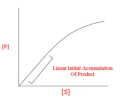
Mary Ann Clark, Matthew Douglas, and Jung Choi, Biology 2E, OpenStax. · CC BY-NC-SA 4.0 · Original image ↗ · Access for free ↗

If the were not able to carry out its function, what other cellular would be affected?

The Plasma

Like , cells have a plasma (Figure 4.9), a bilayer with embedded that separates the internal contents of the cell from its surrounding environment. A is a with two chains and a -containing group. The plasma the passage of organic , , water, and oxygen into and out of the cell. Wastes (such as carbon dioxide and ammonia) also leave the cell by passing through the plasma .
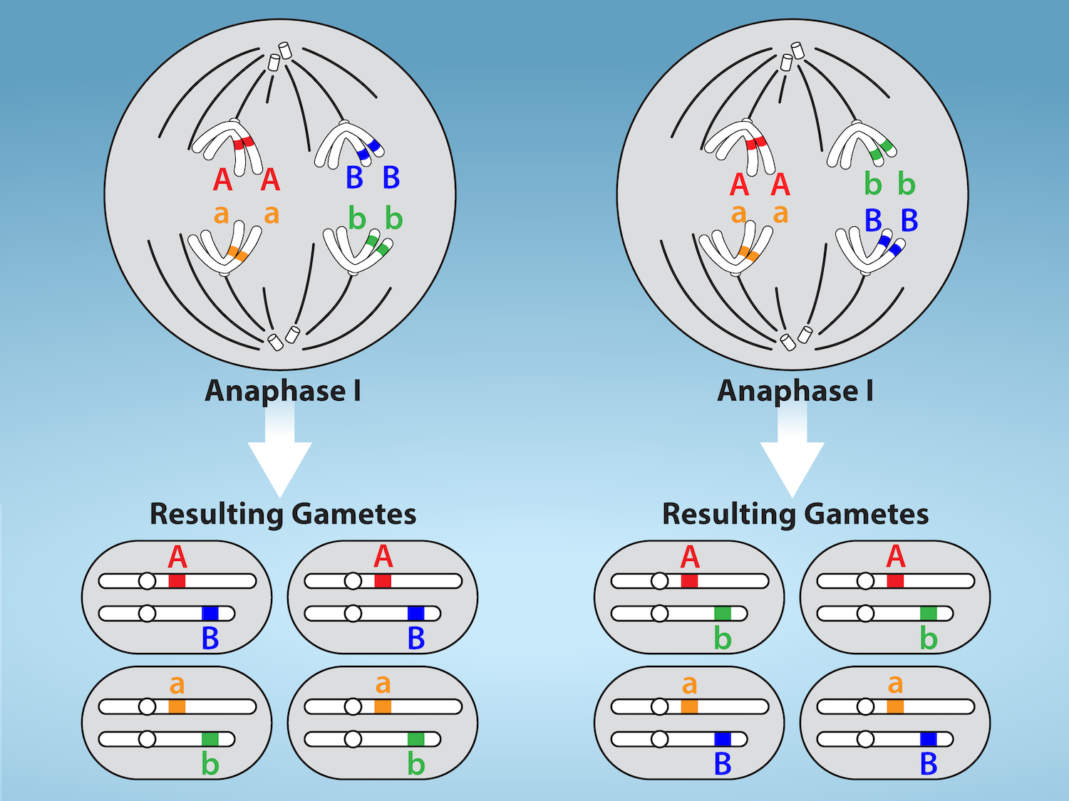
The plasma membrane is composed of a phospholipid bilayer. In the bilayer, the two long hydrophobic tails of phospholipids face toward the center, and the hydrophilic head group faces the exterior. Integral membrane proteins and protein channels span the entire bilayer. Protein channels have a pore in the middle. Peripheral membrane proteins sit on the surface of the phospholipids, and are associated with the phospholipid head groups. On the exterior side of the membrane, carbohydrates are attached to certain proteins and lipids. Filaments of the cytoskeleton line the interior of the membrane.
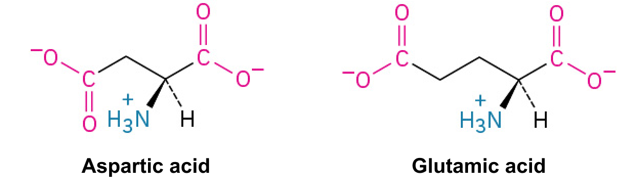

Figure 4.9 — 4.3 Cells

Figure 4.9 The eukaryotic plasma membrane is a phospholipid bilayer with proteins and cholesterol embedded in it.

Mary Ann Clark, Matthew Douglas, and Jung Choi, Biology 2E, OpenStax. · CC BY-NC-SA 4.0 · Original image ↗ · Access for free ↗

The plasma of cells that specialize in absorption fold into fingerlike projections that we call (singular = ); (Figure 4.10). Such cells typically line the small intestine, the organ that absorbs nutrients from digested food. This is an excellent example of form following function. People with celiac disease have an immune response to gluten, which is a in wheat, barley, and rye. The immune response damages , and thus, afflicted individuals cannot absorb nutrients. This leads to malnutrition, cramping, and diarrhea. Patients suffering from celiac disease must follow a gluten-free diet.

The left part of this figure is a transmission electron micrograph of microvilli, which appear as long, slender stalks extending from the plasma membrane. The right side illustrates cells containing microvilli. The microvilli cover the surface of the cell facing the interior of the small intestine.
Figure 4.10 — 4.3 Cells

Figure 4.10 Microvilli, as they appear on cells lining the small intestine, increase the surface area available for absorption. These microvilli are only on the area of the plasma membrane that faces the cavity from which substances will be absorbed. (credit "micrograph": modification of work by Louisa Howard)

Mary Ann Clark, Matthew Douglas, and Jung Choi, Biology 2E, OpenStax. (credit "micrograph": modification of work by Louisa Howard) · CC BY-NC-SA 4.0 · Original image ↗ · Access for free ↗

The

The is the cell's entire region between the plasma and the (a structure we will discuss shortly). It is comprised of suspended in the gel-like , the , and various chemicals (Figure 4.8). Even though the consists of 70 to 80 percent water, it has a semi-solid consistency, which comes from the within it. However, are not the only organic in the . and other simple sugars, polysaccharides, , , , and derivatives of are also there. of sodium, potassium, calcium, and many other also dissolve in the . Many , including , take place in the .

The

Typically, the is the most prominent in a cell (Figure 4.8). The (plural = ) houses the cell’s and directs the synthesis of and . Let’s look at it in more detail (Figure 4.11).

The two-dimensional image depicts the nucleus of a cell as a circular object with two membranes; several gaps appear in the circle, representing nuclear pores. Surrounding the nucleus are membranous sacks representing the endoplasmic reticulum. Inside the nucleus is another circle, approximately ten percent of the total size of the nucleus, representing the nucleolus.
Figure 4.11 — 4.3 Cells

Figure 4.11 The nucleus stores chromatin (DNA plus proteins) in a gel-like substance called the nucleoplasm. The nucleolus is a condensed chromatin region where ribosome synthesis occurs. We call the nucleus' boundary the nuclear envelope. It consists of two phospholipid bilayers: an outer and an inner membrane. The nuclear membrane is continuous with the endoplasmic reticulum. Nuclear pores allow substances to enter and exit the nucleus.

Mary Ann Clark, Matthew Douglas, and Jung Choi, Biology 2E, OpenStax. · CC BY-NC-SA 4.0 · Original image ↗ · Access for free ↗

The

The is a double- structure that constitutes the ' outermost portion (Figure 4.11). Both the 's inner and outer are bilayers.

The is punctuated with that control the passage of , , and between the nucleoplasm and . The nucleoplasm is the semi-solid fluid inside the , where we find the and the .

and

To understand , it is helpful to first explore , structures within the that are made up of , the hereditary material. You may remember that in , is organized into a single circular . In , are linear structures. Every species has a specific number of in the of each cell. For example, in humans, the number is 46, while in fruit flies, it is eight. are only visible and distinguishable from one another when the cell is getting ready to divide. When the cell is in the growth and maintenance phases of its life cycle, attach to , and they resemble an unwound, jumbled bunch of threads. We call these unwound - complexes (Figure 4.12). describes the material that makes up the both when condensed and decondensed.

Part a: In this illustration, DNA tightly coiled into two thick cylinders is shown in the upper right. A close-up shows how the DNA is coiled around proteins called histones. Part b: This image shows paired chromosomes.  The chromosomes are shown as a collection of slender tubes.
Figure 4.12 — 4.3 Cells

Figure 4.12 (a) This image shows various levels of chromatin's organization (DNA and protein). (b) This image shows paired chromosomes. (credit b: modification of work by NIH; scale-bar data from Matt Russell)

Mary Ann Clark, Matthew Douglas, and Jung Choi, Biology 2E, OpenStax. (credit b: modification of work by NIH; scale-bar data from Matt Russell) · CC BY-NC-SA 4.0 · Original image ↗ · Access for free ↗

The

We already know that the directs the synthesis of , but how does it do this? Some have sections of that encode . A darkly staining area within the called the (plural = ) the with associated to assemble the that are then transported out through the in the to the .

are the cellular structures responsible for . When we view them through an microscope, appear either as clusters (polyribosomes) or single, tiny dots that float freely in the . They may be attached to the plasma 's side or the 's side and the 's outer (Figure 4.8). microscopy shows us that , which are large and complexes, consist of two , large and small (Figure 4.13). receive their “orders” for from the where the transcribes into (). The travels to the , which translate the code provided by the sequence of the in the into a specific order of in a . are the building blocks of .

The ribosome consists of a small subunit and a large subunit, which is about three times as big as the small one. The large subunit sits on top of the small one. A chain of m R N A threads between the large and small subunits. A protein chain extends from the top of the large subunit.
Figure 4.13 — 4.3 Cells

Figure 4.13 A large subunit (top) and a small subunit (bottom) comprise ribosomes. During protein synthesis, ribosomes assemble amino acids into proteins.

Mary Ann Clark, Matthew Douglas, and Jung Choi, Biology 2E, OpenStax. · CC BY-NC-SA 4.0 · Original image ↗ · Access for free ↗

Because is an essential function of all cells (including , , , pigments, structural components, and surface ), there are in practically every cell. are particularly abundant in cells that synthesize large amounts of . For example, the is responsible for creating several digestive and the cells that produce these contain many . Thus, we see another example of form following function.

Scientists often call (singular = ) “powerhouses” or “energy factories” of both plant and animal cells because they are responsible for making (), the cell’s main energy-carrying . represents the cell's short-term stored energy. Cellular respiration is the process of making using the chemical energy in and other nutrients. In , this process uses oxygen and produces carbon dioxide as a waste . In , the carbon dioxide that you exhale with every breath comes from the cellular that produce carbon dioxide as a byproduct.

In keeping with our theme of form following function, it is important to point out that muscle cells have a very high of that produce . Your muscle cells need considerable energy to keep your body moving. When your cells don’t get enough oxygen, they do not make much . Instead, producing lactic accompanies the small amount of they make in the absence of oxygen.

are oval-shaped, double (Figure 4.14) that have their own and . Each is a bilayer embedded with . The inner layer has folds called . We call the area surrounded by the folds the . The and the have different roles in cellular respiration.

This transmission electron micrograph of a mitochondrion shows an oval outer membrane and an inner membrane with many folds called cristae. Inside the inner membrane is a space called the mitochondrial matrix.
Figure 4.14 — 4.3 Cells

Figure 4.14 This electron micrograph shows a mitochondrion through an electron microscope. This organelle has an outer membrane and an inner membrane. The inner membrane contains folds, called cristae, which increase its surface area. We call the space between the two membranes the intermembrane space, and the space inside the inner membrane the mitochondrial matrix. ATP synthesis takes place on the inner membrane. (credit: modification of work by Matthew Britton; scale-bar data from Matt Russell)

Mary Ann Clark, Matthew Douglas, and Jung Choi, Biology 2E, OpenStax. (credit: modification of work by Matthew Britton; scale-bar data from Matt Russell) · CC BY-NC-SA 4.0 · Original image ↗ · Access for free ↗

are small, round enclosed by single . They carry out that break down and . They also detoxify many poisons that may enter the body. (Many of these release hydrogen peroxide, H2O2, which would be damaging to cells; however, when these are confined to , safely break down the H2O2 into oxygen and water.) For example, in liver cells detoxify alcohol. Glyoxysomes, which are specialized in plants, are responsible for converting stored fats into sugars. Plant cells contain many different types of that play a role in , pathogene defense, and stress response, to mention a few.

and Vacuoles

and vacuoles are -bound sacs that function in storage and transport. Other than the that vacuoles are somewhat larger than , there is a very subtle distinction between them. can fuse with either the plasma or other systems within the cell. Additionally, some agents such as within plant vacuoles break down . The vacuole's does not fuse with the of other cellular components.

Animal Cells versus Plant Cells

At this point, you know that each cell has a plasma , , a , , , , and in some, vacuoles, but there are some striking differences between animal and plant cells. While both animal and plant cells have organizing centers (MTOCs), animal cells also have associated with the MTOC: a complex we call the . Animal cells each have a and ; whereas, most plant cells do not. Plant cells have a cell wall, and other specialized plastids, and a large central vacuole; whereas, animal cells do not.

The

The is a -organizing center found near the of animal cells. It contains a pair of , two structures that lie perpendicular to each other (Figure 4.15). Each is a cylinder of nine triplets of .

The image depicts two tube-like structures, one on top of the other, at right angles. Each of the tubes is labeled as the centriole. Each tube is composed of smaller tubes grouped in threes; these are labeled 'microtubule triplet.' Each centriole tube is composed of nine triplets arranged to form the wall of the tube.
Figure 4.15 — 4.3 Cells

Figure 4.15 The centrosome consists of two centrioles that lie at right angles to each other. Each centriole is a cylinder comprised of nine triplets of microtubules. Nontubulin proteins (indicated by the green lines) hold the microtubule triplets together.

Mary Ann Clark, Matthew Douglas, and Jung Choi, Biology 2E, OpenStax. · CC BY-NC-SA 4.0 · Original image ↗ · Access for free ↗

The (the where all originate) replicates itself before a cell divides, and the appear to have some role in pulling the duplicated to opposite ends of the dividing cell. However, the 's exact function in cell division isn’t clear, because cells that have had the removed can still divide, and plant cells, which lack , are capable of cell division.

Animal cells have another set of that most plant cells do not: . The are the cell’s “garbage disposal.” In plant cells, the digestive processes take place in vacuoles. within the aid in breaking down , polysaccharides, , , and even worn-out . These are active at a much lower than the 's. Therefore, the within is more than the 's . Many that take place in the could not occur at a low , so again, the advantage of compartmentalizing the cell into is apparent.

The Cell Wall

If you examine Figure 4.8, the plant cell diagram, you will see a structure external to the plasma . This is the cell wall, a rigid covering that protects the cell, provides structural support, and gives shape to the cell. Fungal and some protistan cells also have cell walls. While the cell walls' chief component is , the major organic in the plant (and some protists') cell wall is (Figure 4.16), a polysaccharide comprised of units. Have you ever noticed that when you bite into a raw vegetable, like celery, it crunches? That’s because you are tearing the celery cells' rigid cell walls with your teeth.

This illustration shows three glucose subunits that are attached together. Dashed lines at each end indicate that many more subunits make up an entire cellulose fiber. Each glucose subunit is a closed ring composed of carbon, hydrogen, and oxygen atoms.
Figure 4.16 — 4.3 Cells

Figure 4.16 Cellulose is a long chain of β-glucose molecules connected by a 1-4 linkage. The dashed lines at each end of the figure indicate a series of many more glucose units. The size of the page makes it impossible to portray an entire cellulose molecule.

Mary Ann Clark, Matthew Douglas, and Jung Choi, Biology 2E, OpenStax. · CC BY-NC-SA 4.0 · Original image ↗ · Access for free ↗

Like the , have their own and , but have an entirely different function. are plant cell that carry out photosynthesis. Photosynthesis is the series of that use carbon dioxide, water, and light energy to make and oxygen. This is a major difference between plants and animals. Plants () are able to make their own food, like sugars used in cellular respiration to provide energy generated in the plant . Animals () must ingest their food.

Like , have outer and inner , but within the space enclosed by a ’s inner is a set of interconnected and stacked fluid-filled sacs we call thylakoids (Figure 4.17). Each thylakoid stack is a granum (plural = grana). We call the fluid enclosed by the inner that surrounds the grana the stroma.

This illustration shows a chloroplast, which has an outer membrane and an inner membrane. The space between the outer and inner membranes is called the intermembrane space. Inside the inner membrane are flat, pancake-like structures called thylakoids. The thylakoids form stacks called grana. The liquid inside the inner membrane is called the stroma, and the space inside the thylakoids is called the thylakoid space.
Figure 4.17 — 4.3 Cells

Figure 4.17 The chloroplast has an outer membrane, an inner membrane, and membrane structures - thylakoids that are stacked into grana. We call the space inside the thylakoid membranes the thylakoid space. The light harvesting reactions take place in the thylakoid membranes, and sugar synthesis takes place in the fluid inside the inner membrane, which we call the stroma. Chloroplasts also have their own genome, which is contained on a single circular chromosome.

Mary Ann Clark, Matthew Douglas, and Jung Choi, Biology 2E, OpenStax. · CC BY-NC-SA 4.0 · Original image ↗ · Access for free ↗

The contain a green pigment, chlorophyll, which captures the light energy that drives the of photosynthesis. Like plant cells, photosynthetic protists also have . Some perform photosynthesis, but their chlorophyll is not relegated to an .

Evolution Connection

Endosymbiosis

We have mentioned that both and contain and . Have you wondered why? Strong evidence points to endosymbiosis as the explanation.

Symbiosis is any type of close and long term relationship between two organisms from different species depend on each other for their survival. In some cases both organisms benefit from the relationship. Endosymbiosis (endo- = “within”) is a mutually beneficial relationship in which one organism lives inside the other. Endosymbiotic relationships abound in nature. We have already mentioned that microbes that produce vitamin K live inside the human gut. This relationship is beneficial for us because we are unable to synthesize vitamin K. It is also beneficial for the microbes because they are protected from other organisms and from drying out, and they receive abundant food from the environment of the large intestine.

Scientists have long noticed that , , and are similar in size. We also know that have and , just like and . Scientists believe that host cells and formed an endosymbiotic relationship when the host cells ingested both aerobic and autotrophic (cyanobacteria) but did not destroy them. Through many millions of years of evolution, these ingested became more specialized in their functions, with the aerobic becoming and the autotrophic becoming .

The Central Vacuole

Previously, we mentioned vacuoles as essential components of plant cells. If you look at Figure 4.8b, you will see that plant cells each have a large central vacuole that occupies most of the cell's volume. The central vacuole plays a key role in regulating the cell’s of water in changing environmental conditions. Have you ever noticed that if you forget to water a plant for a few days, it wilts? That’s because as the water in the soil becomes lower than the water in the plant, water moves out of the central vacuoles and . As the central vacuole shrinks, it leaves the cell wall unsupported. This loss of support to the plant's cell walls results in the wilted appearance.

The central vacuole also supports the cell's expansion. When the central vacuole holds more water, the cell becomes larger without having to invest considerable energy in synthesizing new .