Bacterial populations grow exponentially only while conditions support it; metabolism, movement, and selection shape environmental responses.
Binary fission and population growth
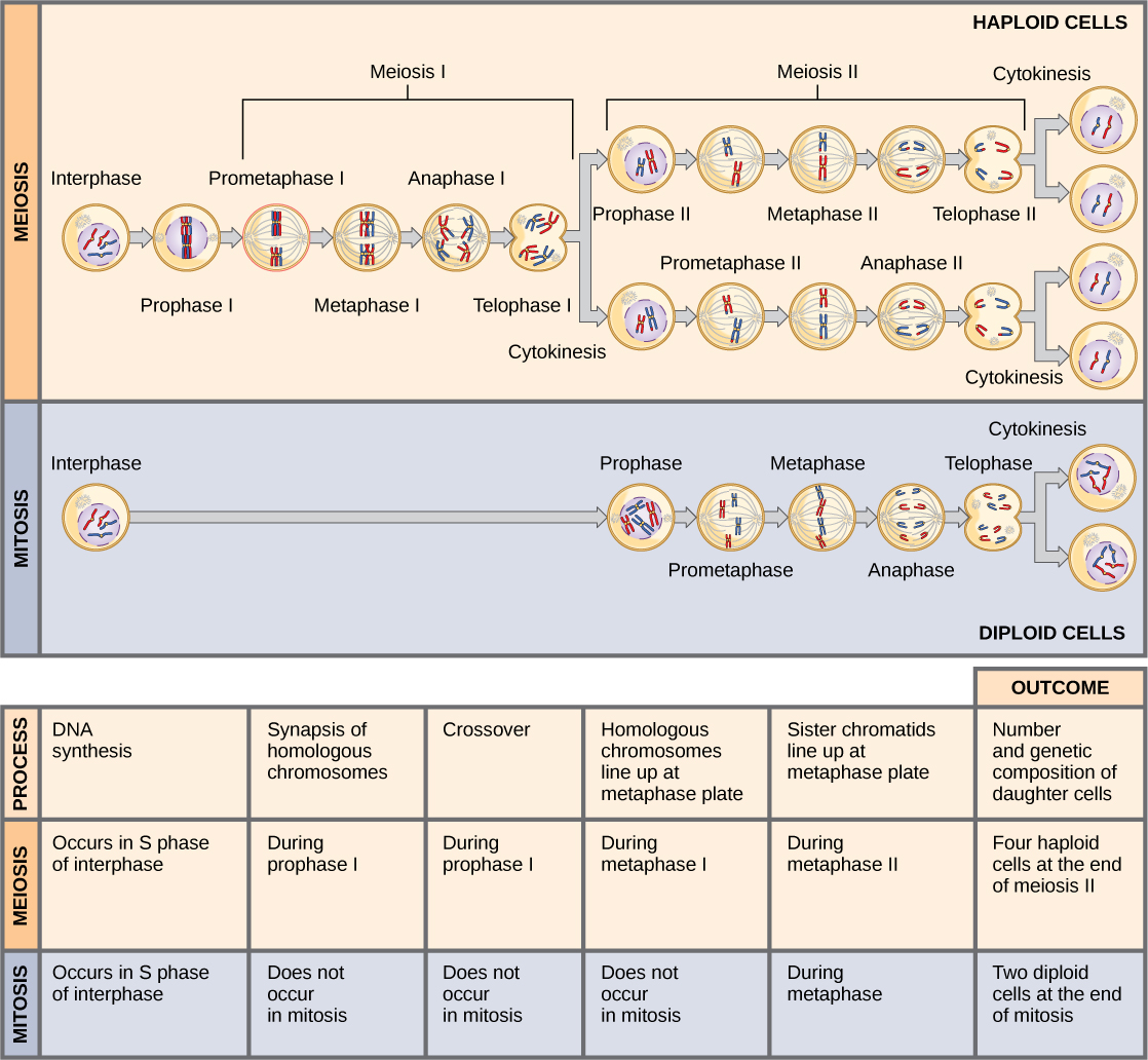
During binary fission a bacterium replicates its chromosome, segregates DNA as the cell grows, and forms a division septum to produce daughters. This is not mitosis because there is no nuclear mitotic apparatus. Under ideal conditions N=N0×2^(t/g), where g is generation time. A 20-minute generation time gives three doublings per hour, not a threefold increase.
A closed culture typically passes through lag, exponential, stationary, and death phases. Stationary phase reflects resource limitation and waste accumulation, with division and death approximately balancing. Optical density estimates turbidity rather than directly counting viable cells; colony-forming units measure reproductive survivors and may count cell clumps as one colony.
Source: 22.2 Structure of Prokaryotes: Bacteria and Archaea · 22.3 Prokaryotic Metabolism
Oxygen use and ecological relationships
Obligate aerobes require oxygen-supported metabolism; obligate anaerobes are harmed by oxygen under their normal growth conditions. Facultative anaerobes can use oxygen when available and switch to fermentation or anaerobic respiration; aerotolerant anaerobes tolerate oxygen without using it for respiration. Anaerobic respiration uses alternative terminal acceptors such as nitrate, whereas fermentation regenerates carriers without a respiratory chain.
Phototrophs obtain energy from light and chemotrophs from chemical reactions; autotrophs use inorganic carbon and heterotrophs organic carbon. These classifications describe separate energy and carbon dimensions. Parasitism benefits one organism at another’s expense, mutualism benefits both, and commensalism benefits one without a substantial effect on the other.
Source: 22.2 Structure of Prokaryotes: Bacteria and Archaea · 22.3 Prokaryotic Metabolism
Chemotaxis and antibiotic resistance
Chemotaxis biases movement in response to chemical gradients. Many motile bacteria alternate runs and tumbles, comparing current signals with recent conditions; favorable changes extend runs rather than requiring a cell to calculate an absolute direction across its length. Receptor adaptation lets the system respond over changing background concentrations.
Antibiotic resistance arises through inherited variation and selection, including target alteration, drug inactivation, reduced uptake, and efflux. Mutations or acquired genes may exist before exposure; antibiotics select survivors rather than causing a directed, useful mutation. Horizontal gene transfer accelerates spread across lineages. Resistance is genetically distinct from transient tolerance or persistence, although both can reduce killing.
Source: 22.2 Structure of Prokaryotes: Bacteria and Archaea · 22.3 Prokaryotic Metabolism
Practice: apply the mechanism
Question. A culture starts with 500 cells and doubles every 30 minutes for 3 hours under exponential conditions. How many cells are expected? Why might viable counts fall below the prediction?
Show reasoning
There are 6 doublings, so N=500×2^6=32,000 cells. Nutrient depletion, waste, temperature changes, death, or a lag phase violate constant exponential growth. Clumping can also make colony-forming units lower than the actual number of viable cells. State which measurement is being compared with the mathematical model.
Source: 22.2 Structure of Prokaryotes: Bacteria and Archaea · 22.3 Prokaryotic Metabolism
22.2 Structure of Prokaryotes: Bacteria and Archaea — figures and tables
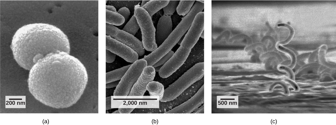
Figure 22.9 Common prokaryotic cell types. Prokaryotes fall into three basic categories based on their shape, visualized here using scanning electron microscopy: (a) cocci, or spherical (a pair is shown); (b) bacilli, or rod-shaped; and (c) spirilli, or spiral-shaped. (credit a: modification of work by Janice Haney Carr, Dr. Richard Facklam, CDC; credit c: modification of work by Dr. David Cox; scale-bar data from Matt Russell)
Mary Ann Clark, Matthew Douglas, and Jung Choi, Biology 2E, OpenStax. (credit a: modification of work by Janice Haney Carr, Dr. Richard Facklam, CDC; credit c: modification of work by Dr. David Cox; scale-bar data from Matt Russell) · CC BY-NC-SA 4.0 · Original image ↗ · Access for free ↗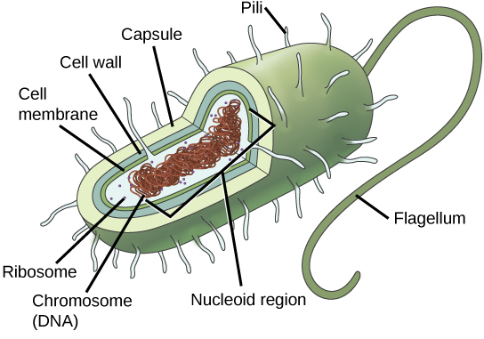
Figure 22.10 The features of a typical prokaryotic cell. Flagella, capsules, and pili are not found in all prokaryotes.
Mary Ann Clark, Matthew Douglas, and Jung Choi, Biology 2E, OpenStax. · CC BY-NC-SA 4.0 · Original image ↗ · Access for free ↗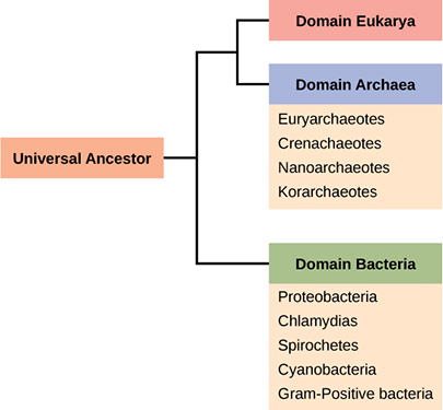
Figure 22.11 The three domains of living organisms. Bacteria and Archaea are both prokaryotes but differ enough to be placed in separate domains. An ancestor of modern Archaea is believed to have given rise to Eukarya, the third domain of life. Major groups of Archaea and Bacteria are shown.
Mary Ann Clark, Matthew Douglas, and Jung Choi, Biology 2E, OpenStax. · CC BY-NC-SA 4.0 · Original image ↗ · Access for free ↗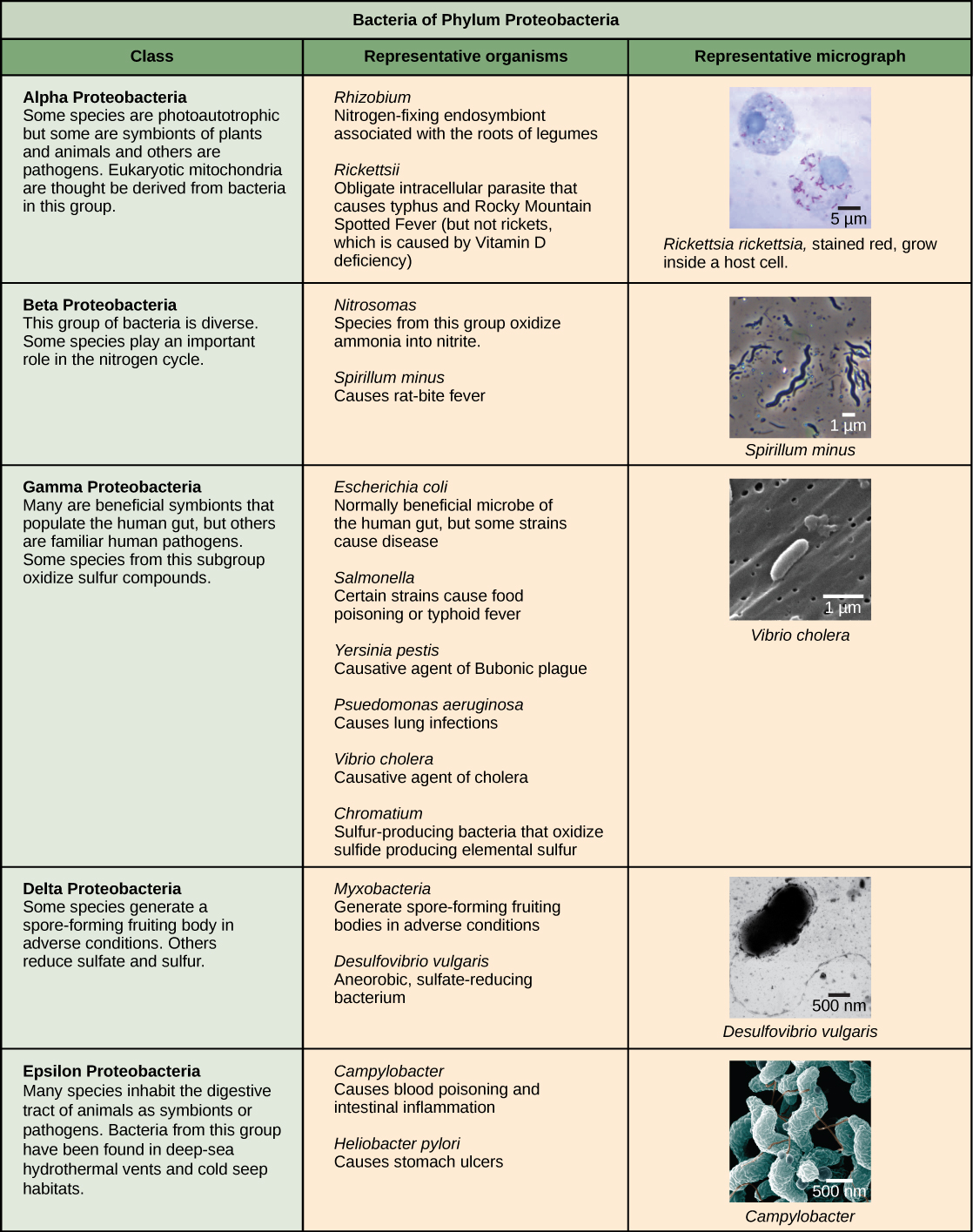
Figure 22.12 The Proteobacteria. Phylum Proteobacteria is one of up to 52 bacteria phyla. Proteobacteria is further subdivided into five classes, Alpha through Epsilon. (credit “Rickettsia rickettsii”: modification of work by CDC; credit “Spirillum minus”: modification of work by Wolframm Adlassnig; credit “Vibrio cholera”: modification of work by Janice Haney Carr, CDC; credit “Desulfovibrio vulgaris”: modification of work by Graham Bradley; credit “Campylobacter”: modification of work by De Wood, Pooley, USDA, ARS, EMU; scale-bar data from Matt Russell)
Mary Ann Clark, Matthew Douglas, and Jung Choi, Biology 2E, OpenStax. (credit “Rickettsia rickettsii”: modification of work by CDC; credit “Spirillum minus”: modification of work by Wolframm Adlassnig; credit “Vibrio cholera”: modification of work by Janice Haney Carr, CDC; credit “Desulfovibrio vulgaris”: modification of work by Graham Bradley; credit “Campylobacter”: modification of work by De Wood, Pooley, USDA, ARS, EMU; scale-bar data from Matt Russell) · CC BY-NC-SA 4.0 · Original image ↗ · Access for free ↗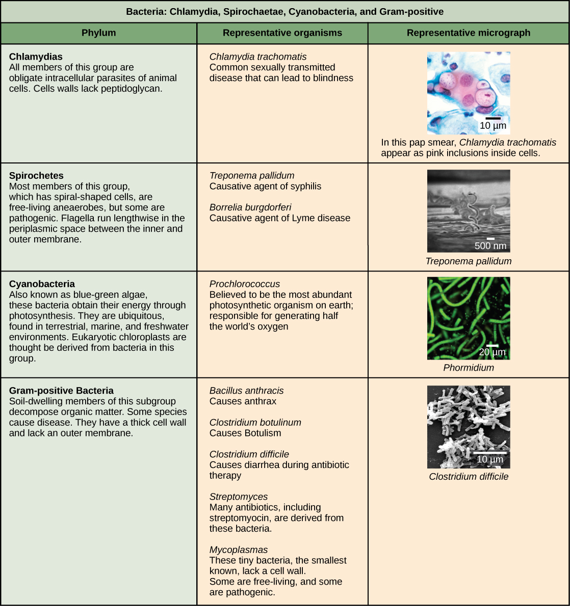
Figure 22.13 Other bacterial phyla. Chlamydia, Spirochetes, Cyanobacteria, and Gram-positive bacteria are described in this table. Note that bacterial shape is not phylum-dependent; bacteria within a phylum may be cocci, rod-shaped, or spiral. (credit “Chlamydia trachomatis” : modification of work by Dr. Lance Liotta Laboratory, NCI; credit “Treponema pallidum” : modification of work by Dr. David Cox, CDC; credit “Phormidium” : modification of work by USGS; credit “Clostridium difficile” : modification of work by Lois S. Wiggs, CDC; scale-bar data from Matt Russell)
Mary Ann Clark, Matthew Douglas, and Jung Choi, Biology 2E, OpenStax. (credit “Chlamydia trachomatis” : modification of work by Dr. Lance Liotta Laboratory, NCI; credit “Treponema pallidum” : modification of work by Dr. David Cox, CDC; credit “Phormidium” : modification of work by USGS; credit “Clostridium difficile” : modification of work by Lois S. Wiggs, CDC; scale-bar data from Matt Russell) · CC BY-NC-SA 4.0 · Original image ↗ · Access for free ↗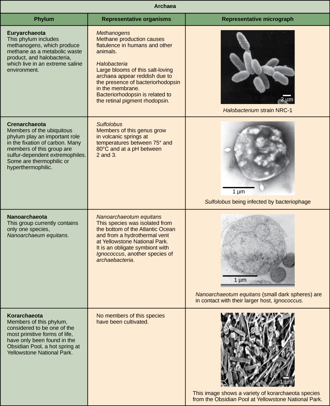
Figure 22.14 Archaeal phyla. Archaea are separated into four phyla: the Korarchaeota, Euryarchaeota, Crenarchaeota, and Nanoarchaeota. (credit “Halobacterium” : modification of work by NASA; credit “Nanoarchaeotum equitans” : modification of work by Karl O. Stetter; credit “Korarchaeota”: modification of work by Office of Science of the U.S. Dept. of Energy; scale-bar data from Matt Russell)
Mary Ann Clark, Matthew Douglas, and Jung Choi, Biology 2E, OpenStax. (credit “Halobacterium” : modification of work by NASA; credit “Nanoarchaeotum equitans” : modification of work by Karl O. Stetter; credit “Korarchaeota”: modification of work by Office of Science of the U.S. Dept. of Energy; scale-bar data from Matt Russell) · CC BY-NC-SA 4.0 · Original image ↗ · Access for free ↗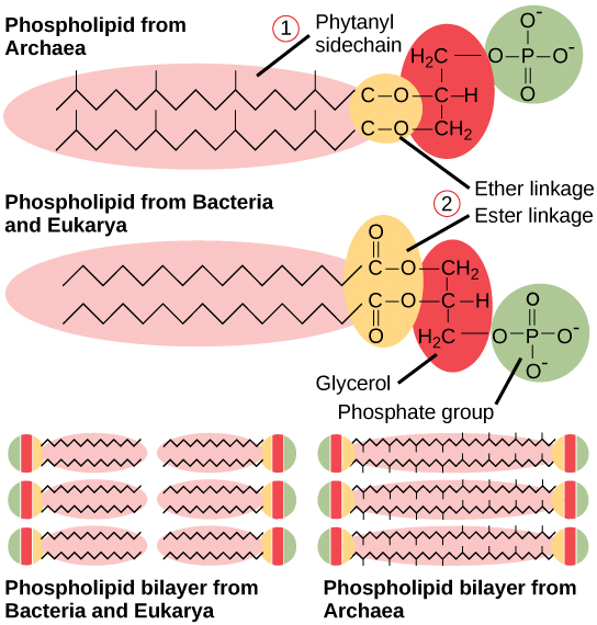
Figure 22.15 Bacterial and archaeal phospholipids. Archaeal phospholipids differ from those found in Bacteria and Eukarya in two ways. First, they have branched phytanyl sidechains instead of linear ones. Second, an ether bond instead of an ester bond connects the lipid to the glycerol.
Mary Ann Clark, Matthew Douglas, and Jung Choi, Biology 2E, OpenStax. · CC BY-NC-SA 4.0 · Original image ↗ · Access for free ↗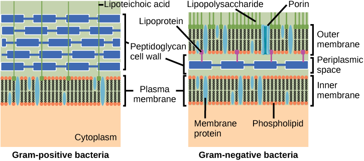
Figure 22.16 Cell walls in Gram-positive and Gram-negative bacteria. Bacteria are divided into two major groups: Gram positive and Gram negative. Both groups have a cell wall composed of peptidoglycan: in Gram-positive bacteria, the wall is thick, whereas in Gram-negative bacteria, the wall is thin. In Gram-negative bacteria, the cell wall is surrounded by an outer membrane that contains lipopolysaccharides and lipoproteins. Porins are proteins in this cell membrane that allow substances to pass through the outer membrane of Gram-negative bacteria. In Gram-positive bacteria, lipoteichoic acid anchors the cell wall to the cell membrane. (credit: modification of work by "Franciscosp2"/Wikimedia Commons)
Mary Ann Clark, Matthew Douglas, and Jung Choi, Biology 2E, OpenStax. (credit: modification of work by "Franciscosp2"/Wikimedia Commons) · CC BY-NC-SA 4.0 · Original image ↗ · Access for free ↗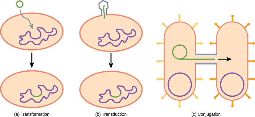
Figure 22.17 Gene transfer mechanisms in prokaryotes. There are three mechanisms by which prokaryotes can exchange DNA. In (a) transformation, the cell takes up prokaryotic DNA directly from the environment. The DNA may remain separate as plasmid DNA or be incorporated into the host genome. In (b) transduction, a bacteriophage injects DNA into the cell that contains a small fragment of DNA from a different prokaryote. In (c) conjugation, DNA is transferred from one cell to another via a mating bridge, or pilus, that connects the two cells after the sex pilus draws the two bacteria close enough to form the bridge.
Mary Ann Clark, Matthew Douglas, and Jung Choi, Biology 2E, OpenStax. · CC BY-NC-SA 4.0 · Original image ↗ · Access for free ↗| Structural Characteristic | Bacteria | Archaea |
|---|---|---|
| Cell type | Prokaryotic | Prokaryotic |
| Cell morphology | Variable | Variable |
| Cell wall | Contains peptidoglycan | Does not contain peptidoglycan |
| Cell membrane type | Lipid bilayer | Lipid bilayer or lipid monolayer |
| Plasma membrane lipids | Fatty acids-glycerol ester | Phytanyl-glycerol ethers |
| Chromosome | Typically circular | Typically circular |
| Replication origins | Single | Multiple |
| RNA polymerase | Single | Multiple |
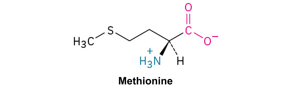
| Initiator tRNA | Formyl-methionine | Methionine |
| Streptomycin inhibition | Sensitive | Resistant |
| Calvin cycle | Yes | No |
Table 22.2. Mary Ann Clark, Matthew Douglas, and Jung Choi, Biology 2E, OpenStax.
22.3 Prokaryotic Metabolism — figures and tables
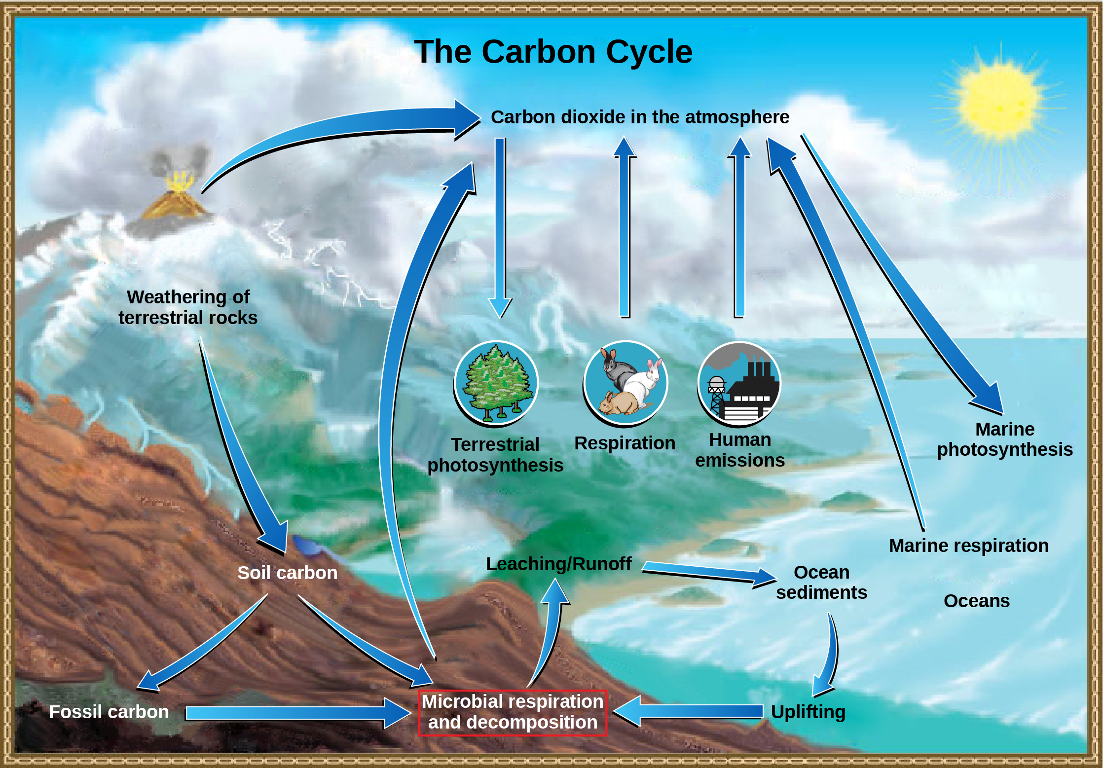
Figure 22.18 The carbon cycle. Prokaryotes play a significant role in continuously moving carbon through the biosphere. (credit: modification of work by John M. Evans and Howard Perlman, USGS)
Mary Ann Clark, Matthew Douglas, and Jung Choi, Biology 2E, OpenStax. (credit: modification of work by John M. Evans and Howard Perlman, USGS) · CC BY-NC-SA 4.0 · Original image ↗ · Access for free ↗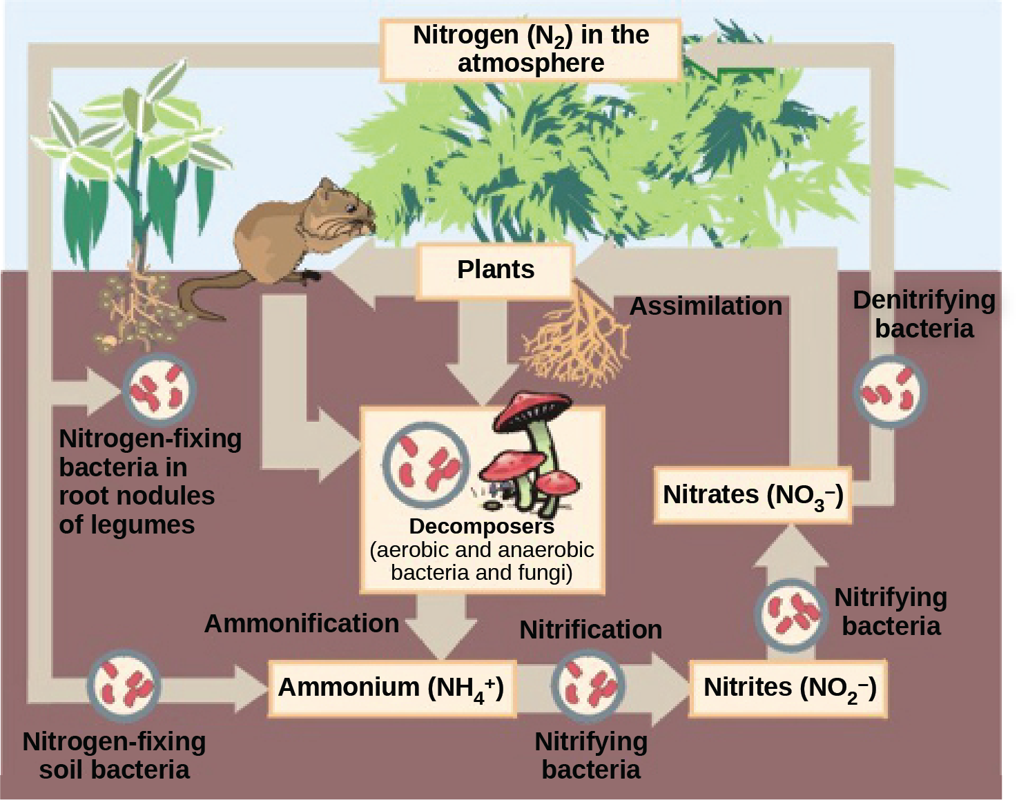
Figure 22.19 The nitrogen cycle. Prokaryotes play a key role in the nitrogen cycle. (credit: Environmental Protection Agency)
Mary Ann Clark, Matthew Douglas, and Jung Choi, Biology 2E, OpenStax. (credit: Environmental Protection Agency) · CC BY-NC-SA 4.0 · Original image ↗ · Access for free ↗| Energy Source | Electron Source | Carbon Source | Nutritional Type |
|---|---|---|---|
| Light (phototroph) |
Organic material (organotroph) |
Organic material (heterotroph) |
Photoorganoheterotroph |
| Carbon dioxide (autotroph) |
|||
| Inorganic material (lithotroph) |
Organic material (heterotroph) |
||
| Carbon dioxide (autotroph) |
Photolithoautotroph | ||
| Chemicals (chemotroph) |
Organic material (organotroph) |
Organic material (heterotroph) |
Chemoorganoheterotroph |
| Carbon dioxide (autotroph) |
|||
| Inorganic material (lithotroph) |
Organic material (heterotroph) |
Chemolithoheterotroph | |
| Carbon dioxide (autotroph) |
Chemolithoautotroph |
Table 22.3. Mary Ann Clark, Matthew Douglas, and Jung Choi, Biology 2E, OpenStax.
Source: 22.3 Prokaryotic Metabolism