Actin, microtubules, and intermediate filaments form distinct systems for force, transport, shape, and mechanical resilience.
Three filament families
Actin microfilaments are polarized polymers that support the cortex, protrusions, and force generation with myosin. Microtubules are hollow, polarized polymers of α/β-tubulin dimers whose dynamic growth and shrinkage organize transport tracks and the mitotic spindle. Intermediate filaments, including keratins and nuclear lamins, provide tensile strength and are generally nonpolar.
Actin assembly is coupled to ATP-bound subunits, while microtubule dynamics depend on GTP-bound tubulin and the stability of the GTP cap. Their distinct properties explain selective drug effects: disrupting microtubules can block spindle function and long-range vesicle transport without directly eliminating every actin-based contractile process.
Source: 4.5 The Cytoskeleton
Motors convert chemical energy into directed motion
Kinesins commonly move cargo toward microtubule plus ends, while cytoplasmic dynein commonly moves toward minus ends; individual motor families have exceptions. Vesicles and organelles use adaptors to connect to motors, and the organization of tracks determines the spatial destination. ATP hydrolysis drives stepping.
Myosin interacts with actin to produce contraction and transport. In animal-cell cytokinesis, an actin–myosin contractile ring constricts the cleavage furrow, separating daughter cytoplasms after chromosomes segregate. Cell crawling combines actin polymerization at the front, substrate adhesion, and contractile forces. Filaments supply tracks or force-bearing structures, while motors and regulated assembly provide direction and work.
Source: 4.5 The Cytoskeleton
Centrosomes, cilia, and flagella
The centrosome is a major microtubule-organizing center in many animal cells and usually contains two centrioles surrounded by nucleating material. A centriole has nine microtubule triplets. Basal bodies anchor cilia and eukaryotic flagella; typical motile axonemes contain nine peripheral doublets and two central microtubules. Dynein-driven sliding is converted to bending by structural constraints.
Most primary sensory cilia have a 9+0 organization and are not motile; some specialized 9+0 cilia can move, so the pattern is a useful generalization rather than an absolute rule. Eukaryotic flagella bend using microtubules, whereas bacterial flagella rotate through an ion-driven motor and do not use a 9+2 axoneme.
Source: 4.5 The Cytoskeleton
Practice: apply the mechanism
Question. A drug disrupts microtubule polymerization. Predict effects on chromosome segregation, a typical cilium, and an already assembled actin–myosin contractile ring.
Show reasoning
Spindle assembly and chromosome segregation will be impaired, and ciliary structure or maintenance can be disrupted. The drug does not directly target actin or myosin, so a contractile ring is a mechanistically different target, although whole-cell disruption can cause indirect effects. A second assay of actin integrity helps distinguish direct specificity from generalized toxicity.
Source: 4.5 The Cytoskeleton
4.5 The Cytoskeleton — figures and tables
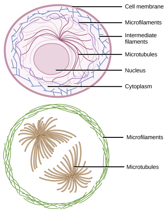
Figure 4.22 Microfilaments thicken the cortex around the cell's inner edge. Like rubber bands, they resist tension. There are microtubules in the cell's interior where they maintain their shape by resisting compressive forces. There are intermediate filaments throughout the cell that hold organelles in place.
Mary Ann Clark, Matthew Douglas, and Jung Choi, Biology 2E, OpenStax. · CC BY-NC-SA 4.0 · Original image ↗ · Access for free ↗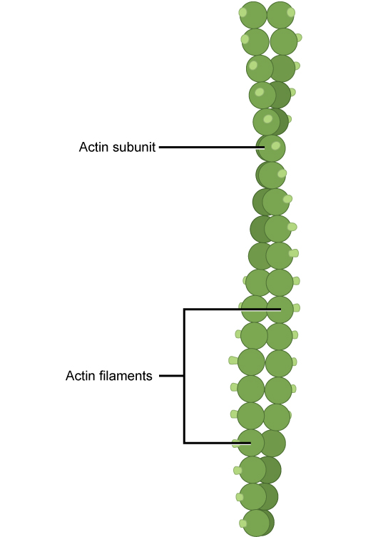
Figure 4.23 Two intertwined actin strands comprise microfilaments.
Mary Ann Clark, Matthew Douglas, and Jung Choi, Biology 2E, OpenStax. · CC BY-NC-SA 4.0 · Original image ↗ · Access for free ↗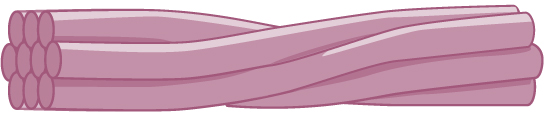
Figure 4.24 Intermediate filaments consist of several intertwined strands of fibrous proteins.
Mary Ann Clark, Matthew Douglas, and Jung Choi, Biology 2E, OpenStax. · CC BY-NC-SA 4.0 · Original image ↗ · Access for free ↗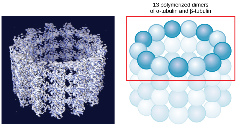
Figure 4.25 Microtubules are hollow. Their walls consist of 13 polymerized dimers of α-tubulin and β-tubulin (right image). The left image shows the tube's molecular structure.
Mary Ann Clark, Matthew Douglas, and Jung Choi, Biology 2E, OpenStax. · CC BY-NC-SA 4.0 · Original image ↗ · Access for free ↗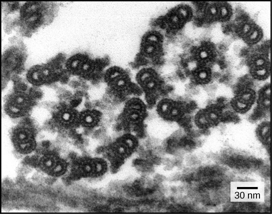
Figure 4.26 This transmission electron micrograph of two flagella shows the microtubules' 9 + 2 array: nine microtubule doublets surround a single microtubule doublet. (credit: modification of work by Dartmouth Electron Microscope Facility, Dartmouth College; scale-bar data from Matt Russell)
Mary Ann Clark, Matthew Douglas, and Jung Choi, Biology 2E, OpenStax. (credit: modification of work by Dartmouth Electron Microscope Facility, Dartmouth College; scale-bar data from Matt Russell) · CC BY-NC-SA 4.0 · Original image ↗ · Access for free ↗| Cell Component | Function | Present in Prokaryotes? | Present in Animal Cells? | Present in Plant Cells? |
|---|---|---|---|---|
| Plasma membrane | Separates cell from external environment; controls passage of organic molecules, ions, water, oxygen, and wastes into and out of cell | Yes | Yes | Yes |
| Cytoplasm | Provides turgor pressure to plant cells as fluid inside the central vacuole; site of many metabolic reactions; medium in which organelles are found | Yes | Yes | Yes |
| Nucleolus | Darkened area within the nucleus where ribosomal subunits are synthesized. | No | Yes | Yes |
| Nucleus | Cell organelle that houses DNA and directs synthesis of ribosomes and proteins | No | Yes | Yes |
| Ribosomes | Protein synthesis | Yes | Yes | Yes |
| Mitochondria | ATP production/cellular respiration | No | Yes | Yes |
| Peroxisomes | Oxidize and thus break down fatty acids and amino acids, and detoxify poisons | No | Yes | Yes |
| Vesicles and vacuoles | Storage and transport; digestive function in plant cells | No | Yes | Yes |
| Centrosome | Unspecified role in cell division in animal cells; microtubule source in animal cells | No | Yes | No |
| Lysosomes | Digestion of macromolecules; recycling of worn-out organelles | No | Yes | Some |
| Cell wall | Protection, structural support, and maintenance of cell shape | Yes, primarily peptidoglycan | No | Yes, primarily cellulose |
| Chloroplasts | Photosynthesis | No | No | Yes |
| Endoplasmic reticulum | Modifies proteins and synthesizes lipids | No | Yes | Yes |
| Golgi apparatus | Modifies, sorts, tags, packages, and distributes lipids and proteins | No | Yes | Yes |
| Cytoskeleton | Maintains cell’s shape, secures organelles in specific positions, allows cytoplasm and vesicles to move within cell, and enables unicellular organisms to move independently | Yes | Yes | Yes |
| Flagella | Cellular locomotion | Some | Some | No, except for some plant sperm cells |
| Cilia | Cellular locomotion, movement of particles along plasma membrane's extracellular surface, and filtration | Some | Some | No |
Table 4.1. Mary Ann Clark, Matthew Douglas, and Jung Choi, Biology 2E, OpenStax.
Source: 4.5 The Cytoskeleton