Foundation 2 · 2A · Cell structure and function

transport, voltage, and

Sources ↗

Predict transport by combining permeability with chemical and electrical , then identify the energy source and route.

, , and effective

Simple moves permeant down their chemical ; flux increases with difference and surface area and decreases with thickness. uses selective channels or carriers but still moves a down its . transport can saturate and show competition.

moves water toward the side with lower water chemical potential, often the side with more effectively nonpenetrating . For a dilute , π≈iMRT, where i counts dissolved particles. predicts cell volume and depends on nonpenetrating ; counts all particles. A permeant such as can make a yet after equilibration.

Source: 5.2 Passive Transport · 5.3 Active Transport · 5.4 Bulk Transport

Direct and indirect energy coupling

directly couples an energy source, commonly , to uphill movement. The exports three Na+ and imports two K+ per , maintaining and producing a small direct electrogenic effect. uses a pre-existing rather than hydrolyzing at the cotransporter.

A Na+– lets inward Na+ movement drive accumulation; an moves coupled in opposite directions. Inhibiting the eventually weakens Na+-coupled uptake even if the cotransporter remains intact. The pump’s immediate electrical effect and the slower dissipation of stored are different time scales.

Source: 5.2 Passive Transport · 5.3 Active Transport · 5.4 Bulk Transport

Electrochemical determine movement

For an , direction depends on and voltage together. The is Eion=(RT/zF) ln([]outside/[]inside); at 37°C this is approximately 61.5 mV/z × log10 of the ratio. At Eion, electrical and chemical forces balance for that , although individual still move both ways.

Resting voltage reflects several permeabilities and , with high K+ permeability often pulling voltage toward EK. Opening a selective channel drives voltage toward that ’s potential. A large need not produce net movement if the opposing electrical force balances it; the pump maintains the used by channels.

Source: 5.2 Passive Transport · 5.3 Active Transport · 5.4 Bulk Transport

Signals cross a without the crossing

Cell-surface bind an extracellular and change intracellular activity. A -gated channel changes permeability directly; a activates a heterotrimeric G that regulates or channels; a promotes and recruitment of signaling . such as or Ca2+ can amplify the response.

Specificity depends on expression and , so the same can produce different responses in different cells. removal, , , and internalization terminate or adapt signaling. A response does not require the signaling itself to enter the .

Source: 5.2 Passive Transport · 5.3 Active Transport · 5.4 Bulk Transport · 9.1 Signaling Molecules and Cellular Receptors · 9.2 Propagation of the Signal

and preserve orientation

engulfs large particles; samples extracellular fluid; -mediated concentrates selected in coated . sort cargo for recycling, transport, or lysosomal degradation. fuses with the plasma to secrete cargo and deliver .

lumens remain topologically equivalent to the extracellular space: a facing the ultimately faces outside after surface fusion. removes surface and adds it, so coordinated recycling maintains area and composition. Vesicular transport can move large cargo that cannot pass through channels or .

Source: 5.2 Passive Transport · 5.3 Active Transport · 5.4 Bulk Transport

Practice: apply the mechanism

Question. A cell has 140 mM K+ inside and 14 mM outside at 37°C. Estimate EK. What happens to Na+-coupled uptake over time if production stops?

Show reasoning

EK≈61.5 log10(14/140)=−61.5 mV. This is the potential for K+, not necessarily the exact resting voltage of a permeable to other . Loss of stops effective Na+/K+ pumping; as the Na+ dissipates, Na+-coupled accumulation declines even though the does not directly use .

Source: 5.2 Passive Transport · 5.3 Active Transport · 5.4 Bulk Transport

5.2 Passive Transport — figures and tables
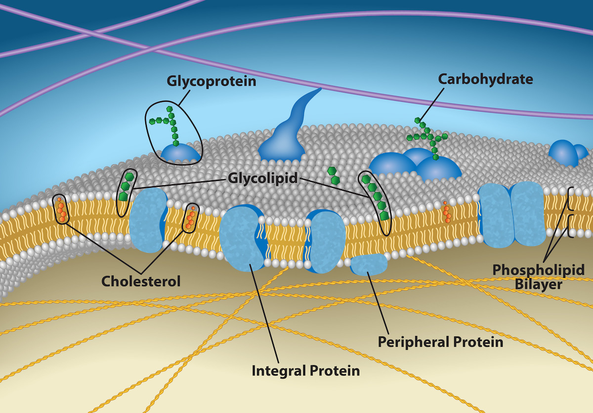
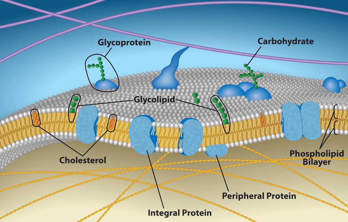
This illustration shows that the inside and outside of a plasma membrane are different, with the exterior covered in the spherical heads, and the interior filled with the strandlike tails.
Figure 5.7 — 5.2 Passive Transport

Figure 5.7 The plasma membrane's exterior surface is not identical to its interior surface. Credit: Rao, A., Ryan, K., Fletcher, S., Hawkins, A. and Tag, A. Department of Biology, Texas A&M University.

Mary Ann Clark, Matthew Douglas, and Jung Choi, Biology 2E, OpenStax. · CC BY-NC-SA 4.0 · Original image ↗ · Access for free ↗
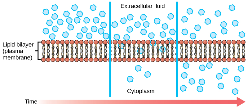
The left part of this illustration shows a substance on one side of a membrane only, in the extracellular fluid. The middle part shows that, after some time, some of the substance has diffused across the plasma membrane, from the extracellular fluid and into the cytoplasm. The right part shows that, after more time, an equal amount of the substance is on each side of the membrane.
Figure 5.8 — 5.2 Passive Transport

Figure 5.8 Diffusion through a permeable membrane moves a substance from a high concentration area (extracellular fluid, in this case) down its concentration gradient (into the cytoplasm). (credit: modification of work by Mariana Ruiz Villareal)

Mary Ann Clark, Matthew Douglas, and Jung Choi, Biology 2E, OpenStax. (credit: modification of work by Mariana Ruiz Villareal) · CC BY-NC-SA 4.0 · Original image ↗ · Access for free ↗
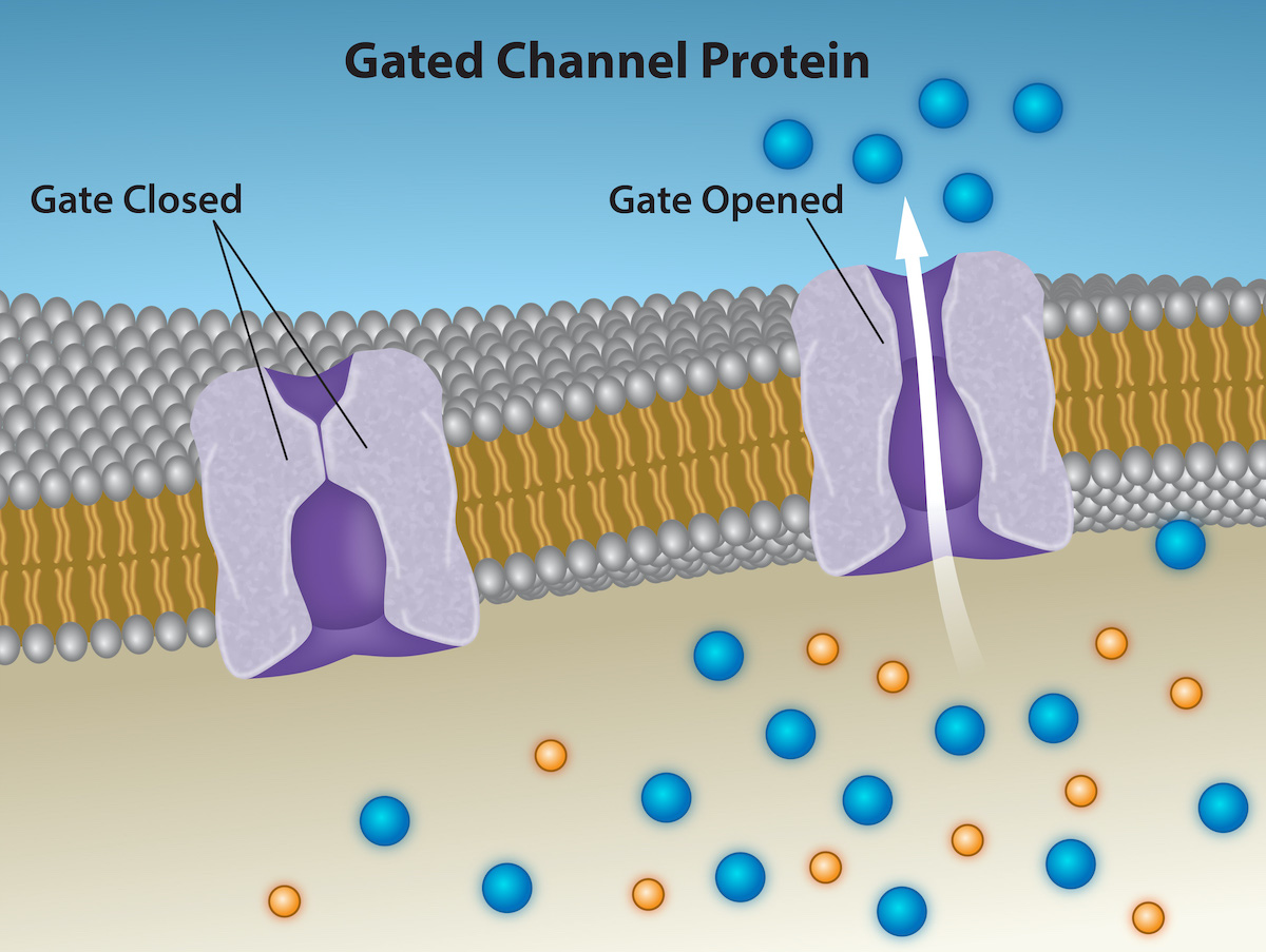
This illustration shows two channel proteins in the plasma membrane. One protein is labeled gate closed, and there is no space for particles to pass through.  The other is labeled gate open, and there is a space for substances to pass through.  Substances of the same type appear on both sides of the membrane near the open-gated protein.
Figure 5.9 — 5.2 Passive Transport

Figure 5.9 Ion Channel Proteins. Ion channel proteins are gated. When they are closed, no ions can pass through them. However, when a channel opens, select ions diffuse through the channel. Channel proteins are highly specific, letting only a specific ion or subset of ions pass. Credit: Rao, A., Ryan, K., Tag, A. and Fletcher, S. Department of Biology, Texas A&M University.

Mary Ann Clark, Matthew Douglas, and Jung Choi, Biology 2E, OpenStax. · CC BY-NC-SA 4.0 · Original image ↗ · Access for free ↗
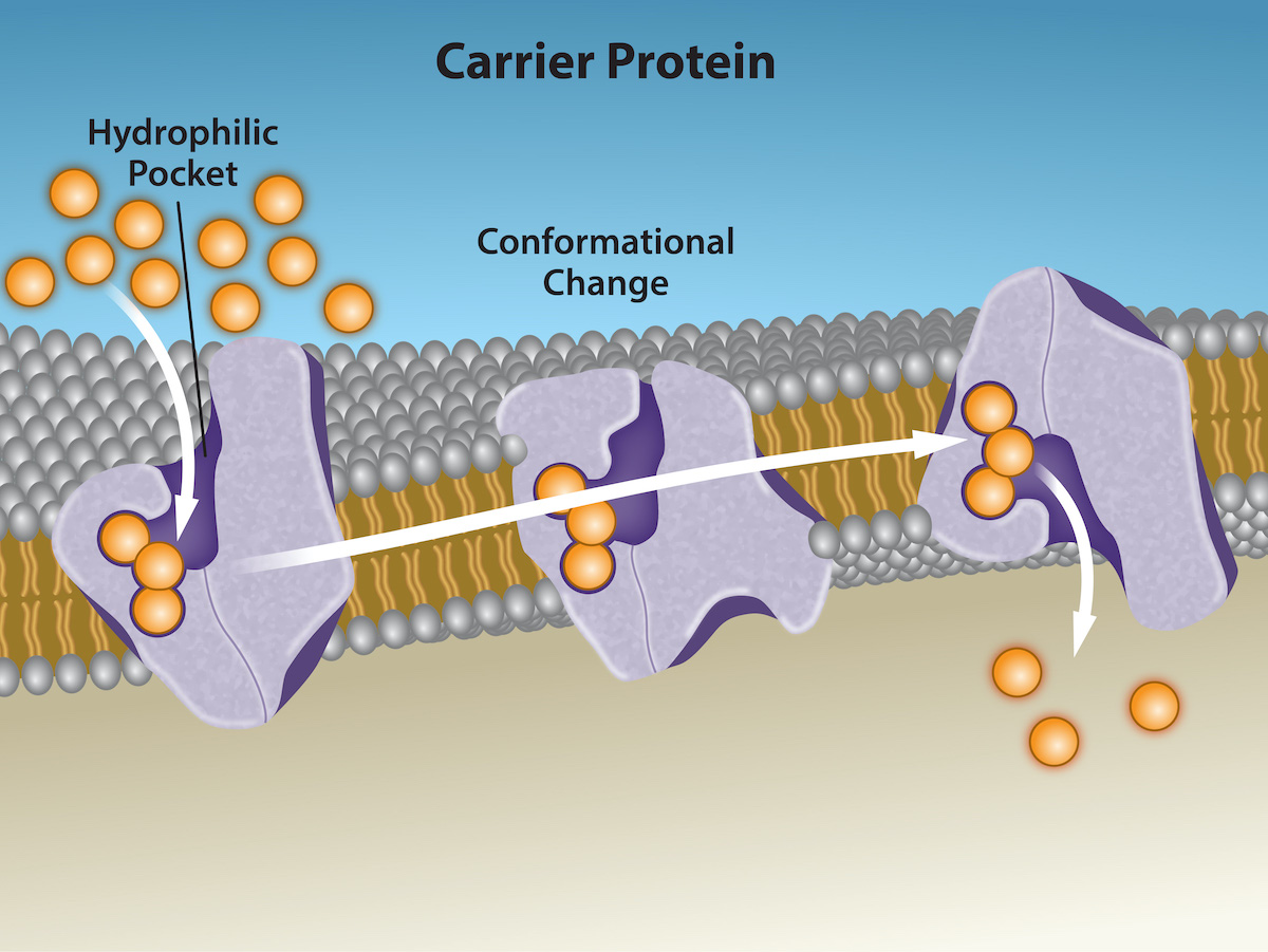
This illustration shows a carrier protein embedded in the membrane with an opening that initially faces the extracellular surface, labeled the hydrophilic pocket. After a substance binds the carrier, it changes shape so that the opening faces the cytoplasm. The change is labeled conformational change. After, the substance is released.
Figure 5.10 — 5.2 Passive Transport

Figure 5.10 Some substances are able to move down their concentration gradient across the plasma membrane with the aid of carrier proteins. Carrier proteins change shape as they move molecules across the membrane. Credit: Rao, A., Tag, A. and Fletcher, S. Department of Biology, Texas A&M University.

Mary Ann Clark, Matthew Douglas, and Jung Choi, Biology 2E, OpenStax. · CC BY-NC-SA 4.0 · Original image ↗ · Access for free ↗
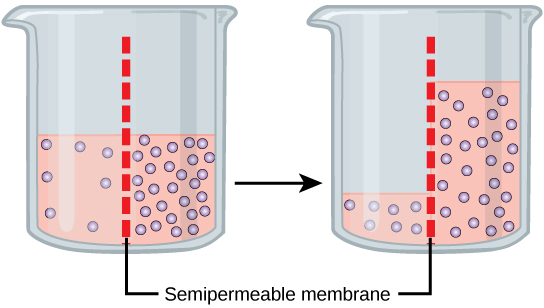
This illustration shows a container whose contents are separated by a semipermeable membrane. Initially, there is a high concentration of solute on the right side of the membrane and a low concentration of the left. Over time, water diffuses across the membrane toward the side of the container that initially had a higher concentration of solute (lower concentration of water). As a result of osmosis, the water level is higher on this side of the membrane, and the solute concentration is the same on both sides.
Figure 5.11 — 5.2 Passive Transport

Figure 5.11 In osmosis, water always moves from an area of higher water concentration to one of lower concentration. In the diagram, the solute cannot pass through the selectively permeable membrane, but the water can.

Mary Ann Clark, Matthew Douglas, and Jung Choi, Biology 2E, OpenStax. · CC BY-NC-SA 4.0 · Original image ↗ · Access for free ↗
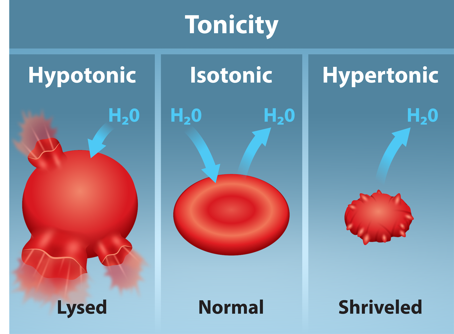
The left part shows bloated red blood cells, lysed, bathed in a hypotonic solution. The middle part shows healthy red blood cells bathed in an isotonic solution, and the right part of this illustration shows shriveled red blood cells bathed in a hypertonic solution.
Figure 5.12 — 5.2 Passive Transport

Figure 5.12 Three different scenarios involving red blood cells (RBC) are shown. Left: A RBC placed in a hypotonic solution, where the concentration of solutes in the surrounding fluid is lower than those in the cell, will cause water to rush into the RBC and lead to lysis of the cell. Middle: there is no net water movement into or out of the cell as the concentration of the solutes inside the cell equal or is isotonic to that of the surrounding fluid. Right: a RBC placed in a hypertonic solution, where the concentration of solutes in the surrounding fluid is greater than that in the cell will cause water to rush out of the cell and into the surrounding fluid. This will cause the RBC to shrivel. Credit: Tag, A., Rao, A., Hawkins, A and Fletcher, S. Department of Biology, Texas A&M University.

Mary Ann Clark, Matthew Douglas, and Jung Choi, Biology 2E, OpenStax. · CC BY-NC-SA 4.0 · Original image ↗ · Access for free ↗
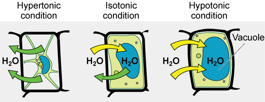
The left part of this image shows a plant cell bathed in a hypertonic solution so that the plasma membrane has pulled away completely from the cell wall, and the central vacuole has shrunk. Water moves out of the plant cell. The middle part shows a plant cell bathed in an isotonic solution; the plasma membrane has pulled away from the cell wall a bit, and the central vacuole size is unchanged. Water moves both in and out of the plant cell. The right part shows a plant cell in a hypotonic solution. The central vacuole is large, and the plasma membrane is pressed against the cell wall. Water is moving into the plant cell.
Figure 5.13 — 5.2 Passive Transport

Figure 5.13 The turgor pressure within a plant cell depends on the solution's tonicity in which it is bathed. (credit: modification of work by Mariana Ruiz Villareal)

Mary Ann Clark, Matthew Douglas, and Jung Choi, Biology 2E, OpenStax. (credit: modification of work by Mariana Ruiz Villareal) · CC BY-NC-SA 4.0 · Original image ↗ · Access for free ↗
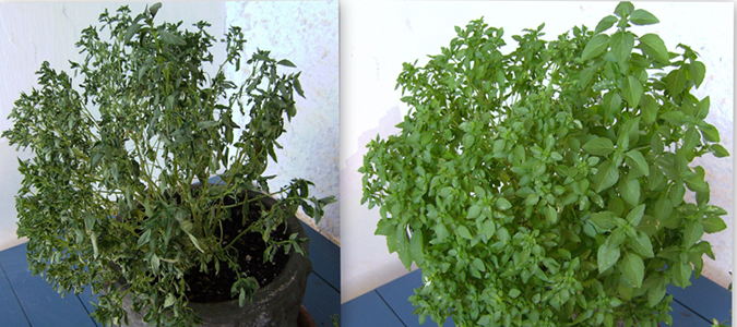
The left photo shows a plant that has wilted, with dark green leaves that are shriveled, and appear dry.  The photo on the right shows a healthy plant, with broad light green leaves that appear soft and pliable.
Figure 5.14 — 5.2 Passive Transport

Figure 5.14 Without adequate water, the plant on the left has lost turgor pressure, visible in its wilting. Watering the plant (right) will restore the turgor pressure. (credit: Victor M. Vicente Selvas)

Mary Ann Clark, Matthew Douglas, and Jung Choi, Biology 2E, OpenStax. (credit: Victor M. Vicente Selvas) · CC BY-NC-SA 4.0 · Original image ↗ · Access for free ↗
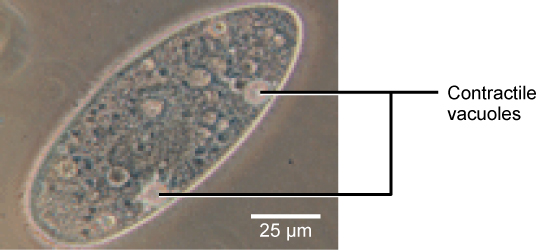
A bright field light microscopy shows an oval-shaped cell. Contractile vacuoles are prominent structures embedded in the cell membrane that pump out water.
Figure 5.15 — 5.2 Passive Transport

Figure 5.15 A paramecium’s contractile vacuole, here visualized using bright field light microscopy at 480x magnification, continuously pumps water out of the organism’s body to keep it from bursting in a hypotonic medium. (credit: modification of work by NIH; scale-bar data from Matt Russell)

Mary Ann Clark, Matthew Douglas, and Jung Choi, Biology 2E, OpenStax. (credit: modification of work by NIH; scale-bar data from Matt Russell) · CC BY-NC-SA 4.0 · Original image ↗ · Access for free ↗

Source: 5.2 Passive Transport

5.3 Active Transport — figures and tables
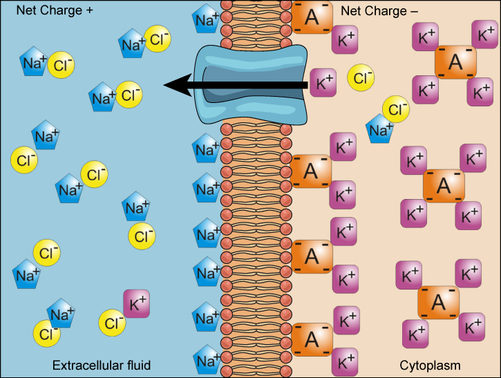
This illustration shows a membrane bilayer with a potassium channel embedded in it. The cytoplasm has a high concentration of potassium associated with a negatively charged molecule. The extracellular fluid has a high concentration of sodium associated with chlorine ions.
Figure 5.16 — 5.3 Active Transport

Figure 5.16 Electrochemical gradients arise from the combined effects of concentration gradients and electrical gradients. Na + ions are at higher concentration outside the cell, and K + ions are at higher concentration inside of the cell, and yet the inside of the cell has negative net charge compared to the other side of the membrane. This is due to the presence of K + binding proteins and other negatively charged molecules. The difference in electrical charges attracts the positively charged Na ions toward the inside of the cell, the electrical gradient, while the K ions tend to flow through K channels toward the outside of the cell due to the concentration difference, the concentration gradient. Structures labeled A represent proteins. (credit: “Synaptitude”/Wikimedia Commons)

Mary Ann Clark, Matthew Douglas, and Jung Choi, Biology 2E, OpenStax. (credit: “Synaptitude”/Wikimedia Commons) · CC BY-NC-SA 4.0 · Original image ↗ · Access for free ↗
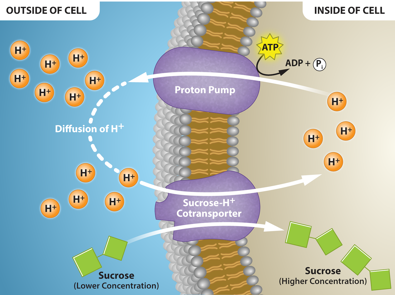
This illustration shows a membrane bilayer with a potassium channel called a proton pump embedded in it. Hydrogen ions flow out of the cell through a proton pump, then back into the cell as part of the sucrose-proton cotransporter. Sucrose flows with the protons from the lower concentration outside the cell to the higher concentration inside the cell, which is against the concentration gradient.
Figure 5.17 — 5.3 Active Transport

Figure 5.17 Proton Gradient provides energy for a secondary active transporter. The proton pump creates an electrochemical gradient of protons (hydrogen ions, H+) using ATP to drive primary active transport. This gradient allows for cotransport/secondary transport of sucrose against its concentration gradient as protons come down their concentration gradient via their membrane cotransporter protein. Credit: Rao, A. Tag, A. and Ryan, K. Department of Biology, Texas A&M University.

Mary Ann Clark, Matthew Douglas, and Jung Choi, Biology 2E, OpenStax. · CC BY-NC-SA 4.0 · Original image ↗ · Access for free ↗
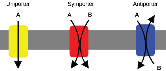
This illustration shows a plasma membrane with three transport proteins embedded in it. The left image shows a uniporter that transports a substance in one direction. The middle image shows a symporter that transports two different substances in the same direction. The right image shows an antiporter that transports two different substances in opposite directions.
Figure 5.18 — 5.3 Active Transport

Figure 5.18 A uniporter carries one molecule or ion. A symporter carries two different molecules or ions, both in the same direction. An antiporter also carries two different molecules or ions, but in different directions. (credit: modification of work by “Lupask”/Wikimedia Commons)

Mary Ann Clark, Matthew Douglas, and Jung Choi, Biology 2E, OpenStax. (credit: modification of work by “Lupask”/Wikimedia Commons) · CC BY-NC-SA 4.0 · Original image ↗ · Access for free ↗
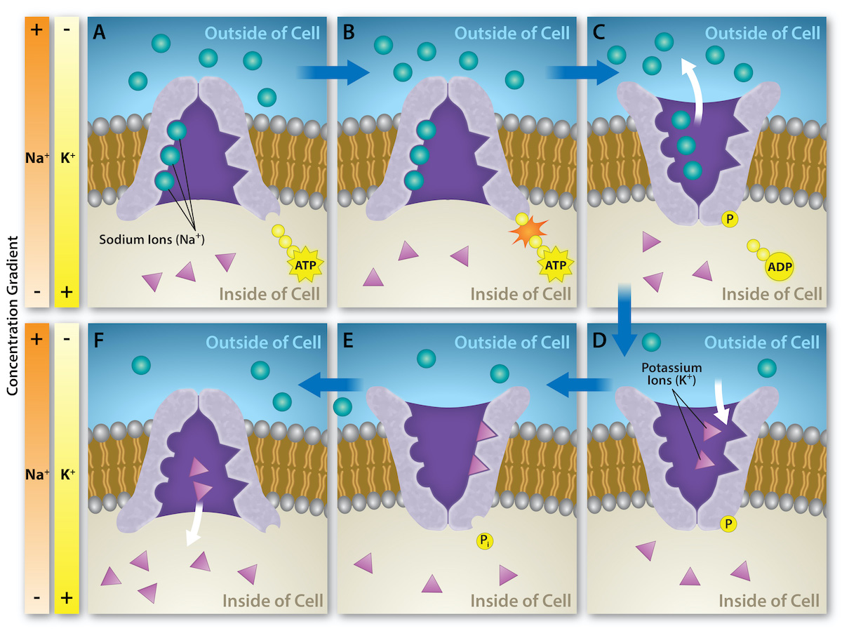
This illustration shows the sodium-potassium pump. Initially, the pumps opening faces the cytoplasm, where three sodium ions bind to it. The antiporter hydrolyzes an A T P to A D P and, as a result, undergoes a conformational change. The sodium ions are released into the extracellular space. Two potassium ions from the extracellular space now bind the antiporter, which changes conformation again, releasing the potassium ions into the cytoplasm.
Figure 5.19 — 5.3 Active Transport

Figure 5.19 The sodium-potassium pump is an example of primary active transport that moves ions, sodium and potassium ions in this instance, across a membrane against their concentration gradients. The energy is provided by the hydrolysis of ATP. Three sodium ions are moved out of the cell for every 2 potassium ions that are brought into the cell. This creates an electrochemical gradient that is crucial for living cells. Credit: Rao, A., Ryan, K. and Fletcher, S. Department of Biology, Texas A&M University.

Mary Ann Clark, Matthew Douglas, and Jung Choi, Biology 2E, OpenStax. · CC BY-NC-SA 4.0 · Original image ↗ · Access for free ↗
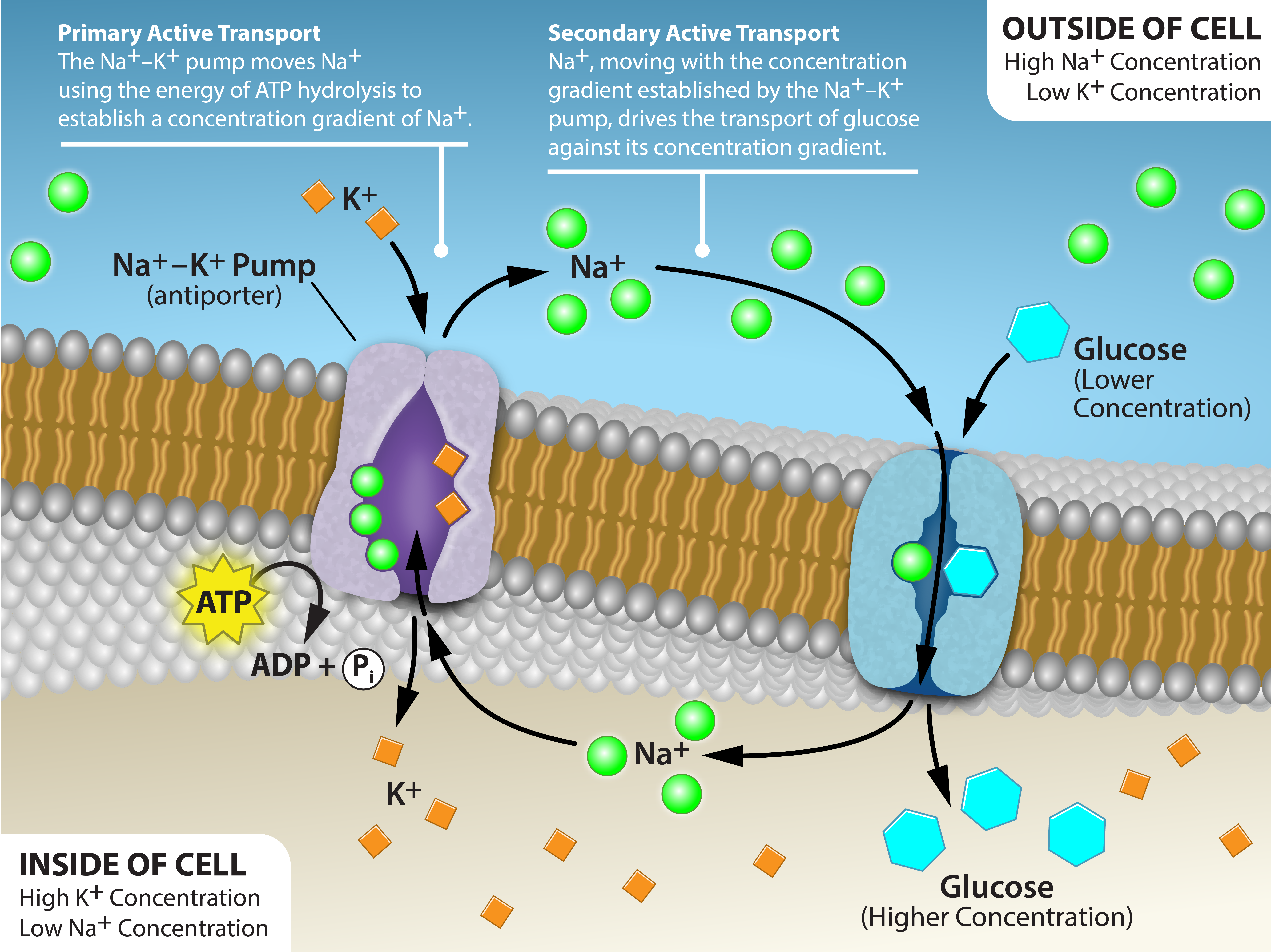
This illustration shows a membrane bilayer with two integral membrane proteins embedded in it. The first, a sodium-potassium pump, uses energy from A T P hydrolysis to pump three sodium ions out of the cell for every two potassium ions it pumps into the cell. The result is a high concentration of sodium outside the cell and a high concentration of potassium inside the cell. There is also a low concentration of glucose outside the cell, and a high concentration inside. A sodium-amino acid co-transporter simultaneously transports sodium and the glucose into the cell. Text within the image reads as follows: Primary Active Transport: the sodium potassium pump moves sodium ions using the energy of ATP hydrolysis to establish a concentration gradient of sodium ions. Secondary active transport: Sodium ions moving with the concentration gradient established by the sodium potassium pump drives the transport of glucose against its concentration gradient.
Figure 5.20 — 5.3 Active Transport

Figure 5.20 An electrochemical gradient (Na + concentration - green), is generated by primary active transport. The energy stored in the Na + gradient provides the energy to move other substances against their concentration gradients (Glucose - blue), a process called co-transport or secondary active transport. Credit: Rao, A., Ryan, K., Tag, A. and Fletcher, S. Department of Biology, Texas A&M University.

Mary Ann Clark, Matthew Douglas, and Jung Choi, Biology 2E, OpenStax. · CC BY-NC-SA 4.0 · Original image ↗ · Access for free ↗

Source: 5.3 Active Transport

5.4 Bulk Transport — figures and tables
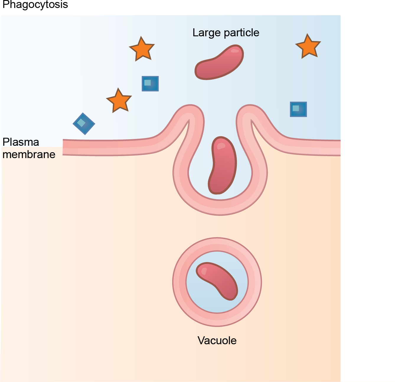
This illustration shows a plasma membrane forming a pocket around a particle in the extracellular fluid. The membrane subsequently engulfs the particle, which becomes trapped in a vacuole inside the cell.
Figure 5.21 — 5.4 Bulk Transport

Figure 5.21 In phagocytosis, the cell membrane surrounds the particle and engulfs it. (credit: modification of work by Mariana Ruiz Villareal)

Mary Ann Clark, Matthew Douglas, and Jung Choi, Biology 2E, OpenStax. (credit: modification of work by Mariana Ruiz Villareal) · CC BY-NC-SA 4.0 · Original image ↗ · Access for free ↗
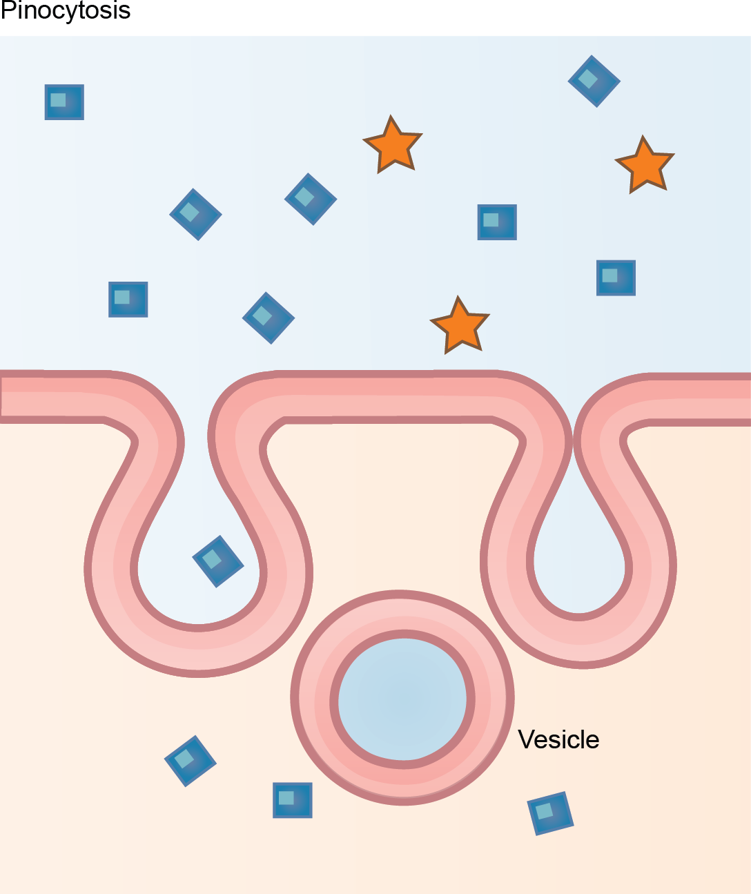
This illustration shows a plasma membrane forming a pocket around fluid in the extracellular fluid. The membrane subsequently engulfs the fluid, which becomes trapped in a vacuole.
Figure 5.22 — 5.4 Bulk Transport

Figure 5.22 In pinocytosis, the cell membrane invaginates, surrounds a small volume of fluid, and pinches off. (credit: modification of work by Mariana Ruiz Villareal)

Mary Ann Clark, Matthew Douglas, and Jung Choi, Biology 2E, OpenStax. (credit: modification of work by Mariana Ruiz Villareal) · CC BY-NC-SA 4.0 · Original image ↗ · Access for free ↗
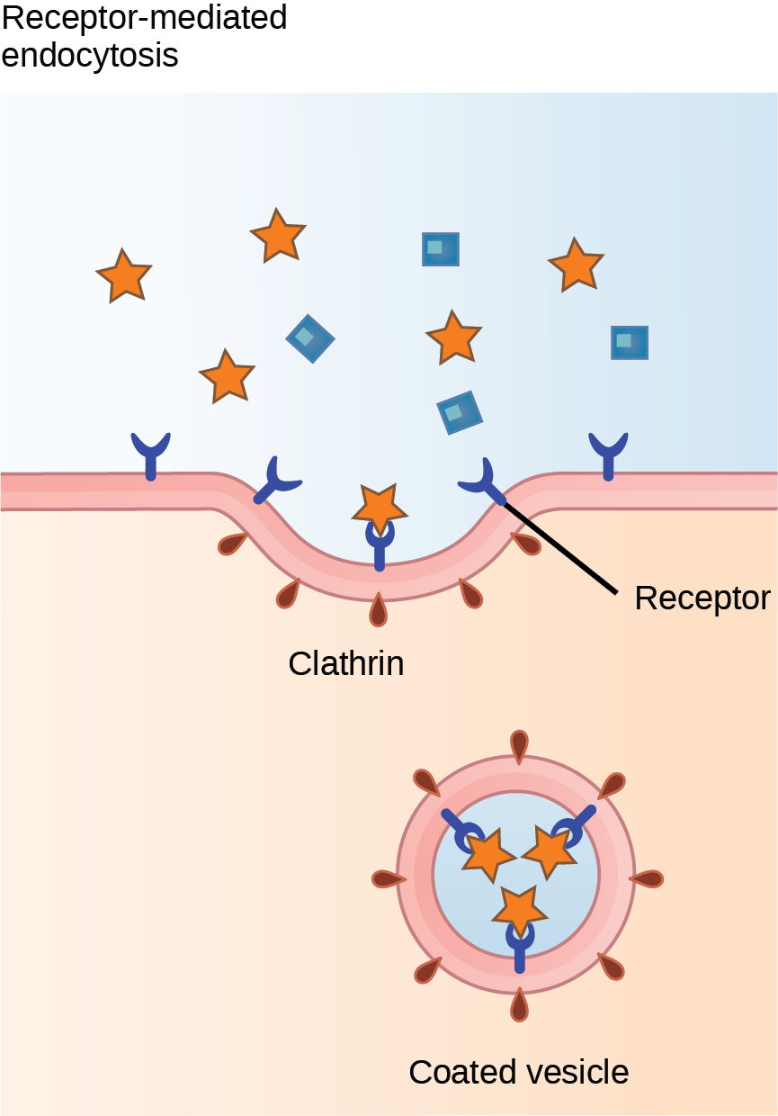
This illustration shows a part of the plasma membrane that is clathrin-coated on the cytoplasmic side and has receptors on the extracellular side. The receptors bind a substance, then pinch off to form a vesicle.
Figure 5.23 — 5.4 Bulk Transport

Figure 5.23 In receptor-mediated endocytosis, the cell's uptake of substances targets a single type of substance that binds to the receptor on the cell membrane's external surface. (credit: modification of work by Mariana Ruiz Villareal)

Mary Ann Clark, Matthew Douglas, and Jung Choi, Biology 2E, OpenStax. (credit: modification of work by Mariana Ruiz Villareal) · CC BY-NC-SA 4.0 · Original image ↗ · Access for free ↗
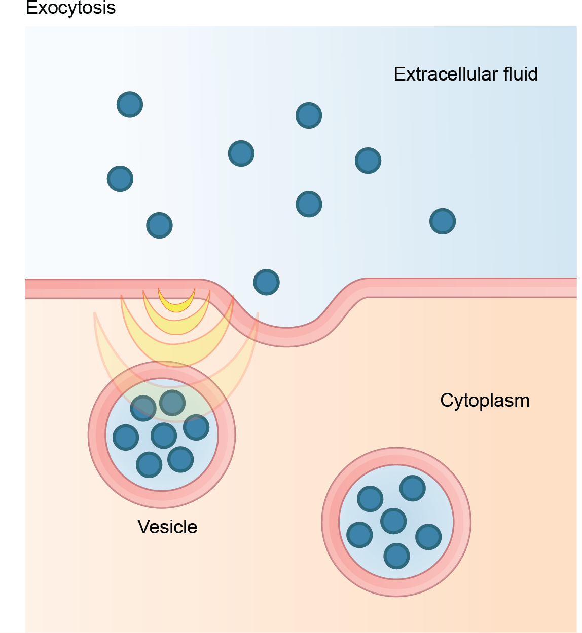
This illustration shows vesicles fusing with the plasma membrane and releasing their contents to the extracellular fluid.
Figure 5.24 — 5.4 Bulk Transport

Figure 5.24 In exocytosis, vesicles containing substances fuse with the plasma membrane. The contents then release to the cell's exterior. (credit: modification of work by Mariana Ruiz Villareal)

Mary Ann Clark, Matthew Douglas, and Jung Choi, Biology 2E, OpenStax. (credit: modification of work by Mariana Ruiz Villareal) · CC BY-NC-SA 4.0 · Original image ↗ · Access for free ↗
Transport MethodActive/PassiveMaterial Transported
PassiveSmall- material
PassiveWater
Facilitated transport/PassiveSodium, potassium, calcium,
ActiveSodium, potassium, calcium
Active,
ActiveLarge , whole cells, or cellular structures
and potocytosisActiveSmall (liquids/water)
-mediated ActiveLarge quantities of

Table 5.2. Mary Ann Clark, Matthew Douglas, and Jung Choi, Biology 2E, OpenStax.

Source: 5.4 Bulk Transport

9.1 Signaling and Cellular — figures and tables

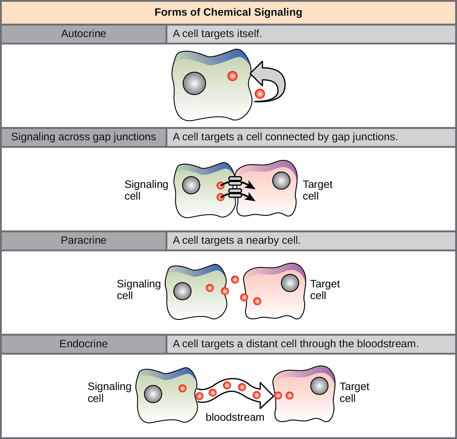
The illustration shows four forms of chemical signaling. In autocrine signaling, a cell targets itself. In signaling across a gap junction, a cell targets a cell connected via gap junctions. In paracrine signaling, a cell targets a nearby cell. In endocrine signaling, a cell targets a distant cell via the bloodstream
Figure 9.2 — 9.1 Signaling and Cellular

Figure 9.2 In chemical signaling, a cell may target itself (autocrine signaling), a cell connected by gap junctions, a nearby cell (paracrine signaling), or a distant cell (endocrine signaling). Paracrine signaling acts on nearby cells, endocrine signaling uses the circulatory system to transport ligands, and autocrine signaling acts on the signaling cell. Signaling via gap junctions involves signaling molecules moving directly between adjacent cells.

Mary Ann Clark, Matthew Douglas, and Jung Choi, Biology 2E, OpenStax. · CC BY-NC-SA 4.0 · Original image ↗ · Access for free ↗
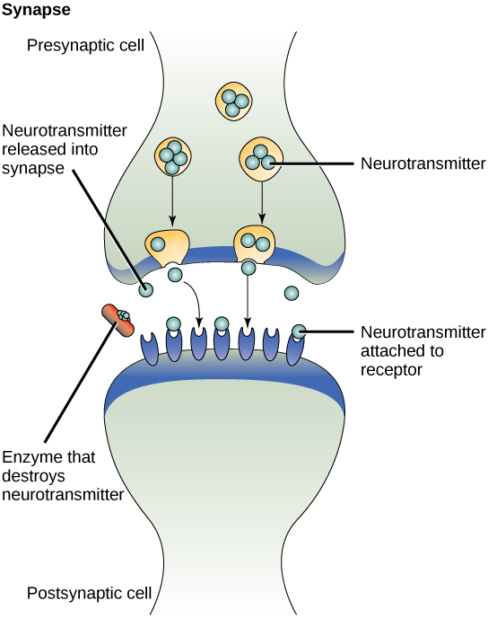
This illustration shows closely juxtaposed bulbous protrusions of presynaptic and postsynaptic cells. The presynaptic cell stores neurotransmitter in synaptic vesicles. When signaling occurs, the vesicles fuse with the cell membrane, thereby releasing the neutrotransmitter, which then binds to receptors on the postsynaptic cell. An enzyme on the surface of the postsynaptic cell destroys the neurotrasmitter, thereby terminating the signal.
Figure 9.3 — 9.1 Signaling and Cellular

Figure 9.3 The distance between the presynaptic cell and the postsynaptic cell—called the synaptic gap—is very small and allows for rapid diffusion of the neurotransmitter. Enzymes in the synapatic gap degrade some types of neurotransmitters to terminate the signal.

Mary Ann Clark, Matthew Douglas, and Jung Choi, Biology 2E, OpenStax. · CC BY-NC-SA 4.0 · Original image ↗ · Access for free ↗
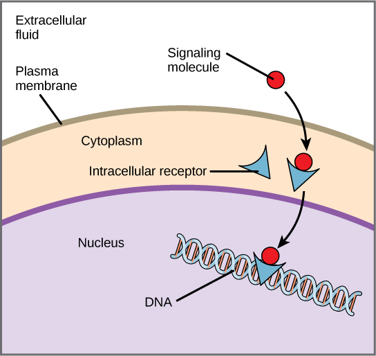
This illustration shows a hydrophobic signaling molecule that diffuses across the plasma membrane and binds an intracellular receptor in the cytoplasm. The intracellular receptor-signaling molecule complex then travels to the nucleus and binds D N A.
Figure 9.4 — 9.1 Signaling and Cellular

Figure 9.4 Hydrophobic signaling molecules typically diffuse across the plasma membrane and interact with intracellular receptors in the cytoplasm. Many intracellular receptors are transcription factors that interact with DNA in the nucleus and regulate gene expression.

Mary Ann Clark, Matthew Douglas, and Jung Choi, Biology 2E, OpenStax. · CC BY-NC-SA 4.0 · Original image ↗ · Access for free ↗
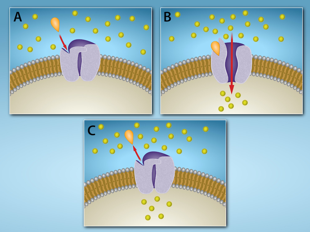
A gated channel is closed within the plasma membrane. A signaling molecule binds to a site on the channel protein, and the channel opens. When the signaling molecule is no longer bound, the channel closes.
Figure 9.5 — 9.1 Signaling and Cellular

Figure 9.5 Gated ion channels located in the plasma membrane allow for the controlled flow of ions into and out of the cell. The channel proteins remain closed until a signaling molecule (orange teardrop) binds to the channel protein. Then the channel protein changes conformation and allows ions (yellow circles) to flow into (or out of) the cell. When the signaling molecule is released, the channel protein resumes its closed conformation, preventing ion flow. Credit: Rao, A. and Fletcher, S. Department of Biology, Texas A&M University.

Mary Ann Clark, Matthew Douglas, and Jung Choi, Biology 2E, OpenStax. · CC BY-NC-SA 4.0 · Original image ↗ · Access for free ↗
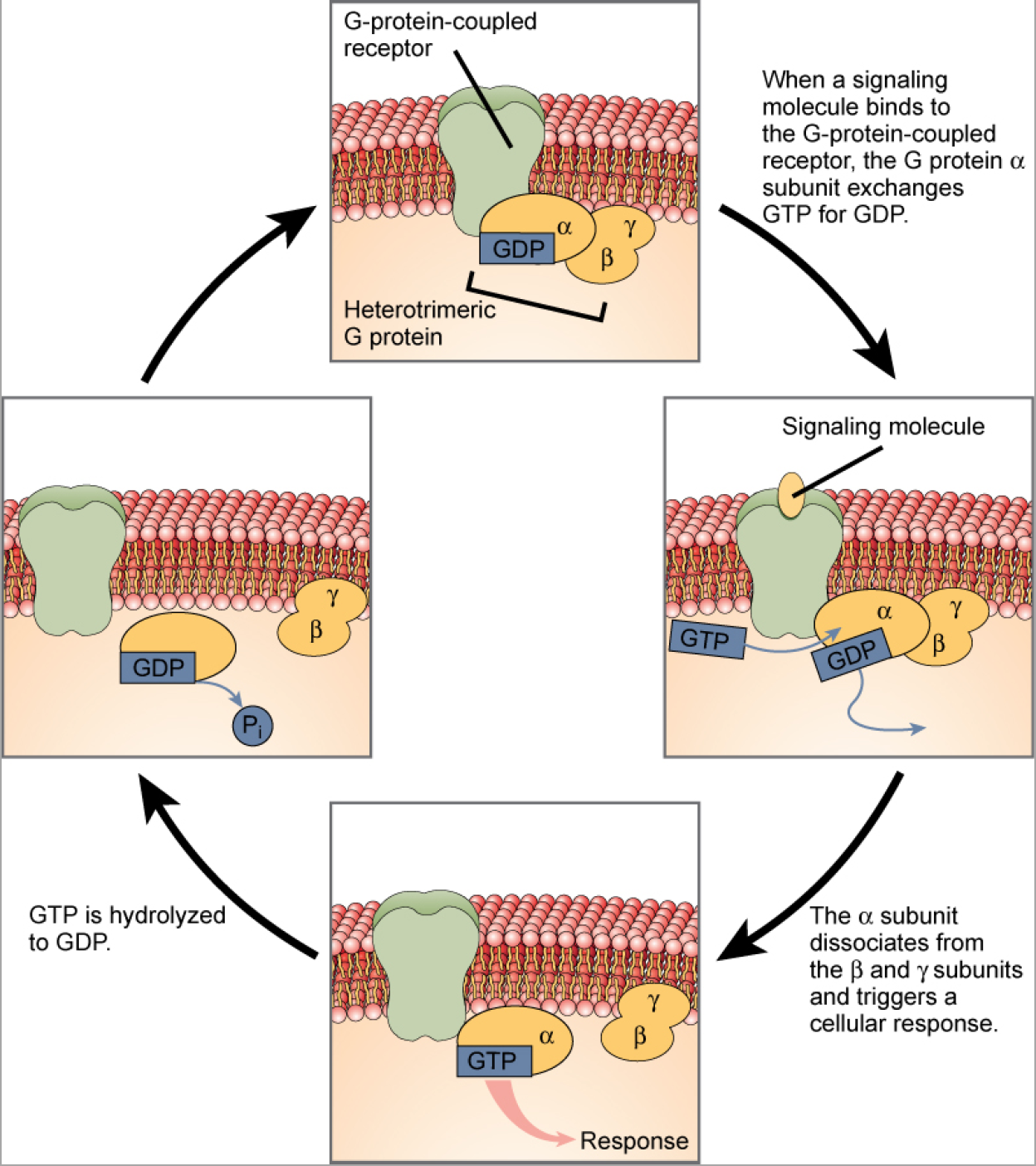
This illustration shows the activation pathway for a heterotrimeric G protein, which has three subunits: alpha, beta, and gamma, all associated with the inside of the plasma membrane. When a signaling molecule binds to a G protein-coupled receptor in the plasma membrane, a G D P molecule associated with the alpha subunit is exchanged for G T P. The alpha subunit dissociates from the beta and gamma subunits and triggers a cellular response. Hydrolysis of G T P to G D P terminates the signal.
Figure 9.6 — 9.1 Signaling and Cellular

Figure 9.6 Heterotrimeric G-proteins have three subunits: α , β , and γ . When a signaling molecule binds to a G-protein-coupled receptor in the plasma membrane, a GDP molecule associated with the α subunit is exchanged for GTP. The β and γ subunits dissociate from the α subunit, and a cellular response is triggered either by the α subunit or the dissociated βγ pair. Hydrolysis of GTP to GDP terminates the signal.

Mary Ann Clark, Matthew Douglas, and Jung Choi, Biology 2E, OpenStax. · CC BY-NC-SA 4.0 · Original image ↗ · Access for free ↗
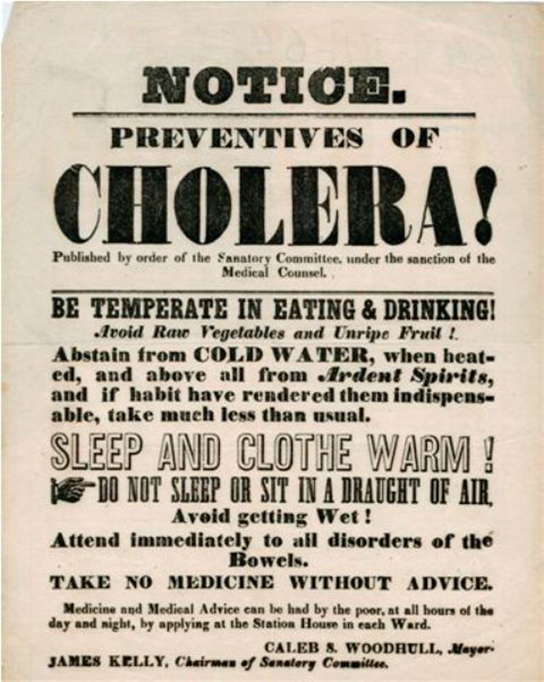
This 1866 poster warns people about a cholera epidemic and gives advice for preventing the disease.
Figure 9.7 — 9.1 Signaling and Cellular

Figure 9.7 Transmitted primarily through contaminated drinking water, cholera is a major cause of death in the developing world and in areas where natural disasters interrupt the availability of clean water. The cholera bacterium, Vibrio cholerae , creates a toxin that modifies G-protein-mediated cell signaling pathways in the intestines. Modern sanitation eliminates the threat of cholera outbreaks, such as the one that swept through New York City in 1866. This poster from that era shows how, at that time, the way that the disease was transmitted was not understood. (credit: New York City Sanitary Commission)

Mary Ann Clark, Matthew Douglas, and Jung Choi, Biology 2E, OpenStax. (credit: New York City Sanitary Commission) · CC BY-NC-SA 4.0 · Original image ↗ · Access for free ↗
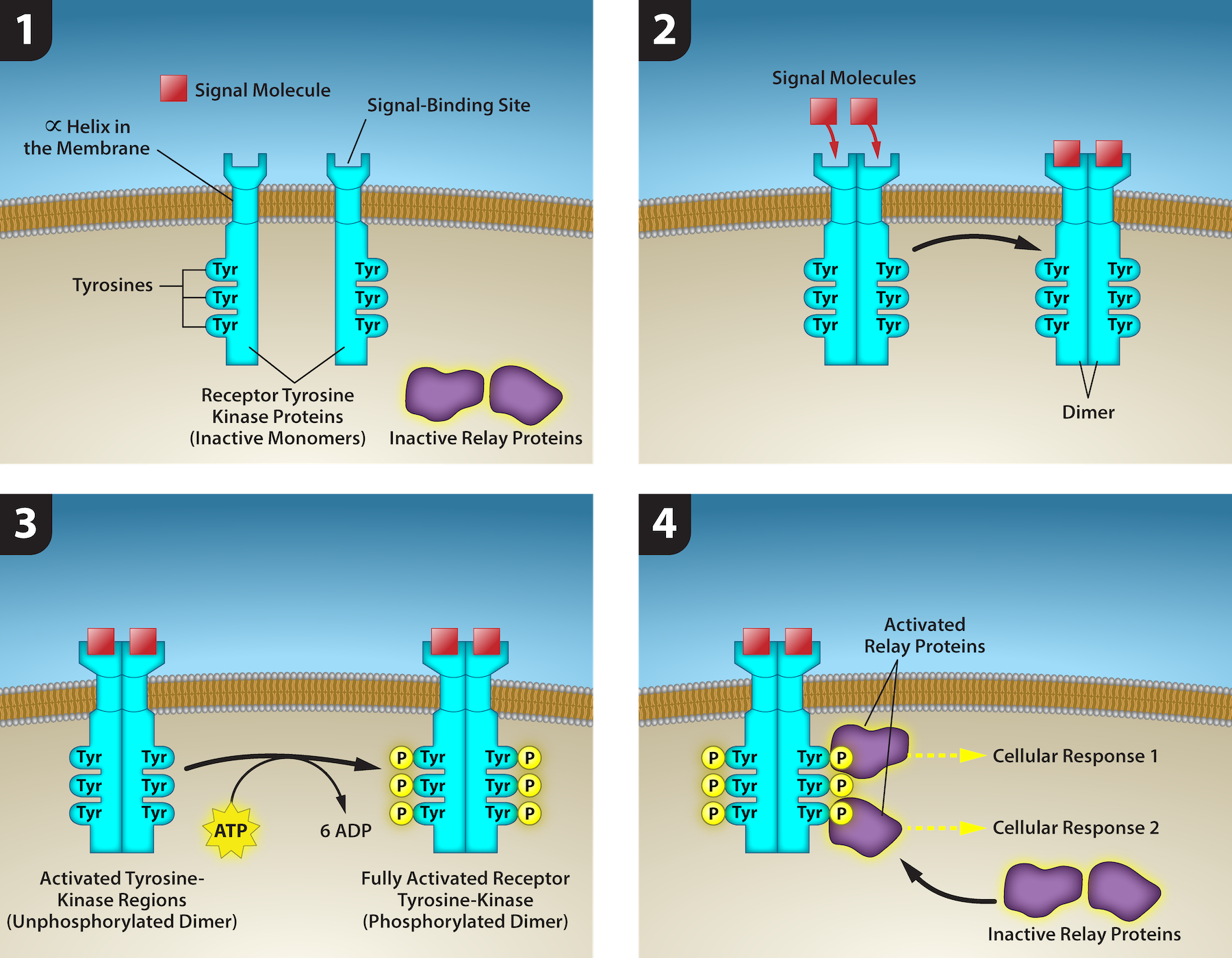
This illustration shows two receptor tyrosine kinase monomers embedded in the plasma membrane. Upon binding of a signaling molecule to the extracellular domain, the receptors dimerize. Tyrosine residues on the intracellular surface are then phosphorylated, triggering a cellular response. The cellular response involves binding of relay proteins, which result in Cellular Response 1 and Cellular Response 2.
Figure 9.8 — 9.1 Signaling and Cellular

Figure 9.8 1) A receptor tyrosine kinase is an enzyme-linked receptor with a single helical transmembrane region, and extracellular and intracellular domains. 2) Binding of a signaling molecule to the extracellular domain causes the receptor to dimerize. 3) Tyrosine residues on the intracellular domain are then autophosphorylated, 4) triggering a downstream cellular response. The signal is terminated by a phosphatase that removes the phosphates from the phosphotyrosine residues. Credit: Rao, A., Ryan, K., Tag, A., Fletcher, S. and Hawkins, A. Department of Biology, Texas A&M University.

Mary Ann Clark, Matthew Douglas, and Jung Choi, Biology 2E, OpenStax. · CC BY-NC-SA 4.0 · Original image ↗ · Access for free ↗
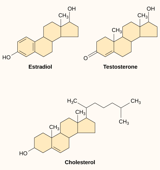
The molecular structures of estradiol, testosterone, and cholesterol are shown. All three molecules share a four-ring structure but differ in the types of functional groups attached to it.
Figure 9.9 — 9.1 Signaling and Cellular

Figure 9.9 Steroid hormones have similar chemical structures to their precursor, cholesterol. Because these molecules are small and hydrophobic, they can diffuse directly across the plasma membrane into the cell, where they interact with internal receptors.

Mary Ann Clark, Matthew Douglas, and Jung Choi, Biology 2E, OpenStax. · CC BY-NC-SA 4.0 · Original image ↗ · Access for free ↗

Source: 9.1 Signaling Molecules and Cellular Receptors

9.2 Propagation of the Signal — figures and tables
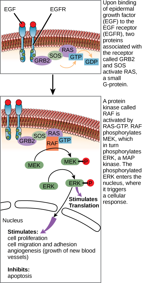
This illustration shows the epidermal growth factor receptor, which is embedded in the plasma membrane. Upon binding of a signaling molecule to the receptor's extracellular domain, the receptor dimerizes, and intracellular residues are phosphorylated. Phosphorylation of the receptor triggers the phosphorylation of a protein called M E K by R A F. M E K, in turn, phosphorylates E R K. E R K stimulates protein translation in the cytoplasm, and transcription in the nucleus. Activation of E R K stimulates cell proliferation, cell migration and adhesion, and angiogenesis, which is the growth of new blood vessels. E R K inhibits apoptosis.
Figure 9.10 — 9.2 Propagation of the Signal

Figure 9.10 The epidermal growth factor (EGF) receptor (EGFR) is a receptor tyrosine kinase involved in the regulation of cell growth, wound healing, and tissue repair. When EGF binds to the EGFR, a cascade of downstream events causes the cell to grow and divide. If EGFR is activated at inappropriate times, uncontrolled cell growth (cancer) may occur.

Mary Ann Clark, Matthew Douglas, and Jung Choi, Biology 2E, OpenStax. · CC BY-NC-SA 4.0 · Original image ↗ · Access for free ↗
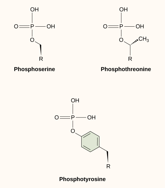
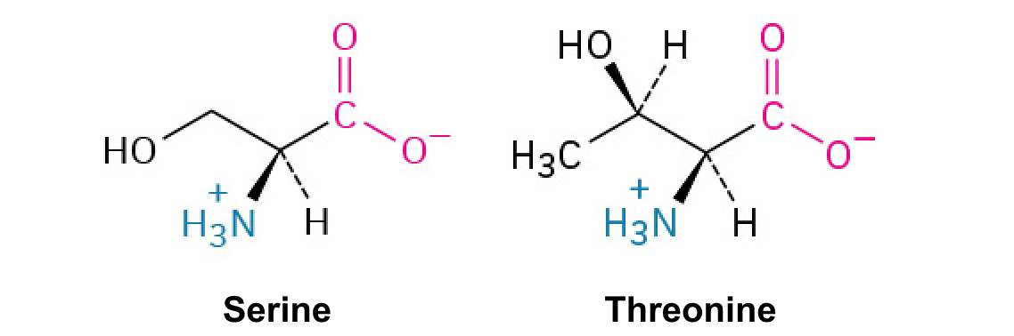
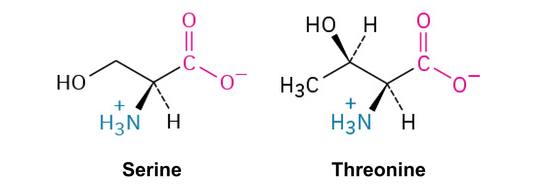
Molecular structures of phosphoserine, phosphothreonine and phosphotyrosine are shown. In each molecule, a phosphate is attached to an oxygen on the amino acid.
Figure 9.11 — 9.2 Propagation of the Signal

Figure 9.11 In protein phosphorylation, a phosphate group (PO 4 -3 ) is added to residues of the amino acids serine, threonine, and tyrosine.

Mary Ann Clark, Matthew Douglas, and Jung Choi, Biology 2E, OpenStax. · CC BY-NC-SA 4.0 · Original image ↗ · Access for free ↗
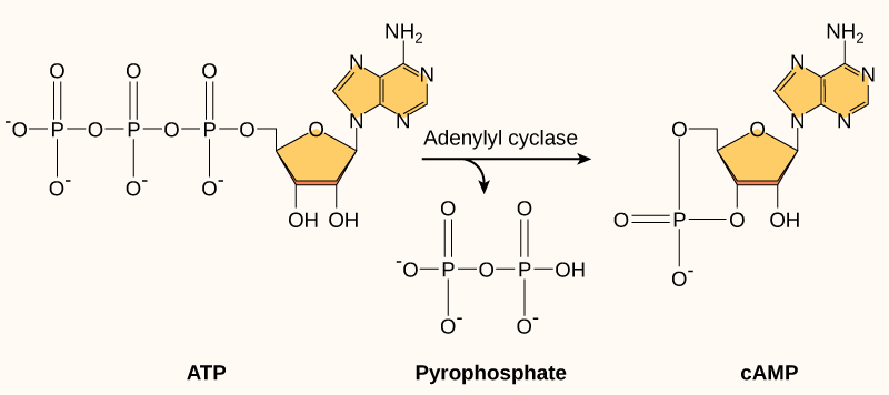
Cyclic A M P is made from A T P by the enzyme adenylyl cyclase. In the process, a pyrophosphate molecule composed of two phosphate residues is released. Cyclic A M P gets its name because the phosphate group is attached to the ribose ring in two places, forming a circle.
Figure 9.12 — 9.2 Propagation of the Signal

Figure 9.12 This diagram shows the mechanism for the formation of cyclic AMP (cAMP). cAMP serves as a second messenger to activate or inactivate proteins within the cell. Termination of the signal occurs when an enzyme called phosphodiesterase converts cAMP into AMP.

Mary Ann Clark, Matthew Douglas, and Jung Choi, Biology 2E, OpenStax. · CC BY-NC-SA 4.0 · Original image ↗ · Access for free ↗
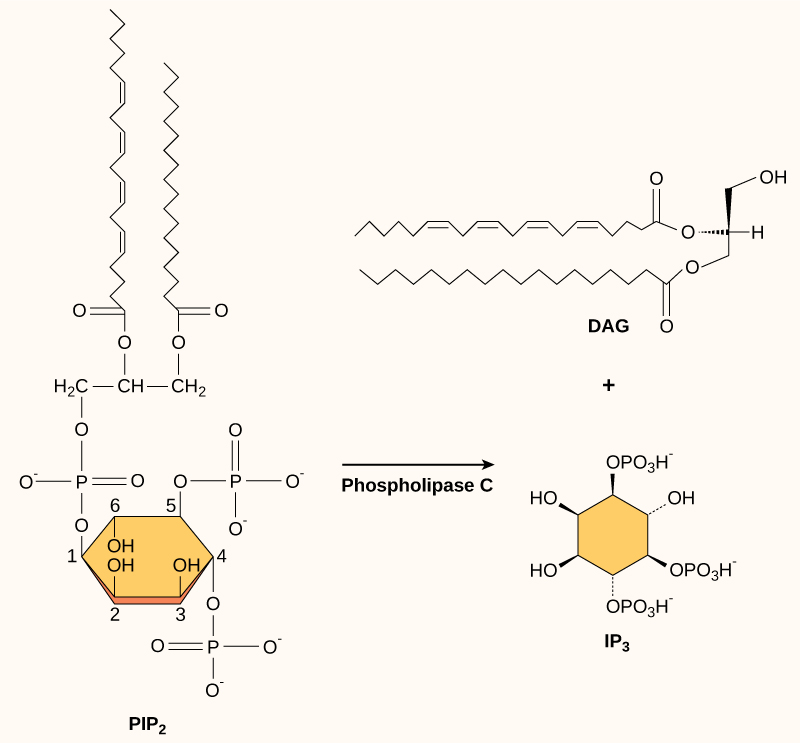
The molecular structures of P I P subscript 2 baseline, D A G, and I P subscript 3 baseline are shown. P I P subscript 2 baseline is a phospholipid that is cleaved by phospholipase C to form D A G, which has a long hydrophobic tail, and I P subscript 3 baseline, a ring structure with three phosphates attached.
Figure 9.13 — 9.2 Propagation of the Signal

Figure 9.13 The enzyme phospholipase C breaks down PIP 2 into IP 3 and DAG, both of which serve as second messengers.

Mary Ann Clark, Matthew Douglas, and Jung Choi, Biology 2E, OpenStax. · CC BY-NC-SA 4.0 · Original image ↗ · Access for free ↗

Source: 9.2 Propagation of the Signal

311 terms, alphabetically ordered

3′ end · ·

The end of a nucleic- strand associated with the sugar’s 3′ position. extends a strand at a free .

Read explanation · OpenStax ↗
3′ hydroxyl group · · ·

The at the sugar’s 3′ carbon that attacks the incoming ’s during strand extension.

Read explanation · OpenStax ↗
A site · ·

The site that accepts most incoming during elongation and at termination.

Read explanation · OpenStax ↗
Acid · ·

In the Brønsted–Lowry model, a substance that can donate a . Acidic have a higher activity than water.

Read explanation · OpenStax ↗
action potential

A regenerative -voltage change produced by voltage-dependent -channel activity.

Read explanation · OpenStax ↗
Activation energy ·

The energy barrier that must overcome to reach a and undergo .

Read explanation · OpenStax ↗
Active catalytic-site concentration ·

The total of active sites used to calculate from . It equals active when each has one site.

Read explanation · Biology LibreTexts ↗
Active site

The region of an that binds its and carries out .

Read explanation · OpenStax ↗
adaptation

A heritable feature shaped by selection because it improves reproductive success in a relevant environment.

Read explanation · OpenStax ↗
Adenine

A that pairs with in and in .

Read explanation · OpenStax ↗
Adenosine triphosphate

An energy-carrying ; its powers many cellular processes, including cycles.

Read explanation · OpenStax ↗
adenylyl cyclase

An converting to in cell signaling.

Read explanation · OpenStax ↗
ADH ·

Antidiuretic hormone, or vasopressin, synthesized in hypothalamic and released from the posterior pituitary to promote renal water conservation.

Read explanation · OpenStax ↗
Albumin

A family of ; serum albumin transports in blood and helps maintain plasma .

Read explanation · OpenStax ↗
Allolactose

A isomer produced by that binds and relieves lac repression.

Read explanation · OpenStax ↗
Alpha carbon · ·

The central carbon next to the carboxyl carbon. In a standard it binds an , hydrogen, and .

Read explanation · OpenStax ↗
Alpha helix · · · · · ·

A helical stabilized by between a oxygen and an hydrogen four later.

Read explanation · OpenStax ↗
Alternative splicing ·

Regulated or context-dependent use of different or combinations to generate different mature from one precursor.

Read explanation · OpenStax ↗
Amide · · ·

A group containing a carbon bonded to nitrogen. are amide linkages.

Read explanation · OpenStax ↗
Amino acid

A building block containing amino and and a characteristic .

Read explanation · OpenStax ↗
Amino acid residue · ·

The part of an incorporated into a chain. Residue can also describe another building block within a larger .

Read explanation · OpenStax ↗
Amino group · · ·

A nitrogen-containing . A free ’s amino group is typically (NH₃⁺) near physiological ; ’s ring nitrogen is an exception to this formula.

Read explanation · OpenStax ↗
Aminoacyl-tRNA · ·

A covalently linked to an at its , ready to deliver that during .

Read explanation · OpenStax ↗
Amphipathic

Having both a region and a region, as in a or .

Read explanation · OpenStax ↗
Amylose

The largely unbranched component of , made mainly of linked by alpha-1,4 .

Read explanation · OpenStax ↗
Analyte

A substance being measured or separated within a sample; the source table uses the term for the mixture under analysis.

Read explanation · Waters ↗
Anion ·

An with a negative net .

Read explanation · OpenStax ↗
anomeric carbon

The former carbon of a sugar, which becomes the or hemiketal center upon cyclization.

Read explanation · OpenStax ↗
Anticodon

A triplet that pairs to an . The anticodon for 5′-AUG-3′ is aligned 3′-UAC-5′, or written 5′-CAU-3′.

Read explanation · NCBI Bookshelf / Garland Science ↗
Antiparallel

Running in opposite directions, as with two strands or neighboring strands in some .

Read explanation · OpenStax ↗
antiporter

A transporter moving coupled in opposite directions.

Read explanation · OpenStax ↗
Apoptosis

A regulated process of cell death that removes cells in development, tissue maintenance, and responses to damage.

Read explanation · OpenStax ↗
aquaporin

A channel facilitating water movement.

Read explanation · OpenStax ↗
Aqueous solution ·

A in which water is the .

Read explanation · OpenStax ↗
Archaea ·

A of distinct from , with characteristic chemistry and -expression machinery.

Read explanation · OpenStax ↗
Aromatic

Containing a cyclic, with . , , and have aromatic .

Read explanation · OpenStax ↗
Atom

The smallest unit of an element that retains its chemical identity; it has a surrounded by .

Read explanation · OpenStax ↗
ATP synthase

A rotary that commonly uses to synthesize from ADP and .

Read explanation · OpenStax ↗
axon

Neuronal process that usually conducts toward terminals.

Read explanation · OpenStax ↗
Bacteria ·

A of single-celled . Bacterial cells lack a -bound and are distinct from .

Read explanation · OpenStax ↗
bacteriophage · ·

A virus that infects .

Read explanation · OpenStax ↗
Benzene ring

A six-carbon ring with .

Read explanation · OpenStax ↗
Benzyl group

A attached through a –CH₂– group; part of ’s .

Read explanation · OpenStax ↗
Beta sheet · · · · · ·

A made of aligned segments connected by ; strands may be parallel or .

Read explanation · OpenStax ↗
Binding affinity ·

The strength of a binding interaction between . For simple binding, a smaller means higher affinity.

Read explanation · OpenStax ↗
Binding site

A region of a where another interacts selectively.

Read explanation · OpenStax ↗
Biomolecule

A made by or involved in living systems, including , , , and .

Read explanation · OpenStax ↗
Blotting ·

Transfer of separated or to a so a specific target can be detected.

Read explanation · Thermo Fisher ↗
Brønsted–Lowry base · · · · · · · · · · ·

In the Brønsted–Lowry model, a substance that can accept a . Basic solutions have lower activity than water.

Read explanation · OpenStax ↗
Cancer

A group of diseases involving abnormal cells that can proliferate without normal restraints and invade tissues.

Read explanation · OpenStax ↗
Carbohydrate

A class of including sugars and their , used for energy storage, structure, and recognition.

Read explanation · OpenStax ↗
Carbonyl group · ·

A carbon double-bonded to oxygen, C=O; present in and many other .

Read explanation · OpenStax ↗
Carboxyl group · ·

An , COOH. In free it is usually to COO⁻ near physiological .

Read explanation · OpenStax ↗
Catalyst · ·

A substance that speeds a by providing a pathway with lower and is regenerated rather than consumed overall.

Read explanation · OpenStax ↗
Cation ·

An with a positive net .

Read explanation · OpenStax ↗
Cellulose

A structural in plant cell walls, with beta-1,4 linkages; also used as a support.

Read explanation · OpenStax ↗
Chemical compound ·

A substance containing of two or more different chemically combined in a definite composition.

Read explanation · OpenStax ↗
Chemical element

A substance defined by having the same number of , such as carbon or oxygen.

Read explanation · OpenStax ↗
Chemical reaction ·

A process that rearranges and chemical bonds, converting into .

Read explanation · OpenStax ↗
cholesterol

An animal that modulates packing and serves as a precursor for and bile .

Read explanation · OpenStax ↗
Chromatography

Separation of mixture components by their different interactions with a and a moving .

Read explanation · Waters ↗
Chromatography resin ·

The solid or beads in a column, often carrying chemical groups that determine separation behavior.

Read explanation · Cytiva ↗
Chromosome ·

An organized with associated that carries . A replicated chromosome has two .

Read explanation · OpenStax ↗
Codon

A sequence of three that specifies an or a stop signal.

Read explanation · OpenStax ↗
Concentration

The amount of a substance in a specified volume or mass of mixture.

Read explanation · OpenStax ↗
Configuration

A stereochemical arrangement that cannot be changed into another configuration by ordinary single-bond rotations alone.

Read explanation · OpenStax ↗
Conformation ·

A spatial arrangement accessible by rotations and other motions that do not require breaking .

Read explanation · NCBI Bookshelf ↗
Conjugated system ·

Adjacent orbitals that allow to be distributed across several , often through alternating single and multiple bonds.

Read explanation · OpenStax ↗
Cooperativity · ·

An interaction in which binding at one site changes binding at other sites. Positive cooperativity can produce a response.

Read explanation · Biology LibreTexts ↗
Covalent bond · ·

A bond in which share pairs. and are covalent.

Read explanation · OpenStax ↗
Cyclic AMP ·

A signal. In the simplified E. coli lac model, low raises cAMP, which activates CAP.

Read explanation · OpenStax ↗
cyclic nucleotide

whose forms a cyclic within its sugar, such as .

Read explanation · OpenStax ↗
Cysteine ·

An with a . Two cysteines can form a .

Read explanation · OpenStax ↗
Cytoplasm

Cell contents inside the plasma and outside the , where one is present.

Read explanation · OpenStax ↗
Cytosine

A in and that pairs with . can convert it to .

Read explanation · OpenStax ↗
Cytoskeleton

A network of filaments that supports cell shape, organizes components, and enables movement.

Read explanation · OpenStax ↗
Cytosol

The aqueous part of the outside -enclosed .

Read explanation · OpenStax ↗
DAG

Diacylglycerol, a -associated messenger that helps activate .

Read explanation · OpenStax ↗
Dalton

A unit of mass equal to one twelfth of the mass of a carbon-12 ; used to describe .

Read explanation · OpenStax ↗
Deamination

Removal of an . Deamination of produces and can change genetic information if unrepaired.

Read explanation · Biology LibreTexts / Oregon State University ↗
dendrite

Branching neuronal process receiving many synaptic inputs.

Read explanation · OpenStax ↗
Deoxyribonucleic acid

The that stores genetic information used to specify and sequences.

Read explanation · OpenStax ↗
Deprotonation

Removal of a from a or group; this changes its by minus one.

Read explanation · OpenStax ↗
Detergent

An substance that can associate with both water and material; detergents can disrupt or interactions.

Read explanation · Thermo Fisher ↗
differentiation

Acquisition of specialized cellular structures and functions through regulated and other changes.

Read explanation · OpenStax ↗
Diffusion

Net spreading of particles down a due to random molecular motion.

Read explanation · OpenStax ↗
Dissociation · ·

Separation of a chemical species or molecular complex into smaller components; dissociation releases a .

Read explanation · OpenStax ↗
Dissociation constant · ·

An for complex . For E + S ⇌ , Kd = [E]/[]; a smaller value indicates tighter binding under the stated conditions.

Read explanation · IUBMB ↗
Disulfide bond · · ·

A covalent sulfur–sulfur bond formed by of two , which can stabilize structure.

Read explanation · NCBI Bookshelf ↗
DNA polymerase ·

An that synthesizes 5′ to 3′ using a template and a with a free .

Read explanation · OpenStax ↗
DNA replication

Copying so genetic information can be passed to daughter cells. Each guides a complementary new strand.

Read explanation · OpenStax ↗
Downstream

Later along a directed sequence or process; in , toward positions following a reference site in the direction of synthesis.

Read explanation · OpenStax ↗
dynamic equilibrium

State in which forward and reverse processes continue at equal rates with no net macroscopic change.

Read explanation · OpenStax ↗
Effector

A that changes a ’s activity or binding behavior, often by stabilizing a particular .

Read explanation · Biology LibreTexts ↗
Electric charge · · ·

An electrical property that can be positive or negative. Opposite charges attract and like charges repel; net charge is the sum of all charges on a particle.

Read explanation · OpenStax ↗
electrochemical gradient

The combined and electrical driving forces acting on an across space or a .

Read explanation · OpenStax ↗
Electron

A negatively subatomic particle. Electron sharing and transfer determine much of chemical bonding.

Read explanation · OpenStax ↗
Electron delocalization ·

Distribution of across several rather than confinement to one or bond.

Read explanation · OpenStax ↗
Electronegativity ·

An ’s tendency to attract shared in a chemical bond. Unequal attraction can make a bond .

Read explanation · OpenStax ↗
Endocrine signaling ·

Communication in which enter the circulation and act on distant target cells.

Read explanation · OpenStax ↗
endocytosis

Uptake of extracellular material or surface through inward budding of .

Read explanation · OpenStax ↗
Endomembrane system

The interacting and compartments involved in processes such as modification, transport, and .

Read explanation · OpenStax ↗
Endoplasmic reticulum

An interconnected network continuous with the ; it includes regions involved in processing and synthesis.

Read explanation · OpenStax ↗
endosome

A compartment sorting endocytosed material for recycling or further transport.

Read explanation · OpenStax ↗
Enzyme

A biological that speeds a without being consumed. Most enzymes are .

Read explanation · OpenStax ↗
Enzyme saturation · ·

The high- limit in which sites spend almost all their time processing and the rate approaches .

Read explanation · Biology LibreTexts ↗
Enzyme–substrate complex · · ·

The bound association of an and its during a cycle. Often abbreviated ES in schemes.

Read explanation · OpenStax ↗
Equilibrium

A state where opposing processes occur at equal rates, so macroscopic or other properties remain constant.

Read explanation · OpenStax ↗
Equilibrium constant

A ratio of to activities at , each raised to its stoichiometric coefficient. At a fixed temperature, a does not change this ratio.

Read explanation · OpenStax ↗
estradiol

A major estrogen produced by ovarian follicles and other tissues, influencing reproductive tissues and hormonal feedback.

Read explanation · OpenStax ↗
Eukaryote ·

An organism whose cells have a -enclosed ; animals, plants, fungi, and protists are eukaryotes.

Read explanation · OpenStax ↗
exocytosis

Fusion of an intracellular with the plasma , releasing contents and adding components.

Read explanation · OpenStax ↗
Exon

A segment retained in a mature spliced . In an exon may contain coding sequence, untranslated sequence, or both.

Read explanation · OpenStax ↗
Experimental control ·

A comparison sample or condition used to distinguish the effect being tested from other influences.

Read explanation · Thermo Fisher ↗
extracellular matrix

The network of and surrounding cells and providing structural and signaling functions.

Read explanation · OpenStax ↗
facilitated diffusion

Passive movement down an through a selective channel or .

Read explanation · OpenStax ↗
Fatty acid

A attached to a chain; fatty acids are components of many .

Read explanation · OpenStax ↗
First-order kinetics

A rate dependence proportional to the first power of a . At low , a is approximately first-order in .

Read explanation · Biology LibreTexts ↗
forebrain

Major developmental brain division including cerebral hemispheres, , and .

Read explanation · OpenStax ↗
Functional group

A group of whose chemical behavior gives a characteristic and properties.

Read explanation · OpenStax ↗
G-protein-coupled receptor

A that relays binding through a heterotrimeric G .

Read explanation · OpenStax ↗
Galactose

A six-carbon monosaccharide released along with when is hydrolyzed.

Read explanation · OpenStax ↗
gap junction

A junction containing intercellular channels for and small .

Read explanation · OpenStax ↗
Gene

A sequence that contributes to a functional or , together with the information needed to express it.

Read explanation · OpenStax ↗
Gene expression

Production of a functional from a , including and, for -coding , .

Read explanation · NCBI Bookshelf ↗
Genome

The complete genetic material of an organism or cell.

Read explanation · OpenStax ↗
Glucagon

A pancreatic that helps raise , especially by promoting release from the liver.

Read explanation · OpenStax ↗
Glucose

A six-carbon sugar that is a major cellular energy source and a building block of , , and .

Read explanation · OpenStax ↗
glycogen

A highly branched used for storage in animals.

Read explanation · Biology LibreTexts / Oregon State University ↗
Glycosidic bond

A involving a sugar’s . In a , an N-glycosidic bond links the sugar to the .

Read explanation · Biology LibreTexts / Oregon State University ↗
Golgi apparatus ·

A stack of compartments that modifies and sorts and traveling through the .

Read explanation · OpenStax ↗
Gradient

A change in a quantity, such as , , or density, across a distance or over time.

Read explanation · Thermo Fisher ↗
Guanine

A that pairs with through three .

Read explanation · OpenStax ↗
Guanosine diphosphate

A with two phosphates, produced when is hydrolyzed by many signaling and factors.

Read explanation · The International Journal of Biochemistry & Cell Biology / PubMed ↗
Guanosine triphosphate

A -containing whose supports factor-driven steps in and other cellular processes.

Read explanation · OpenStax ↗
Half-life

The time required for half of a population or amount to remain during decay; a constant half-life is characteristic of decay.

Read explanation · OpenStax ↗
hemiacetal

Carbon bonded to both and alkoxy groups, usually formed by alcohol addition to a .

Read explanation · OpenStax ↗
Hemoglobin

The oxygen-carrying in , composed of four with heme groups.

Read explanation · OpenStax ↗
HER2 receptor ·

A growth-signaling encoded by ERBB2; amplification of its can cause excess production in some cancers.

Read explanation · NHGRI / NIH ↗
homeostasis

Dynamic regulation that maintains physiological variables within functional ranges despite disturbances.

Read explanation · OpenStax ↗
Hormone

A chemical messenger released by cells that changes the activity of target cells with matching .

Read explanation · OpenStax ↗
Hydrocarbon · ·

A made only of carbon and hydrogen. Hydrocarbon regions are generally .

Read explanation · OpenStax ↗
Hydrogen bond ·

An attraction between a hydrogen bonded to an and another . hydrogen bonds stabilize .

Read explanation · OpenStax ↗
Hydrogen ion · ·

A positively hydrogen species. H⁺ denotes a , which is hydrated in water; its activity determines .

Read explanation · OpenStax ↗
Hydrolysis

Breaking a chemical bond by adding water. hydrolysis separates .

Read explanation · OpenStax ↗
Hydrophilic

Interacting favorably with water, typically because a group is or .

Read explanation · OpenStax ↗
Hydrophobic

Interacting poorly with water. often gather in a ’s interior.

Read explanation · OpenStax ↗
hydrostatic pressure

Pressure in a fluid at rest, increasing with depth under gravity.

Read explanation · OpenStax ↗
Hydroxide ion ·

The negatively OH⁻. Basic contain a higher hydroxide ion activity than water at the same temperature.

Read explanation · OpenStax ↗
Hydroxyl group ·

An oxygen bonded to hydrogen, –OH. Hydroxyl groups can and often increase .

Read explanation · OpenStax ↗
Hyperbolic curve ·

A curve that approaches an asymptote. The standard rate curve is a rectangular hyperbola approaching .

Read explanation · Biology LibreTexts ↗
hypertonic

Describing a that tends to cause cell shrinkage because of its higher effective nonpenetrating- .

Read explanation · OpenStax ↗
hypothalamus

region coordinating homeostatic, , and autonomic functions.

Read explanation · OpenStax ↗
hypotonic

Describing a that tends to cause cell swelling because of its lower effective nonpenetrating- .

Read explanation · OpenStax ↗
Indole

A fused six-membered and five-membered ring system containing nitrogen; found in .

Read explanation · OpenStax ↗
Inducer

A signal that increases expression of a regulated ; does so by disabling the .

Read explanation · OpenStax ↗
Initial reaction rate · · · ·

The early rate of formation, measured before substantial depletion or accumulation, usually after the brief binding transient.

Read explanation · Biology LibreTexts ↗
Insulin

A from that lowers by promoting uptake and storage and reducing production.

Read explanation · OpenStax ↗
Intron ·

A segment removed from a precursor during . Intronic signals or regulatory sequences can affect .

Read explanation · OpenStax ↗
Ion · ·

An or with a net because its numbers of and do not balance.

Read explanation · OpenStax ↗
IP3

Inositol 1,4,5-trisphosphate, a messenger that promotes calcium release from stores.

Read explanation · OpenStax ↗
iso-osmotic

Having equal total osmotic-particle , which need not imply equal .

Read explanation · OpenStax ↗
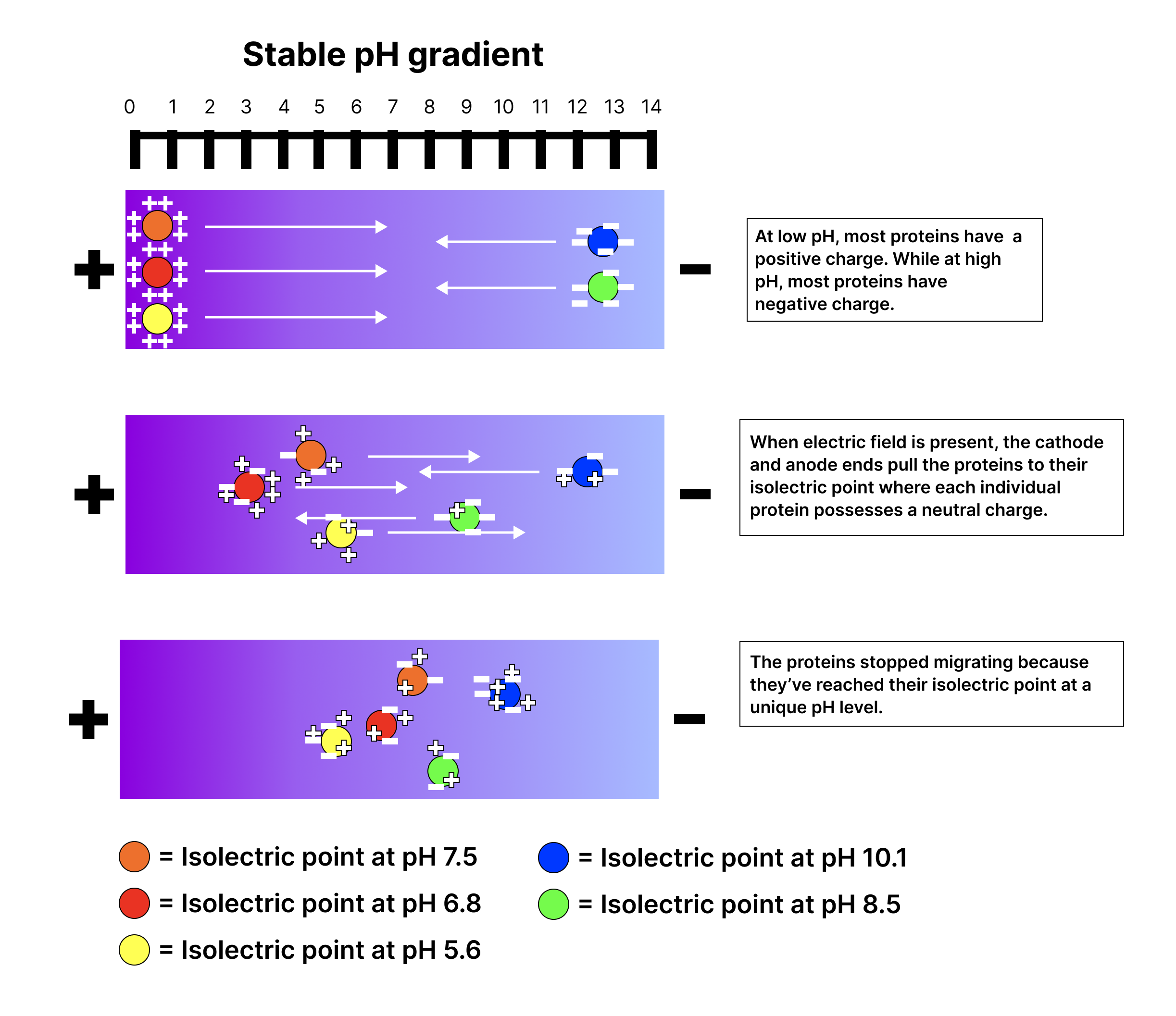
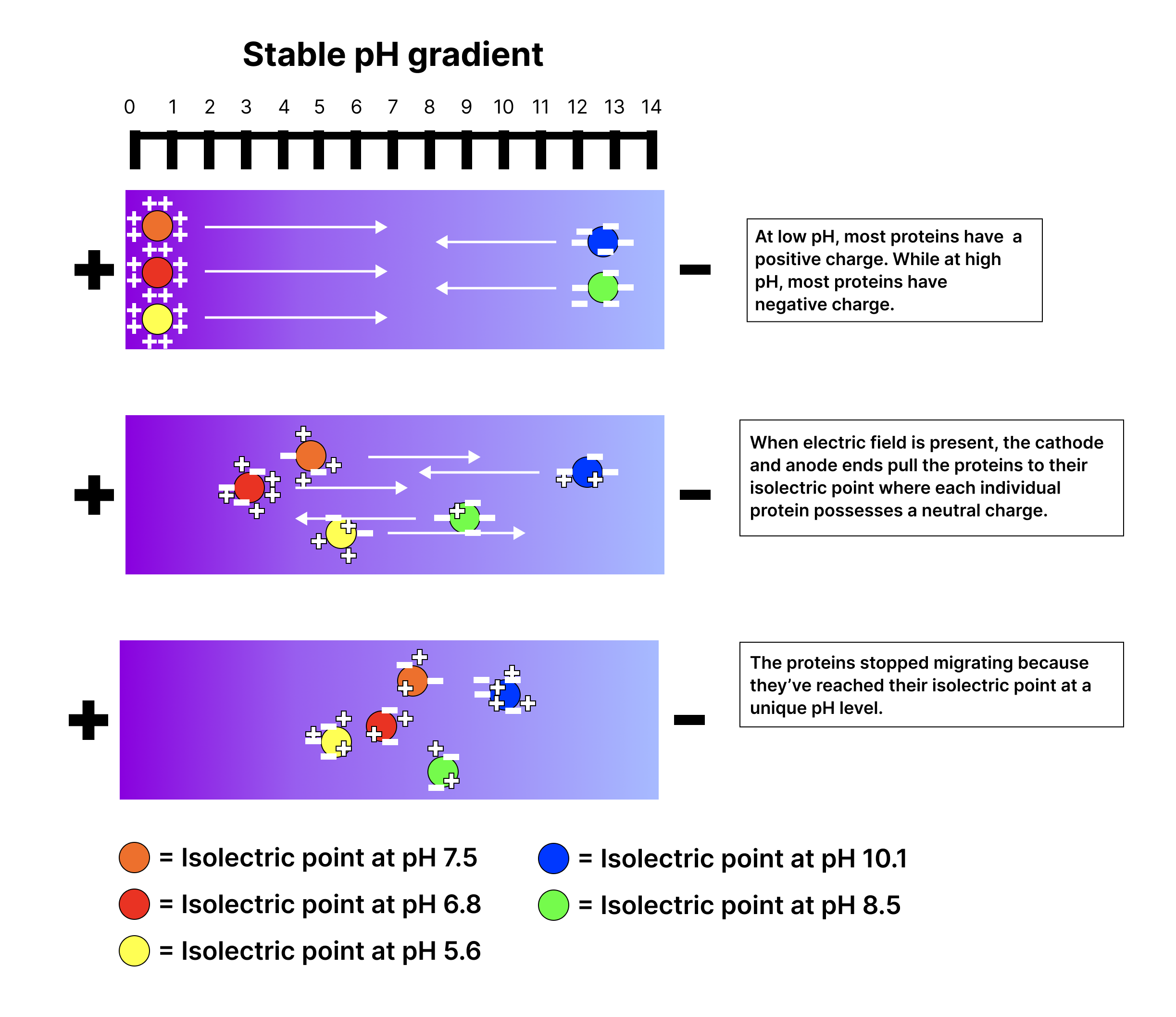
Isoelectric focusing

Separation of in a by their . A stops migrating where it has no net .

Read explanation · Thermo Fisher ↗
Isoelectric point ·

The at which a has zero net . In , a accumulates where local equals its pI.

Read explanation · OpenStax ↗
Isoform

One of several related or forms produced from a , for example through .

Read explanation · OpenStax ↗
Kinase ·

An that transfers a , often from , to a target .

Read explanation · Thermo Fisher ↗
Kinetic energy

Energy associated with motion; higher temperature generally increases average molecular kinetic energy.

Read explanation · OpenStax ↗
Lac operon

The E. coli containing , , and , controlled by repression and CAP– activation.

Read explanation · OpenStax ↗
Lac repressor

The encoded by lacI; it binds lac and represses until an reduces its binding.

Read explanation · OpenStax ↗
lacA

The encoding a galactoside .

Read explanation · OpenStax ↗
Lactose

A disaccharide containing and ; its use by E. coli is supported by .

Read explanation · OpenStax ↗
Lactose permease

The transport that couples uptake to movement.

Read explanation · OpenStax ↗
lacY

The encoding , which transports into the cell.

Read explanation · OpenStax ↗
lacZ

The encoding , often used as an experimental .

Read explanation · OpenStax ↗
leukocyte

A white blood cell involved in defense and immune functions.

Read explanation · OpenStax ↗
Ligand

A or that binds to a particular site on another , such as a or metal .

Read explanation · Cytiva ↗
linkage

A tendency of nearby on the same to be inherited together.

Read explanation · OpenStax ↗
Lipid

A broad class of largely or , including fats, , and steroids.

Read explanation · OpenStax ↗
locus

The specific position of a or other feature.

Read explanation · OpenStax ↗
Logarithm · · ·

The exponent needed to obtain a number from a chosen fixed number. For decimal logarithms, log₁₀(100) = 2 because 10² = 100; uses the negative decimal logarithm of activity.

Read explanation · OpenStax ↗
Lumen · ·

The interior space of an or tube; the ER lumen is enclosed by the .

Read explanation · OpenStax ↗
Lysosome

An -enclosed compartment that uses hydrolytic to digest and recycle cellular material.

Read explanation · OpenStax ↗
Macromolecule

A large , such as a , , or polysaccharide.

Read explanation · OpenStax ↗
Matrix

A supporting material, such as a gel or the solid support in a column. Its properties influence movement and binding of sample .

Read explanation · Thermo Fisher ↗
Maximum reaction rate · ·

The limiting as becomes saturating. For a fixed under given conditions, Vmax = times .

Read explanation · Biology LibreTexts ↗
Membrane · · · ·

A thin boundary or sheet. Cell membranes are bilayers containing ; membranes are synthetic supports that hold or for detection.

Read explanation · OpenStax ↗
Messenger RNA ·

An that carries coding information used by to synthesize a .

Read explanation · OpenStax ↗
Metabolic pathway

A sequence of linked chemical , usually each catalyzed by an .

Read explanation · OpenStax ↗
Metabolism

The chemical that build , break them down, and transform energy in a living system.

Read explanation · OpenStax ↗
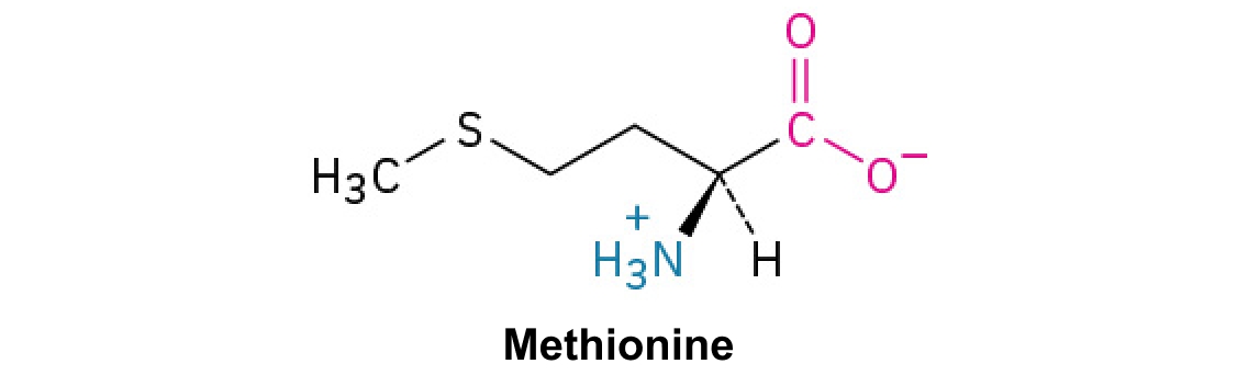
Methionine ·

A with sulfur in a . Unlike , it does not form .

Read explanation · OpenStax ↗
Methyl group ·

A –CH₃ group, consisting of one carbon bonded to three hydrogens.

Read explanation · OpenStax ↗
Michaelis–Menten kinetics · ·

A model with = [S]/(Km + ). Its curve is ; may require a different model.

Read explanation · Biology LibreTexts ↗
Mitochondrion ·

A with its own , central to aerobic production and several other processes.

Read explanation · OpenStax ↗
Mobile phase

The moving fluid that carries a sample through a system.

Read explanation · Waters ↗
Molecular chaperone · ·

A that assists other in folding or prevents , without specifying their sequence.

Read explanation · NCBI Bookshelf ↗
Molecular mass · ·

The mass of a , commonly expressed in for . Molecular weight is often used informally for the same quantity.

Read explanation · OpenStax ↗
Molecule

Two or more joined by . A molecule may be electrically or carry a .

Read explanation · OpenStax ↗
Nernst potential

The voltage at which chemical and electrical forces balance for a particular .

Read explanation · OpenStax ↗
neuron

An electrically excitable cell specialized for receiving and transmitting information.

Read explanation · OpenStax ↗
Neutral

Having no net . For a , neutral instead means equal and activities; its is about 7 at 25 °C.

Read explanation · OpenStax ↗
neutrophil

An abundant innate specialized in rapid antimicrobial responses and .

Read explanation · OpenStax ↗
Nitrogenous base · · ·

A nitrogen-containing ring component of ; its pairing interactions help specify sequences.

Read explanation · OpenStax ↗
Nonpolar ·

Lacking a substantial separation of . Nonpolar groups generally interact poorly with water.

Read explanation · OpenStax ↗
Nuclear envelope

The double enclosing the and continuous with the .

Read explanation · OpenStax ↗
Nucleic acid

A of , such as or , used in information storage, expression, and other cellular functions.

Read explanation · OpenStax ↗
Nucleotide

A joined to a sugar and one or more .

Read explanation · OpenStax ↗
Nucleus ·

A -enclosed compartment containing most of a cell’s .

Read explanation · OpenStax ↗
Operator · ·

A regulatory sequence bound by a , often overlapping or near a .

Read explanation · OpenStax ↗
Operon

A group of from a shared into one , with regulatory that coordinates their expression.

Read explanation · OpenStax ↗
Organelle ·

A specialized structure within a cell that carries out particular functions, such as a or .

Read explanation · OpenStax ↗
osmolarity

of osmotically active dissolved particles per liter of .

Read explanation · OpenStax ↗
osmoreceptor

A sensory cell responding to osmotic conditions and helping regulate thirst and .

Read explanation · OpenStax ↗
osmosis

Net water movement across a selectively permeable in response to water chemical-potential differences.

Read explanation · OpenStax ↗
osmotic pressure

The pressure required to prevent net across a under specified conditions.

Read explanation · OpenStax ↗
Oxidation ·

Loss of or an increase in oxidation state. Oxidizing two can form a .

Read explanation · OpenStax ↗
Pancreas

An organ with digestive and functions; its islets contain cells that secrete and .

Read explanation · OpenStax ↗
Pancreatic beta cell ·

An cell in a pancreatic islet that secretes .

Read explanation · OpenStax ↗
Peptide

A chain of joined by ; the term commonly describes chains shorter than a typical .

Read explanation · OpenStax ↗
Peptide bond ·

A covalent between the carboxyl carbon of one and the amino nitrogen of the next.

Read explanation · OpenStax ↗
Peptide hormone

An amino--based chemical messenger released by ; and are examples.

Read explanation · OpenStax ↗
pH

A measure of , defined as the negative of activity. Lower pH means greater ; pH influences .

Read explanation · OpenStax ↗
phagocytosis

Engulfment of large particles or cells into -bound compartments.

Read explanation · OpenStax ↗
Phenylalanine ·

A with an .

Read explanation · OpenStax ↗
phosphatase

An removing a by .

Read explanation · OpenStax ↗
Phosphate group ·

A phosphorus-containing group bonded to oxygens; phosphate groups occur in , , and modified .

Read explanation · OpenStax ↗
phospholipase C

An that cleaves phosphatidylinositol 4,5-bisphosphate to form and .

Read explanation · OpenStax ↗
Phospholipid

An with a -containing region and tails; a major component of cellular .

Read explanation · OpenStax ↗
Phosphorylation

Addition of a , often to , , or in ; can regulate activity.

Read explanation · Thermo Fisher ↗
pinocytosis

Vesicular uptake of extracellular fluid and dissolved material.

Read explanation · OpenStax ↗
PKC

Protein kinase C, a family regulated by signals and, for conventional , calcium.

Read explanation · OpenStax ↗
Polar

Having an uneven distribution of . Polar groups often interact favorably with water.

Read explanation · OpenStax ↗
Polymer

A large made from many linked molecular building blocks.

Read explanation · OpenStax ↗
Polypeptide

A chain of connected by . A functional can contain one or several such chains.

Read explanation · OpenStax ↗
Porous material · · · ·

A material containing small spaces that can admit of suitable size.

Read explanation · Cytiva ↗
Potential energy

Stored energy associated with position or molecular arrangement.

Read explanation · OpenStax ↗
primary active transport

Uphill transport directly coupled to an energy source such as .

Read explanation · OpenStax ↗
Primer · ·

A short nucleic- strand paired with a template that provides a free for polymerase extension.

Read explanation · OpenStax ↗
Prokaryote ·

An organism whose cells lack a -bound ; and are prokaryotes.

Read explanation · OpenStax ↗
Proline ·

An whose loops back to the nitrogen, restricting rotation and often creating bends.

Read explanation · OpenStax ↗
Promoter

A region that recruits machinery and helps specify where and in which direction begins.

Read explanation · OpenStax ↗
Protein

One or more folded chains that perform biological roles such as , transport, signaling, or structural support.

Read explanation · OpenStax ↗
Protein aggregation · ·

Association of into larger clusters, often when exposed surfaces of unfolded stick together.

Read explanation · NCBI Bookshelf ↗
Protein backbone ·

The repeating nitrogen–alpha-carbon– chain of a , excluding its variable .

Read explanation · OpenStax ↗
Protein domain · ·

A region of a that forms a recognizable structural or functional unit.

Read explanation · OpenStax ↗
Proton

A positively subatomic particle. In – chemistry, donating a proton means transferring a .

Read explanation · OpenStax ↗
proton-motive force

The combined chemical and electrical driving force for movement across a .

Read explanation · OpenStax ↗
Protonation

Addition of a to an , , or ; this changes its by plus one.

Read explanation · OpenStax ↗
Purine · ·

A with two fused rings. and are purines.

Read explanation · OpenStax ↗
Pyrimidine · ·

A with one six-membered ring. , , and are pyrimidines.

Read explanation · OpenStax ↗
R group · ·

The variable side chain attached to an ’s . It determines identity, , , and chemical behavior.

Read explanation · OpenStax ↗
Reactant

A starting substance transformed during a .

Read explanation · OpenStax ↗
Reaction intermediate · ·

A species formed in one step and consumed in another. It occupies a local energy minimum between .

Read explanation · OpenStax ↗
Reaction product ·

A substance formed by a .

Read explanation · OpenStax ↗
Reaction rate · ·

How quickly are consumed or form, usually expressed as a change per unit time.

Read explanation · OpenStax ↗
Receptor

A that recognizes a particular signal and helps produce a cellular response.

Read explanation · OpenStax ↗
receptor tyrosine kinase

A that activates - signaling following appropriate stimulation.

Read explanation · OpenStax ↗
Red blood cell · ·

A blood cell that transports oxygen using ; mature human red blood cells lack a .

Read explanation · OpenStax ↗
refractive index

Ratio of vacuum light speed to phase speed in a medium.

Read explanation · OpenStax ↗
Refractive index detector

A detector that measures changes in when pass through it.

Read explanation · Waters ↗
Release factor ·

A that recognizes a stop signal and promotes of the – to release the .

Read explanation · OpenStax ↗
Reporter gene · ·

A with a readily measured used to study regulation; reporter output can depend on synthesis, decay, , and behavior.

Read explanation · NCBI Bookshelf / Sinauer Associates ↗
Ribonucleic acid ·

A with roles in , regulation, and ; carries -coding information.

Read explanation · OpenStax ↗
Ribosome ·

A complex of and that translates into a .

Read explanation · OpenStax ↗
RNA splicing · ·

Removal of and joining of in a precursor .

Read explanation · NCBI Bookshelf / Garland Science ↗
Salt

An electrically made from and . Dissolved salts can screen or compete with – interactions.

Read explanation · Cytiva ↗
second messenger

An intracellular signal or relaying and often amplifying activation.

Read explanation · OpenStax ↗
secondary active transport

Uphill movement of one substance coupled to downhill movement of another across a .

Read explanation · OpenStax ↗
Secondary structure

Local folding, such as and , stabilized mainly by .

Read explanation · OpenStax ↗
Secretory pathway ·

The route through the , , and used to deliver many to the cell surface, outside the cell, or compartments.

Read explanation · OpenStax ↗
Serine ·

A , uncharged with a -containing .

Read explanation · OpenStax ↗
Sigmoidal curve ·

An S-shaped curve. A sigmoidal rate-versus- curve can indicate rather than simple behavior.

Read explanation · Biology LibreTexts ↗
Sister chromatid

One of the two copies of a produced by . An intact sister chromatid can supply a template for accurate repair.

Read explanation · Biology LibreTexts / Oregon State University ↗
sodium–potassium ATPase ·

The -driven pump exporting three Na+ and importing two K+ per cycle.

Read explanation · OpenStax ↗
Solubility ·

The amount of a substance that can dissolve in a under specified conditions. Soluble means able to dissolve; insoluble means only very slightly soluble in that .

Read explanation · OpenStax ↗
Solute

A dissolved component of a .

Read explanation · OpenStax ↗
Solution

A homogeneous mixture in which are dissolved in a .

Read explanation · OpenStax ↗
Solvent

The component of a that dissolves other substances, usually the component present in the greatest amount.

Read explanation · OpenStax ↗
Splice site · · ·

A sequence at an – boundary recognized during ; changing it can alter use or removal.

Read explanation · OpenStax ↗
Starch

A plant storage made of chains; its two main forms are and amylopectin.

Read explanation · OpenStax ↗
Stationary phase

The material that remains fixed in a system.

Read explanation · Waters ↗
steroid hormone

A -derived with fused-ring structure, commonly acting through intracellular .

Read explanation · OpenStax ↗
sterol

A containing fused rings and a ; is a major animal example.

Read explanation · OpenStax ↗
Substrate

A that an binds and chemically transforms.

Read explanation · OpenStax ↗
Substrate concentration

The amount of per volume. Square brackets denote ; [S] is varied to measure an curve.

Read explanation · Biology LibreTexts ↗
Subunit ·

An individual chain within a containing multiple chains.

Read explanation · OpenStax ↗
symporter

A transporter moving coupled in the same direction.

Read explanation · OpenStax ↗
synapse

A specialized connection through which a cell communicates with another electrically or chemically.

Read explanation · OpenStax ↗
Template strand · · ·

A preexisting strand whose sequence guides complementary nucleic- synthesis.

Read explanation · OpenStax ↗
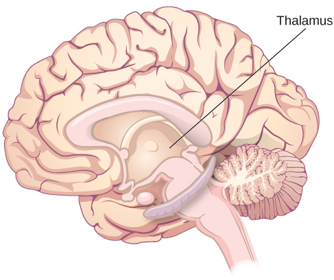
thalamus

structure containing relays and circuits connecting sensory, motor, and cortical systems.

Read explanation · OpenStax ↗
Thioether

A sulfur bonded to two carbon groups, as in ’s . It has no hydrogen.

Read explanation · OpenStax ↗
Thiol group · · · ·

A sulfur bonded to hydrogen, –SH. thiols can form through .

Read explanation · OpenStax ↗
Threonine ·

A , uncharged with a and a on its .

Read explanation · OpenStax ↗
Thymine

A normally used in ; it pairs with through two .

Read explanation · OpenStax ↗
tonicity

A ’s effect on cell volume, determined mainly by that do not effectively penetrate the relevant .

Read explanation · OpenStax ↗
Transacetylase

An transferring an acetyl group; the acetylates certain galactosides.

Read explanation · OpenStax ↗
Transcription

Synthesis of using a template.

Read explanation · OpenStax ↗
Transcriptional repressor · ·

A regulatory that reduces , for example by obstructing polymerase access or recruiting other repressing factors.

Read explanation · OpenStax ↗
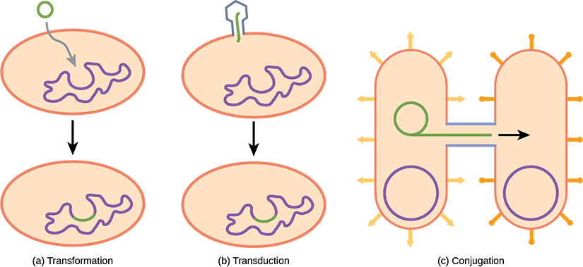
transduction

Transfer of mediated by a .

Read explanation · OpenStax ↗
Transfer RNA ·

An adaptor that carries an and pairs its with a on .

Read explanation · OpenStax ↗
Transition state

A fleeting, high-energy arrangement at a barrier. It is an energy maximum along the path, rather than a stable .

Read explanation · OpenStax ↗
Translation ·

Synthesis of a on a using the sequence of in .

Read explanation · OpenStax ↗
Tryptophan ·

An with a bulky ; commonly grouped as .

Read explanation · OpenStax ↗
Tumor · ·

An abnormal mass of tissue; tumors can be benign or malignant.

Read explanation · OpenStax ↗
Turnover number · · ·

The maximum number of cycles per per unit time, calculated as /. Units are inverse time, commonly s⁻¹.

Read explanation · IUBMB ↗
Tyrosine ·

An with a phenolic . Usually uncharged near physiological .

Read explanation · OpenStax ↗
Upstream

Toward positions preceding a reference site in the direction opposite ; positions before +1 have negative numbering.

Read explanation · OpenStax ↗
Uracil

A normally used in in place of . Uracil produced by in is a repair target.

Read explanation · OpenStax ↗
Urea

A small nitrogen-containing that, at sufficiently high , can destabilize folds.

Read explanation · NCBI Bookshelf ↗
Vesicle · · ·

A small -enclosed compartment that can transport material between cellular locations.

Read explanation · OpenStax ↗
Volatility · ·

The tendency of a substance to enter the gas phase; volatile vaporize relatively readily under given conditions.

Read explanation · Agilent ↗
β-Galactosidase ·

The -encoded that hydrolyzes and can also convert to .

Read explanation · OpenStax ↗

5.2 Passive Transport

OpenStax · 5.2 Passive Transport ↗
Instructional page including learning objectives, examples, figures, source tables and questions. Site navigation and embedded media players are omitted; source media links remain available.

Learning Objectives

By the end of this section, you will be able to do the following:

  • Explain why and how passive transport occurs
  • Understand the and processes
  • Define and its relevance to passive transport

Plasma must allow certain substances to enter and leave a cell, and prevent some harmful materials from entering and some essential materials from leaving. In other words, plasma are selectively permeable (semipermeable)—they allow some substances to pass through, but not others. If they were to lose this selectivity, the cell would no longer be able to sustain itself, and it would be destroyed. Some cells require larger amounts of specific substances. They must have a way of obtaining these materials from extracellular fluids. This may happen passively, as certain materials move back and forth, or the cell may have special mechanisms that facilitate transport. Some materials are so important to a cell that it spends some of its energy, hydrolyzing (), to obtain these materials. use some of their energy doing just that. Most cells spend the majority of their energy to maintain an imbalance of sodium and potassium between the cell's interior and exterior, as well as on .

The most direct forms of transport are passive. Passive transport is a naturally occurring phenomenon and does not require the cell to exert any of its energy to accomplish the movement. In passive transport, substances move from an area of higher to an area of lower . A physical space in which there is a single substance range has a .

Selective Permeability

Plasma are asymmetric: the 's interior is not identical to its exterior. There is a considerable difference between the array of and between the two leaflets that form a . On the 's interior, some serve to anchor the to 's fibers. There are peripheral on the 's exterior that bind . , attached to or , are also on the plasma 's exterior surface. These complexes help the cell bind required substances in the extracellular fluid. This adds considerably to plasma 's selective nature (Figure 5.7).

This illustration shows that the inside and outside of a plasma membrane are different, with the exterior covered in the spherical heads, and the interior filled with the strandlike tails.
Figure 5.7 — 5.2 Passive Transport

Figure 5.7 The plasma membrane's exterior surface is not identical to its interior surface. Credit: Rao, A., Ryan, K., Fletcher, S., Hawkins, A. and Tag, A. Department of Biology, Texas A&M University.

Mary Ann Clark, Matthew Douglas, and Jung Choi, Biology 2E, OpenStax. · CC BY-NC-SA 4.0 · Original image ↗ · Access for free ↗

Recall that plasma are : They have and regions. This characteristic helps move some materials through the and hinders the movement of others. and - material with a low can easily slip through the 's core. Substances such as the fat- vitamins A, D, E, and K readily pass through the plasma in the digestive tract and other tissues. Fat- drugs and also gain easy entry into cells and readily transport themselves into the body’s tissues and organs. Oxygen and carbon dioxide have no and pass through by simple .

substances present problems for the . While some connect easily with the cell's outside, they cannot readily pass through the plasma 's core. Additionally, while small could easily slip through the spaces in the 's mosaic, their prevents them from doing so. such as sodium, potassium, calcium, and chloride must have special means of penetrating plasma . Simple sugars and also need the help of various transmembrane (channels) to transport themselves across plasma .

is a passive process of transport. A single substance moves from a high to a low area until the is equal across a space. You are familiar with of substances through the air. For example, think about someone opening a bottle of ammonia in a room filled with people. The ammonia gas is at its highest in the bottle. Its lowest is at the room's edges. The ammonia vapor will diffuse, or spread away, from the bottle, and gradually, increasingly more people will smell the ammonia as it spreads. Materials move within the cell’s by , and certain materials move through the plasma by (Figure 5.8). expends no energy. On the contrary, are a form of , which dissipates as the is eliminated.

The left part of this illustration shows a substance on one side of a membrane only, in the extracellular fluid. The middle part shows that, after some time, some of the substance has diffused across the plasma membrane, from the extracellular fluid and into the cytoplasm. The right part shows that, after more time, an equal amount of the substance is on each side of the membrane.
Figure 5.8 — 5.2 Passive Transport

Figure 5.8 Diffusion through a permeable membrane moves a substance from a high concentration area (extracellular fluid, in this case) down its concentration gradient (into the cytoplasm). (credit: modification of work by Mariana Ruiz Villareal)

Mary Ann Clark, Matthew Douglas, and Jung Choi, Biology 2E, OpenStax. (credit: modification of work by Mariana Ruiz Villareal) · CC BY-NC-SA 4.0 · Original image ↗ · Access for free ↗

Each separate substance in a medium, such as the extracellular fluid, has its own , independent of other materials' . In addition, each substance will diffuse according to that . Within a system, there will be different rates of various substances in the medium.

Factors That Affect

move constantly in a random manner, at a rate that depends on their mass, their environment, and the amount of thermal energy they possess, which in turn is a function of temperature. This movement accounts for through whatever medium in which they are localized. A substance moves into any space available to it until it evenly distributes itself throughout. After a substance has diffused completely through a space, removing its , will still move around in the space, but there will be no net movement of the number of from one area to another. We call this lack of a in which the substance has no net movement . While will go forward in the presence of a substance's , several factors affect the rate.

  • Extent of the : The greater the difference in , the more rapid the . The closer the distribution of the material gets to , the slower the rate.
  • Mass of the diffusing: Heavier move more slowly; therefore, they diffuse more slowly. The reverse is true for lighter .
  • Temperature: Higher temperatures increase the energy and therefore the ' movement, increasing the rate. Lower temperatures decrease the ' energy, thus decreasing the rate.
  • density: As the density of a increases, the rate decreases. The slow down because they have a more difficult time passing through the denser medium. If the medium is less dense, increases. Because cells primarily use to move materials within the , any increase in the ’s density will inhibit the movement of the materials. An example of this is a person experiencing dehydration. As the body’s cells lose water, the rate decreases in the , and the cells’ functions deteriorate. tend to be very sensitive to this effect. Dehydration frequently leads to unconsciousness and possibly coma because of the decrease in rate within the cells.
  • : As we discussed earlier, or - materials pass through plasma more easily than materials, allowing a faster rate.
  • Surface area and plasma thickness: Increased surface area increases the rate; whereas, a thicker reduces it.
  • Distance travelled: The greater the distance that a substance must travel, the slower the rate. This places an upper limitation on cell size. A large, spherical cell will die because nutrients or waste cannot reach or leave the cell's center, respectively. Therefore, cells must either be small in size, as in the case of many , or be flattened, as with many single-celled .

A variation of is the process of filtration. In filtration, material moves according to its through a . Sometimes pressure enhances the rate, causing the substances to filter more rapidly. This occurs in the kidney, where blood pressure forces large amounts of water and accompanying dissolved substances, or , out of the blood and into the renal tubules. The rate in this instance is almost totally dependent on pressure. One of the effects of high blood pressure is the appearance of in the urine, which abnormally high pressure "squeezes through".

Facilitated transport

In facilitated transport, or , materials diffuse across the plasma with the help of . A exists that would allow these materials to diffuse into the cell without expending cellular energy. However, these materials are that the 's parts repel. Facilitated transport shield these materials from the 's repulsive force, allowing them to diffuse into the cell.

The transported material first attaches to or glycoprotein on the plasma 's exterior surface. This allows removal of material from the extracellular fluid that the cell needs. The substances then pass to specific integral that facilitate their passage. Some of these integral are collections of beta-pleated sheets that form a or channel through the bilayer. Others are which bind with the substance and aid its through the .

Channels

The integral involved in facilitated transport are transport , and they function as either channels for the material or carriers. In both cases, they are transmembrane . Channels are specific for the transported substance. Channel have exposed to the intracellular and extracellular fluids. In addition, they have a channel through their core that provides a hydrated opening through the layers (Figure 5.9). Passage through the channel allows to avoid the plasma 's central layer that would otherwise slow or prevent their entry into the cell. are channel that allow water to pass through the at a very high rate.

This illustration shows two channel proteins in the plasma membrane. One protein is labeled gate closed, and there is no space for particles to pass through.  The other is labeled gate open, and there is a space for substances to pass through.  Substances of the same type appear on both sides of the membrane near the open-gated protein.
Figure 5.9 — 5.2 Passive Transport

Figure 5.9 Ion Channel Proteins. Ion channel proteins are gated. When they are closed, no ions can pass through them. However, when a channel opens, select ions diffuse through the channel. Channel proteins are highly specific, letting only a specific ion or subset of ions pass. Credit: Rao, A., Ryan, K., Tag, A. and Fletcher, S. Department of Biology, Texas A&M University.

Mary Ann Clark, Matthew Douglas, and Jung Choi, Biology 2E, OpenStax. · CC BY-NC-SA 4.0 · Original image ↗ · Access for free ↗

Channel are either open at all times or they are “gated,” which the channel's opening. When a particular attaches to the channel it may control the opening, or other mechanisms or substances may be involved. In some tissues, sodium and chloride pass freely through open channels; whereas, in other tissues a gate must open to allow passage. An example of this occurs in the kidney, where there are both channel forms in different parts of the renal tubules. Cells involved in transmitting electrical impulses, such as nerve and muscle cells, have gated channels for sodium, potassium, and calcium in their . Opening and closing these channels changes the relative on opposing sides of the of these , resulting in facilitating electrical transmission along (in the case of nerve cells) or in muscle contraction (in the case of muscle cells).

Another type of embedded in the plasma is a . This aptly named binds a substance and, thus triggers a change of its own shape, moving the bound from the cell's outside to its interior (Figure 5.10); depending on the , the material may move in the opposite direction. are typically specific for a single substance. This selectivity adds to the plasma 's overall selectivity. Scientists poorly understand the exact mechanism for the change of shape. can change shape when their are affected, but this may not fully explain this mechanism. Each is specific to one substance, and there are a finite number of these in any . This can cause problems in transporting enough material for the cell to function properly. When all of the are bound to their , they are saturated and the rate of transport is at its maximum. Increasing the at this point will not result in an increased transport rate.

This illustration shows a carrier protein embedded in the membrane with an opening that initially faces the extracellular surface, labeled the hydrophilic pocket. After a substance binds the carrier, it changes shape so that the opening faces the cytoplasm. The change is labeled conformational change. After, the substance is released.
Figure 5.10 — 5.2 Passive Transport

Figure 5.10 Some substances are able to move down their concentration gradient across the plasma membrane with the aid of carrier proteins. Carrier proteins change shape as they move molecules across the membrane. Credit: Rao, A., Tag, A. and Fletcher, S. Department of Biology, Texas A&M University.

Mary Ann Clark, Matthew Douglas, and Jung Choi, Biology 2E, OpenStax. · CC BY-NC-SA 4.0 · Original image ↗ · Access for free ↗

An example of this process occurs in the kidney. In one part, the kidney filters , water, , , and that the body requires. This filtrate, which includes , then reabsorbs in another part of the kidney. Because there are only a finite number of for , if more is present than the can handle, the excess is not transported and the body excretes this through urine. In a diabetic individual, the term is “spilling into the urine.” A different group of , transport , or GLUTs, are involved in transporting and other hexose sugars through plasma within the body.

Channel and transport material at different rates. Channel transport much more quickly than . Channel facilitate at a rate of tens of millions of per second; whereas, work at a rate of a thousand to a million per second.

is the movement of free water through a semipermeable according to the water's across the , which is inversely proportional to the ' . While transports material across and within cells, transports only water across a and the limits the ' in the water. Not surprisingly, the that facilitate water movement play a large role in , most prominently in and the of kidney tubules.

Mechanism

is a special case of . Water, like other substances, moves from an area of high of free water to one of low free water . An obvious question is what makes water move at all? Imagine a beaker with a semipermeable separating the two sides or halves (Figure 5.11). On both sides of the the water level is the same, but there are different dissolved substance , or , that cannot cross the (otherwise the crossing the would balance on each side). If the 's volume on both sides of the is the same, but the 's are different, then there are different amounts of water, the , on either side of the .

This illustration shows a container whose contents are separated by a semipermeable membrane. Initially, there is a high concentration of solute on the right side of the membrane and a low concentration of the left. Over time, water diffuses across the membrane toward the side of the container that initially had a higher concentration of solute (lower concentration of water). As a result of osmosis, the water level is higher on this side of the membrane, and the solute concentration is the same on both sides.
Figure 5.11 — 5.2 Passive Transport

Figure 5.11 In osmosis, water always moves from an area of higher water concentration to one of lower concentration. In the diagram, the solute cannot pass through the selectively permeable membrane, but the water can.

Mary Ann Clark, Matthew Douglas, and Jung Choi, Biology 2E, OpenStax. · CC BY-NC-SA 4.0 · Original image ↗ · Access for free ↗

To illustrate this, imagine two full water glasses. One has a single teaspoon of sugar in it; whereas, the second one contains one-quarter cup of sugar. If the total volume of the in both cups is the same, which cup contains more water? Because the large sugar amount in the second cup takes up much more space than the teaspoon of sugar in the first cup, the first cup has more water in it.

Returning to the beaker example, recall that it has a mixture on either side of the . A principle of is that the move around and will spread evenly throughout the medium if they can. However, only the material capable of getting through the will diffuse through it. In this example, the cannot diffuse through the , but the water can. Water has a in this system. Thus, water will diffuse down its , crossing the to the side where it is less concentrated. This of water through the ——will continue until the water's goes to zero or until the water's balances the . proceeds constantly in living systems.

describes how an extracellular can change a cell's volume by affecting . A 's often directly correlates with the 's . describes the 's total . A with low has a greater number of water relative to the number of particles. A with high has fewer water with respect to particles. In a situation in which a permeable to water, though not to the separates two different osmolarities, water will move from the 's side with lower (and more water) to the side with higher (and less water). This effect makes sense if you remember that the cannot move across the , and thus the only component in the system that can move—the water—moves along its own . An important distinction that concerns living systems is that measures the number of particles (which may be ) in a . Therefore, a that is cloudy with cells may have a lower than a that is clear, if the second contains more dissolved than there are cells.

Scientists use three terms—, isotonic, and —to relate the cell's to the extracellular fluid's that contains the cells. In a situation, the extracellular fluid has lower than the fluid inside the cell, and water enters the cell. (In living systems, the point of reference is always the , so the prefix hypo- means that the extracellular fluid has a lower , or a lower , than the cell .) It also means that the extracellular fluid has a higher water in the than does the cell. In this situation, water will follow its and enter the cell.

As for a , the prefix hyper- refers to the extracellular fluid having a higher than the cell’s ; therefore, the fluid contains less water than the cell does. Because the cell has a relatively higher water , water will leave the cell.

Isotonic

In an isotonic , the extracellular fluid has the same as the cell. If the cell's matches that of the extracellular fluid, there will be no net movement of water into or out of the cell, although water will still move in and out. Blood cells and plant cells in , isotonic, and take on characteristic appearances (Figure 5.12).

Visual Connection

The left part shows bloated red blood cells, lysed, bathed in a hypotonic solution. The middle part shows healthy red blood cells bathed in an isotonic solution, and the right part of this illustration shows shriveled red blood cells bathed in a hypertonic solution.
Figure 5.12 — 5.2 Passive Transport

Figure 5.12 Three different scenarios involving red blood cells (RBC) are shown. Left: A RBC placed in a hypotonic solution, where the concentration of solutes in the surrounding fluid is lower than those in the cell, will cause water to rush into the RBC and lead to lysis of the cell. Middle: there is no net water movement into or out of the cell as the concentration of the solutes inside the cell equal or is isotonic to that of the surrounding fluid. Right: a RBC placed in a hypertonic solution, where the concentration of solutes in the surrounding fluid is greater than that in the cell will cause water to rush out of the cell and into the surrounding fluid. This will cause the RBC to shrivel. Credit: Tag, A., Rao, A., Hawkins, A and Fletcher, S. Department of Biology, Texas A&M University.

Mary Ann Clark, Matthew Douglas, and Jung Choi, Biology 2E, OpenStax. · CC BY-NC-SA 4.0 · Original image ↗ · Access for free ↗

A doctor injects a patient with what the doctor thinks is an isotonic saline . The patient dies, and an autopsy reveals that many have been destroyed. Do you think the the doctor injected was really isotonic?

Link to Learning

For a video illustrating the process in , visit this site.

in Living Systems

In a environment, water enters a cell, and the cell swells. In an isotonic condition, the relative and are equal on both sides. There is no net water movement; therefore, there is no change in the cell's size. In a , water leaves a cell and the cell shrinks. If either the hypo- or hyper- condition goes to excess, the cell’s functions become compromised, and the cell may be destroyed.

A will burst, or lyse, when it swells beyond the plasma ’s capability to expand. Remember, the resembles a mosaic, with discrete spaces between the comprising it. If the cell swells, and the spaces between the and become too large, the cell will break apart.

In contrast, when excessive water amounts leave a , the cell shrinks, or crenates. This has the effect of concentrating the left in the cell, making the denser and interfering with within the cell. The cell’s ability to function will be compromised and may also result in the cell's death.

Various living things have ways of controlling the effects of —a mechanism we call osmoregulation. Some organisms, such as plants, fungi, , and some protists, have cell walls that surround the plasma and prevent cell lysis in a . The plasma can only expand to the cell wall's limit, so the cell will not lyse. The in plants is always slightly to the cellular environment, and water will always enter a cell if water is available. This water inflow produces turgor pressure, which stiffens the plant's cell walls (Figure 5.13). In nonwoody plants, turgor pressure supports the plant. Conversely, if you do not water the plant, the extracellular fluid will become , causing water to leave the cell. In this condition, the cell does not shrink because the cell wall is not flexible. However, the detaches from the wall and constricts the . We call this plasmolysis. Plants lose turgor pressure in this condition and wilt (Figure 5.14).

The left part of this image shows a plant cell bathed in a hypertonic solution so that the plasma membrane has pulled away completely from the cell wall, and the central vacuole has shrunk. Water moves out of the plant cell. The middle part shows a plant cell bathed in an isotonic solution; the plasma membrane has pulled away from the cell wall a bit, and the central vacuole size is unchanged. Water moves both in and out of the plant cell. The right part shows a plant cell in a hypotonic solution. The central vacuole is large, and the plasma membrane is pressed against the cell wall. Water is moving into the plant cell.
Figure 5.13 — 5.2 Passive Transport

Figure 5.13 The turgor pressure within a plant cell depends on the solution's tonicity in which it is bathed. (credit: modification of work by Mariana Ruiz Villareal)

Mary Ann Clark, Matthew Douglas, and Jung Choi, Biology 2E, OpenStax. (credit: modification of work by Mariana Ruiz Villareal) · CC BY-NC-SA 4.0 · Original image ↗ · Access for free ↗
The left photo shows a plant that has wilted, with dark green leaves that are shriveled, and appear dry.  The photo on the right shows a healthy plant, with broad light green leaves that appear soft and pliable.
Figure 5.14 — 5.2 Passive Transport

Figure 5.14 Without adequate water, the plant on the left has lost turgor pressure, visible in its wilting. Watering the plant (right) will restore the turgor pressure. (credit: Victor M. Vicente Selvas)

Mary Ann Clark, Matthew Douglas, and Jung Choi, Biology 2E, OpenStax. (credit: Victor M. Vicente Selvas) · CC BY-NC-SA 4.0 · Original image ↗ · Access for free ↗

is a concern for all living things. For example, paramecia and amoebas, which are protists that lack cell walls, have contractile vacuoles. This collects excess water from the cell and pumps it out, keeping the cell from lysing as it takes on water from its environment (Figure 5.15).

A bright field light microscopy shows an oval-shaped cell. Contractile vacuoles are prominent structures embedded in the cell membrane that pump out water.
Figure 5.15 — 5.2 Passive Transport

Figure 5.15 A paramecium’s contractile vacuole, here visualized using bright field light microscopy at 480x magnification, continuously pumps water out of the organism’s body to keep it from bursting in a hypotonic medium. (credit: modification of work by NIH; scale-bar data from Matt Russell)

Mary Ann Clark, Matthew Douglas, and Jung Choi, Biology 2E, OpenStax. (credit: modification of work by NIH; scale-bar data from Matt Russell) · CC BY-NC-SA 4.0 · Original image ↗ · Access for free ↗

Many marine invertebrates have internal levels matched to their environments, making them isotonic with the water in which they live. Fish, however, must spend approximately five percent of their energy maintaining osmotic . Freshwater fish live in an environment that is to their cells. These fish actively take in through their gills and excrete diluted urine to themselves of excess water. Saltwater fish live in the reverse environment, which is to their cells, and they secrete through their gills and excrete highly concentrated urine.

In vertebrates, the kidneys regulate the water amount in the body. are specialized cells in the brain that monitor in the blood. If the levels increase beyond a certain range, a releases that slows water loss through the kidney and dilutes the blood to safer levels. Animals also have high , which the liver produces, in their blood. This is too large to pass easily through plasma and is a major factor in controlling the osmotic pressures applied to tissues.

5.3 Active Transport

OpenStax · 5.3 Active Transport ↗
Instructional page including learning objectives, examples, figures, source tables and questions. Site navigation and embedded media players are omitted; source media links remain available.

Learning Objectives

By the end of this section, you will be able to do the following:

Active transport mechanisms require the cell’s energy, usually in the form of (). If a substance must move into the cell against its —that is, if the substance's inside the cell is greater than its in the extracellular fluid (and vice versa)—the cell must use energy to move the substance. Some active transport mechanisms move small- materials, such as , through the . Other mechanisms transport much larger .

We have discussed simple —a substance's differential across a space or a —but in living systems, are more complex. Because move into and out of cells and because cells contain that do not move across the and are mostly negatively , there is also an electrical , a difference of , across the plasma . The interior of living cells is electrically negative with respect to the extracellular fluid in which they are bathed, and at the same time, cells have higher of potassium (K+) and lower of sodium (Na+) than the extracellular fluid. Thus in a living cell, the of Na+ tends to drive it into the cell, and its electrical (a positive ) also drives it inward to the negatively interior. However, the situation is more complex for other such as potassium. The electrical of K+, a positive , also drives it into the cell, but the of K+ drives K+ out of the cell (Figure 5.16). We call the combined and that affects an its .

Visual Connection

This illustration shows a membrane bilayer with a potassium channel embedded in it. The cytoplasm has a high concentration of potassium associated with a negatively charged molecule. The extracellular fluid has a high concentration of sodium associated with chlorine ions.
Figure 5.16 — 5.3 Active Transport

Figure 5.16 Electrochemical gradients arise from the combined effects of concentration gradients and electrical gradients. Na + ions are at higher concentration outside the cell, and K + ions are at higher concentration inside of the cell, and yet the inside of the cell has negative net charge compared to the other side of the membrane. This is due to the presence of K + binding proteins and other negatively charged molecules. The difference in electrical charges attracts the positively charged Na ions toward the inside of the cell, the electrical gradient, while the K ions tend to flow through K channels toward the outside of the cell due to the concentration difference, the concentration gradient. Structures labeled A represent proteins. (credit: “Synaptitude”/Wikimedia Commons)

Mary Ann Clark, Matthew Douglas, and Jung Choi, Biology 2E, OpenStax. (credit: “Synaptitude”/Wikimedia Commons) · CC BY-NC-SA 4.0 · Original image ↗ · Access for free ↗

Injecting a potassium into a person’s blood is lethal. This is how capital punishment and euthanasia subjects die. Why do you think a potassium injection is lethal?

This illustration shows a membrane bilayer with a potassium channel called a proton pump embedded in it. Hydrogen ions flow out of the cell through a proton pump, then back into the cell as part of the sucrose-proton cotransporter. Sucrose flows with the protons from the lower concentration outside the cell to the higher concentration inside the cell, which is against the concentration gradient.
Figure 5.17 — 5.3 Active Transport

Figure 5.17 Proton Gradient provides energy for a secondary active transporter. The proton pump creates an electrochemical gradient of protons (hydrogen ions, H+) using ATP to drive primary active transport. This gradient allows for cotransport/secondary transport of sucrose against its concentration gradient as protons come down their concentration gradient via their membrane cotransporter protein. Credit: Rao, A. Tag, A. and Ryan, K. Department of Biology, Texas A&M University.

Mary Ann Clark, Matthew Douglas, and Jung Choi, Biology 2E, OpenStax. · CC BY-NC-SA 4.0 · Original image ↗ · Access for free ↗

Moving Against a

To move substances against a or , the cell must use energy. This energy comes from generated through the cell’s . Active transport mechanisms, or pumps, work against electrochemical . Small substances constantly pass through plasma . Active transport maintains of and other substances that living cells require in the face of these passive movements. A cell may spend much of its energy supply maintaining these processes. (A uses most of its energy to maintain the imbalance between exterior and interior sodium and potassium levels that the cell requires.) Because active transport mechanisms depend on a cell’s for energy, they are sensitive to many poisons that interfere with the supply.

Two mechanisms exist for transporting small- material and small . moves across a and creates a difference in across that , which is directly dependent on . does not directly require : instead, it is the movement of material due to the established by (Figure 5.19).

for Active Transport

An important for active transport is the presence of specific or pumps to facilitate movement: there are three types or transporters (Figure 5.18). A uniporter carries one specific or . A carries two different or , both in the same direction. An also carries two different or , but in different directions. All of these transporters can also transport small, uncharged organic like . These three types of are also in , but they do not require to work in that process. Some examples of pumps for active transport are Na+-K+ ATPase, which carries sodium and potassium , and H+-K+ ATPase, which carries hydrogen and potassium . Both of these are . Two other are Ca2+ ATPase and H+ ATPase, which carry only calcium and only , respectively. Both are pumps.

This illustration shows a plasma membrane with three transport proteins embedded in it. The left image shows a uniporter that transports a substance in one direction. The middle image shows a symporter that transports two different substances in the same direction. The right image shows an antiporter that transports two different substances in opposite directions.
Figure 5.18 — 5.3 Active Transport

Figure 5.18 A uniporter carries one molecule or ion. A symporter carries two different molecules or ions, both in the same direction. An antiporter also carries two different molecules or ions, but in different directions. (credit: modification of work by “Lupask”/Wikimedia Commons)

Mary Ann Clark, Matthew Douglas, and Jung Choi, Biology 2E, OpenStax. (credit: modification of work by “Lupask”/Wikimedia Commons) · CC BY-NC-SA 4.0 · Original image ↗ · Access for free ↗

The that functions with the active transport of sodium and potassium allows to occur. The second transport method is still active because it depends on using energy as does primary transport (Figure 5.19).

This illustration shows the sodium-potassium pump. Initially, the pumps opening faces the cytoplasm, where three sodium ions bind to it. The antiporter hydrolyzes an A T P to A D P and, as a result, undergoes a conformational change. The sodium ions are released into the extracellular space. Two potassium ions from the extracellular space now bind the antiporter, which changes conformation again, releasing the potassium ions into the cytoplasm.
Figure 5.19 — 5.3 Active Transport

Figure 5.19 The sodium-potassium pump is an example of primary active transport that moves ions, sodium and potassium ions in this instance, across a membrane against their concentration gradients. The energy is provided by the hydrolysis of ATP. Three sodium ions are moved out of the cell for every 2 potassium ions that are brought into the cell. This creates an electrochemical gradient that is crucial for living cells. Credit: Rao, A., Ryan, K. and Fletcher, S. Department of Biology, Texas A&M University.

Mary Ann Clark, Matthew Douglas, and Jung Choi, Biology 2E, OpenStax. · CC BY-NC-SA 4.0 · Original image ↗ · Access for free ↗

One of the most important pumps in animal cells is the (Na+-K+ ATPase), which maintains the (and the correct of Na+ and K+) in living cells. The moves K+ into the cell while moving Na+ out at the same time, at a ratio of three Na+ for every two K+ moved in. The Na+-K+ ATPase exists in two forms, depending on its orientation to the cell's interior or exterior and its for either sodium or potassium . The process consists of the following six steps.

  1. With the oriented towards the cell's interior, the has a high for sodium . Three bind to the .
  2. The hydrolyzes and a low-energy attaches to it.
  3. As a result, the changes shape and reorients itself towards the 's exterior. The ’s for sodium decreases and the three sodium leave the .
  4. The shape change increases the ’s for potassium , and two such attach to the . Subsequently, the low-energy detaches from the .
  5. With the removed and potassium attached, the repositions itself towards the cell's interior.
  6. The , in its new , has a decreased for potassium, and the two moves into the . The now has a higher for sodium , and the process starts again.

Several things have happened as a result of this process. At this point, there are more sodium outside the cell than inside and more potassium inside than out. For every three sodium that move out, two potassium move in. This results in the interior being slightly more negative relative to the exterior. This difference in is important in creating the conditions necessary for the secondary process. The is, therefore, an electrogenic pump (a pump that creates a imbalance), creating an electrical imbalance across the and contributing to the potential.

Link to Learning

Watch this video to see an active transport simulation in a sodium-potassium ATPase.

(Co-transport)

uses the of the sodium to bring other , against their into the cell. As sodium build outside of the plasma because of the process, this creates an . If a channel exists and is open, the sodium will move down its across the . This movement transports other substances that must be attached to the same transport in order for the sodium to move across the (Figure 5.20). Many , as well as , enter a cell this way. This secondary process also stores high-energy in the of plant and animal cells in order to produce . The that accumulates in the stored translates into as the surge through the channel , and that energy then converts ADP into .

Visual Connection

This illustration shows a membrane bilayer with two integral membrane proteins embedded in it. The first, a sodium-potassium pump, uses energy from A T P hydrolysis to pump three sodium ions out of the cell for every two potassium ions it pumps into the cell. The result is a high concentration of sodium outside the cell and a high concentration of potassium inside the cell. There is also a low concentration of glucose outside the cell, and a high concentration inside. A sodium-amino acid co-transporter simultaneously transports sodium and the glucose into the cell. Text within the image reads as follows: Primary Active Transport: the sodium potassium pump moves sodium ions using the energy of ATP hydrolysis to establish a concentration gradient of sodium ions. Secondary active transport: Sodium ions moving with the concentration gradient established by the sodium potassium pump drives the transport of glucose against its concentration gradient.
Figure 5.20 — 5.3 Active Transport

Figure 5.20 An electrochemical gradient (Na + concentration - green), is generated by primary active transport. The energy stored in the Na + gradient provides the energy to move other substances against their concentration gradients (Glucose - blue), a process called co-transport or secondary active transport. Credit: Rao, A., Ryan, K., Tag, A. and Fletcher, S. Department of Biology, Texas A&M University.

Mary Ann Clark, Matthew Douglas, and Jung Choi, Biology 2E, OpenStax. · CC BY-NC-SA 4.0 · Original image ↗ · Access for free ↗

If the outside the cell decreases, would you expect the amount of transported into the cell to increase or decrease?

5.4 Bulk Transport

OpenStax · 5.4 Bulk Transport ↗
Instructional page including learning objectives, examples, figures, source tables and questions. Site navigation and embedded media players are omitted; source media links remain available.

Learning Objectives

By the end of this section, you will be able to do the following:

In addition to moving small and through the , cells also need to remove and take in larger and particles (see Table 5.2 for examples). Some cells are even capable of engulfing entire unicellular microorganisms. You might have correctly hypothesized that when a cell uptakes and releases large particles, it requires energy. A large particle, however, cannot pass through the , even with energy that the cell supplies.

is a type of active transport that moves particles, such as large , parts of cells, and even whole cells, into a cell. There are different variations, but all share a common characteristic: the cell's plasma invaginates, forming a pocket around the target particle. The pocket pinches off, resulting in the particle containing itself in a newly created intracellular formed from the plasma .

(the condition of “cell eating”) is the process by which a cell takes in large particles, such as other cells or relatively large particles. For example, when microorganisms invade the human body, a type of white blood cell, a , will remove the invaders through this process, surrounding and engulfing the microorganism, which the then destroys (Figure 5.21).

This illustration shows a plasma membrane forming a pocket around a particle in the extracellular fluid. The membrane subsequently engulfs the particle, which becomes trapped in a vacuole inside the cell.
Figure 5.21 — 5.4 Bulk Transport

Figure 5.21 In phagocytosis, the cell membrane surrounds the particle and engulfs it. (credit: modification of work by Mariana Ruiz Villareal)

Mary Ann Clark, Matthew Douglas, and Jung Choi, Biology 2E, OpenStax. (credit: modification of work by Mariana Ruiz Villareal) · CC BY-NC-SA 4.0 · Original image ↗ · Access for free ↗

In preparation for , a portion of the plasma 's inward-facing surface becomes coated with the clathrin, which stabilizes this 's section. The 's coated portion then extends from the cell's body and surrounds the particle, eventually enclosing it. Once the containing the particle is enclosed within the cell, the clathrin disengages from the and the merges with a for breaking down the material in the newly formed compartment (). When accessible nutrients from the vesicular contents' degradation have been extracted, the newly formed merges with the plasma and releases its contents into the extracellular fluid. The endosomal again becomes part of the plasma .

A variation of is . This literally means “cell drinking”. Discovered by Warren Lewis in 1929, this American embryologist and cell biologist described a process whereby he assumed that the cell was purposefully taking in extracellular fluid. In reality, this is a process that takes in , including water, which the cell needs from the extracellular fluid. results in a much smaller than does , and the does not need to merge with a (Figure 5.22).

This illustration shows a plasma membrane forming a pocket around fluid in the extracellular fluid. The membrane subsequently engulfs the fluid, which becomes trapped in a vacuole.
Figure 5.22 — 5.4 Bulk Transport

Figure 5.22 In pinocytosis, the cell membrane invaginates, surrounds a small volume of fluid, and pinches off. (credit: modification of work by Mariana Ruiz Villareal)

Mary Ann Clark, Matthew Douglas, and Jung Choi, Biology 2E, OpenStax. (credit: modification of work by Mariana Ruiz Villareal) · CC BY-NC-SA 4.0 · Original image ↗ · Access for free ↗

A variation of is potocytosis. This process uses a coating , caveolin, on the plasma 's side, which performs a similar function to clathrin. The cavities in the plasma that form the vacuoles have and rafts in addition to caveolin. The vacuoles or formed in caveolae (singular caveola) are smaller than those in . Potocytosis brings small into the cell and transports them through the cell for their release on the other side, a process we call transcytosis. In some cases, the caveolae deliver their cargo to membranous like the .

-mediated

A targeted variation of employs in the plasma that have a specific for certain substances (Figure 5.23).

This illustration shows a part of the plasma membrane that is clathrin-coated on the cytoplasmic side and has receptors on the extracellular side. The receptors bind a substance, then pinch off to form a vesicle.
Figure 5.23 — 5.4 Bulk Transport

Figure 5.23 In receptor-mediated endocytosis, the cell's uptake of substances targets a single type of substance that binds to the receptor on the cell membrane's external surface. (credit: modification of work by Mariana Ruiz Villareal)

Mary Ann Clark, Matthew Douglas, and Jung Choi, Biology 2E, OpenStax. (credit: modification of work by Mariana Ruiz Villareal) · CC BY-NC-SA 4.0 · Original image ↗ · Access for free ↗

In -mediated , as in , clathrin attaches to the plasma 's side. If a 's uptake is dependent on -mediated and the process is ineffective, the material will not be removed from the tissue fluids or blood. Instead, it will stay in those fluids and increase in . The failure of -mediated causes some human diseases. For example, mediated removes low density lipoprotein or LDL (or "bad" ) from the blood. In the human genetic disease familial hypercholesterolemia, the LDL are defective or missing entirely. People with this condition have life-threatening levels of in their blood, because their cells cannot clear LDL particles.

Although -mediated is designed to bring specific substances that are normally in the extracellular fluid into the cell, other substances may gain entry into the cell at the same site. Flu viruses, diphtheria, and cholera toxin all have sites that cross-react with normal - and gain entry into cells.

Link to Learning

See -mediated in action, and click on different parts for a focused animation.

The reverse process of moving material into a cell is the process of . is the opposite of the processes we discussed above in that its purpose is to expel material from the cell into the extracellular fluid. Waste material is enveloped in a and fuses with the plasma 's interior. This fusion opens the membranous envelope on the cell's exterior, and the waste material expels into the extracellular space (Figure 5.24). Other examples of cells releasing via include and neurotransmitter into the synaptic cleft by synaptic .

This illustration shows vesicles fusing with the plasma membrane and releasing their contents to the extracellular fluid.
Figure 5.24 — 5.4 Bulk Transport

Figure 5.24 In exocytosis, vesicles containing substances fuse with the plasma membrane. The contents then release to the cell's exterior. (credit: modification of work by Mariana Ruiz Villareal)

Mary Ann Clark, Matthew Douglas, and Jung Choi, Biology 2E, OpenStax. (credit: modification of work by Mariana Ruiz Villareal) · CC BY-NC-SA 4.0 · Original image ↗ · Access for free ↗
Methods of Transport, Energy Requirements, and Types of Transported Material
Transport MethodActive/PassiveMaterial Transported
PassiveSmall- material
PassiveWater
Facilitated transport/PassiveSodium, potassium, calcium,
ActiveSodium, potassium, calcium
Active,
ActiveLarge , whole cells, or cellular structures
and potocytosisActiveSmall (liquids/water)
-mediated ActiveLarge quantities of
Table 5.2

9.1 Signaling Molecules and Cellular Receptors

OpenStax · 9.1 Signaling Molecules and Cellular Receptors ↗
Instructional page including learning objectives, examples, figures, source tables and questions. Site navigation and embedded media players are omitted; source media links remain available.

Learning Objectives

By the end of this section, you will be able to do the following:

  • Describe four types of signaling mechanisms found in multicellular organisms
  • Compare internal with cell-surface
  • Recognize the relationship between a ’s structure and its mechanism of action

There are two kinds of communication in the world of living cells. Communication between cells is called intercellular signaling, and communication within a cell is called intracellular signaling. An easy way to remember the distinction is by understanding the Latin origin of the prefixes: inter- means "between" (for example, intersecting lines are those that cross each other) and intra- means "inside" (as in intravenous).

Chemical signals are released by signaling cells in the form of small, usually or called . A is a that binds another specific , in some cases, delivering a signal in the process. can thus be thought of as signaling . interact with in target cells, which are cells that are affected by chemical signals; these are also called . and exist in several varieties; however, a specific will have a specific that typically binds only that .

Forms of Signaling

There are four categories of chemical signaling found in multicellular organisms: paracrine signaling, , autocrine signaling, and direct signaling across (Figure 9.2). The main difference between the different categories of signaling is the distance that the signal travels through the organism to reach the target cell. We should note here that not all cells are affected by the same signals.

The illustration shows four forms of chemical signaling. In autocrine signaling, a cell targets itself. In signaling across a gap junction, a cell targets a cell connected via gap junctions. In paracrine signaling, a cell targets a nearby cell. In endocrine signaling, a cell targets a distant cell via the bloodstream
Figure 9.2 — 9.1 Signaling and Cellular

Figure 9.2 In chemical signaling, a cell may target itself (autocrine signaling), a cell connected by gap junctions, a nearby cell (paracrine signaling), or a distant cell (endocrine signaling). Paracrine signaling acts on nearby cells, endocrine signaling uses the circulatory system to transport ligands, and autocrine signaling acts on the signaling cell. Signaling via gap junctions involves signaling molecules moving directly between adjacent cells.

Mary Ann Clark, Matthew Douglas, and Jung Choi, Biology 2E, OpenStax. · CC BY-NC-SA 4.0 · Original image ↗ · Access for free ↗

Paracrine Signaling

Signals that act locally between cells that are close together are called paracrine signals. Paracrine signals move by through the . These types of signals usually elicit quick responses that last only a short period of time. In order to keep the response localized, paracrine are normally quickly degraded by or removed by neighboring cells. Removing the signals will reestablish the for the signal, allowing them to quickly diffuse through the intracellular space if released again.

One example of paracrine signaling is the transfer of signals across between nerve cells. A nerve cell consists of a cell body, several short, branched extensions called that receive stimuli, and a long extension called an , which transmits signals to other nerve cells or muscle cells. The junction between nerve cells where signal transmission occurs is called a . A synaptic signal is a chemical signal that travels between nerve cells. Signals within the nerve cells are propagated by fast-moving electrical impulses. When these impulses reach the end of the , the signal continues on to a of the next cell by the release of chemical called neurotransmitters from the presynaptic cell (the cell emitting the signal). The neurotransmitters are transported across the very small distances (20–40 nanometers) between nerve cells, which are called chemical (Figure 9.3). The small distance between nerve cells allows the signal to travel quickly; this enables an immediate response, such as, "Take your hand off the stove!"

When the neurotransmitter binds the on the surface of the postsynaptic cell, the electrochemical potential of the target cell changes, and the next electrical impulse is launched. The neurotransmitters that are released into the chemical are degraded quickly or get reabsorbed by the presynaptic cell so that the recipient nerve cell can recover quickly and be prepared to respond rapidly to the next synaptic signal.

This illustration shows closely juxtaposed bulbous protrusions of presynaptic and postsynaptic cells. The presynaptic cell stores neurotransmitter in synaptic vesicles. When signaling occurs, the vesicles fuse with the cell membrane, thereby releasing the neutrotransmitter, which then binds to receptors on the postsynaptic cell. An enzyme on the surface of the postsynaptic cell destroys the neurotrasmitter, thereby terminating the signal.
Figure 9.3 — 9.1 Signaling and Cellular

Figure 9.3 The distance between the presynaptic cell and the postsynaptic cell—called the synaptic gap—is very small and allows for rapid diffusion of the neurotransmitter. Enzymes in the synapatic gap degrade some types of neurotransmitters to terminate the signal.

Mary Ann Clark, Matthew Douglas, and Jung Choi, Biology 2E, OpenStax. · CC BY-NC-SA 4.0 · Original image ↗ · Access for free ↗

Signals from distant cells are called signals, and they originate from . (In the body, many are located in glands, such as the thyroid gland, the , and the pituitary gland.) These types of signals usually produce a slower response but have a longer-lasting effect. The released in are called , signaling that are produced in one part of the body but affect other body regions some distance away.

travel the large distances between and their target cells via the bloodstream, which is a relatively slow way to move throughout the body. Because of their form of transport, become diluted and are present in low when they act on their target cells. This is different from paracrine signaling, in which local of can be very high.

Autocrine Signaling

Autocrine signals are produced by signaling cells that can also bind to the that is released. This means the signaling cell and the target cell can be the same or a similar cell (the prefix auto- means self, a reminder that the signaling cell sends a signal to itself). This type of signaling often occurs during the early development of an organism to ensure that cells develop into the correct tissues and take on the proper function. Autocrine signaling also regulates pain sensation and inflammatory responses. Further, if a cell is infected with a virus, the cell can signal itself to undergo , killing the virus in the process. In some cases, neighboring cells of the same type are also influenced by the released . In embryological development, this process of stimulating a group of neighboring cells may help to direct the of identical cells into the same cell type, thus ensuring the proper developmental outcome.

Direct Signaling Across

in animals and plasmodesmata in plants are connections between the plasma of neighboring cells. These fluid-filled channels allow small signaling , called intracellular mediators, to diffuse between the two cells. Small or , such as calcium (Ca2+), are able to move between cells, but large like and cannot fit through the channels. The specificity of the channels ensures that the cells remain independent but can quickly and easily transmit signals. The transfer of signaling communicates the current state of the cell that is directly next to the target cell; this allows a group of cells to coordinate their response to a signal that only one of them may have received. In plants, plasmodesmata are ubiquitous, making the entire plant into a giant communication network.

Types of

are in the target cell or on its surface that bind . There are two types of , internal and cell-surface .

Internal

Internal , also known as intracellular or , are found in the of the cell and respond to that are able to travel across the plasma . Once inside the cell, many of these bind to that act as regulators of synthesis () to mediate . is the cellular process of transforming the information in a cell's into a sequence of , which ultimately forms a . When the binds to the internal , a change is triggered that exposes a - on the . The - complex moves into the , then binds to specific regulatory regions of the and promotes the initiation of (Figure 9.4). is the process of copying the information in a cell's into a special form of called (); the cell uses information in the (which moves out into the and associates with ) to link specific in the correct order, producing a . Internal can directly influence without having to pass the signal on to other or messengers.

This illustration shows a hydrophobic signaling molecule that diffuses across the plasma membrane and binds an intracellular receptor in the cytoplasm. The intracellular receptor-signaling molecule complex then travels to the nucleus and binds D N A.
Figure 9.4 — 9.1 Signaling and Cellular

Figure 9.4 Hydrophobic signaling molecules typically diffuse across the plasma membrane and interact with intracellular receptors in the cytoplasm. Many intracellular receptors are transcription factors that interact with DNA in the nucleus and regulate gene expression.

Mary Ann Clark, Matthew Douglas, and Jung Choi, Biology 2E, OpenStax. · CC BY-NC-SA 4.0 · Original image ↗ · Access for free ↗

Cell-Surface

Cell-surface , also known as transmembrane , are cell surface, -anchored (integral) that bind to external . This type of spans the plasma and performs signal , through which an extracellular signal is converted into an intracellular signal. that interact with cell-surface do not have to enter the cell that they affect. Cell-surface are also called cell-specific or markers because they are specific to individual cell types.

Because cell-surface are fundamental to normal cell functioning, it should come as no surprise that a malfunction in any one of these could have severe consequences. Errors in the structures of certain have been shown to play a role in hypertension (high blood pressure), asthma, heart disease, and .

Each cell-surface has three main components: an external -binding called the extracellular , a -spanning region called a transmembrane , and an intracellular inside the cell. The size and extent of each of these vary widely, depending on the type of .

Evolution Connection

How Viruses Recognize a Host

Unlike living cells, many viruses do not have a plasma or any of the structures necessary to sustain life. Some viruses are simply composed of an inert shell enclosing or . To reproduce, viruses must invade a living cell, which serves as a host, and then take over the hosts cellular apparatus. But how does a virus recognize its host?

Viruses often bind to cell-surface on the host cell. For example, the virus that causes human influenza (flu) binds specifically to on of cells of the respiratory system. Chemical differences in the cell-surface among hosts mean that a virus that infects a specific species (for example, humans) often cannot infect another species (for example, chickens).

However, viruses have very small amounts of or compared to humans, and, as a result, viral reproduction can occur rapidly. Viral reproduction invariably produces errors that can lead to changes in newly produced viruses; these changes mean that the viral that interact with cell-surface may evolve in such a way that they can bind to in a new host. Such changes happen randomly and quite often in the reproductive cycle of a virus, but the changes only matter if a virus with new binding properties comes into contact with a suitable host. In the case of influenza, this situation can occur in settings where animals and people are in close contact, such as poultry and swine farms.1 Once a virus jumps the former "species barrier" to a new host, it can spread quickly. Scientists watch newly appearing viruses (called emerging viruses) closely in the hope that such monitoring can reduce the likelihood of global viral epidemics.

Cell-surface are involved in most of the signaling in multicellular organisms. There are three general categories of cell-surface : channel-linked , G--linked , and -linked .

channel-linked bind a and open a channel through the that allows specific to pass through. To form a channel, this type of cell-surface has an extensive -spanning region. In order to interact with the double layer of tails that form the center of the plasma , many of the in the -spanning region are in nature. Conversely, the that line the inside of the channel are to allow for the passage of water or . When a binds to the extracellular region of the channel, there is a change in the 's structure that allows such as sodium, calcium, magnesium, and hydrogen to pass through (Figure 9.5).

A gated channel is closed within the plasma membrane. A signaling molecule binds to a site on the channel protein, and the channel opens. When the signaling molecule is no longer bound, the channel closes.
Figure 9.5 — 9.1 Signaling and Cellular

Figure 9.5 Gated ion channels located in the plasma membrane allow for the controlled flow of ions into and out of the cell. The channel proteins remain closed until a signaling molecule (orange teardrop) binds to the channel protein. Then the channel protein changes conformation and allows ions (yellow circles) to flow into (or out of) the cell. When the signaling molecule is released, the channel protein resumes its closed conformation, preventing ion flow. Credit: Rao, A. and Fletcher, S. Department of Biology, Texas A&M University.

Mary Ann Clark, Matthew Douglas, and Jung Choi, Biology 2E, OpenStax. · CC BY-NC-SA 4.0 · Original image ↗ · Access for free ↗

G--linked bind a and activate a called a G-. The activated G- then interacts with either an channel or an in the (Figure 9.6). All G--linked have seven transmembrane , but each has its own specific extracellular and G--.

Cell signaling using G--linked occurs as a cyclic series of events. Before the binds, the inactive G- can bind to on the specific for its binding. Once the binds to the , the resulting change in shape activates the G-, which releases () and picks up guanosine 3- (). The of the G- then split into the α and the βγ . One or both of these G- fragments may be able to activate other as a result. After awhile, the on the active α of the G- is hydrolyzed to and the βγ is deactivated. The reassociate to form the inactive G- and the cycle begins anew.

This illustration shows the activation pathway for a heterotrimeric G protein, which has three subunits: alpha, beta, and gamma, all associated with the inside of the plasma membrane. When a signaling molecule binds to a G protein-coupled receptor in the plasma membrane, a G D P molecule associated with the alpha subunit is exchanged for G T P. The alpha subunit dissociates from the beta and gamma subunits and triggers a cellular response. Hydrolysis of G T P to G D P terminates the signal.
Figure 9.6 — 9.1 Signaling and Cellular

Figure 9.6 Heterotrimeric G-proteins have three subunits: α , β , and γ . When a signaling molecule binds to a G-protein-coupled receptor in the plasma membrane, a GDP molecule associated with the α subunit is exchanged for GTP. The β and γ subunits dissociate from the α subunit, and a cellular response is triggered either by the α subunit or the dissociated βγ pair. Hydrolysis of GTP to GDP terminates the signal.

Mary Ann Clark, Matthew Douglas, and Jung Choi, Biology 2E, OpenStax. · CC BY-NC-SA 4.0 · Original image ↗ · Access for free ↗

G--linked have been extensively studied and much has been learned about their roles in maintaining health. that are pathogenic to humans can release poisons that interrupt specific G--linked function, leading to illnesses such as pertussis, botulism, and cholera. In cholera (Figure 9.7), for example, the water-borne Vibrio cholerae produces a toxin, choleragen, that binds to cells lining the small intestine. The toxin then enters these intestinal cells, where it modifies a G- that the opening of a chloride channel and causes it to remain continuously active, resulting in large losses of fluids from the body and potentially fatal dehydration as a result.

This 1866 poster warns people about a cholera epidemic and gives advice for preventing the disease.
Figure 9.7 — 9.1 Signaling and Cellular

Figure 9.7 Transmitted primarily through contaminated drinking water, cholera is a major cause of death in the developing world and in areas where natural disasters interrupt the availability of clean water. The cholera bacterium, Vibrio cholerae , creates a toxin that modifies G-protein-mediated cell signaling pathways in the intestines. Modern sanitation eliminates the threat of cholera outbreaks, such as the one that swept through New York City in 1866. This poster from that era shows how, at that time, the way that the disease was transmitted was not understood. (credit: New York City Sanitary Commission)

Mary Ann Clark, Matthew Douglas, and Jung Choi, Biology 2E, OpenStax. (credit: New York City Sanitary Commission) · CC BY-NC-SA 4.0 · Original image ↗ · Access for free ↗

-linked are cell-surface with intracellular that are associated with an . In some cases, the intracellular of the itself is an . Other -linked have a small intracellular that interacts directly with an . The -linked normally have large extracellular and intracellular , but the -spanning region consists of a single region of the strand. When a binds to the extracellular , a signal is transferred through the , activating the . Activation of the sets off a chain of events within the cell that eventually leads to a response. One example of this type of -linked is the (Figure 9.8). A is an that transfers from to another . The transfers to ( ). First, signaling bind to the extracellular of two nearby . The two neighboring then bond together, or dimerize. Phosphates are then added to on the intracellular of the (). The can then transmit the signal to the next messenger within the .

Visual Connection

This illustration shows two receptor tyrosine kinase monomers embedded in the plasma membrane. Upon binding of a signaling molecule to the extracellular domain, the receptors dimerize. Tyrosine residues on the intracellular surface are then phosphorylated, triggering a cellular response. The cellular response involves binding of relay proteins, which result in Cellular Response 1 and Cellular Response 2.
Figure 9.8 — 9.1 Signaling and Cellular

Figure 9.8 1) A receptor tyrosine kinase is an enzyme-linked receptor with a single helical transmembrane region, and extracellular and intracellular domains. 2) Binding of a signaling molecule to the extracellular domain causes the receptor to dimerize. 3) Tyrosine residues on the intracellular domain are then autophosphorylated, 4) triggering a downstream cellular response. The signal is terminated by a phosphatase that removes the phosphates from the phosphotyrosine residues. Credit: Rao, A., Ryan, K., Tag, A., Fletcher, S. and Hawkins, A. Department of Biology, Texas A&M University.

Mary Ann Clark, Matthew Douglas, and Jung Choi, Biology 2E, OpenStax. · CC BY-NC-SA 4.0 · Original image ↗ · Access for free ↗

is a . In 30 percent of human breast cancers, is permanently activated, resulting in unregulated cell division. Lapatinib, a drug used to treat breast , inhibits autophosphorylation (the process by which the adds phosphates onto itself), thus reducing growth by 50 percent. Besides autophosphorylation, which of the following steps would be inhibited by Lapatinib?

  1. Signaling binding, dimerization, and the cellular response
  2. Dimerization, and the cellular response
  3. The cellular response
  4. activity, dimerization, and the downsteam cellular response

Signaling

Produced by signaling cells and the subsequent binding to in target cells, act as chemical signals that travel to the target cells to coordinate responses. The types of that serve as are incredibly varied and range from small to small like calcium (Ca2+).

Small

Small can directly diffuse through the plasma and interact with internal . Important members of this class of are the . Steroids are that have a skeleton with four fused rings; different steroids have different attached to the carbon skeleton. include the female sex , , which is a type of estrogen; the male sex , testosterone; and , which is an important structural component of and a precursor of (Figure 9.9). Other include thyroid and vitamin D. In order to be in blood, must bind to while they are being transported through the bloodstream.

The molecular structures of estradiol, testosterone, and cholesterol are shown. All three molecules share a four-ring structure but differ in the types of functional groups attached to it.
Figure 9.9 — 9.1 Signaling and Cellular

Figure 9.9 Steroid hormones have similar chemical structures to their precursor, cholesterol. Because these molecules are small and hydrophobic, they can diffuse directly across the plasma membrane into the cell, where they interact with internal receptors.

Mary Ann Clark, Matthew Douglas, and Jung Choi, Biology 2E, OpenStax. · CC BY-NC-SA 4.0 · Original image ↗ · Access for free ↗

Water-

Water- are and, therefore, cannot pass through the plasma unaided; sometimes, they are too large to pass through the at all. Instead, most water- bind to the extracellular of cell-surface . This group of is quite diverse and includes small , , and .

Other

Nitric oxide (NO) is a gas that also acts as a . It is able to diffuse directly across the plasma , and one of its roles is to interact with in smooth muscle and induce relaxation of the tissue. NO has a very short and, therefore, only functions over short distances. Nitroglycerin, a treatment for heart disease, acts by triggering the release of NO, which causes blood vessels to dilate (expand), thus restoring blood flow to the heart. NO has become better known recently because the pathway that it affects is targeted by prescription medications for erectile dysfunction, such as Viagra (erection involves dilated blood vessels).

9.2 Propagation of the Signal

OpenStax · 9.2 Propagation of the Signal ↗

Instructional page including learning objectives, examples, figures, source tables and questions. Site navigation and embedded media players are omitted; source media links remain available.

Learning Objectives

By the end of this section, you will be able to do the following:

Once a binds to a , the signal is transmitted through the and into the . Continuation of a signal in this manner is called signal . Signal only occurs with cell-surface , which cannot interact with most components of the cell such as . Only internal are able to interact directly with in the to initiate .

When a binds to its , changes occur that affect the ’s intracellular . changes of the extracellular upon binding can propagate through the region of the and lead to activation of the intracellular or its associated . In some cases, binding of the causes dimerization of the , which means that two bind to each other to form a stable complex called a dimer. A dimer is a formed when two (often identical) join together. The binding of the in this manner enables their intracellular to come into close contact and activate each other.

Binding Initiates a Signaling Pathway

After the binds to the cell-surface , the activation of the ’s intracellular components sets off a chain of events that is called a signaling pathway, sometimes called a signaling cascade. In a signaling pathway, ––and activated interact with specific , which are in turn activated in a chain that eventually leads to a change in the cell’s environment (Figure 9.10), such as an increase in or specific . The events in the cascade occur in a series, much like a current flows in a river. Interactions that occur before a certain point are defined as events, and events after that point are called events.

Visual Connection

This illustration shows the epidermal growth factor receptor, which is embedded in the plasma membrane. Upon binding of a signaling molecule to the receptor's extracellular domain, the receptor dimerizes, and intracellular residues are phosphorylated. Phosphorylation of the receptor triggers the phosphorylation of a protein called M E K by R A F. M E K, in turn, phosphorylates E R K. E R K stimulates protein translation in the cytoplasm, and transcription in the nucleus. Activation of E R K stimulates cell proliferation, cell migration and adhesion, and angiogenesis, which is the growth of new blood vessels. E R K inhibits apoptosis.
Figure 9.10 — 9.2 Propagation of the Signal
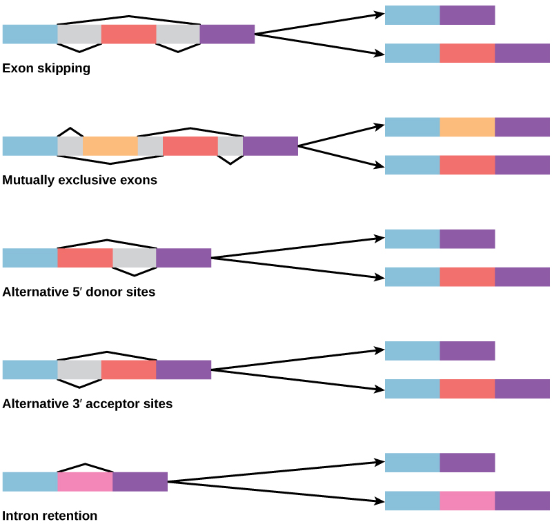
Figure 9.10 The epidermal growth factor (EGF) receptor (EGFR) is a receptor tyrosine kinase involved in the regulation of cell growth, wound healing, and tissue repair. When EGF binds to the EGFR, a cascade of downstream events causes the cell to grow and divide. If EGFR is activated at inappropriate times, uncontrolled cell growth (cancer) may occur.

Mary Ann Clark, Matthew Douglas, and Jung Choi, Biology 2E, OpenStax. · CC BY-NC-SA 4.0 · Original image ↗ · Access for free ↗

In certain cancers, the GTPase activity of the RAS G- is inhibited. This means that the RAS can no longer hydrolyze into . What effect would this have on cellular events?

You can see that signaling pathways can get very complicated very quickly because most cellular can affect different events, depending on the conditions within the cell. A single pathway can branch off toward different endpoints based on the interplay between two or more signaling pathways, and the same are often used to initiate different signals in different cell types. This variation in response is due to differences in in different cell types. Another complicating element is signal integration of the pathways, in which signals from two or more different cell-surface merge to activate the same response in the cell. This process can ensure that multiple external requirements are before a cell commits to a specific response.

The effects of extracellular signals can also be amplified by enzymatic cascades. At the initiation of the signal, a single binds to a single . However, activation of a -linked can activate many copies of a component of the signaling cascade, which amplifies the signal.

Link to Learning

Observe an animation of cell signaling at this site.

Methods of Intracellular Signaling

The induction of a signaling pathway depends on the modification of a cellular component by an . There are numerous enzymatic modifications that can occur, and they are recognized in turn by the next component . The following are some of the more common events in intracellular signaling.

One of the most common chemical modifications that occurs in signaling pathways is the addition of a (PO4–3) to a such as a in a process called . The can be added to a such as GMP to form or . Phosphates are also often added to , , and of , where they replace the of the (Figure 9.11). The transfer of the is catalyzed by an called a . Various are named for the they phosphorylate. of and often activates . of can either affect the activity of an or create a that interacts with components in the signaling cascade. may activate or inactivate , and the reversal of , dephosphorylation by a , will reverse the effect.

Molecular structures of phosphoserine, phosphothreonine and phosphotyrosine are shown. In each molecule, a phosphate is attached to an oxygen on the amino acid.
Figure 9.11 — 9.2 Propagation of the Signal
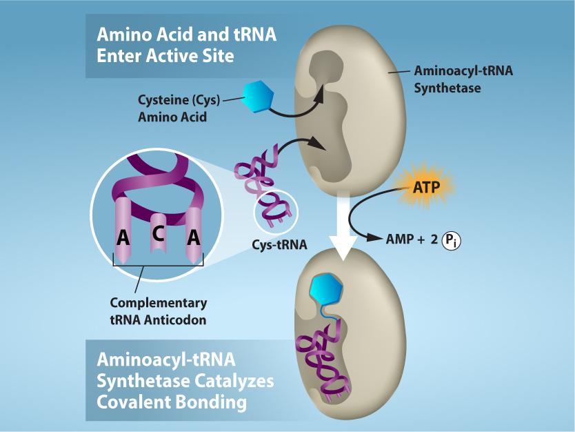
Figure 9.11 In protein phosphorylation, a phosphate group (PO 4 -3 ) is added to residues of the amino acids serine, threonine, and tyrosine.

Mary Ann Clark, Matthew Douglas, and Jung Choi, Biology 2E, OpenStax. · CC BY-NC-SA 4.0 · Original image ↗ · Access for free ↗

are small that propagate a signal after it has been initiated by the binding of the signaling to the . These help to spread a signal through the by altering the behavior of certain cellular .

Calcium is a widely used . The free of calcium (Ca2+) within a cell is very low because pumps in the plasma continuously remove it by using adenosine-5'-triphosphate (). For signaling purposes, Ca2+ is stored in , such as the , or accessed from outside the cell. When signaling occurs, -gated calcium channels allow the higher levels of Ca2+ that are present outside the cell (or in intracellular storage compartments) to flow into the , which raises the of Ca2+. The response to the increase in Ca2+ varies and depends on the cell type involved. For example, in the β-cells of the , Ca2+ signaling leads to the release of , and in muscle cells, an increase in Ca2+ leads to muscle contractions.

Another utilized in many different cell types is (). is synthesized by the from (Figure 9.12). The main role of in cells is to bind to and activate an called -dependent (A-). A- regulates many vital : It phosphorylates and of its target , activating them in the process. A- is found in many different types of cells, and the target in each kind of cell are different. Differences give rise to the variation of the responses to in different cells.

Cyclic A M P is made from A T P by the enzyme adenylyl cyclase. In the process, a pyrophosphate molecule composed of two phosphate residues is released. Cyclic A M P gets its name because the phosphate group is attached to the ribose ring in two places, forming a circle.
Figure 9.12 — 9.2 Propagation of the Signal

Figure 9.12 This diagram shows the mechanism for the formation of cyclic AMP (cAMP). cAMP serves as a second messenger to activate or inactivate proteins within the cell. Termination of the signal occurs when an enzyme called phosphodiesterase converts cAMP into AMP.

Mary Ann Clark, Matthew Douglas, and Jung Choi, Biology 2E, OpenStax. · CC BY-NC-SA 4.0 · Original image ↗ · Access for free ↗

Present in small in the plasma , inositol are that can also be converted into . Because these are components, they are located near -bound and can easily interact with them. Phosphatidylinositol () is the main that plays a role in cellular signaling. known as phosphorylate to form - (PIP) and -bisphosphate (PIP2).

The cleaves PIP2 to form diacylglycerol () and inositol triphosphate (IP3) (Figure 9.13). These of the cleavage of PIP2 serve as . Diacylglycerol () remains in the plasma and activates (), which then phosphorylates and in its target . IP3 diffuses into the and binds to -gated calcium channels in the to release Ca2+ that continues the signal cascade.

The molecular structures of P I P subscript 2 baseline, D A G, and I P subscript 3 baseline are shown. P I P subscript 2 baseline is a phospholipid that is cleaved by phospholipase C to form D A G, which has a long hydrophobic tail, and I P subscript 3 baseline, a ring structure with three phosphates attached.
Figure 9.13 — 9.2 Propagation of the Signal

Figure 9.13 The enzyme phospholipase C breaks down PIP 2 into IP 3 and DAG, both of which serve as second messengers.

Mary Ann Clark, Matthew Douglas, and Jung Choi, Biology 2E, OpenStax. · CC BY-NC-SA 4.0 · Original image ↗ · Access for free ↗