Junctions connect or seal cells, extracellular matrix transmits support and signals, and tissue architecture reflects each tissue’s job.
Seal, communicate, or resist tension
Tight junctions restrict passage between neighboring epithelial cells and help maintain distinct apical and basolateral membrane domains. Their permeability varies by tissue rather than forming a universal impermeable seal. Gap junctions contain paired channels that pass ions and small molecules directly between cells, enabling electrical and metabolic coupling.
Desmosomes are strong cell–cell anchoring junctions linked to intermediate filaments and resist mechanical stress. Adherens junctions connect actin networks, while hemidesmosomes anchor cells to basement membrane. Predict a defect by its function: a leaky epithelial barrier suggests altered sealing, poor electrical coupling suggests gap-junction disruption, and mechanical separation suggests impaired anchoring.
Source: 4.6 Connections between Cells and Cellular Activities · 4.2 Epithelial Tissue · 4.3 Connective Tissue Supports and Protects
Extracellular support also conveys information
Animal extracellular matrix contains fibrous proteins such as collagen and elastin, adhesive glycoproteins such as fibronectin and laminin, and hydrated proteoglycans. Collagen resists tension, elastin supports recoil, and glycosaminoglycan-rich material retains water and resists compression. The basement membrane is a specialized matrix beneath epithelia and around some other cells.
Integrins link extracellular ligands to the cytoskeleton and transmit signals in both directions. Matrix stiffness, adhesion, and composition can influence migration, survival, and differentiation. Cells continually synthesize and remodel matrix, so it is an active environment rather than an inert filler between otherwise independent cells.
Source: 4.6 Connections between Cells and Cellular Activities · 4.2 Epithelial Tissue · 4.3 Connective Tissue Supports and Protects
Compare epithelial and connective organization
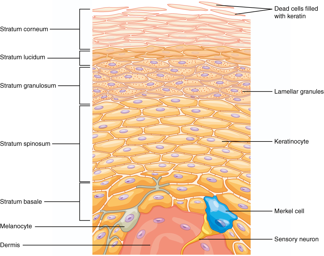
Epithelia form tightly packed sheets with polarity and little intervening matrix. Simple epithelia favor exchange or secretion; stratified epithelia protect against abrasion. Squamous, cuboidal, and columnar describe cell shape, and apical specializations such as microvilli or cilia support absorption or movement. Epithelia are avascular and obtain nutrients from underlying tissues.
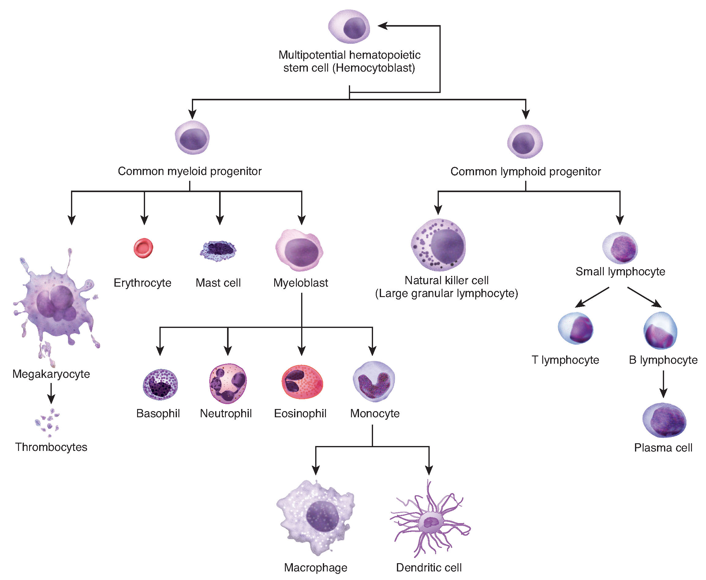
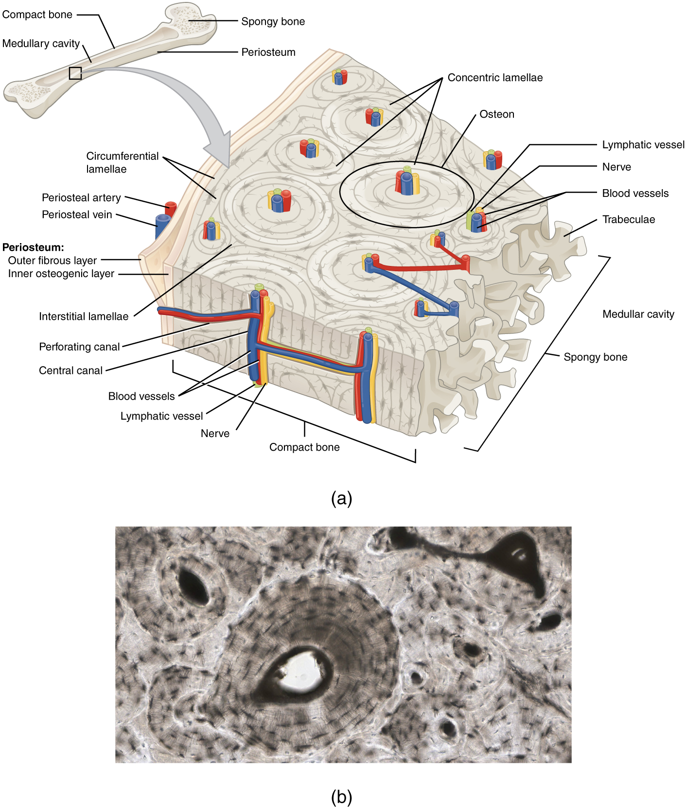
Connective tissues typically contain more extracellular matrix and more widely separated cells. Fibroblasts produce fibers and ground substance; other specialized cells produce adipose, cartilage, bone, or blood functions. Loose connective tissue supports exchange and attachment, dense connective tissue resists tension, cartilage provides flexible support, and mineralized bone supports load and mineral storage.
Source: 4.6 Connections between Cells and Cellular Activities · 4.2 Epithelial Tissue · 4.3 Connective Tissue Supports and Protects
Practice: apply the mechanism
Question. A mutation disrupts epithelial desmosomes while tight junctions initially remain intact. Predict the earliest defect during mechanical stretching and explain why it differs from a primary gap-junction defect.
Show reasoning
Cells are more likely to separate under tension because desmosomes connect intermediate-filament networks between neighbors. A primary gap-junction defect instead impairs passage of ions or small signaling molecules and thus coordination between cells. Either can eventually damage tissue broadly, but their immediate mechanisms differ.
Source: 4.6 Connections between Cells and Cellular Activities · 4.2 Epithelial Tissue · 4.3 Connective Tissue Supports and Protects
4.6 Connections between Cells and Cellular Activities — figures and tables

Figure 4.27 The extracellular matrix consists of a network of proteins and carbohydrates.
Mary Ann Clark, Matthew Douglas, and Jung Choi, Biology 2E, OpenStax. · CC BY-NC-SA 4.0 · Original image ↗ · Access for free ↗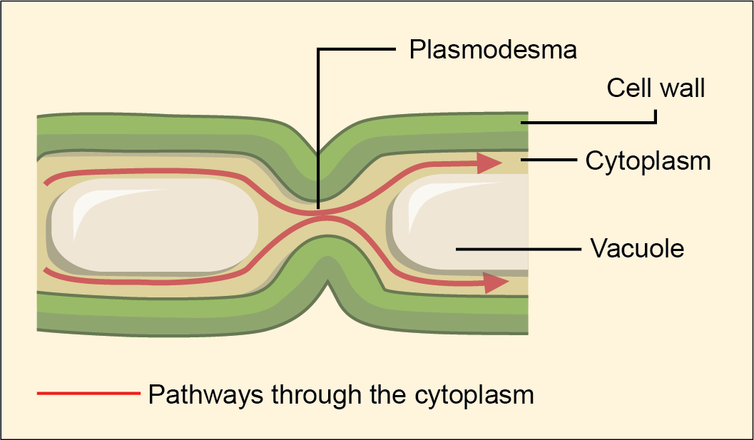
Figure 4.28 A plasmodesma is a channel between two adjacent plant cells' cell walls. Plasmodesmata allow materials to pass from one plant cell's cytoplasm to an adjacent cell's cytoplasm.
Mary Ann Clark, Matthew Douglas, and Jung Choi, Biology 2E, OpenStax. · CC BY-NC-SA 4.0 · Original image ↗ · Access for free ↗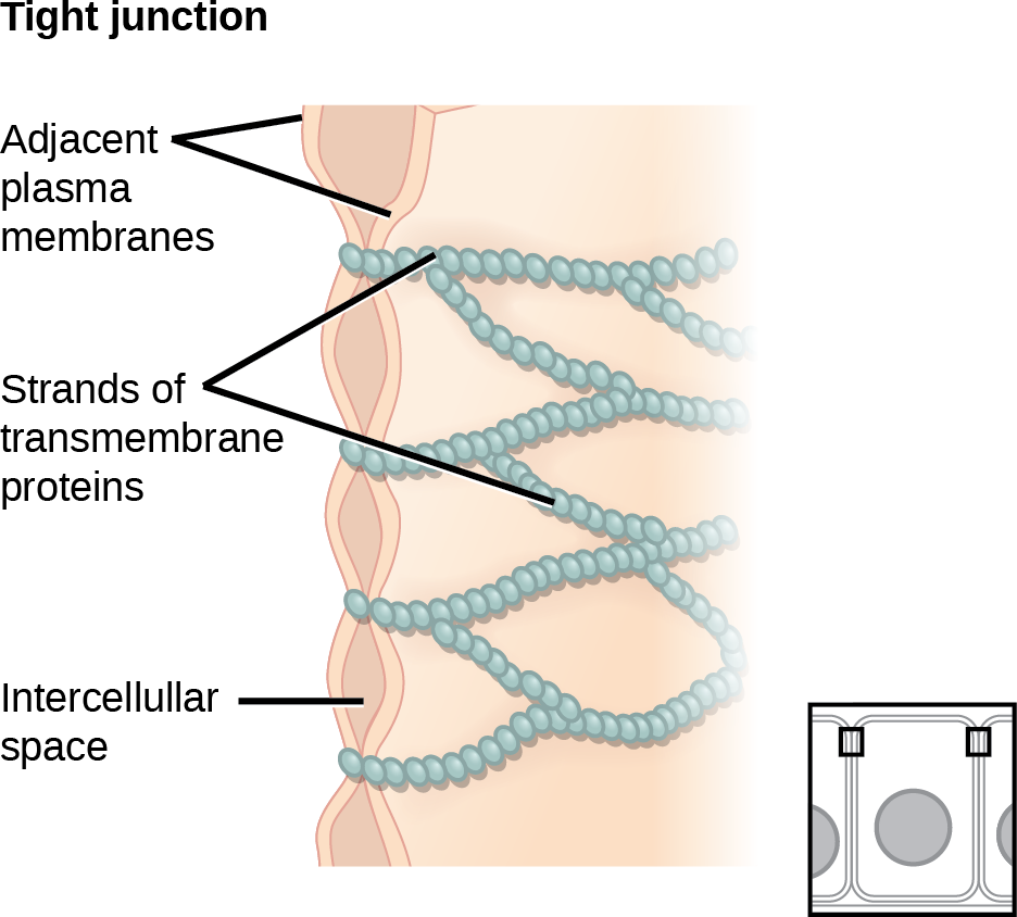
Figure 4.29 Tight junctions form watertight connections between adjacent animal cells. Proteins create tight junction adherence. (credit: modification of work by Mariana Ruiz Villareal)
Mary Ann Clark, Matthew Douglas, and Jung Choi, Biology 2E, OpenStax. (credit: modification of work by Mariana Ruiz Villareal) · CC BY-NC-SA 4.0 · Original image ↗ · Access for free ↗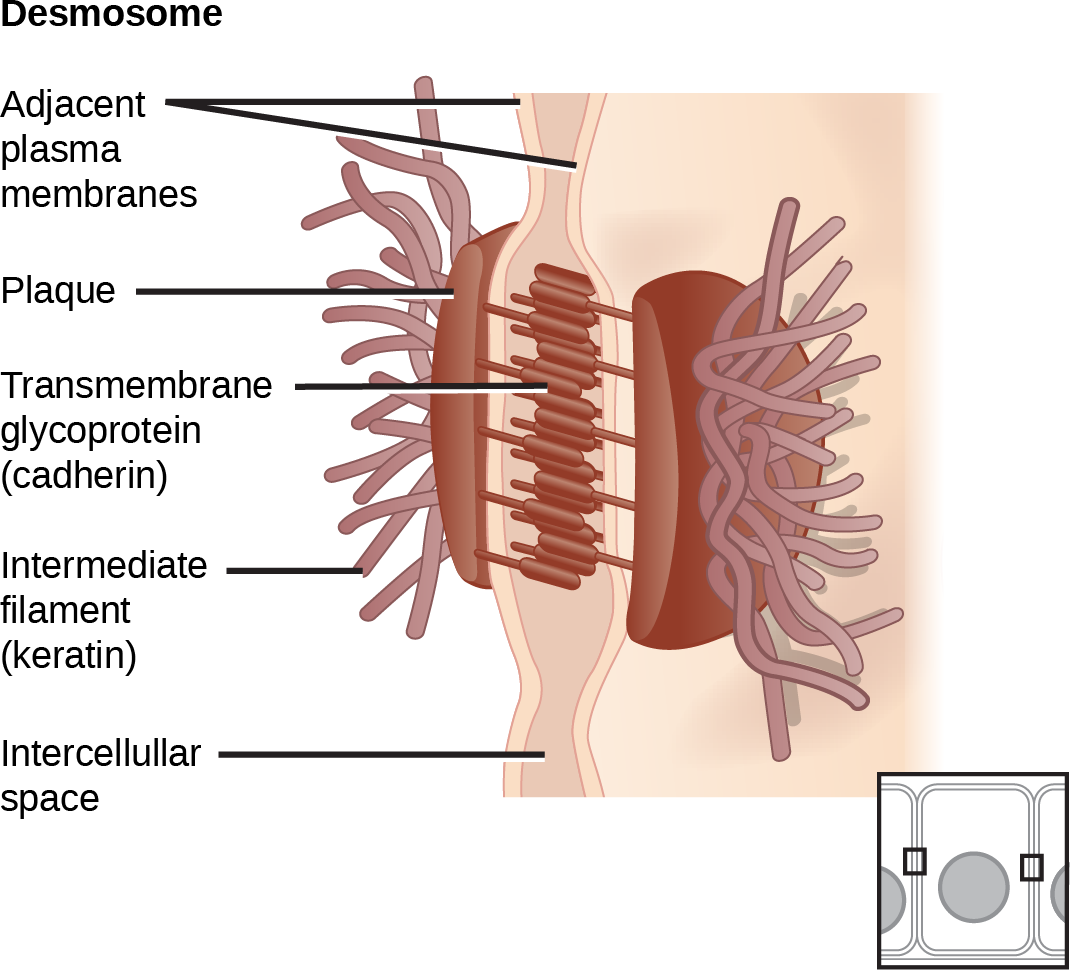
Figure 4.30 A desmosome forms a very strong spot weld between cells. Linking cadherins and intermediate filaments create it. (credit: modification of work by Mariana Ruiz Villareal)
Mary Ann Clark, Matthew Douglas, and Jung Choi, Biology 2E, OpenStax. (credit: modification of work by Mariana Ruiz Villareal) · CC BY-NC-SA 4.0 · Original image ↗ · Access for free ↗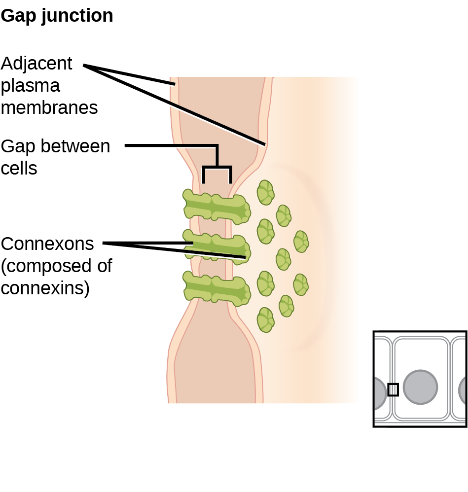
Figure 4.31 A gap junction is a protein-lined pore that allows water and small molecules to pass between adjacent animal cells. (credit: modification of work by Mariana Ruiz Villareal)
Mary Ann Clark, Matthew Douglas, and Jung Choi, Biology 2E, OpenStax. (credit: modification of work by Mariana Ruiz Villareal) · CC BY-NC-SA 4.0 · Original image ↗ · Access for free ↗Source: 4.6 Connections between Cells and Cellular Activities
4.2 Epithelial Tissue — figures and tables
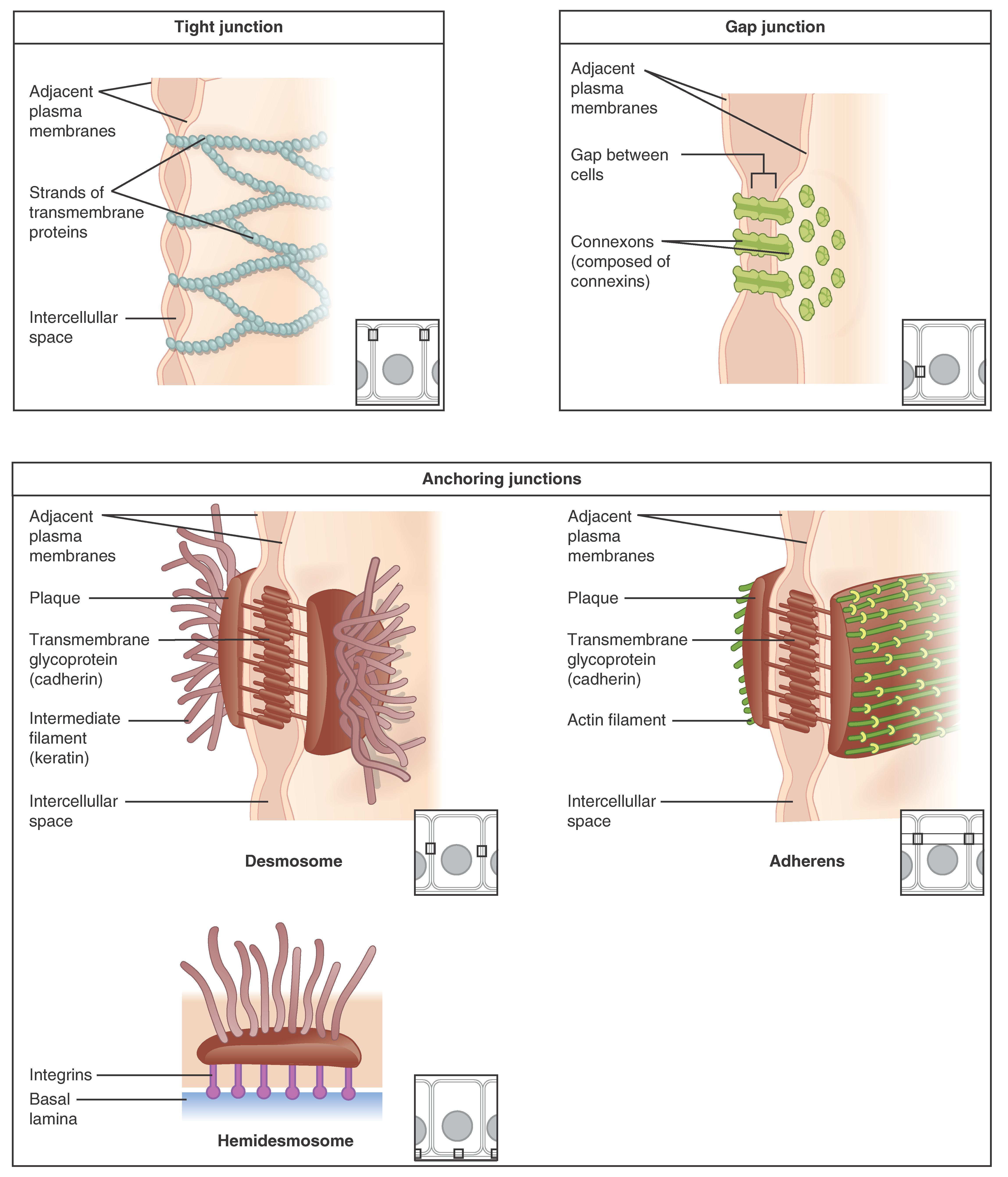
Figure 4.5 Types of Cell Junctions The three basic types of cell-to-cell junctions are tight junctions, gap junctions, and anchoring junctions.
J. Gordon Betts and colleagues, Anatomy And Physiology 2E, OpenStax. · CC BY-NC-SA 4.0 · Original image ↗ · Access for free ↗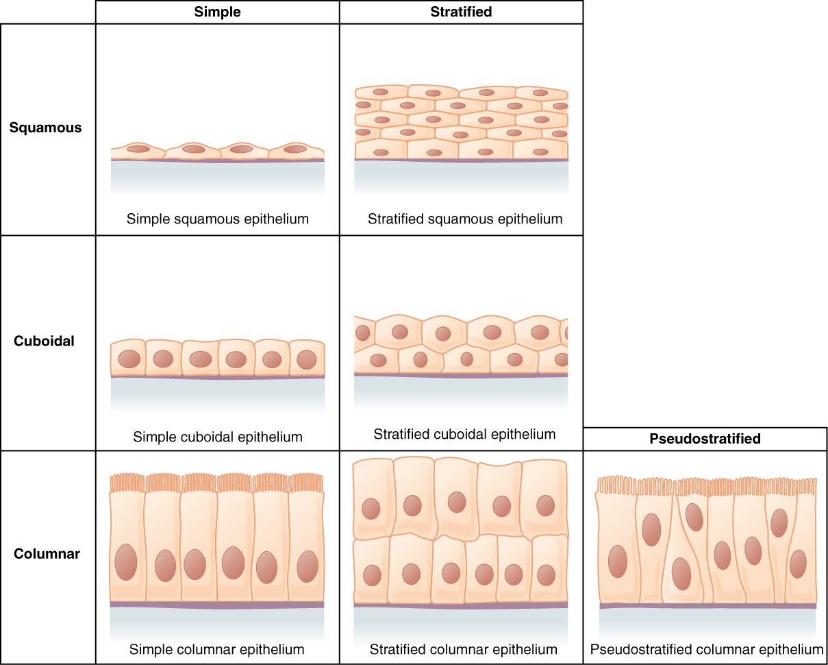
Figure 4.6 Cells of Epithelial Tissue Simple epithelial tissue is organized as a single layer of cells and stratified epithelial tissue is formed by several layers of cells.
J. Gordon Betts and colleagues, Anatomy And Physiology 2E, OpenStax. · CC BY-NC-SA 4.0 · Original image ↗ · Access for free ↗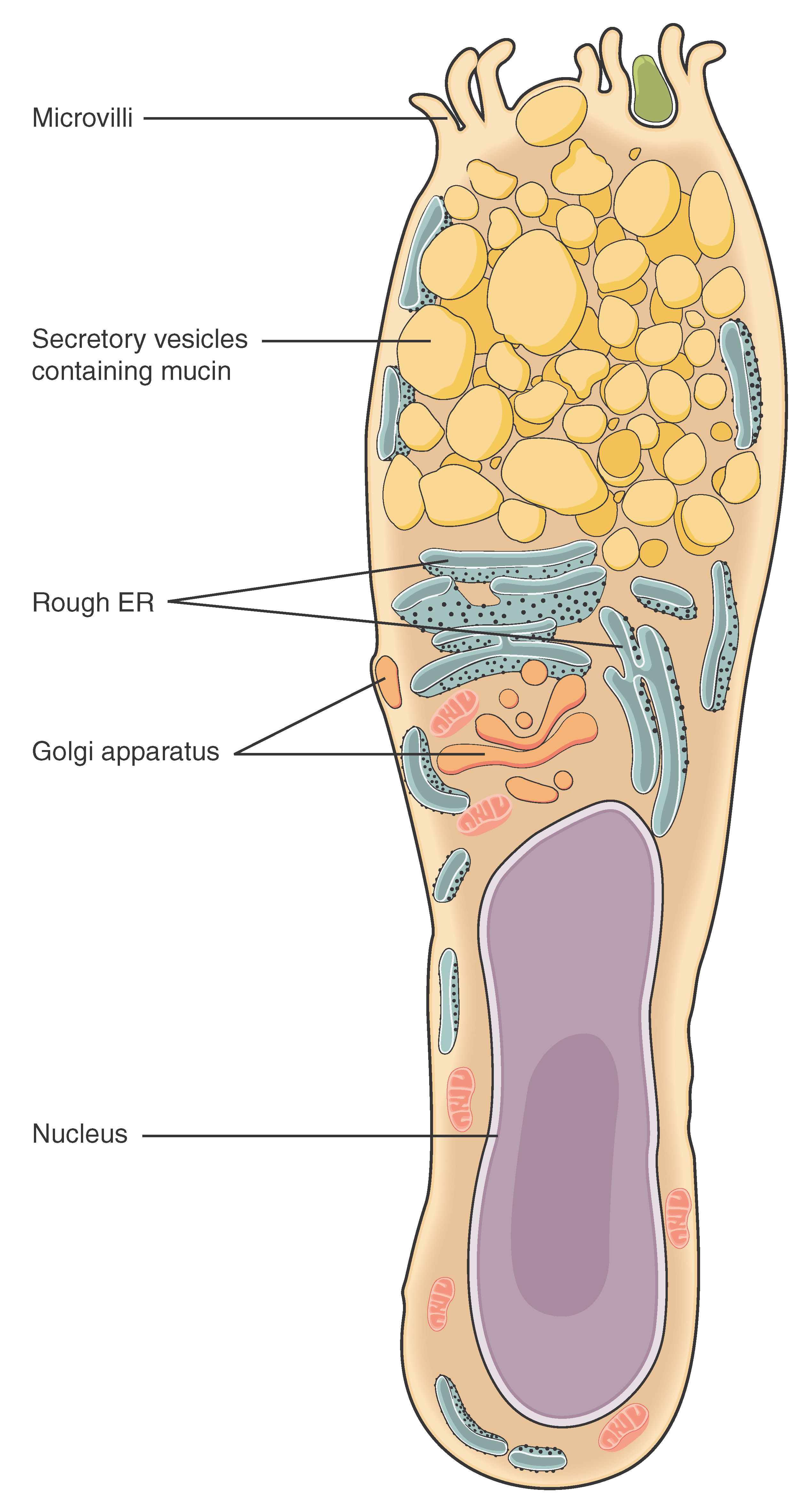
Figure 4.7 Goblet Cell (a) In the lining of the small intestine, columnar epithelium cells are interspersed with goblet cells. (b) The arrows in this micrograph point to the mucous-secreting goblet cells. LM × 1600. (Micrograph provided by the Regents of University of Michigan Medical School © 2012)
J. Gordon Betts and colleagues, Anatomy And Physiology 2E, OpenStax. · CC BY-NC-SA 4.0 · Original image ↗ · Access for free ↗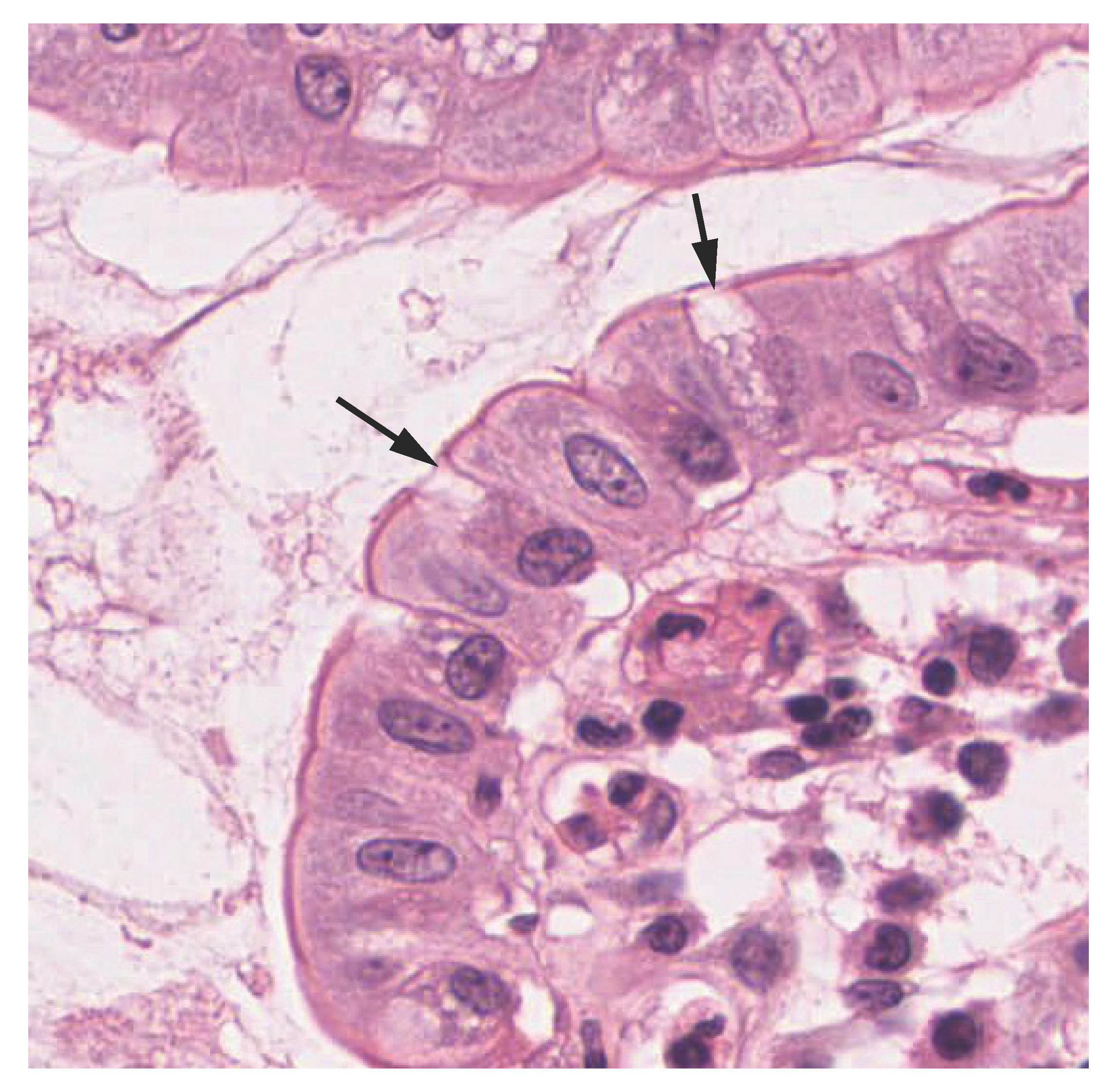
Figure 4.7 Goblet Cell (a) In the lining of the small intestine, columnar epithelium cells are interspersed with goblet cells. (b) The arrows in this micrograph point to the mucous-secreting goblet cells. LM × 1600. (Micrograph provided by the Regents of University of Michigan Medical School © 2012)
J. Gordon Betts and colleagues, Anatomy And Physiology 2E, OpenStax. · CC BY-NC-SA 4.0 · Original image ↗ · Access for free ↗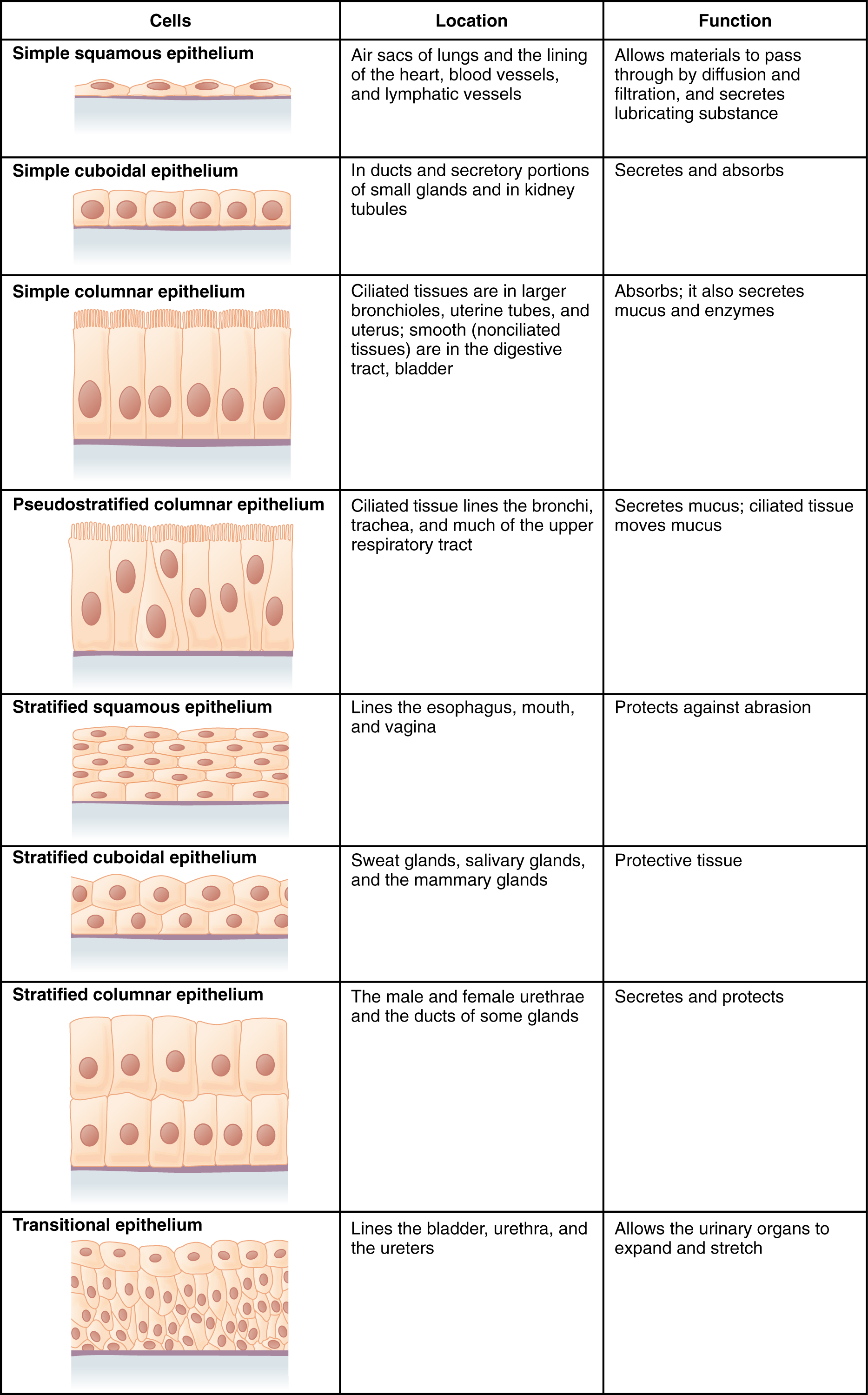
Figure 4.8 Summary of Epithelial Tissue Cells
J. Gordon Betts and colleagues, Anatomy And Physiology 2E, OpenStax. · CC BY-NC-SA 4.0 · Original image ↗ · Access for free ↗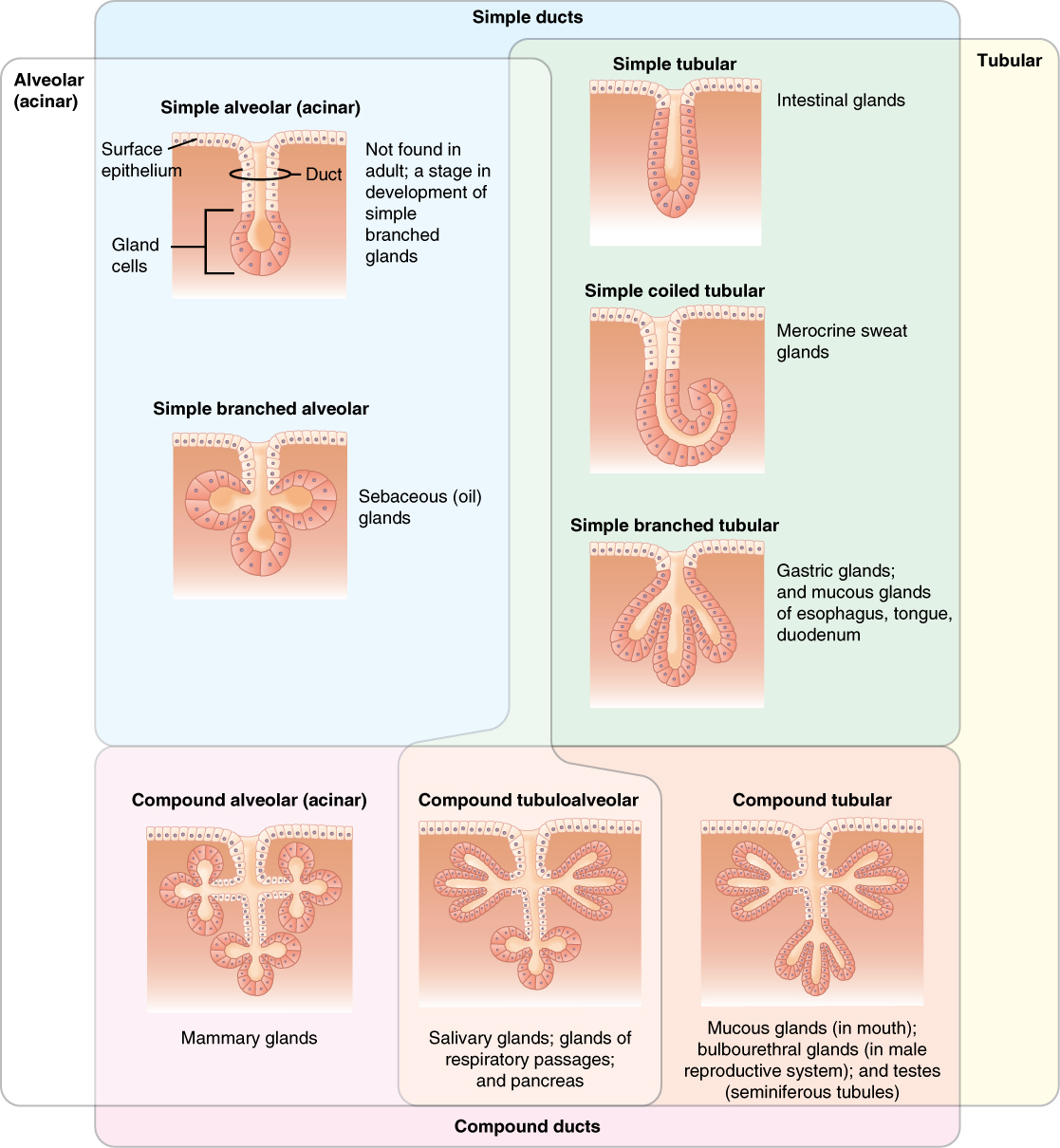
Figure 4.9 Types of Exocrine Glands Exocrine glands are classified by their structure.
J. Gordon Betts and colleagues, Anatomy And Physiology 2E, OpenStax. · CC BY-NC-SA 4.0 · Original image ↗ · Access for free ↗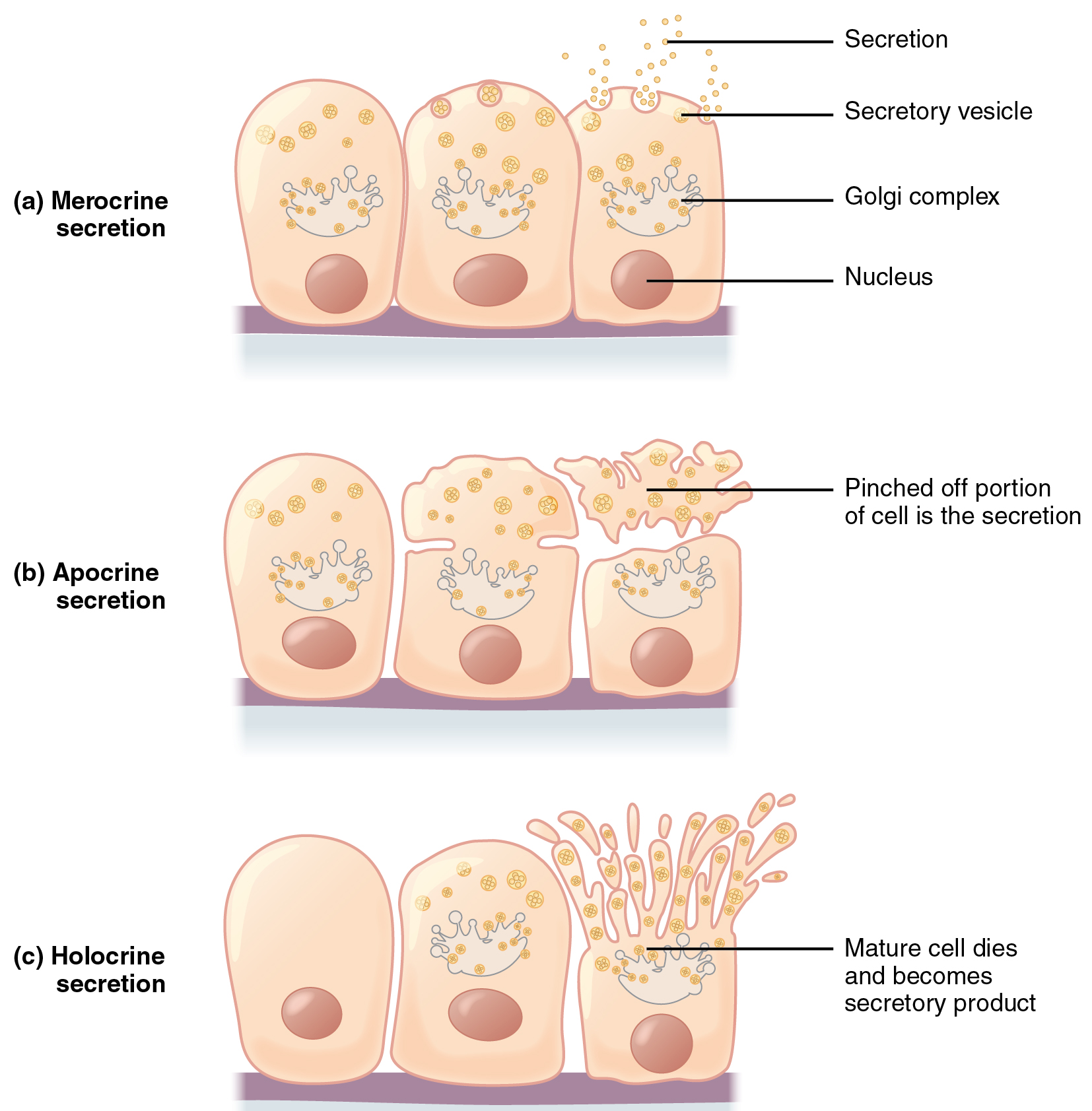
Figure 4.10 Modes of Glandular Secretion (a) In merocrine secretion, the cell remains intact. (b) In apocrine secretion, the apical portion of the cell is released, as well. (c) In holocrine secretion, the cell is destroyed as it releases its product and the cell itself becomes part of the secretion.
J. Gordon Betts and colleagues, Anatomy And Physiology 2E, OpenStax. · CC BY-NC-SA 4.0 · Original image ↗ · Access for free ↗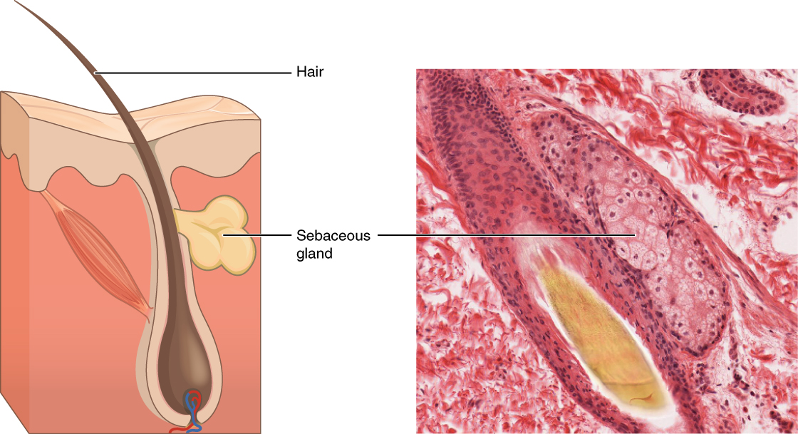
Figure 4.11 Sebaceous Glands These glands secrete oils that lubricate and protect the skin. They are holocrine glands and they are destroyed after releasing their contents. New glandular cells form to replace the cells that are lost. LM × 400. (Micrograph provided by the Regents of University of Michigan Medical School © 2012)
J. Gordon Betts and colleagues, Anatomy And Physiology 2E, OpenStax. · CC BY-NC-SA 4.0 · Original image ↗ · Access for free ↗Source: 4.2 Epithelial Tissue
4.3 Connective Tissue Supports and Protects — figures and tables

Figure 4.12 Connective Tissue Proper Fibroblasts produce this fibrous tissue. Connective tissue proper includes the fixed cells fibrocytes, adipocytes, and mesenchymal cells. LM × 400. (Micrograph provided by the Regents of University of Michigan Medical School © 2012)
J. Gordon Betts and colleagues, Anatomy And Physiology 2E, OpenStax. · CC BY-NC-SA 4.0 · Original image ↗ · Access for free ↗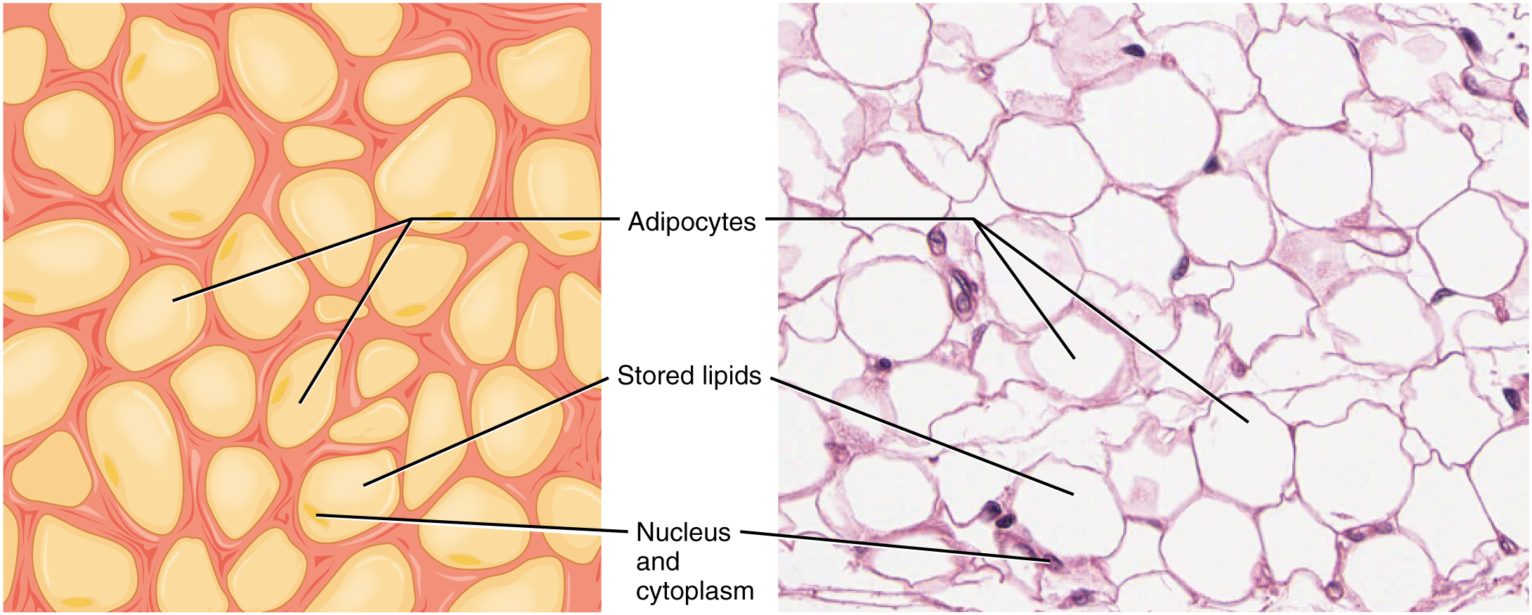
Figure 4.13 Adipose Tissue This is a loose connective tissue that consists of fat cells with little extracellular matrix. It stores fat for energy and provides insulation. LM × 800. (Micrograph provided by the Regents of University of Michigan Medical School © 2012)
J. Gordon Betts and colleagues, Anatomy And Physiology 2E, OpenStax. · CC BY-NC-SA 4.0 · Original image ↗ · Access for free ↗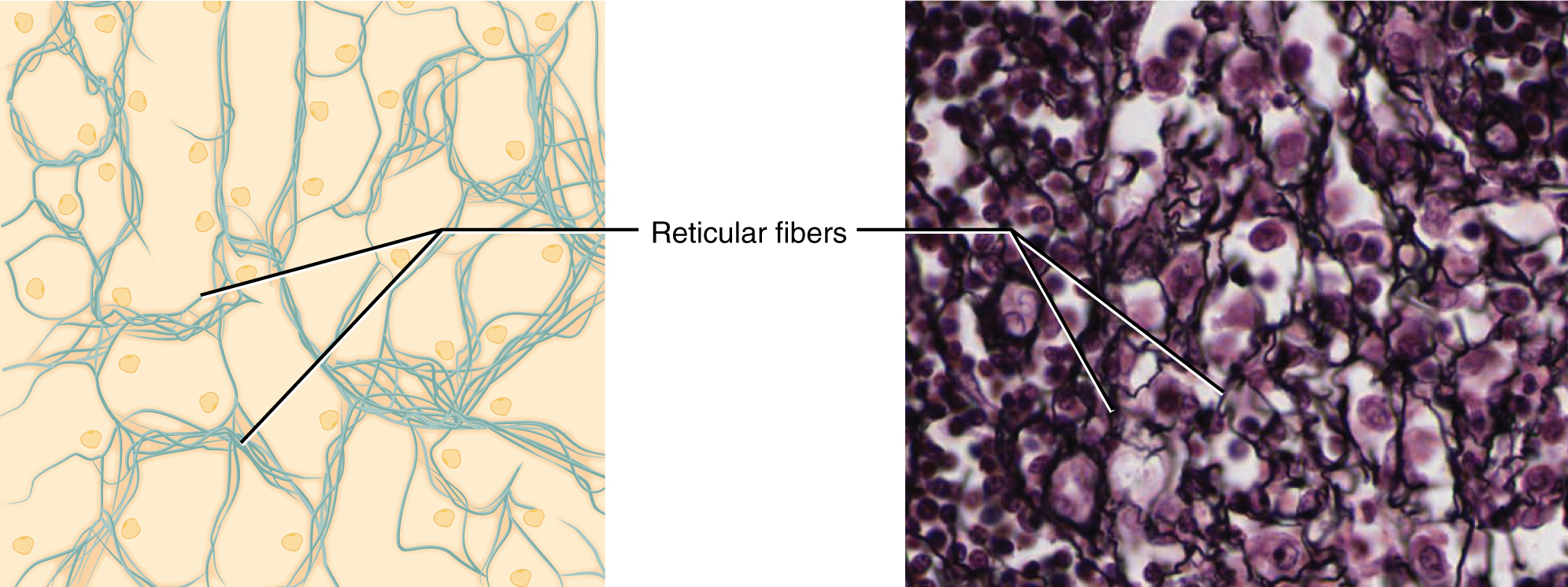
Figure 4.14 Reticular Tissue This is a loose connective tissue made up of a network of reticular fibers that provides a supportive framework for soft organs. LM × 1600. (Micrograph provided by the Regents of University of Michigan Medical School © 2012)
J. Gordon Betts and colleagues, Anatomy And Physiology 2E, OpenStax. · CC BY-NC-SA 4.0 · Original image ↗ · Access for free ↗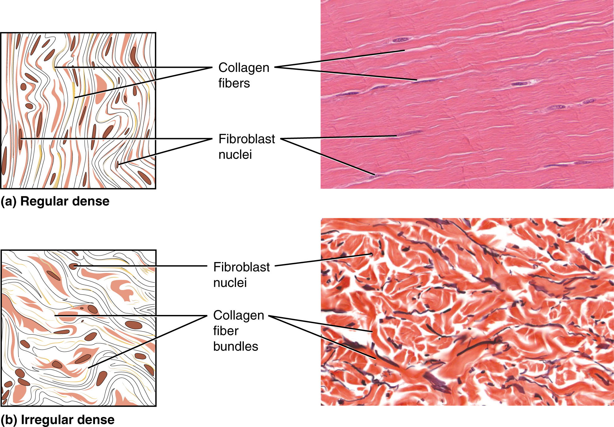
Figure 4.15 Dense Connective Tissue (a) Dense regular connective tissue consists of collagenous fibers packed into parallel bundles. (b) Dense irregular connective tissue consists of collagenous fibers interwoven into a mesh-like network. From top, LM × 1000, LM × 200. (Micrographs provided by the Regents of University of Michigan Medical School © 2012)
J. Gordon Betts and colleagues, Anatomy And Physiology 2E, OpenStax. · CC BY-NC-SA 4.0 · Original image ↗ · Access for free ↗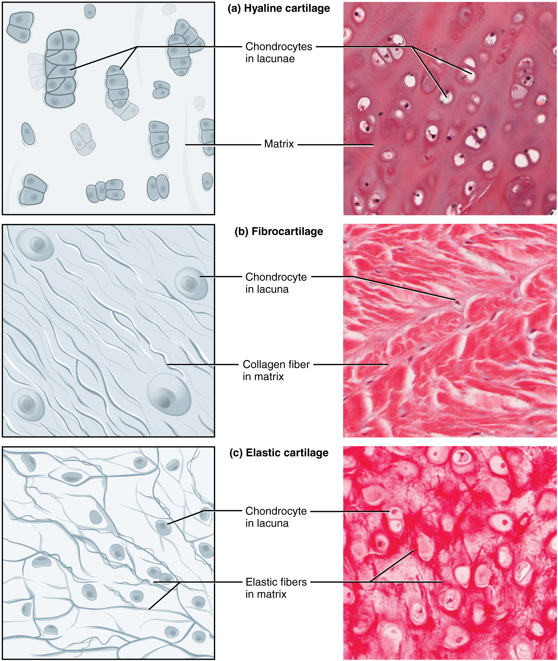
Figure 4.16 Types of Cartilage Cartilage is a connective tissue consisting of collagenous fibers embedded in a firm matrix of chondroitin sulfates. (a) Hyaline cartilage provides support with some flexibility. The example is from dog tissue. (b) Fibrocartilage provides some compressibility and can absorb pressure. (c) Elastic cartilage provides firm but elastic support. From top, LM × 300, LM × 1200, LM × 1016. (Micrographs provided by the Regents of University of Michigan Medical School © 2012)
J. Gordon Betts and colleagues, Anatomy And Physiology 2E, OpenStax. · CC BY-NC-SA 4.0 · Original image ↗ · Access for free ↗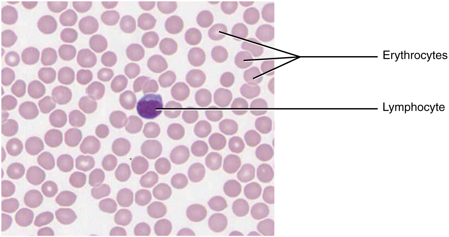
Figure 4.17 Blood: A Fluid Connective Tissue Blood is a fluid connective tissue containing erythrocytes and various types of leukocytes that circulate in a liquid extracellular matrix. LM × 1600. (Micrograph provided by the Regents of University of Michigan Medical School © 2012)
J. Gordon Betts and colleagues, Anatomy And Physiology 2E, OpenStax. · CC BY-NC-SA 4.0 · Original image ↗ · Access for free ↗| Connective tissue proper | Supportive connective tissue | Fluid connective tissue |
|---|---|---|
Loose connective tissue
|
Cartilage
|
Blood
Lymph |
Table 4.1. J. Gordon Betts and colleagues, Anatomy And Physiology 2E, OpenStax.