Compartments separate incompatible reactions; targeting signals and vesicle routes connect protein synthesis, secretion, energy conversion, and degradation.
Nuclear organization and ribosome assembly
The nuclear envelope has two membranes and is continuous with the endoplasmic reticulum. Nuclear pores allow small molecules to exchange and selectively transport larger proteins and RNA complexes using localization signals and transport factors. This separates nuclear transcription and RNA processing from cytosolic translation in eukaryotes.
The nucleolus assembles around rRNA gene regions: much rRNA is transcribed and processed there, and imported ribosomal proteins combine with rRNA to form precursor ribosomal subunits. Subunits are exported separately before participating in translation. The nucleolus is not enclosed by its own membrane, and ribosomal proteins are synthesized on cytosolic ribosomes rather than translated inside the nucleolus.
Source: 4.3 Eukaryotic Cells · 4.4 The Endomembrane System and Proteins
Two membranes create an energy-converting compartment
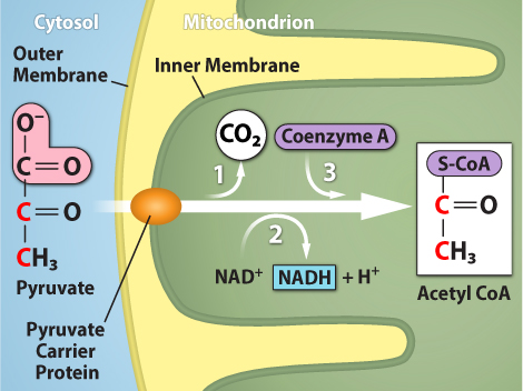
The relatively permeable outer mitochondrial membrane surrounds a highly selective inner membrane folded into cristae. The matrix contains enzymes for pyruvate oxidation, most citric-cycle reactions, and β-oxidation; the inner membrane houses electron transport and ATP synthase. Proton separation across that membrane links fuel oxidation to ATP production.
Mitochondria contain their own DNA and ribosomes and grow and divide from existing organelles, consistent with endosymbiotic ancestry. They are not genetically autonomous: most mitochondrial proteins are nuclear-encoded, synthesized in the cytosol, and imported. Organelle number, fusion, fission, and selective removal change with cellular demand and damage.
Source: 4.3 Eukaryotic Cells · 4.4 The Endomembrane System and Proteins
Rough ER, smooth ER, and Golgi routes
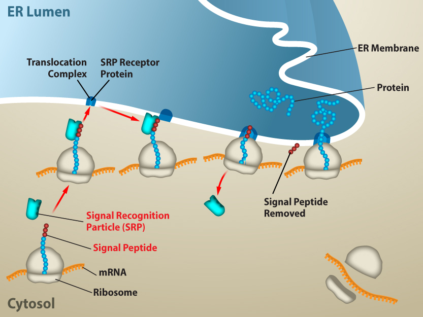
An ER signal sequence recruits translating ribosomes to rough ER, allowing secreted and many membrane proteins to enter the lumen or membrane through a translocon. Folding, quality control, disulfide formation, and some glycosylation occur there. Transport vesicles carry cargo to the cis Golgi, through processing compartments, and toward the trans Golgi for sorting to secretion, plasma membrane, or endosomal routes.
Smooth ER lacks attached ribosomes and participates in lipid synthesis, detoxification, and calcium storage; specialized muscle smooth ER is the sarcoplasmic reticulum. Free and attached ribosomes are the same basic machinery, with their location determined by the protein currently being synthesized.
Source: 4.3 Eukaryotic Cells · 4.4 The Endomembrane System and Proteins
Lysosomes and peroxisomes use different chemistry
Lysosomes contain acid hydrolases and maintain an acidic lumen using proton pumps. They digest endocytosed material and cellular components delivered through autophagy; products return to cellular metabolism. Failure of a lysosomal enzyme or targeting pathway can cause substrate accumulation.
Peroxisomes perform oxidative reactions, including shortening very-long-chain fatty acids, and contain catalase to handle hydrogen peroxide. Their oxidative chemistry differs from acidic lysosomal hydrolysis and from mitochondrial respiratory ATP synthesis. Peroxisomal electrons can be transferred directly to oxygen, producing peroxide; the organelle does not use a respiratory proton gradient to capture this energy as ATP.
Source: 4.3 Eukaryotic Cells · 4.4 The Endomembrane System and Proteins
Practice: apply the mechanism
Question. A newly made secreted protein is found in the ER but never reaches the extracellular medium. Would this establish failed translation? Name two downstream explanations and one useful measurement.
Show reasoning
No. ER-associated protein demonstrates that at least some translation and targeting occurred. Defective folding can retain cargo in ER quality control, or disrupted vesicle traffic/Golgi sorting can prevent export. A pulse–chase experiment or time-resolved localization can follow movement from ER to Golgi and medium; measure intracellular abundance as well as secretion to distinguish retention from degradation.
Source: 4.3 Eukaryotic Cells · 4.4 The Endomembrane System and Proteins
4.3 Eukaryotic Cells — figures and tables
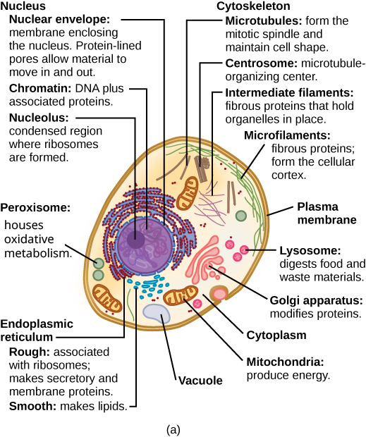
Figure 4.8 These figures show the major organelles and other cell components of (a) a typical animal cell and (b) a typical eukaryotic plant cell. The plant cell has a cell wall, chloroplasts, plastids, and a central vacuole—structures not in animal cells. Most cells do not have lysosomes or centrosomes.
Mary Ann Clark, Matthew Douglas, and Jung Choi, Biology 2E, OpenStax. · CC BY-NC-SA 4.0 · Original image ↗ · Access for free ↗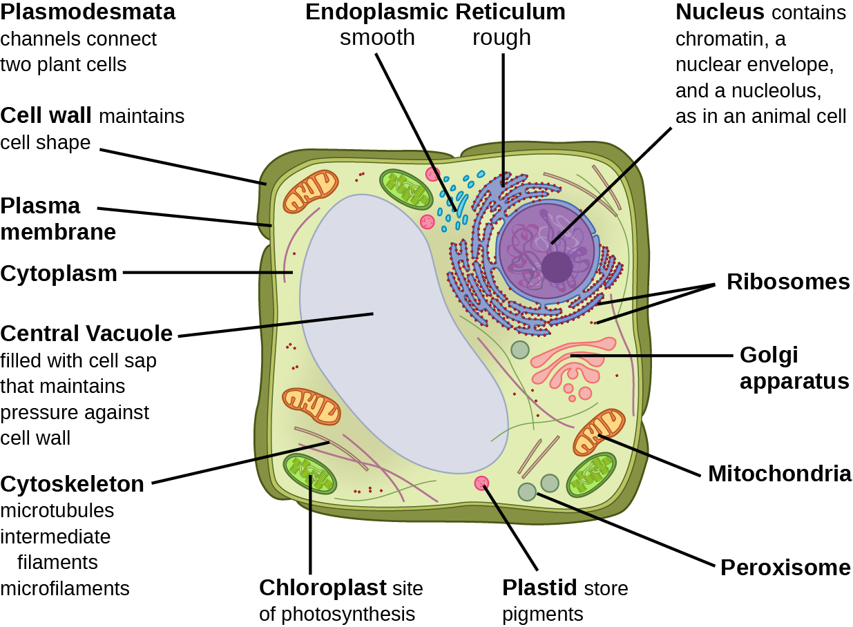
Figure 4.8 These figures show the major organelles and other cell components of (a) a typical animal cell and (b) a typical eukaryotic plant cell. The plant cell has a cell wall, chloroplasts, plastids, and a central vacuole—structures not in animal cells. Most cells do not have lysosomes or centrosomes.
Mary Ann Clark, Matthew Douglas, and Jung Choi, Biology 2E, OpenStax. · CC BY-NC-SA 4.0 · Original image ↗ · Access for free ↗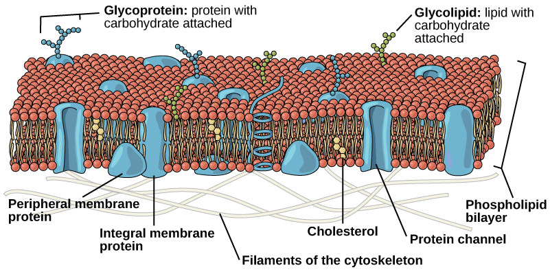
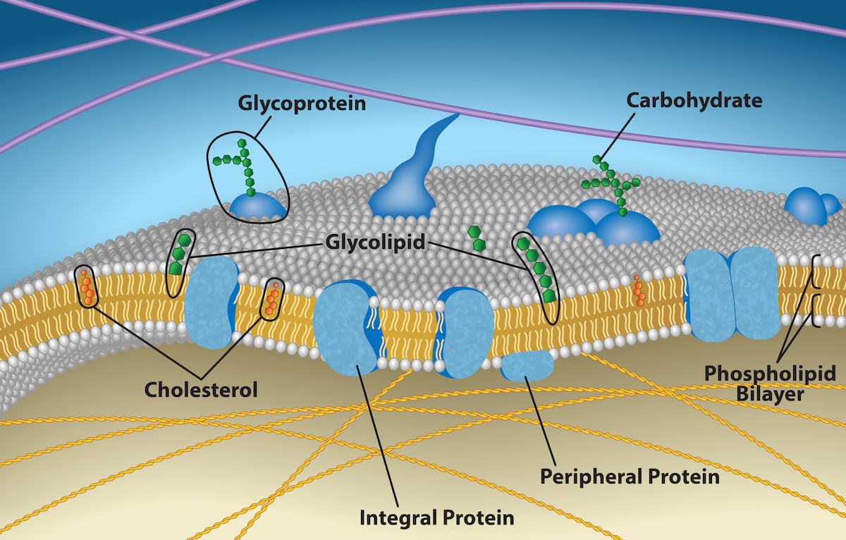
Figure 4.9 The eukaryotic plasma membrane is a phospholipid bilayer with proteins and cholesterol embedded in it.
Mary Ann Clark, Matthew Douglas, and Jung Choi, Biology 2E, OpenStax. · CC BY-NC-SA 4.0 · Original image ↗ · Access for free ↗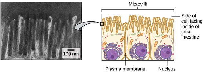
Figure 4.10 Microvilli, as they appear on cells lining the small intestine, increase the surface area available for absorption. These microvilli are only on the area of the plasma membrane that faces the cavity from which substances will be absorbed. (credit "micrograph": modification of work by Louisa Howard)
Mary Ann Clark, Matthew Douglas, and Jung Choi, Biology 2E, OpenStax. (credit "micrograph": modification of work by Louisa Howard) · CC BY-NC-SA 4.0 · Original image ↗ · Access for free ↗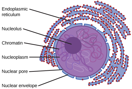
Figure 4.11 The nucleus stores chromatin (DNA plus proteins) in a gel-like substance called the nucleoplasm. The nucleolus is a condensed chromatin region where ribosome synthesis occurs. We call the nucleus' boundary the nuclear envelope. It consists of two phospholipid bilayers: an outer and an inner membrane. The nuclear membrane is continuous with the endoplasmic reticulum. Nuclear pores allow substances to enter and exit the nucleus.
Mary Ann Clark, Matthew Douglas, and Jung Choi, Biology 2E, OpenStax. · CC BY-NC-SA 4.0 · Original image ↗ · Access for free ↗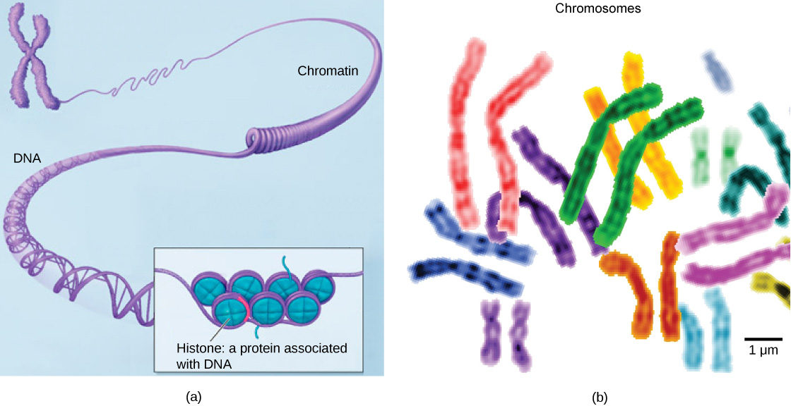
Figure 4.12 (a) This image shows various levels of chromatin's organization (DNA and protein). (b) This image shows paired chromosomes. (credit b: modification of work by NIH; scale-bar data from Matt Russell)
Mary Ann Clark, Matthew Douglas, and Jung Choi, Biology 2E, OpenStax. (credit b: modification of work by NIH; scale-bar data from Matt Russell) · CC BY-NC-SA 4.0 · Original image ↗ · Access for free ↗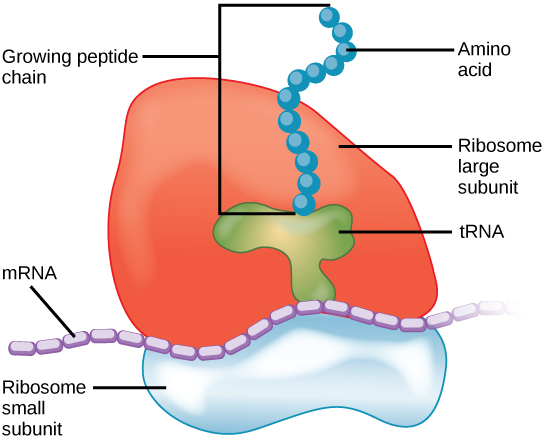
Figure 4.13 A large subunit (top) and a small subunit (bottom) comprise ribosomes. During protein synthesis, ribosomes assemble amino acids into proteins.
Mary Ann Clark, Matthew Douglas, and Jung Choi, Biology 2E, OpenStax. · CC BY-NC-SA 4.0 · Original image ↗ · Access for free ↗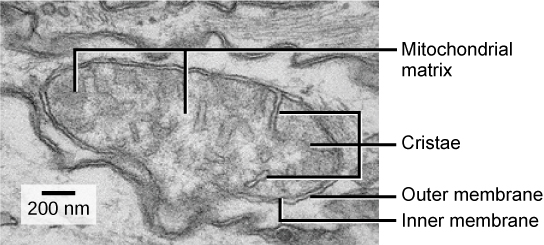
Figure 4.14 This electron micrograph shows a mitochondrion through an electron microscope. This organelle has an outer membrane and an inner membrane. The inner membrane contains folds, called cristae, which increase its surface area. We call the space between the two membranes the intermembrane space, and the space inside the inner membrane the mitochondrial matrix. ATP synthesis takes place on the inner membrane. (credit: modification of work by Matthew Britton; scale-bar data from Matt Russell)
Mary Ann Clark, Matthew Douglas, and Jung Choi, Biology 2E, OpenStax. (credit: modification of work by Matthew Britton; scale-bar data from Matt Russell) · CC BY-NC-SA 4.0 · Original image ↗ · Access for free ↗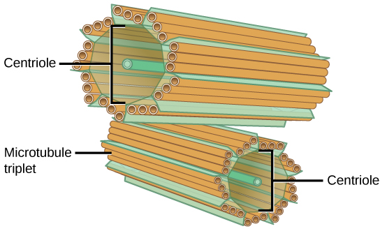
Figure 4.15 The centrosome consists of two centrioles that lie at right angles to each other. Each centriole is a cylinder comprised of nine triplets of microtubules. Nontubulin proteins (indicated by the green lines) hold the microtubule triplets together.
Mary Ann Clark, Matthew Douglas, and Jung Choi, Biology 2E, OpenStax. · CC BY-NC-SA 4.0 · Original image ↗ · Access for free ↗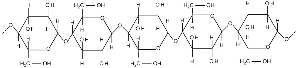
Figure 4.16 Cellulose is a long chain of β-glucose molecules connected by a 1-4 linkage. The dashed lines at each end of the figure indicate a series of many more glucose units. The size of the page makes it impossible to portray an entire cellulose molecule.
Mary Ann Clark, Matthew Douglas, and Jung Choi, Biology 2E, OpenStax. · CC BY-NC-SA 4.0 · Original image ↗ · Access for free ↗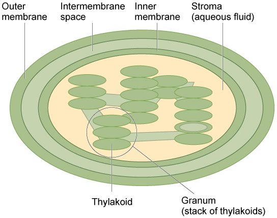
Figure 4.17 The chloroplast has an outer membrane, an inner membrane, and membrane structures - thylakoids that are stacked into grana. We call the space inside the thylakoid membranes the thylakoid space. The light harvesting reactions take place in the thylakoid membranes, and sugar synthesis takes place in the fluid inside the inner membrane, which we call the stroma. Chloroplasts also have their own genome, which is contained on a single circular chromosome.
Mary Ann Clark, Matthew Douglas, and Jung Choi, Biology 2E, OpenStax. · CC BY-NC-SA 4.0 · Original image ↗ · Access for free ↗Source: 4.3 Eukaryotic Cells
4.4 The Endomembrane System and Proteins — figures and tables
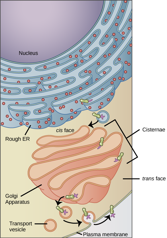
Figure 4.18 Membrane and secretory proteins are synthesized in the rough endoplasmic reticulum (RER). The RER also sometimes modifies proteins. In this illustration, a (green) integral membrane protein is modified by attachment of a (purple) carbohydrate in the ER. Vesicles with the integral protein bud from the ER and fuse with the Golgi apparatus' cis face. As the protein passes along the Golgi’s cisternae, the addition of more carbohydrates further modifies it. After its synthesis is complete, it exits as an integral membrane protein of the vesicle that buds from the Golgi’s trans face. When the vesicle fuses with the cell membrane, the protein becomes an integral portion of that cell membrane. (credit: modification of work by Magnus Manske)
Mary Ann Clark, Matthew Douglas, and Jung Choi, Biology 2E, OpenStax. (credit: modification of work by Magnus Manske) · CC BY-NC-SA 4.0 · Original image ↗ · Access for free ↗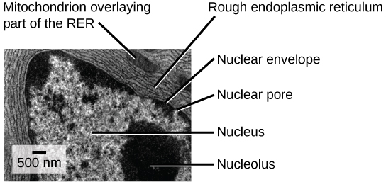
Figure 4.19 This transmission electron micrograph shows the rough endoplasmic reticulum and other organelles in a pancreatic cell. (credit: modification of work by Louisa Howard)
Mary Ann Clark, Matthew Douglas, and Jung Choi, Biology 2E, OpenStax. (credit: modification of work by Louisa Howard) · CC BY-NC-SA 4.0 · Original image ↗ · Access for free ↗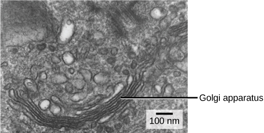
Figure 4.20 The Golgi apparatus in this white blood cell is visible as a stack of semicircular, flattened rings in the lower portion of the image. You can see several vesicles near the Golgi apparatus. (credit: modification of work by Louisa Howard)
Mary Ann Clark, Matthew Douglas, and Jung Choi, Biology 2E, OpenStax. (credit: modification of work by Louisa Howard) · CC BY-NC-SA 4.0 · Original image ↗ · Access for free ↗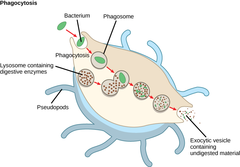
Figure 4.21 A macrophage has engulfed (phagocytized) a potentially pathogenic bacterium and then fuses with lysosomes within the cell to destroy the pathogen. Other organelles are present in the cell but for simplicity we do not show them.
Mary Ann Clark, Matthew Douglas, and Jung Choi, Biology 2E, OpenStax. · CC BY-NC-SA 4.0 · Original image ↗ · Access for free ↗