Relate outer, middle, and inner ear structures to transmission of sound
The pinna gathers sound into the auditory canal. Air-pressure changes vibrate the tympanic membrane, and the malleus, incus, and stapes transmit motion to the oval window. The middle ear improves energy transfer from air to inner-ear fluid. Fluid movement produces a traveling wave along the cochlear basilar membrane; the round window permits pressure relief. High frequencies peak near the stiff, narrow base, whereas low frequencies peak nearer the flexible apex. Frequency relates to pitch and amplitude to perceived loudness, but perception also depends on frequency and context. Conductive loss impairs sound transmission; sensorineural loss involves hair cells or neural pathways.
Explain hair-cell transduction and auditory pathways in the brain
Deflection of a cochlear hair bundle toward its tallest stereocilia increases opening of mechanically gated channels; movement in the opposite direction reduces opening. Potassium enters from potassium-rich endolymph, depolarizing the cell; calcium entry then promotes transmitter release onto auditory-nerve fibers. Inner hair cells supply most sensory output, whereas outer hair cells amplify and sharpen cochlear motion. Signals pass through brainstem nuclei, inferior colliculus, medial geniculate nucleus, and auditory cortex, with substantial bilateral representation. Place coding contributes to pitch across frequencies; temporal firing patterns contribute especially at lower frequencies. Interaural timing and intensity differences help localize sound. Loudness cannot be inferred from frequency alone.
Worked question. A sound has twice the frequency but the same physical amplitude as another. Predict pitch, and explain why a damaged middle ear differs from loss of basal cochlear hair cells.
The higher-frequency sound generally has higher pitch; equal amplitude does not guarantee equal perceived loudness. Middle-ear damage impairs mechanical transmission across frequencies according to the lesion. Basal hair-cell damage especially disrupts high-frequency transduction. Comparing air and bone conduction helps distinguish transmission loss from inner-ear loss.
Figure 5.18 The ear is divided into outer (pinna and tympanic membrane), middle (the three ossicles: malleus, incus, and stapes), and inner (cochlea and basilar membrane) divisions.
Figure 5.19 Localizing sound involves the use of both monaural and binaural cues. (credit "plane": modification of work by Max Pfandl)
Rose M. Spielman, William J. Jenkins, and Marilyn D. Lovett, Psychology 2E, OpenStax. (credit "plane": modification of work by Max Pfandl) · CC BY-NC-SA 4.0
· Original image ↗ · Access for free ↗Figure 5.20 — 5.4 Hearing
Figure 5.20 Environmental factors that can lead to sensorineural hearing loss include regular exposure to loud music or construction equipment. (a) Musical performers and (b) construction workers are at risk for this type of hearing loss. (credit a: modification of work by "GillyBerlin_Flickr"/Flickr; credit b: modification of work by Nick Allen)
Rose M. Spielman, William J. Jenkins, and Marilyn D. Lovett, Psychology 2E, OpenStax. (credit a: modification of work by "GillyBerlin_Flickr"/Flickr; credit b: modification of work by Nick Allen) · CC BY-NC-SA 4.0
· Original image ↗ · Access for free ↗
Figure 14.7 DNA has (a) a double helix structure and (b) phosphodiester bonds; the dotted lines between Thymine and Adenine and Guanine and Cytosine represent hydrogen bonds. The (c) major and minor grooves are binding sites for DNA binding proteins during processes such as transcription (the copying of RNA from DNA) and replication.
Figure 14.7 DNA has (a) a double helix structure and (b) phosphodiester bonds; the dotted lines between Thymine and Adenine and Guanine and Cytosine represent hydrogen bonds. The (c) major and minor grooves are binding sites for DNA binding proteins during processes such as transcription (the copying of RNA from DNA) and replication.
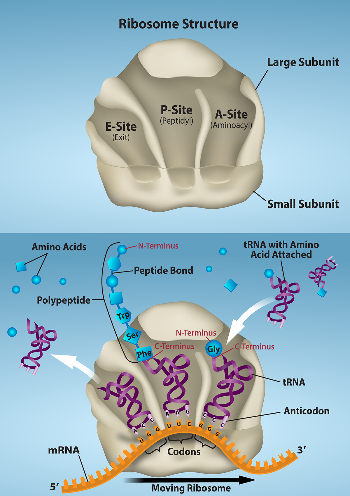
Figure 15.16 The ribosome and its function. The ribosome is responsible for translating the mRNA into protein. A. The ribosome consists of a large and small ribosomal subunit. Assembly of the subunits on the mRNA forms three tRNA binding sites. B. During translation, charged tRNAs enter the Acceptor site, and the anticodon on the tRNA base pairs with the codon in the mRNA. After the incoming amino acid forms a peptide bond with the growing polypeptide chain, the ribosome will move three nucleotides toward the 3’ end of the mRNA. This movement will transfer the tRNA with the growing polypeptide to the Peptidyl-tRNA binding site and allow the empty tRNA to exit at the Exit site. Credit: Rao, A., Ryan, K. and Fletcher, S. Department of Biology, Texas A&M University.
Rao, A., Ryan, K. and Fletcher, S. Department of Biology, Texas A&M University.; Mary Ann Clark, Matthew Douglas, and Jung Choi, Biology 2e, OpenStax. · CC BY-NC-SA 4.0
· Original image ↗ · Access for free ↗
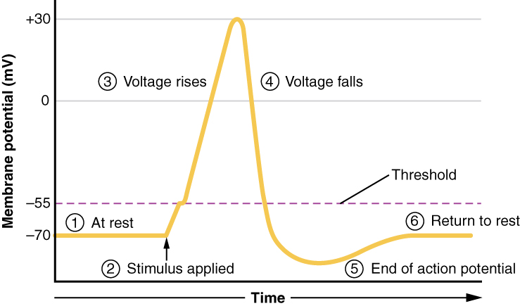
Figure 12.24 Stages of an Action Potential Plotting voltage measured across the cell membrane against time, the events of the action potential can be related to specific changes in the membrane voltage. (1) At rest, the membrane voltage is -70 mV. (2) The membrane begins to depolarize when an external stimulus is applied. (3) The membrane voltage begins a rapid rise toward +30 mV. (4) The membrane voltage starts to return to a negative value. (5) Repolarization continues past the resting membrane voltage, resulting in hyperpolarization. (6) The membrane voltage returns to the resting value shortly after hyperpolarization.
A nitrogen-containing functional group. A free amino acid’s amino group is typically protonated (NH₃⁺) near physiological pH; proline’s ring nitrogen is an exception to this formula.
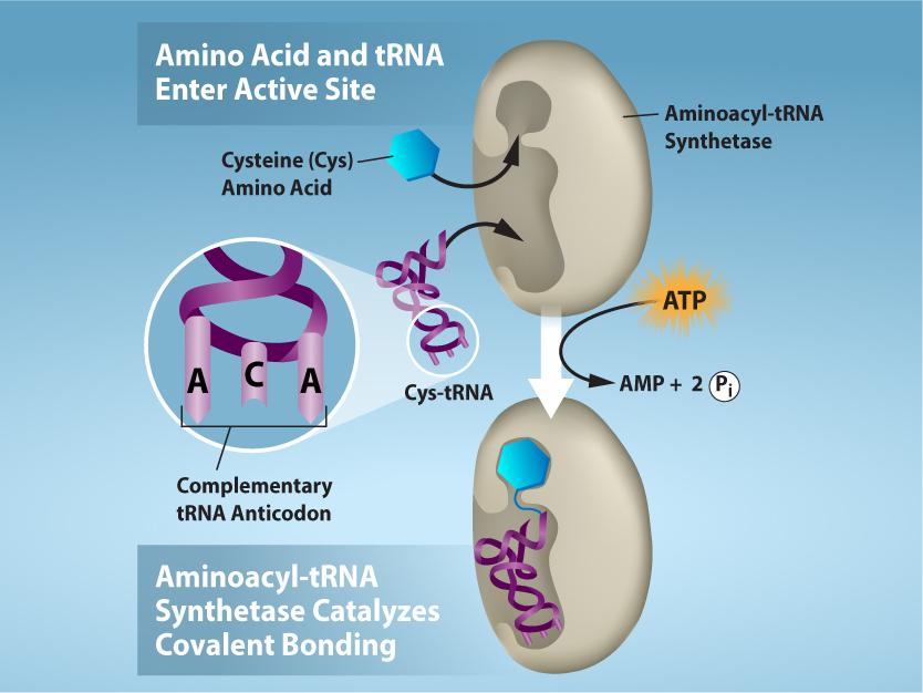
Figure 15.17 Charging of tRNAs with correct amino acids. Aminoacyl-tRNA synthetases catalyze covalent bond formation between the tRNA and the correct amino acid in preparation for translation. Because there are multiple amino acids, there are multiple different tRNA synthetases. All of the synthetases require energy, in the form of ATP, to make sure the correct amino acid is attached to the tRNA with the correct anticodon sequence. Credit: Rao, A., Ryan, K. and Tag, A. Department of Biology, Texas A&M University.
Rao, A., Ryan, K. and Tag, A. Department of Biology, Texas A&M University.; Mary Ann Clark, Matthew Douglas, and Jung Choi, Biology 2e, OpenStax. · CC BY-NC-SA 4.0
· Original image ↗ · Access for free ↗
Figure 15.14 This is a space-filling model of a tRNA molecule that adds the amino acid phenylalanine to a growing polypeptide chain. The anticodon AAG binds the Codon UUC on the mRNA. The amino acid phenylalanine is attached to the other end of the tRNA.
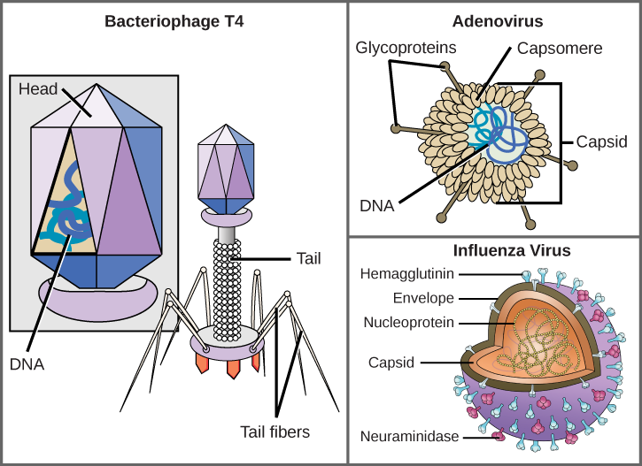
Figure 21.5 Complex Viruses. Viruses can be either complex or relatively simple in shape. This figure shows three relatively complex virions: the bacteriophage T4, with its DNA-containing head group and tail fibers that attach to host cells; adenovirus, which uses spikes from its capsid to bind to host cells; and the influenza virus, which uses glycoproteins embedded in its envelope to bind to host cells. The influenza virus also has matrix proteins, internal to the envelope, which help stabilize the virion’s shape. (credit “bacteriophage, adenovirus”: modification of work by NCBI, NIH; credit "influenza virus": modification of work by Dan Higgins, Centers for Disease Control and Prevention)
Mary Ann Clark, Matthew Douglas, and Jung Choi, Biology 2E, OpenStax. (credit “bacteriophage, adenovirus”: modification of work by NCBI, NIH; credit "influenza virus": modification of work by Dan Higgins, Centers for Disease Control and Prevention) · CC BY-NC-SA 4.0
· Original image ↗ · Access for free ↗
Figure 5.18 The ear is divided into outer (pinna and tympanic membrane), middle (the three ossicles: malleus, incus, and stapes), and inner (cochlea and basilar membrane) divisions.
Figure 5.18 The ear is divided into outer (pinna and tympanic membrane), middle (the three ossicles: malleus, incus, and stapes), and inner (cochlea and basilar membrane) divisions.
Figure 15.4 This figure shows the genetic code for translating each nucleotide triplet in mRNA into an amino acid or a termination signal in a protein. (credit: modification of work by NIH)
Figure 14.19 Nucleotide excision repairs thymine dimers. When exposed to UV light, thymines lying adjacent to each other can form thymine dimers. In normal cells, they are excised and replaced. Credit: Rao, A., Fletcher, S. and Tag, A. Department of Biology, Texas A&M University.
Figure 14.14 First Components of DNA Replication. As DNA replication begins, DNA Helicase, a large enzyme, separates the two strands of DNA so that they can act as templates for replication. Single-strand binding proteins bind to each strand to stabilize and prevent them from reforming the double helix. Primase, an RNA polymerase, binds to the single stranded DNA and synthesizes a short RNA primer in the 5’ to 3’ direction that is antiparallel to the parental strand. This RNA primer allows for DNA polymerase to begin replicating the DNA. Topoisomerase binds to the double helix upstream of the replication fork to prevent additional coiling by making small cuts in one of the DNA strands.
Credit: Rao, A., Ryan, K. Fletcher, S. and Tag, A. Department of Biology, Texas A&M University.
An electrical property that can be positive or negative. Opposite charges attract and like charges repel; net charge is the sum of all charges on a particle.
Figure 4.18 Membrane and secretory proteins are synthesized in the rough endoplasmic reticulum (RER). The RER also sometimes modifies proteins. In this illustration, a (green) integral membrane protein is modified by attachment of a (purple) carbohydrate in the ER. Vesicles with the integral protein bud from the ER and fuse with the Golgi apparatus' cis face. As the protein passes along the Golgi’s cisternae, the addition of more carbohydrates further modifies it. After its synthesis is complete, it exits as an integral membrane protein of the vesicle that buds from the Golgi’s trans face. When the vesicle fuses with the cell membrane, the protein becomes an integral portion of that cell membrane. (credit: modification of work by Magnus Manske)
Figure 15.10 A generalized promoter of a gene transcribed by RNA polymerase II is shown. Transcription factors recognize the promoter. RNA polymerase II then binds and forms the transcription initiation complex.
Figure 5.18 The ear is divided into outer (pinna and tympanic membrane), middle (the three ossicles: malleus, incus, and stapes), and inner (cochlea and basilar membrane) divisions.
The exponent needed to obtain a number from a chosen fixed number. For decimal logarithms, log₁₀(100) = 2 because 10² = 100; pH uses the negative decimal logarithm of hydrogen ion activity.
Figure 4.18 Membrane and secretory proteins are synthesized in the rough endoplasmic reticulum (RER). The RER also sometimes modifies proteins. In this illustration, a (green) integral membrane protein is modified by attachment of a (purple) carbohydrate in the ER. Vesicles with the integral protein bud from the ER and fuse with the Golgi apparatus' cis face. As the protein passes along the Golgi’s cisternae, the addition of more carbohydrates further modifies it. After its synthesis is complete, it exits as an integral membrane protein of the vesicle that buds from the Golgi’s trans face. When the vesicle fuses with the cell membrane, the protein becomes an integral portion of that cell membrane. (credit: modification of work by Magnus Manske)
Figure 4.18 Membrane and secretory proteins are synthesized in the rough endoplasmic reticulum (RER). The RER also sometimes modifies proteins. In this illustration, a (green) integral membrane protein is modified by attachment of a (purple) carbohydrate in the ER. Vesicles with the integral protein bud from the ER and fuse with the Golgi apparatus' cis face. As the protein passes along the Golgi’s cisternae, the addition of more carbohydrates further modifies it. After its synthesis is complete, it exits as an integral membrane protein of the vesicle that buds from the Golgi’s trans face. When the vesicle fuses with the cell membrane, the protein becomes an integral portion of that cell membrane. (credit: modification of work by Magnus Manske)
Figure 5.18 The ear is divided into outer (pinna and tympanic membrane), middle (the three ossicles: malleus, incus, and stapes), and inner (cochlea and basilar membrane) divisions.
A thin boundary or sheet. Cell membranes are lipid bilayers containing proteins; blotting membranes are synthetic supports that hold proteins or nucleic acids for detection.
Figure 15.11 Eukaryotic pre-mRNA processing. In addition to 5’ Cap and 3’ Poly-A Tail addition, introns must be precisely removed and exons joined to generate a functional mRNA. Nucleotides upstream (towards the 5’cap) of the translation START codon are part of the 5’ untranslated region (5’ UTR). Nucleotides downstream (towards 3’end) of the STOP codon form the 3’ UTR. Both 5’ and 3’ UTRs are important for regulating translation initiation and mRNA stability. Credit: Rao, A., Ryan, K. Fletcher, S. and Tag, A. Department of Biology, Texas A&M University.
Rao, A., Ryan, K. Fletcher, S. and Tag, A. Department of Biology, Texas A&M University.; Mary Ann Clark, Matthew Douglas, and Jung Choi, Biology 2e, OpenStax. · CC BY-NC-SA 4.0
· Original image ↗ · Access for free ↗
Figure 4.18 Membrane and secretory proteins are synthesized in the rough endoplasmic reticulum (RER). The RER also sometimes modifies proteins. In this illustration, a (green) integral membrane protein is modified by attachment of a (purple) carbohydrate in the ER. Vesicles with the integral protein bud from the ER and fuse with the Golgi apparatus' cis face. As the protein passes along the Golgi’s cisternae, the addition of more carbohydrates further modifies it. After its synthesis is complete, it exits as an integral membrane protein of the vesicle that buds from the Golgi’s trans face. When the vesicle fuses with the cell membrane, the protein becomes an integral portion of that cell membrane. (credit: modification of work by Magnus Manske)
Figure 4.18 Membrane and secretory proteins are synthesized in the rough endoplasmic reticulum (RER). The RER also sometimes modifies proteins. In this illustration, a (green) integral membrane protein is modified by attachment of a (purple) carbohydrate in the ER. Vesicles with the integral protein bud from the ER and fuse with the Golgi apparatus' cis face. As the protein passes along the Golgi’s cisternae, the addition of more carbohydrates further modifies it. After its synthesis is complete, it exits as an integral membrane protein of the vesicle that buds from the Golgi’s trans face. When the vesicle fuses with the cell membrane, the protein becomes an integral portion of that cell membrane. (credit: modification of work by Magnus Manske)
Figure 5.18 The ear is divided into outer (pinna and tympanic membrane), middle (the three ossicles: malleus, incus, and stapes), and inner (cochlea and basilar membrane) divisions.
Figure 14.14 First Components of DNA Replication. As DNA replication begins, DNA Helicase, a large enzyme, separates the two strands of DNA so that they can act as templates for replication. Single-strand binding proteins bind to each strand to stabilize and prevent them from reforming the double helix. Primase, an RNA polymerase, binds to the single stranded DNA and synthesizes a short RNA primer in the 5’ to 3’ direction that is antiparallel to the parental strand. This RNA primer allows for DNA polymerase to begin replicating the DNA. Topoisomerase binds to the double helix upstream of the replication fork to prevent additional coiling by making small cuts in one of the DNA strands.
Credit: Rao, A., Ryan, K. Fletcher, S. and Tag, A. Department of Biology, Texas A&M University.
Figure 12.19 Reaction diagrams for an endothermic process in the absence (red curve) and presence (blue curve) of a catalyst. The catalyzed pathway involves a two-step mechanism (note the presence of two transition states) and an intermediate species (represented by the valley between the two transitions states).
Figure 15.19 Translation Termination is Active. Translation is terminated when a STOP codon is in the A-site of the ribosome. Since there are no tRNAs corresponding to the STOP codons, the Release Factor protein enters and catalyzes the hydrolysis between the last amino acid and its tRNA. This hydrolysis releases the free carboxyl terminus (C-term) of the protein. Additional factors use the energy in GTP hydrolysis to disassemble the large and small ribosomal subunits and mRNA. Credit: Rao, A. and Ryan, K. Department of Biology, Texas A&M University.
Rao, A. and Ryan, K. Department of Biology, Texas A&M University.; Mary Ann Clark, Matthew Douglas, and Jung Choi, Biology 2e, OpenStax. · CC BY-NC-SA 4.0
· Original image ↗ · Access for free ↗
Figure 7.45 - Non-homologous end joining (left) versus homologous recombination (right) - Wikipedia
Wikipedia, as credited by Kevin Ahern, Indira Rajagopal, and Taralyn Tan, Biochemistry Free For All, Oregon State University, via Biology LibreTexts. · Source attribution
· Original image ↗ · Access for free ↗
The amount of a substance that can dissolve in a solvent under specified conditions. Soluble means able to dissolve; insoluble means only very slightly soluble in that solvent.
Figure 5.18 The ear is divided into outer (pinna and tympanic membrane), middle (the three ossicles: malleus, incus, and stapes), and inner (cochlea and basilar membrane) divisions.
Figure 15.7 Messenger RNA is a copy of protein-coding information in the coding strand of DNA, with the substitution of U in the RNA for T in the coding sequence. However, new RNA nucleotides base pair with the nucleotides of the template strand. RNA is synthesized in its 5'-3' direction, using the enzyme RNA polymerase. As the template is read, the DNA unwinds ahead of the polymerase and then rewinds behind it.
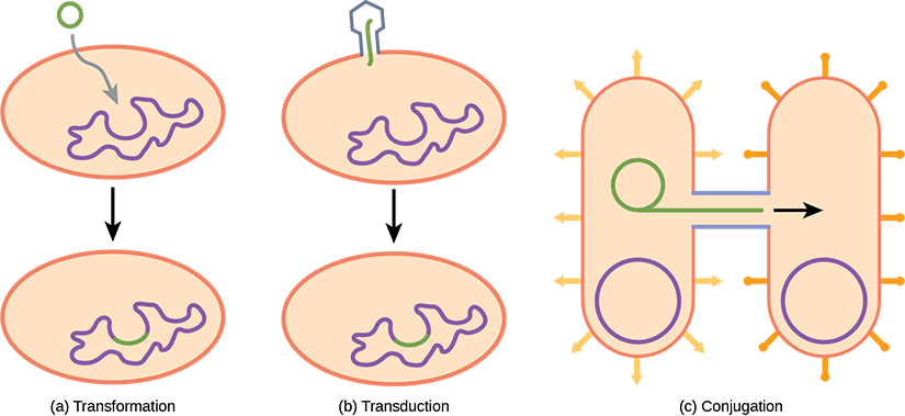
Figure 22.17 Gene transfer mechanisms in prokaryotes. There are three mechanisms by which prokaryotes can exchange DNA. In (a) transformation, the cell takes up prokaryotic DNA directly from the environment. The DNA may remain separate as plasmid DNA or be incorporated into the host genome. In (b) transduction, a bacteriophage injects DNA into the cell that contains a small fragment of DNA from a different prokaryote. In (c) conjugation, DNA is transferred from one cell to another via a mating bridge, or pilus, that connects the two cells after the sex pilus draws the two bacteria close enough to form the bridge.
Figure 15.14 This is a space-filling model of a tRNA molecule that adds the amino acid phenylalanine to a growing polypeptide chain. The anticodon AAG binds the Codon UUC on the mRNA. The amino acid phenylalanine is attached to the other end of the tRNA.
A fleeting, high-energy arrangement at a reaction barrier. It is an energy maximum along the reaction path, rather than a stable reaction intermediate.
Figure 12.19 Reaction diagrams for an endothermic process in the absence (red curve) and presence (blue curve) of a catalyst. The catalyzed pathway involves a two-step mechanism (note the presence of two transition states) and an intermediate species (represented by the valley between the two transitions states).
Figure 15.18 Translation begins when an initiator tRNA anticodon recognizes a start codon on mRNA bound to a small ribosomal subunit. The large ribosomal subunit joins the small subunit, and a second tRNA is recruited. As the mRNA moves relative to the ribosome, successive tRNAs move through the ribosome and the polypeptide chain is formed. Entry of a release factor into the A site terminates translation and the components dissociate.
Figure 10.15 Cell Division and Apoptosis. In an adult organism, normal cell division is balanced by apoptosis (programmed cell death) to maintain a constant cell number in homeostasis. Either an increase in cell division or a decrease in apoptosis leads an increase in the number of cells and tumor formation. Credit: Rao, A. and Ryan, K. Department of Biology, Texas A&M University.
Rao, A. and Ryan, K. Department of Biology, Texas A&M University. Mary Ann Clark, Matthew Douglas, and Jung Choi, Biology 2e, OpenStax. · CC BY-NC-SA 4.0
· Original image ↗ · Access for free ↗
Figure 5.18 The ear is divided into outer (pinna and tympanic membrane), middle (the three ossicles: malleus, incus, and stapes), and inner (cochlea and basilar membrane) divisions.
No matching terms. Try a different name or abbreviation.
5.4 Hearing
OpenStax · 5.4 Hearing ↗ Instructional page including learning objectives, examples, figures, source tables and questions. Site navigation and embedded media players are omitted; source media links remain available.
Learning Objectives
By the end of this section, you will be able to:
Describe the basic anatomy and function of the auditory system
Explain how we encode and perceive pitch
Discuss how we localize sound
Our auditory system converts pressure waves into meaningful sounds. This translates into our ability to hear the sounds of nature, to appreciate the beauty of music, and to communicate with one another through spoken language. This section will provide an overview of the basic anatomy and function of the auditory system. It will include a discussion of how the sensory stimulus is translated into neural impulses, where in the brain that information is processed, how we perceive pitch, and how we know where sound is coming from.
Anatomy of the Auditory System
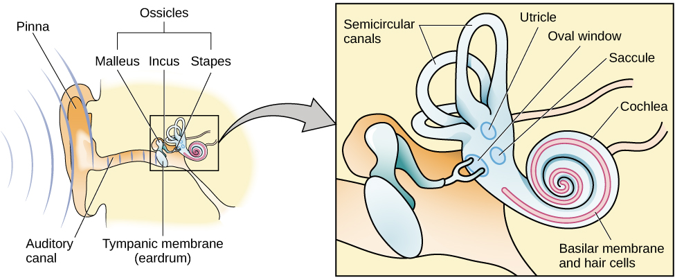
The ear can be separated into multiple sections. The outer ear includes the pinna, which is the visible part of the ear that protrudes from our heads, the auditory canal, and the tympanic membrane, or eardrum. The middle ear contains three tiny bones known as the ossicles, which are named the malleus (or hammer), incus (or anvil), and the stapes (or stirrup). The inner ear contains the semi-circular canals, which are involved in balance and movement (the vestibular sense), and the cochlea. The cochlea is a fluid-filled, snail-shaped structure that contains the sensory receptor cells (hair cells) of the auditory system (Figure 5.18).
Figure 5.18 — 5.4 Hearing
Figure 5.18 The ear is divided into outer (pinna and tympanic membrane), middle (the three ossicles: malleus, incus, and stapes), and inner (cochlea and basilar membrane) divisions.
Sound waves travel along the auditory canal and strike the tympanic membrane, causing it to vibrate. This vibration results in movement of the three ossicles. As the ossicles move, the stapes presses into a thin membrane of the cochlea known as the oval window. As the stapes presses into the oval window, the fluid inside the cochlea begins to move, which in turn stimulates hair cells, which are auditory receptor cells of the inner ear embedded in the basilar membrane. The basilar membrane is a thin strip of tissue within the cochlea.
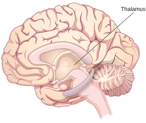
The activation of hair cells is a mechanical process: the stimulation of the hair cell ultimately leads to activation of the cell. As hair cells become activated, they generate neural impulses that travel along the auditory nerve to the brain. Auditory information is shuttled to the inferior colliculus, the medial geniculate nucleus of the thalamus, and finally to the auditory cortex in the temporal lobe of the brain for processing. Like the visual system, there is also evidence suggesting that information about auditory recognition and localization is processed in parallel streams (Rauschecker & Tian, 2000; Renier et al., 2009).
Pitch Perception
Different frequencies of sound waves are associated with differences in our perception of the pitch of those sounds. Low-frequency sounds are lower pitched, and high-frequency sounds are higher pitched. How does the auditory system differentiate among various pitches?
Several theories have been proposed to account for pitch perception. We’ll discuss two of them here: temporal theory and place theory. The temporal theory of pitch perception asserts that frequency is coded by the activity level of a sensory neuron. This would mean that a given hair cell would fire action potentials related to the frequency of the sound wave. While this is a very intuitive explanation, we detect such a broad range of frequencies (20–20,000 Hz) that the frequency of action potentials fired by hair cells cannot account for the entire range. Because of properties related to sodium channels on the neuronal membrane that are involved in action potentials, there is a point at which a cell cannot fire any faster (Shamma, 2001).
The place theory of pitch perception suggests that different portions of the basilar membrane are sensitive to sounds of different frequencies. More specifically, the base of the basilar membrane responds best to high frequencies and the tip of the basilar membrane responds best to low frequencies. Therefore, hair cells that are in the base portion would be labeled as high-pitch receptors, while those in the tip of basilar membrane would be labeled as low-pitch receptors (Shamma, 2001).
In reality, both theories explain different aspects of pitch perception. At frequencies up to about 4000 Hz, it is clear that both the rate of action potentials and place contribute to our perception of pitch. However, much higher frequency sounds can only be encoded using place cues (Shamma, 2001).
Sound Localization
The ability to locate sound in our environments is an important part of hearing. Localizing sound could be considered similar to the way that we perceive depth in our visual fields. Like the monocular and binocular cues that provided information about depth, the auditory system uses both monaural (one-eared) and binaural (two-eared) cues to localize sound.
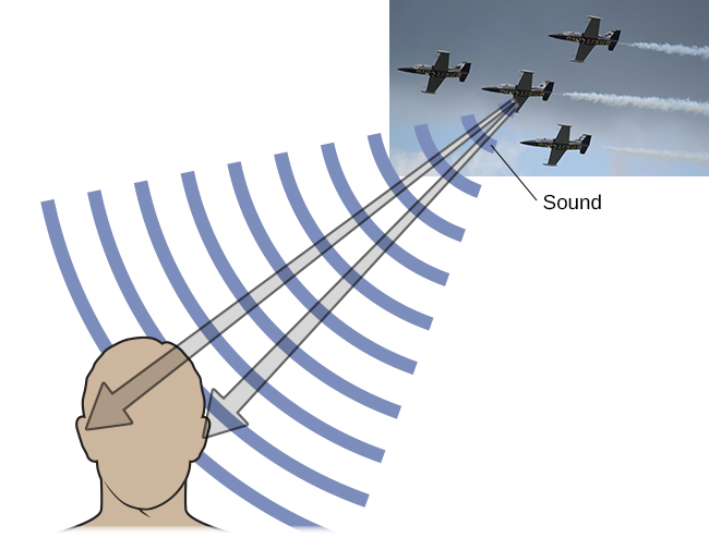
Each pinna interacts with incoming sound waves differently, depending on the sound’s source relative to our bodies. This interaction provides a monaural cue that is helpful in locating sounds that occur above or below and in front or behind us. The sound waves received by your two ears from sounds that come from directly above, below, in front, or behind you would be identical; therefore, monaural cues are essential (Grothe, Pecka, & McAlpine, 2010).
Binaural cues, on the other hand, provide information on the location of a sound along a horizontal axis by relying on differences in patterns of vibration of the eardrum between our two ears. If a sound comes from an off-center location, it creates two types of binaural cues: interaural level differences and interaural timing differences. Interaural level difference refers to the fact that a sound coming from the right side of your body is more intense at your right ear than at your left ear because of the attenuation of the sound wave as it passes through your head. Interaural timing difference refers to the small difference in the time at which a given sound wave arrives at each ear (Figure 5.19). Certain brain areas monitor these differences to construct where along a horizontal axis a sound originates (Grothe et al., 2010).
Figure 5.19 — 5.4 Hearing
Figure 5.19 Localizing sound involves the use of both monaural and binaural cues. (credit "plane": modification of work by Max Pfandl)
Rose M. Spielman, William J. Jenkins, and Marilyn D. Lovett, Psychology 2E, OpenStax. (credit "plane": modification of work by Max Pfandl) · CC BY-NC-SA 4.0
· Original image ↗ · Access for free ↗
Hearing Loss
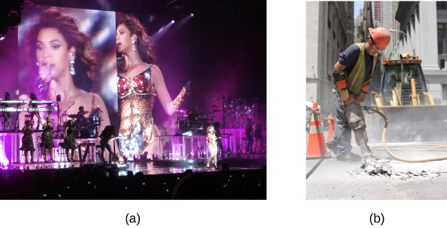
Deafness is the partial or complete inability to hear. Some people are born without hearing, which is known as congenital deafness. Other people suffer from conductive hearing loss, which is due to a problem delivering sound energy to the cochlea. Causes for conductive hearing loss include blockage of the ear canal, a hole in the tympanic membrane, problems with the ossicles, or fluid in the space between the eardrum and cochlea. Another group of people suffer from sensorineural hearing loss, which is the most common form of hearing loss. Sensorineural hearing loss can be caused by many factors, such as aging, head or acoustic trauma, infections and diseases (such as measles or mumps), medications, environmental effects such as noise exposure (noise-induced hearing loss, as shown in Figure 5.20), tumors, and toxins (such as those found in certain solvents and metals).
Figure 5.20 — 5.4 Hearing
Figure 5.20 Environmental factors that can lead to sensorineural hearing loss include regular exposure to loud music or construction equipment. (a) Musical performers and (b) construction workers are at risk for this type of hearing loss. (credit a: modification of work by "GillyBerlin_Flickr"/Flickr; credit b: modification of work by Nick Allen)
Rose M. Spielman, William J. Jenkins, and Marilyn D. Lovett, Psychology 2E, OpenStax. (credit a: modification of work by "GillyBerlin_Flickr"/Flickr; credit b: modification of work by Nick Allen) · CC BY-NC-SA 4.0
· Original image ↗ · Access for free ↗
Given the mechanical nature by which the sound wave stimulus is transmitted from the eardrum through the ossicles to the oval window of the cochlea, some degree of hearing loss is inevitable. With conductive hearing loss, hearing problems are associated with a failure in the vibration of the eardrum and/or movement of the ossicles. These problems are often dealt with through devices like hearing aids that amplify incoming sound waves to make vibration of the eardrum and movement of the ossicles more likely to occur.
When the hearing problem is associated with a failure to transmit neural signals from the cochlea to the brain, it is called sensorineural hearing loss. One disease that results in sensorineural hearing loss is Ménière's disease. Although not well understood, Ménière's disease results in a degeneration of inner ear structures that can lead to hearing loss, tinnitus (constant ringing or buzzing), vertigo (a sense of spinning), and an increase in pressure within the inner ear (Semaan & Megerian, 2011). This kind of loss cannot be treated with hearing aids, but some individuals might be candidates for a cochlear implant as a treatment option. Cochlear implants are electronic devices that consist of a microphone, a speech processor, and an electrode array. The device receives incoming sound information and directly stimulates the auditory nerve to transmit information to the brain.
In the United States and other places around the world, deaf people have their own language, schools, and customs. This is called deaf culture. In the United States, deaf individuals often communicate using American Sign Language (ASL); ASL has no verbal component and is based entirely on visual signs and gestures. The primary mode of communication is signing. One of the values of deaf culture is to continue traditions like using sign language rather than teaching deaf children to try to speak, read lips, or have cochlear implant surgery.
When a child is diagnosed as deaf, parents have difficult decisions to make. Should the child be enrolled in mainstream schools and taught to verbalize and read lips? Or should the child be sent to a school for deaf children to learn ASL and have significant exposure to deaf culture? Do you think there might be differences in the way that parents approach these decisions depending on whether or not they are also deaf?