In the usual introductory detection convention, an absolute threshold is the intensity reported present on 50% of trials, and a difference threshold or just noticeable difference is a change noticed on 50% of trials under specified conditions. The criterion depends on the procedure: in a two-alternative forced-choice task, guessing already gives 50% correct, so a threshold is commonly defined at 75% correct instead. Weber’s law approximates ΔI/I = k: increasing a 100-g load by 2 g and a 200-g load by 4 g gives the same 2% change. The relationship is approximate and can fail near extremes. Sensory adaptation reduces responsiveness to a sustained stimulus, helping emphasize change. A threshold depends on the stimulus, observer, and testing conditions; it is not an immutable boundary between all perception and none.
Interpret signal detection outcomes and separate sensitivity from response bias
Signal detection theory separates sensory sensitivity from the decision criterion. With a signal present, “yes” is a hit and “no” a miss; without it, “yes” is a false alarm and “no” a correct rejection. A liberal criterion increases both hits and false alarms; a conservative criterion decreases both. Higher sensitivity makes signal and noise more distinguishable, allowing more hits at a matched false-alarm rate. Payoffs, expectations, and costs can shift criterion without changing sensory receptors. Thus hit rate alone cannot establish improved sensitivity; compare false alarms or the full receiver operating characteristic as well.
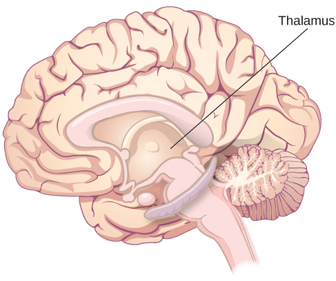
Mechanoreceptors respond to deformation, chemoreceptors to molecules, photoreceptors to light, thermoreceptors to temperature, and nociceptors to potentially damaging stimulation. Transduction converts stimulus energy into a receptor potential. Receptors may themselves generate action potentials or release transmitter onto a sensory neuron, as hair cells do. Stimulus intensity is represented through firing rates and recruited neurons, not larger action potentials. Labeled pathways help preserve modality: activity reaching visual circuits is interpreted differently from activity reaching auditory circuits. Most sensory information relays through the thalamus before cortex; early olfactory processing is a major exception.
Worked question. Each of two blocks contains 100 signal-present and 100 signal-absent trials. A detector reports 80 hits and 40 false alarms in the first, then 95 hits and 70 false alarms in the second after being told that misses are costly. Has sensitivity necessarily increased?
No. More hits together with more false alarms is consistent with a more liberal response criterion. Hold signal and noise distributions constant and compare performance across criteria before attributing the change to sensitivity. Separately, with k = 0.02, Weber’s law predicts a 6-g noticeable increment on a 300-g standard.
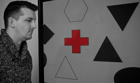
Figure 5.3 Nearly one third of participants in a study did not notice that a red cross passed on the screen because their attention was focused on the black or white figures. (credit: Cory Zanker)
Rose M. Spielman, William J. Jenkins, and Marilyn D. Lovett, Psychology 2E, OpenStax. (credit: Cory Zanker) · CC BY-NC-SA 4.0
· Original image ↗ · Access for free ↗Figure 5.4 — 5.1 Sensation versus Perception
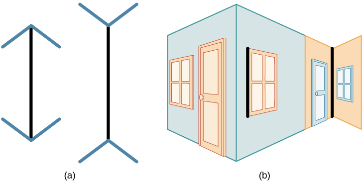
Figure 5.4 In the Müller-Lyer illusion, lines appear to be different lengths although they are identical. (a) Arrows at the ends of lines may make the line on the right appear longer, although the lines are the same length. (b) When applied to a three-dimensional image, the line on the right again may appear longer although both black lines are the same length.
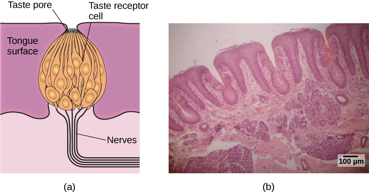
Figure 5.21 (a) Taste buds are composed of a number of individual taste receptors cells that transmit information to nerves. (b) This micrograph shows a close-up view of the tongue’s surface. (credit a: modification of work by Jonas Töle; credit b: scale-bar data from Matt Russell)
Rose M. Spielman, William J. Jenkins, and Marilyn D. Lovett, Psychology 2E, OpenStax. (credit a: modification of work by Jonas Töle; credit b: scale-bar data from Matt Russell) · CC BY-NC-SA 4.0
· Original image ↗ · Access for free ↗Figure 5.22 — 5.5 The Other Senses
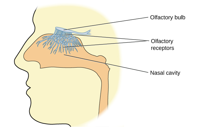
Figure 5.22 Olfactory receptors are the hair-like parts that extend from the olfactory bulb into the mucous membrane of the nasal cavity.
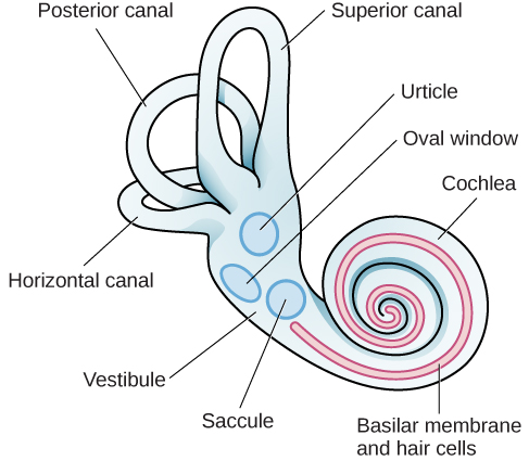
Figure 5.24 The major sensory organs of the vestibular system are located next to the cochlea in the inner ear. These include the utricle, saccule, and the three semicircular canals (posterior, superior, and horizontal).
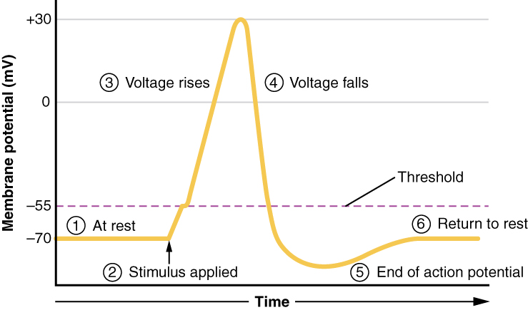
Figure 12.24 Stages of an Action Potential Plotting voltage measured across the cell membrane against time, the events of the action potential can be related to specific changes in the membrane voltage. (1) At rest, the membrane voltage is -70 mV. (2) The membrane begins to depolarize when an external stimulus is applied. (3) The membrane voltage begins a rapid rise toward +30 mV. (4) The membrane voltage starts to return to a negative value. (5) Repolarization continues past the resting membrane voltage, resulting in hyperpolarization. (6) The membrane voltage returns to the resting value shortly after hyperpolarization.
A nitrogen-containing functional group. A free amino acid’s amino group is typically protonated (NH₃⁺) near physiological pH; proline’s ring nitrogen is an exception to this formula.
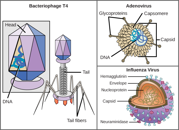
Figure 21.5 Complex Viruses. Viruses can be either complex or relatively simple in shape. This figure shows three relatively complex virions: the bacteriophage T4, with its DNA-containing head group and tail fibers that attach to host cells; adenovirus, which uses spikes from its capsid to bind to host cells; and the influenza virus, which uses glycoproteins embedded in its envelope to bind to host cells. The influenza virus also has matrix proteins, internal to the envelope, which help stabilize the virion’s shape. (credit “bacteriophage, adenovirus”: modification of work by NCBI, NIH; credit "influenza virus": modification of work by Dan Higgins, Centers for Disease Control and Prevention)
Mary Ann Clark, Matthew Douglas, and Jung Choi, Biology 2E, OpenStax. (credit “bacteriophage, adenovirus”: modification of work by NCBI, NIH; credit "influenza virus": modification of work by Dan Higgins, Centers for Disease Control and Prevention) · CC BY-NC-SA 4.0
· Original image ↗ · Access for free ↗
Figure 10.15 Cell Division and Apoptosis. In an adult organism, normal cell division is balanced by apoptosis (programmed cell death) to maintain a constant cell number in homeostasis. Either an increase in cell division or a decrease in apoptosis leads an increase in the number of cells and tumor formation. Credit: Rao, A. and Ryan, K. Department of Biology, Texas A&M University.
Rao, A. and Ryan, K. Department of Biology, Texas A&M University. Mary Ann Clark, Matthew Douglas, and Jung Choi, Biology 2e, OpenStax. · CC BY-NC-SA 4.0
· Original image ↗ · Access for free ↗
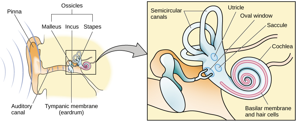
Figure 5.18 The ear is divided into outer (pinna and tympanic membrane), middle (the three ossicles: malleus, incus, and stapes), and inner (cochlea and basilar membrane) divisions.
Figure 15.4 This figure shows the genetic code for translating each nucleotide triplet in mRNA into an amino acid or a termination signal in a protein. (credit: modification of work by NIH)
The stimulus difference meeting a specified discrimination criterion, often described as noticed on half the trials in introductory yes/no procedures; forced-choice accuracy uses a criterion above chance.
An electrical property that can be positive or negative. Opposite charges attract and like charges repel; net charge is the sum of all charges on a particle.
Figure 4.18 Membrane and secretory proteins are synthesized in the rough endoplasmic reticulum (RER). The RER also sometimes modifies proteins. In this illustration, a (green) integral membrane protein is modified by attachment of a (purple) carbohydrate in the ER. Vesicles with the integral protein bud from the ER and fuse with the Golgi apparatus' cis face. As the protein passes along the Golgi’s cisternae, the addition of more carbohydrates further modifies it. After its synthesis is complete, it exits as an integral membrane protein of the vesicle that buds from the Golgi’s trans face. When the vesicle fuses with the cell membrane, the protein becomes an integral portion of that cell membrane. (credit: modification of work by Magnus Manske)
Figure 4.18 Membrane and secretory proteins are synthesized in the rough endoplasmic reticulum (RER). The RER also sometimes modifies proteins. In this illustration, a (green) integral membrane protein is modified by attachment of a (purple) carbohydrate in the ER. Vesicles with the integral protein bud from the ER and fuse with the Golgi apparatus' cis face. As the protein passes along the Golgi’s cisternae, the addition of more carbohydrates further modifies it. After its synthesis is complete, it exits as an integral membrane protein of the vesicle that buds from the Golgi’s trans face. When the vesicle fuses with the cell membrane, the protein becomes an integral portion of that cell membrane. (credit: modification of work by Magnus Manske)
Figure 15.10 A generalized promoter of a gene transcribed by RNA polymerase II is shown. Transcription factors recognize the promoter. RNA polymerase II then binds and forms the transcription initiation complex.
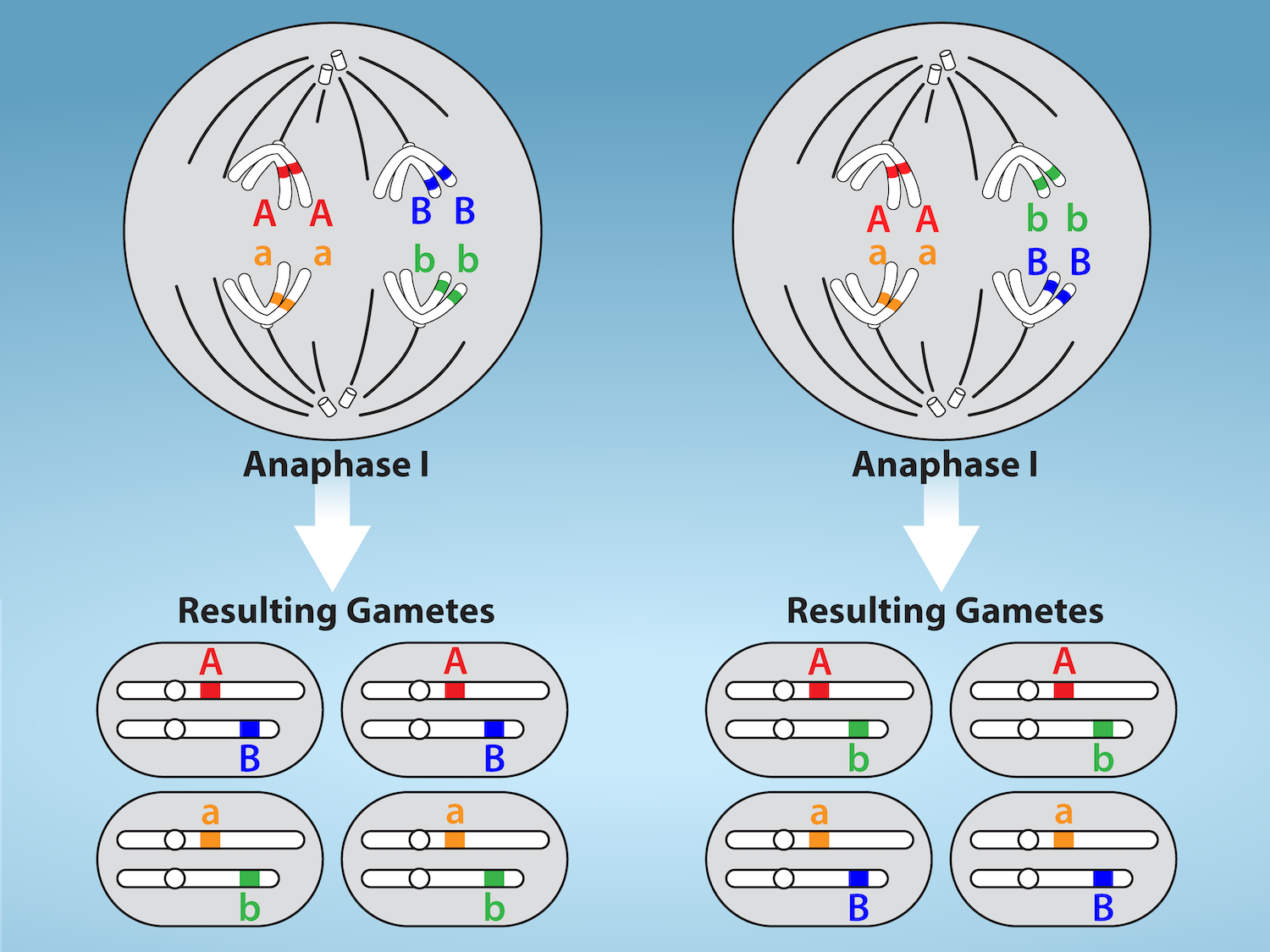
Figure 11.5 Random, independent assortment during metaphase I is demonstrated by considering a cell with a set of two chromosomes (n = 2).
There are two possible homologous chromosome arrangements at the equatorial plane in metaphase I, that are then separated during anaphase I.
The total possible number of different gametes is 2 n , where n equals the number of chromosomes in a set. In this example, there are four possible
genetic combinations for the gametes. With n = 23 in human cells, there are over eight million possible combinations of paternal and maternal
chromosomes. Credit: Rao, A.and Fletcher, S. Department of Biology, Texas A&M University.
Figure 4.18 Membrane and secretory proteins are synthesized in the rough endoplasmic reticulum (RER). The RER also sometimes modifies proteins. In this illustration, a (green) integral membrane protein is modified by attachment of a (purple) carbohydrate in the ER. Vesicles with the integral protein bud from the ER and fuse with the Golgi apparatus' cis face. As the protein passes along the Golgi’s cisternae, the addition of more carbohydrates further modifies it. After its synthesis is complete, it exits as an integral membrane protein of the vesicle that buds from the Golgi’s trans face. When the vesicle fuses with the cell membrane, the protein becomes an integral portion of that cell membrane. (credit: modification of work by Magnus Manske)
The exponent needed to obtain a number from a chosen fixed number. For decimal logarithms, log₁₀(100) = 2 because 10² = 100; pH uses the negative decimal logarithm of hydrogen ion activity.
A thin boundary or sheet. Cell membranes are lipid bilayers containing proteins; blotting membranes are synthetic supports that hold proteins or nucleic acids for detection.
Figure 15.11 Eukaryotic pre-mRNA processing. In addition to 5’ Cap and 3’ Poly-A Tail addition, introns must be precisely removed and exons joined to generate a functional mRNA. Nucleotides upstream (towards the 5’cap) of the translation START codon are part of the 5’ untranslated region (5’ UTR). Nucleotides downstream (towards 3’end) of the STOP codon form the 3’ UTR. Both 5’ and 3’ UTRs are important for regulating translation initiation and mRNA stability. Credit: Rao, A., Ryan, K. Fletcher, S. and Tag, A. Department of Biology, Texas A&M University.
Rao, A., Ryan, K. Fletcher, S. and Tag, A. Department of Biology, Texas A&M University.; Mary Ann Clark, Matthew Douglas, and Jung Choi, Biology 2e, OpenStax. · CC BY-NC-SA 4.0
· Original image ↗ · Access for free ↗
Figure 4.18 Membrane and secretory proteins are synthesized in the rough endoplasmic reticulum (RER). The RER also sometimes modifies proteins. In this illustration, a (green) integral membrane protein is modified by attachment of a (purple) carbohydrate in the ER. Vesicles with the integral protein bud from the ER and fuse with the Golgi apparatus' cis face. As the protein passes along the Golgi’s cisternae, the addition of more carbohydrates further modifies it. After its synthesis is complete, it exits as an integral membrane protein of the vesicle that buds from the Golgi’s trans face. When the vesicle fuses with the cell membrane, the protein becomes an integral portion of that cell membrane. (credit: modification of work by Magnus Manske)
Figure 12.19 Reaction diagrams for an endothermic process in the absence (red curve) and presence (blue curve) of a catalyst. The catalyzed pathway involves a two-step mechanism (note the presence of two transition states) and an intermediate species (represented by the valley between the two transitions states).
Figure 4.18 Membrane and secretory proteins are synthesized in the rough endoplasmic reticulum (RER). The RER also sometimes modifies proteins. In this illustration, a (green) integral membrane protein is modified by attachment of a (purple) carbohydrate in the ER. Vesicles with the integral protein bud from the ER and fuse with the Golgi apparatus' cis face. As the protein passes along the Golgi’s cisternae, the addition of more carbohydrates further modifies it. After its synthesis is complete, it exits as an integral membrane protein of the vesicle that buds from the Golgi’s trans face. When the vesicle fuses with the cell membrane, the protein becomes an integral portion of that cell membrane. (credit: modification of work by Magnus Manske)
Figure 7.45 - Non-homologous end joining (left) versus homologous recombination (right) - Wikipedia
Wikipedia, as credited by Kevin Ahern, Indira Rajagopal, and Taralyn Tan, Biochemistry Free For All, Oregon State University, via Biology LibreTexts. · Source attribution
· Original image ↗ · Access for free ↗
The amount of a substance that can dissolve in a solvent under specified conditions. Soluble means able to dissolve; insoluble means only very slightly soluble in that solvent.
Figure 15.7 Messenger RNA is a copy of protein-coding information in the coding strand of DNA, with the substitution of U in the RNA for T in the coding sequence. However, new RNA nucleotides base pair with the nucleotides of the template strand. RNA is synthesized in its 5'-3' direction, using the enzyme RNA polymerase. As the template is read, the DNA unwinds ahead of the polymerase and then rewinds behind it.
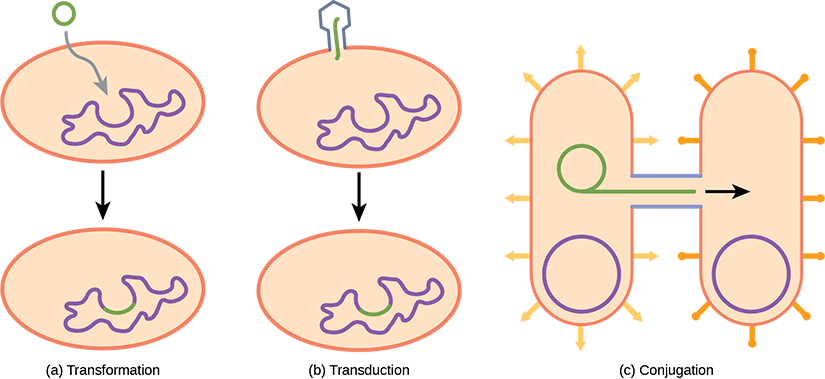
Figure 22.17 Gene transfer mechanisms in prokaryotes. There are three mechanisms by which prokaryotes can exchange DNA. In (a) transformation, the cell takes up prokaryotic DNA directly from the environment. The DNA may remain separate as plasmid DNA or be incorporated into the host genome. In (b) transduction, a bacteriophage injects DNA into the cell that contains a small fragment of DNA from a different prokaryote. In (c) conjugation, DNA is transferred from one cell to another via a mating bridge, or pilus, that connects the two cells after the sex pilus draws the two bacteria close enough to form the bridge.
A fleeting, high-energy arrangement at a reaction barrier. It is an energy maximum along the reaction path, rather than a stable reaction intermediate.
Figure 12.19 Reaction diagrams for an endothermic process in the absence (red curve) and presence (blue curve) of a catalyst. The catalyzed pathway involves a two-step mechanism (note the presence of two transition states) and an intermediate species (represented by the valley between the two transitions states).
Figure 15.18 Translation begins when an initiator tRNA anticodon recognizes a start codon on mRNA bound to a small ribosomal subunit. The large ribosomal subunit joins the small subunit, and a second tRNA is recruited. As the mRNA moves relative to the ribosome, successive tRNAs move through the ribosome and the polypeptide chain is formed. Entry of a release factor into the A site terminates translation and the components dissociate.
Figure 10.15 Cell Division and Apoptosis. In an adult organism, normal cell division is balanced by apoptosis (programmed cell death) to maintain a constant cell number in homeostasis. Either an increase in cell division or a decrease in apoptosis leads an increase in the number of cells and tumor formation. Credit: Rao, A. and Ryan, K. Department of Biology, Texas A&M University.
Rao, A. and Ryan, K. Department of Biology, Texas A&M University. Mary Ann Clark, Matthew Douglas, and Jung Choi, Biology 2e, OpenStax. · CC BY-NC-SA 4.0
· Original image ↗ · Access for free ↗
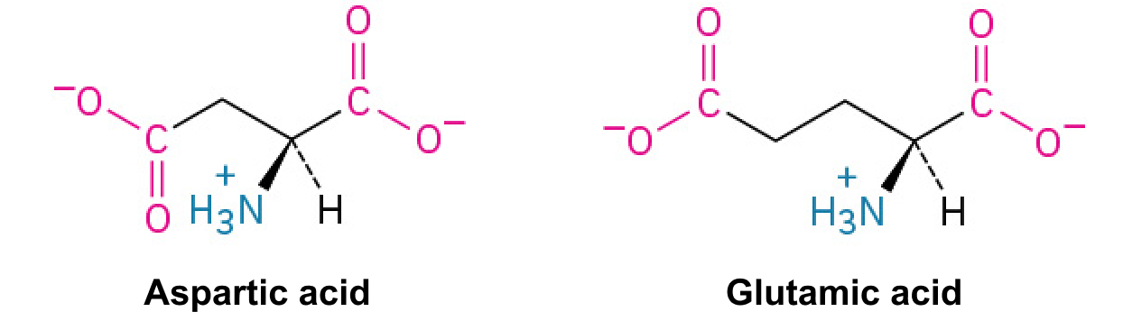
umami
Basic taste associated especially with glutamate and related savory stimuli.
Figure 4.18 Membrane and secretory proteins are synthesized in the rough endoplasmic reticulum (RER). The RER also sometimes modifies proteins. In this illustration, a (green) integral membrane protein is modified by attachment of a (purple) carbohydrate in the ER. Vesicles with the integral protein bud from the ER and fuse with the Golgi apparatus' cis face. As the protein passes along the Golgi’s cisternae, the addition of more carbohydrates further modifies it. After its synthesis is complete, it exits as an integral membrane protein of the vesicle that buds from the Golgi’s trans face. When the vesicle fuses with the cell membrane, the protein becomes an integral portion of that cell membrane. (credit: modification of work by Magnus Manske)
No matching terms. Try a different name or abbreviation.
5.1 Sensation versus Perception
OpenStax · 5.1 Sensation versus Perception ↗ Instructional page including learning objectives, examples, figures, source tables and questions. Site navigation and embedded media players are omitted; source media links remain available.
Discuss the roles attention, motivation, and sensory adaptation play in perception
Sensation
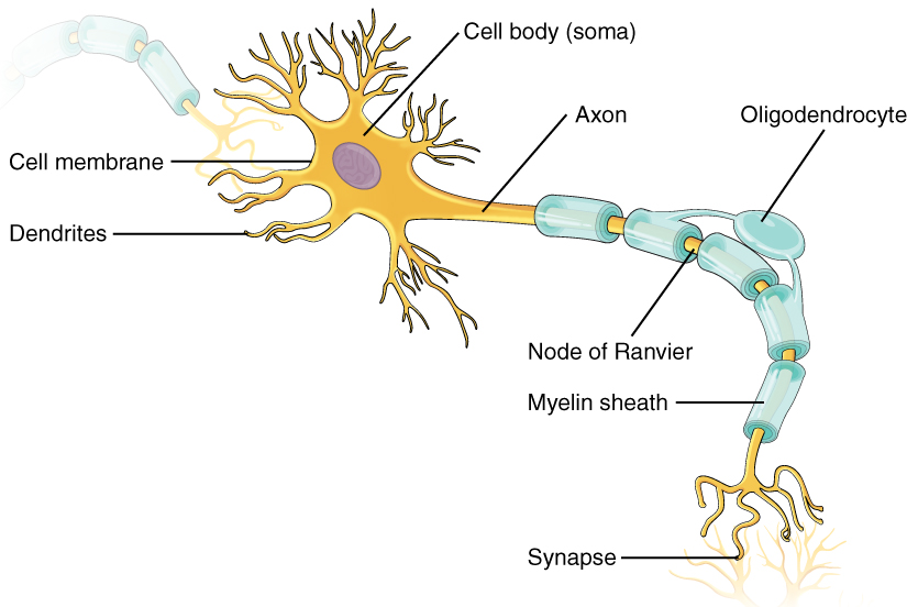
What does it mean to sense something? Sensory receptors are specialized neurons that respond to specific types of stimuli. When sensory information is detected by a sensory receptor, sensation has occurred. For example, light that enters the eye causes chemical changes in cells that line the back of the eye. These cells relay messages, in the form of action potentials (as you learned when studying biopsychology), to the central nervous system. The conversion from sensory stimulus energy to action potential is known as transduction.
You have probably known since elementary school that we have five senses: vision, hearing (audition), smell (olfaction), taste (gustation), and touch (somatosensation). It turns out that this notion of five senses is oversimplified. We also have sensory systems that provide information about balance (the vestibular sense), body position and movement (proprioception and kinesthesia), pain (nociception), and temperature (thermoception).
The sensitivity of a given sensory system to the relevant stimuli can be expressed as an absolute threshold. Absolute threshold refers to the minimum amount of stimulus energy that must be present for the stimulus to be detected 50% of the time. Another way to think about this is by asking how dim can a light be or how soft can a sound be and still be detected half of the time. The sensitivity of our sensory receptors can be quite amazing. It has been estimated that on a clear night, the most sensitive sensory cells in the back of the eye can detect a candle flame 30 miles away (Okawa & Sampath, 2007). Under quiet conditions, the hair cells (the receptor cells of the inner ear) can detect the tick of a clock 20 feet away (Galanter, 1962).
It is also possible for us to get messages that are presented below the threshold for conscious awareness—these are called subliminal messages. A stimulus reaches a physiological threshold when it is strong enough to excite sensory receptors and send nerve impulses to the brain: This is an absolute threshold. A message below that threshold is said to be subliminal: We receive it, but we are not consciously aware of it. Over the years there has been a great deal of speculation about the use of subliminal messages in advertising, rock music, and self-help audio programs. Research evidence shows that in laboratory settings, people can process and respond to information outside of awareness. But this does not mean that we obey these messages like zombies; in fact, hidden messages have little effect on behavior outside the laboratory (Kunst-Wilson & Zajonc, 1980; Rensink, 2004; Nelson, 2008; Radel, Sarrazin, Legrain, & Gobancé, 2009; Loersch, Durso, & Petty, 2013).
Absolute thresholds are generally measured under incredibly controlled conditions in situations that are optimal for sensitivity. Sometimes, we are more interested in how much difference in stimuli is required to detect a difference between them. This is known as the just noticeable difference (jnd) or difference threshold. Unlike the absolute threshold, the difference threshold changes depending on the stimulus intensity. As an example, imagine yourself in a very dark movie theater. If an audience member were to receive a text message that caused the cell phone screen to light up, chances are that many people would notice the change in illumination in the theater. However, if the same thing happened in a brightly lit arena during a basketball game, very few people would notice. The cell phone brightness does not change, but its ability to be detected as a change in illumination varies dramatically between the two contexts. Ernst Weber proposed this theory of change in difference threshold in the 1830s, and it has become known as Weber’s law: The difference threshold is a constant fraction of the original stimulus, as the example illustrates.
Perception
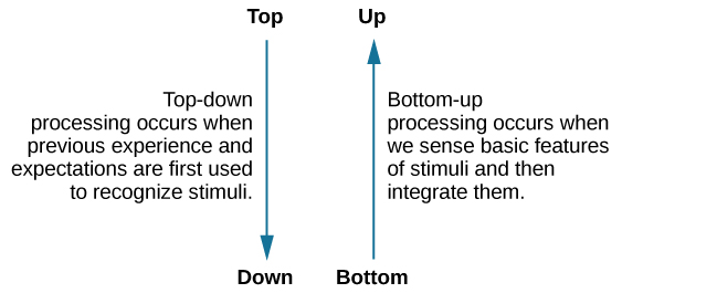
While our sensory receptors are constantly collecting information from the environment, it is ultimately how we interpret that information that affects how we interact with the world. Perception refers to the way sensory information is organized, interpreted, and consciously experienced. Perception involves both bottom-up and top-down processing. Bottom-up processing refers to sensory information from a stimulus in the environment driving a process, and top-down processing refers to knowledge and expectancy driving a process, as shown in Figure 5.2 (Egeth & Yantis, 1997; Fine & Minnery, 2009; Yantis & Egeth, 1999).
Figure 5.2 — 5.1 Sensation versus Perception
Figure 5.2 Top-down and bottom-up are ways we process our perceptions.
Imagine that you and some friends are sitting in a crowded restaurant eating lunch and talking. It is very noisy, and you are concentrating on your friend’s face to hear what they are saying, then the sound of breaking glass and clang of metal pans hitting the floor rings out. The server dropped a large tray of food. Although you were attending to your meal and conversation, that crashing sound would likely get through your attentional filters and capture your attention. You would have no choice but to notice it. That attentional capture would be caused by the sound from the environment: it would be bottom-up.
Alternatively, top-down processes are generally goal directed, slow, deliberate, effortful, and under your control (Fine & Minnery, 2009; Miller & Cohen, 2001; Miller & D'Esposito, 2005). For instance, if you misplaced your keys, how would you look for them? If you had a yellow key fob, you would probably look for yellowness of a certain size in specific locations, such as on the counter, coffee table, and other similar places. You would not look for yellowness on your ceiling fan, because you know keys are not normally lying on top of a ceiling fan. That act of searching for a certain size of yellowness in some locations and not others would be top-down—under your control and based on your experience.
One way to think of this concept is that sensation is a physical process, whereas perception is psychological. For example, upon walking into a kitchen and smelling the scent of baking cinnamon rolls, the sensation is the scent receptors detecting the odor of cinnamon, but the perception may be “Mmm, this smells like the bread Grandma used to bake when the family gathered for holidays.”
Although our perceptions are built from sensations, not all sensations result in perception. In fact, we often don’t perceive stimuli that remain relatively constant over prolonged periods of time. This is known as sensory adaptation. Imagine going to a city that you have never visited. You check in to the hotel, but when you get to your room, there is a road construction sign with a bright flashing light outside your window. Unfortunately, there are no other rooms available, so you are stuck with a flashing light. You decide to watch television to unwind. The flashing light was extremely annoying when you first entered your room. It was as if someone was continually turning a bright yellow spotlight on and off in your room, but after watching television for a short while, you no longer notice the light flashing. The light is still flashing and filling your room with yellow light every few seconds, and the photoreceptors in your eyes still sense the light, but you no longer perceive the rapid changes in lighting conditions. That you no longer perceive the flashing light demonstrates sensory adaptation and shows that while closely associated, sensation and perception are different.
There is another factor that affects sensation and perception: attention. Attention plays a significant role in determining what is sensed versus what is perceived. Imagine you are at a party full of music, chatter, and laughter. You get involved in an interesting conversation with a friend, and you tune out all the background noise. If someone interrupted you to ask what song had just finished playing, you would probably be unable to answer that question.
One of the most interesting demonstrations of how important attention is in determining our perception of the environment occurred in a famous study conducted by Daniel Simons and Christopher Chabris (1999). In this study, participants watched a video of people dressed in black and white passing basketballs. Participants were asked to count the number of times the team dressed in white passed the ball. During the video, a person dressed in a black gorilla costume walks among the two teams. You would think that someone would notice the gorilla, right? Nearly half of the people who watched the video didn’t notice the gorilla at all, despite the fact that he was clearly visible for nine seconds. Because participants were so focused on the number of times the team dressed in white was passing the ball, they completely tuned out other visual information. Inattentional blindness is the failure to notice something that is completely visible because the person was actively attending to something else and did not pay attention to other things (Mack & Rock, 1998; Simons & Chabris, 1999).
In a similar experiment, researchers tested inattentional blindness by asking participants to observe images moving across a computer screen. They were instructed to focus on either white or black objects, disregarding the other color. When a red cross passed across the screen, about one third of subjects did not notice it (Figure 5.3) (Most, Simons, Scholl, & Chabris, 2000).
Figure 5.3 — 5.1 Sensation versus Perception
Figure 5.3 Nearly one third of participants in a study did not notice that a red cross passed on the screen because their attention was focused on the black or white figures. (credit: Cory Zanker)
Motivation can also affect perception. Have you ever been expecting a really important phone call and, while taking a shower, you think you hear the phone ringing, only to discover that it is not? If so, then you have experienced how motivation to detect a meaningful stimulus can shift our ability to discriminate between a true sensory stimulus and background noise. The ability to identify a stimulus when it is embedded in a distracting background is called signal detection theory. This might also explain why a mother is awakened by a quiet murmur from her baby but not by other sounds that occur while she is asleep. Signal detection theory has practical applications, such as increasing air traffic controller accuracy. Controllers need to be able to detect planes among many signals (blips) that appear on the radar screen and follow those planes as they move through the sky. In fact, the original work of the researcher who developed signal detection theory was focused on improving the sensitivity of air traffic controllers to plane blips (Swets, 1964).
Our perceptions can also be affected by our beliefs, values, prejudices, expectations, and life experiences. As you will see later in this chapter, individuals who are deprived of the experience of binocular vision during critical periods of development have trouble perceiving depth (Fawcett, Wang, & Birch, 2005). The shared experiences of people within a given cultural context can have pronounced effects on perception. For example, Marshall Segall, Donald Campbell, and Melville Herskovits (1963) published the results of a multinational study in which they demonstrated that individuals from Western cultures were more prone to experience certain types of visual illusions than individuals from non-Western cultures, and vice versa. One such illusion that Westerners were more likely to experience was the Müller-Lyer illusion (Figure 5.4): The lines appear to be different lengths, but they are actually the same length.
Figure 5.4 — 5.1 Sensation versus Perception
Figure 5.4 In the Müller-Lyer illusion, lines appear to be different lengths although they are identical. (a) Arrows at the ends of lines may make the line on the right appear longer, although the lines are the same length. (b) When applied to a three-dimensional image, the line on the right again may appear longer although both black lines are the same length.
These perceptual differences were consistent with differences in the types of environmental features experienced on a regular basis by people in a given cultural context. People in Western cultures, for example, have a perceptual context of buildings with straight lines, what Segall’s study called a carpentered world (Segall et al., 1966). In contrast, people from certain non-Western cultures with an uncarpentered view, such as the Zulu of South Africa, whose villages are made up of round huts arranged in circles, are less susceptible to this illusion (Segall et al., 1999). It is not just vision that is affected by cultural factors. Indeed, research has demonstrated that the ability to identify an odor, and rate its pleasantness and its intensity, varies cross-culturally (Ayabe-Kanamura, Saito, Distel, Martínez-Gómez, & Hudson, 1998).
Children described as thrill seekers are more likely to show taste preferences for intense sour flavors (Liem, Westerbeek, Wolterink, Kok, & de Graaf, 2004), which suggests that basic aspects of personality might affect perception. Furthermore, individuals who hold positive attitudes toward reduced-fat foods are more likely to rate foods labeled as reduced fat as tasting better than people who have less positive attitudes about these products (Aaron, Mela, & Evans, 1994).
5.5 The Other Senses
OpenStax · 5.5 The Other Senses ↗ Instructional page including learning objectives, examples, figures, source tables and questions. Site navigation and embedded media players are omitted; source media links remain available.
Learning Objectives
By the end of this section, you will be able to:
Describe the basic functions of the chemical senses
Explain the basic functions of the somatosensory, nociceptive, and thermoceptive sensory systems
Describe the basic functions of the vestibular, proprioceptive, and kinesthetic sensory systems
Vision and hearing have received an incredible amount of attention from researchers over the years. While there is still much to be learned about how these sensory systems work, we have a much better understanding of them than of our other sensory modalities. In this section, we will explore our chemical senses (taste and smell) and our body senses (touch, temperature, pain, balance, and body position).
The Chemical Senses
Taste (gustation) and smell (olfaction) are called chemical senses because both have sensory receptors that respond to molecules in the food we eat or in the air we breathe. There is a pronounced interaction between our chemical senses. For example, when we describe the flavor of a given food, we are really referring to both gustatory and olfactory properties of the food working in combination.
Taste (Gustation)
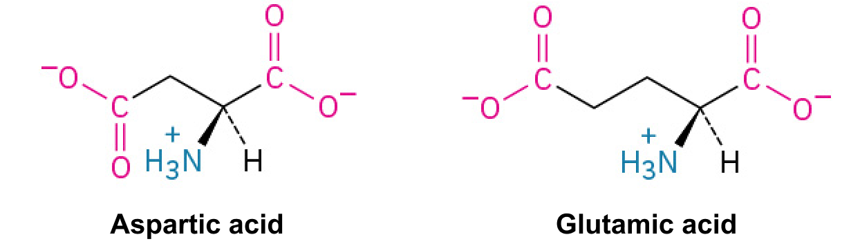
You have learned since elementary school that there are four basic groupings of taste: sweet, salty, sour, and bitter. Research demonstrates, however, that we have at least six taste groupings. Umami is our fifth taste. Umami is actually a Japanese word that roughly translates to yummy, and it is associated with a taste for monosodium glutamate (Kinnamon & Vandenbeuch, 2009). There is also a growing body of experimental evidence suggesting that we possess a taste for the fatty content of a given food (Mizushige, Inoue, & Fushiki, 2007).
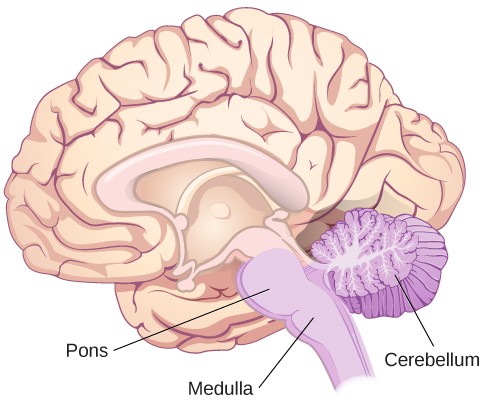
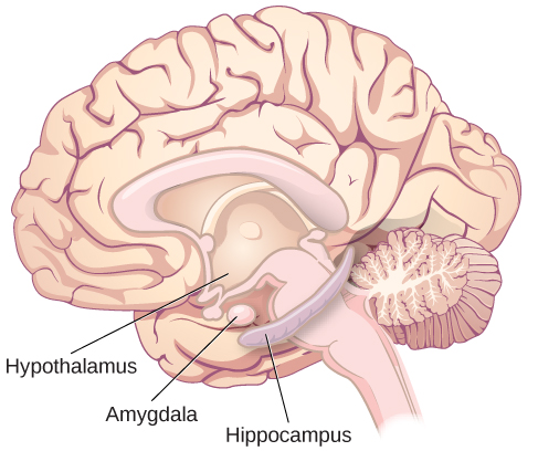
Molecules from the food and beverages we consume dissolve in our saliva and interact with taste receptors on our tongue and in our mouth and throat. Taste buds are formed by groupings of taste receptor cells with hair-like extensions that protrude into the central pore of the taste bud (Figure 5.21). Taste buds have a life cycle of ten days to two weeks, so even destroying some by burning your tongue won’t have any long-term effect; they just grow right back. Taste molecules bind to receptors on this extension and cause chemical changes within the sensory cell that result in neural impulses being transmitted to the brain via different nerves, depending on where the receptor is located. Taste information is transmitted to the medulla, thalamus, and limbic system, and to the gustatory cortex, which is tucked underneath the overlap between the frontal and temporal lobes (Maffei, Haley, & Fontanini, 2012; Roper, 2013).
Figure 5.21 — 5.5 The Other Senses
Figure 5.21 (a) Taste buds are composed of a number of individual taste receptors cells that transmit information to nerves. (b) This micrograph shows a close-up view of the tongue’s surface. (credit a: modification of work by Jonas Töle; credit b: scale-bar data from Matt Russell)
Rose M. Spielman, William J. Jenkins, and Marilyn D. Lovett, Psychology 2E, OpenStax. (credit a: modification of work by Jonas Töle; credit b: scale-bar data from Matt Russell) · CC BY-NC-SA 4.0
· Original image ↗ · Access for free ↗
Olfactory receptor cells are located in a mucous membrane at the top of the nose. Small hair-like extensions from these receptors serve as the sites for odor molecules dissolved in the mucus to interact with chemical receptors located on these extensions (Figure 5.22). Once an odor molecule has bound a given receptor, chemical changes within the cell result in signals being sent to the olfactory bulb: a bulb-like structure at the tip of the frontal lobe where the olfactory nerves begin. From the olfactory bulb, information is sent to regions of the limbic system and to the primary olfactory cortex, which is located very near the gustatory cortex (Lodovichi & Belluscio, 2012; Spors et al., 2013).
Figure 5.22 — 5.5 The Other Senses
Figure 5.22 Olfactory receptors are the hair-like parts that extend from the olfactory bulb into the mucous membrane of the nasal cavity.
There is tremendous variation in the sensitivity of the olfactory systems of different species. We often think of dogs as having far superior olfactory systems than our own, and indeed, dogs can do some remarkable things with their noses. There is some evidence to suggest that dogs can “smell” dangerous drops in blood glucose levels as well as canceroustumors (Wells, 2010). Dogs’ extraordinary olfactory abilities may be due to the increased number of functional genes for olfactory receptors (between 800 and 1200), compared to the fewer than 400 observed in humans and other primates (Niimura & Nei, 2007).
Many species respond to chemical messages, known as pheromones, sent by another individual (Wysocki & Preti, 2004). Pheromonal communication often involves providing information about the reproductive status of a potential mate. So, for example, when a female rat is ready to mate, it secretes pheromonal signals that draw attention from nearby male rats. Pheromonal activation is actually an important component in eliciting sexual behavior in the male rat (Furlow, 1996, 2012; Purvis & Haynes, 1972; Sachs, 1997). There has also been a good deal of research (and controversy) about pheromones in humans (Comfort, 1971; Russell, 1976; Wolfgang-Kimball, 1992; Weller, 1998).
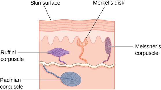
A number of receptors are distributed throughout the skin to respond to various touch-related stimuli (Figure 5.23). These receptors include Meissner’s corpuscles, Pacinian corpuscles, Merkel’s disks, and Ruffini corpuscles. Meissner’s corpuscles respond to pressure and lower frequency vibrations, and Pacinian corpuscles detect transient pressure and higher frequency vibrations. Merkel’s disks respond to light pressure, while Ruffini corpuscles detect stretch (Abraira & Ginty, 2013).
Figure 5.23 — 5.5 The Other Senses
Figure 5.23 There are many types of sensory receptors located in the skin, each attuned to specific touch-related stimuli.
In addition to the receptors located in the skin, there are also a number of free nerve endings that serve sensory functions. These nerve endings respond to a variety of different types of touch-related stimuli and serve as sensory receptors for both thermoception (temperature perception) and nociception (a signal indicating potential harm and maybe pain) (Garland, 2012; Petho & Reeh, 2012; Spray, 1986). Sensory information collected from the receptors and free nerve endings travels up the spinal cord and is transmitted to regions of the medulla, thalamus, and ultimately to the somatosensory cortex, which is located in the postcentral gyrus of the parietal lobe.
Pain Perception
Pain is an unpleasant experience that involves both physical and psychological components. Feeling pain is quite adaptive because it makes us aware of an injury, and it motivates us to remove ourselves from the cause of that injury. In addition, pain also makes us less likely to suffer additional injury because we will be gentler with our injured body parts.
Generally speaking, pain can be considered to be neuropathic or inflammatory in nature. Pain that signals some type of tissue damage is known as inflammatory pain. In some situations, pain results from damage to neurons of either the peripheral or central nervous system. As a result, pain signals that are sent to the brain get exaggerated. This type of pain is known as neuropathic pain. Multiple treatment options for pain relief range from relaxation therapy to the use of analgesic medications to deep brain stimulation. The most effective treatment option for a given individual will depend on a number of considerations, including the severity and persistence of the pain and any medical/psychological conditions.
Some individuals are born without the ability to feel pain. This very rare genetic disorder is known as congenital insensitivity to pain (or congenital analgesia). While those with congenital analgesia can detect differences in temperature and pressure, they cannot experience pain. As a result, they often suffer significant injuries. Young children have serious mouth and tongue injuries because they have bitten themselves repeatedly. Not surprisingly, individuals suffering from this disorder have much shorter life expectancies due to their injuries and secondary infections of injured sites (U.S. National Library of Medicine, 2013).
The vestibular sense contributes to our ability to maintain balance and body posture. As Figure 5.24 shows, the major sensory organs (utricle, saccule, and the three semicircular canals) of this system are located next to the cochlea in the inner ear. The vestibular organs are fluid-filled and have hair cells, similar to the ones found in the auditory system, which respond to movement of the head and gravitational forces. When these hair cells are stimulated, they send signals to the brain via the vestibular nerve. Although we may not be consciously aware of our vestibular system’s sensory information under normal circumstances, its importance is apparent when we experience motion sickness and/or dizziness related to infections of the inner ear (Khan & Chang, 2013).
Figure 5.24 — 5.5 The Other Senses
Figure 5.24 The major sensory organs of the vestibular system are located next to the cochlea in the inner ear. These include the utricle, saccule, and the three semicircular canals (posterior, superior, and horizontal).
In addition to maintaining balance, the vestibular system collects information critical for controlling movement and the reflexes that move various parts of our bodies to compensate for changes in body position. Therefore, both proprioception(perception of body position) and kinesthesia(perception of the body’s movement through space) interact with information provided by the vestibular system.
These sensory systems also gather information from receptors that respond to stretch and tension in muscles, joints, skin, and tendons (Lackner & DiZio, 2005; Proske, 2006; Proske & Gandevia, 2012). Proprioceptive and kinesthetic information travels to the brain via the spinal column. Several cortical regions in addition to the cerebellum receive information from and send information to the sensory organs of the proprioceptive and kinesthetic systems.