Phonemes distinguish speech sounds; morphemes are the smallest meaningful units; syntax organizes words into sentences and semantics concerns meaning.
Compare learning, nativist, and interactionist accounts of language development
Phonemes distinguish speech sounds; morphemes are the smallest meaningful units; syntax organizes words into sentences and semantics concerns meaning. Pragmatics concerns appropriate interpretation in social context. Learning accounts emphasize imitation, reinforcement, and statistical regularities. Nativist accounts propose biological preparedness for language and help explain generative rules that exceed memorized phrases. Interactionist accounts combine maturational constraints with responsive social communication. Overregularization, such as applying a regular past-tense ending to an irregular verb, demonstrates rule use rather than simple repetition. Sensitive developmental periods influence later learning, but age effects do not mean that no useful language learning can occur afterward.
Explain language–cognition relationships and the roles of brain regions in speech and comprehension
Linguistic relativity proposes that language habits influence attention, categorization, or memory; strong linguistic determinism claims language fixes what can be thought and is not supported as a general rule. Nonverbal thought and learning new distinctions argue against absolute determinism. In many people, left-hemisphere networks are especially important for language. Damage near Broca-associated frontal networks can cause effortful, nonfluent production; damage near Wernicke-associated temporal networks can produce fluent but poorly comprehended language. These syndromes involve distributed networks and variable lesions, not isolated word boxes. Reading, prosody, gesture, and context recruit additional bilateral systems.
Worked question. A child says an unfamiliar verb with a grammatically appropriate ending, even though nobody modeled that exact word. Which account is challenged, and does the observation prove one innate grammar theory?
It challenges a pure imitation-only explanation because the child generalizes a rule. It does not uniquely prove a particular nativist mechanism: statistical learning and interactionist accounts can also predict generalization. Test competing accounts using novel forms, controlled input, and developmental comparisons rather than treating one example as decisive.
Figure 7.6 These two works of art depict saudade . (a) Saudade de Nápoles , which is translated into “missing Naples,” was painted by Bertha Worms in 1895. (b) Almeida Júnior painted Saudade in 1899.
3.4 The Brain and Spinal Cord — figures and tables
Figure 3.15 — 3.4 The Brain and Spinal Cord
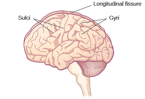
Figure 3.15 The surface of the brain is covered with gyri and sulci. A deep sulcus is called a fissure, such as the longitudinal fissure that divides the brain into left and right hemispheres. (credit: modification of work by Bruce Blaus)
Rose M. Spielman, William J. Jenkins, and Marilyn D. Lovett, Psychology 2E, OpenStax. (credit: modification of work by Bruce Blaus) · CC BY-NC-SA 4.0
· Original image ↗ · Access for free ↗Figure 3.16 — 3.4 The Brain and Spinal Cord
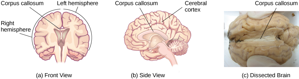
Figure 3.16 (a, b) The corpus callosum connects the left and right hemispheres of the brain. (c) A scientist spreads this dissected sheep brain apart to show the corpus callosum between the hemispheres. (credit c: modification of work by Aaron Bornstein)
Rose M. Spielman, William J. Jenkins, and Marilyn D. Lovett, Psychology 2E, OpenStax. (credit c: modification of work by Aaron Bornstein) · CC BY-NC-SA 4.0
· Original image ↗ · Access for free ↗Figure 3.17 — 3.4 The Brain and Spinal Cord
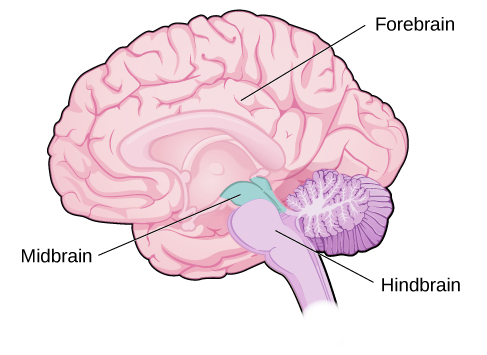
Figure 3.17 The brain and its parts can be divided into three main categories: the forebrain, midbrain, and hindbrain.
Figure 3.19 (a) Phineas Gage holds the iron rod that penetrated his skull in an 1848 railroad construction accident. (b) Gage’s prefrontal cortex was severely damaged in the left hemisphere. The rod entered Gage’s face on the left side, passed behind his eye, and exited through the top of his skull, before landing about 80 feet away. (credit a: modification of work by Jack and Beverly Wilgus)
Rose M. Spielman, William J. Jenkins, and Marilyn D. Lovett, Psychology 2E, OpenStax. (credit a: modification of work by Jack and Beverly Wilgus) · CC BY-NC-SA 4.0
· Original image ↗ · Access for free ↗Figure 3.20 — 3.4 The Brain and Spinal Cord
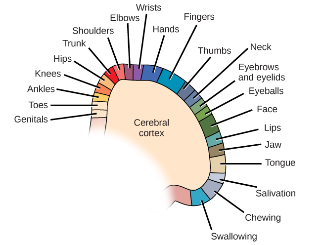
Figure 3.20 Spatial relationships in the body are mirrored in the organization of the somatosensory cortex.
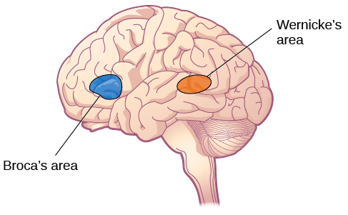
Figure 3.21 Damage to either Broca’s area or Wernicke’s area can result in language deficits. The types of deficits are very different, however, depending on which area is affected.
Figure 3.26 A CT scan can be used to show brain tumors. (a) The image on the left shows a healthy brain, whereas (b) the image on the right indicates a brain tumor in the left frontal lobe. (credit a: modification of work by "Aceofhearts1968"/Wikimedia Commons; credit b: modification of work by Roland Schmitt et al)
Rose M. Spielman, William J. Jenkins, and Marilyn D. Lovett, Psychology 2E, OpenStax. (credit a: modification of work by "Aceofhearts1968"/Wikimedia Commons; credit b: modification of work by Roland Schmitt et al) · CC BY-NC-SA 4.0
· Original image ↗ · Access for free ↗Figure 3.27 — 3.4 The Brain and Spinal Cord
Figure 3.27 A PET scan is helpful for showing activity in different parts of the brain. (credit: Health and Human Services Department, National Institutes of Health)
Rose M. Spielman, William J. Jenkins, and Marilyn D. Lovett, Psychology 2E, OpenStax. (credit: Health and Human Services Department, National Institutes of Health) · CC BY-NC-SA 4.0
· Original image ↗ · Access for free ↗Figure 3.28 — 3.4 The Brain and Spinal Cord
Figure 3.28 An fMRI shows activity in the brain over time. This image represents a single frame from an fMRI. (credit: modification of work by Kim J, Matthews NL, Park S.)
Rose M. Spielman, William J. Jenkins, and Marilyn D. Lovett, Psychology 2E, OpenStax. (credit: modification of work by Kim J, Matthews NL, Park S.) · CC BY-NC-SA 4.0
· Original image ↗ · Access for free ↗Figure 3.29 — 3.4 The Brain and Spinal Cord
Figure 3.29 Using caps with electrodes, modern EEG research can study the precise timing of overall brain activities. (credit: SMI Eye Tracking)
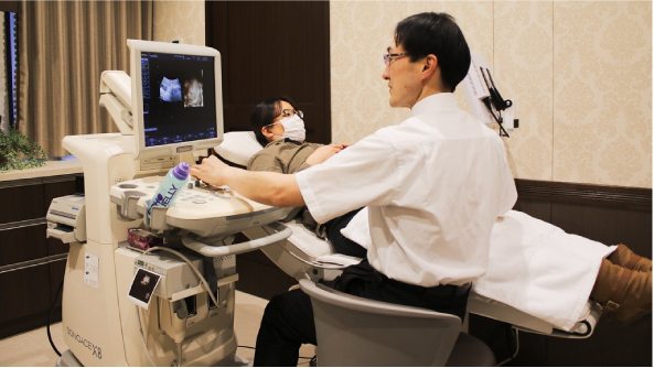
Figure 9.9 A pregnant person receives an ultrasound as part of their prenatal care. (credit: "MIKI Yoshihito_Flickr"/Flickr)
Rose M. Spielman, William J. Jenkins, and Marilyn D. Lovett, Psychology 2E, OpenStax. (credit: "MIKI Yoshihito_Flickr"/Flickr) · CC BY-NC-SA 4.0
· Original image ↗ · Access for free ↗Figure 9.10 — 9.3 Stages of Development
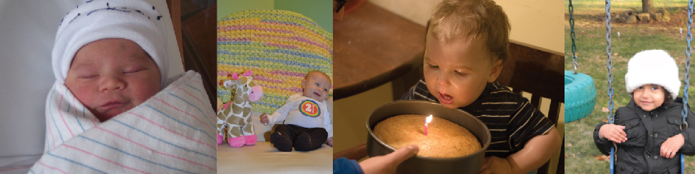
Figure 9.10 Children experience rapid physical changes through infancy and early childhood. (credit "left": modification of work by Kerry Ceszyk; credit "middle-left": modification of work by Kristi Fausel; credit "middle-right": modification of work by "devinf"/Flickr; credit "right": modification of work by Rose Spielman)
Rose M. Spielman, William J. Jenkins, and Marilyn D. Lovett, Psychology 2E, OpenStax. (credit "left": modification of work by Kerry Ceszyk; credit "middle-left": modification of work by Kristi Fausel; credit "middle-right": modification of work by "devinf"/Flickr; credit "right": modification of work by Rose Spielman) · CC BY-NC-SA 4.0
· Original image ↗ · Access for free ↗Figure 9.11 — 9.3 Stages of Development
Figure 9.11 In Baillargeon’s study, infants observed a truck (a) roll down an unobstructed track, (b) roll down an unobstructed track with an obstruction (box) beside it, and (c) roll down and pass through what appeared to be an obstruction.
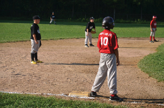
Figure 9.12 Because they understand luck and fairness, children in middle and late childhood (6–11 years old) are able to follow rules for games. (credit: Edwin Martinez)
Rose M. Spielman, William J. Jenkins, and Marilyn D. Lovett, Psychology 2E, OpenStax. (credit: Edwin Martinez) · CC BY-NC-SA 4.0
· Original image ↗ · Access for free ↗Figure 9.13 — 9.3 Stages of Development
Rose M. Spielman, William J. Jenkins, and Marilyn D. Lovett, Psychology 2E, OpenStax. (credit: "balouriarajesh_Pixabay"/Pixabay) · CC BY-NC-SA 4.0
· Original image ↗ · Access for free ↗Figure 9.14 — 9.3 Stages of Development
Figure 9.14 In secure attachment, the parent provides a secure base for the toddler, allowing him to securely explore his environment. (credit: Kerry Ceszyk)
Rose M. Spielman, William J. Jenkins, and Marilyn D. Lovett, Psychology 2E, OpenStax. (credit: Kerry Ceszyk) · CC BY-NC-SA 4.0
· Original image ↗ · Access for free ↗Figure 9.15 — 9.3 Stages of Development
Figure 9.15 Peers are a primary influence on our development in adolescence. (credit: "manseok_Pixabay"/ Pixabay)
Rose M. Spielman, William J. Jenkins, and Marilyn D. Lovett, Psychology 2E, OpenStax. (credit: "manseok_Pixabay"/ Pixabay) · CC BY-NC-SA 4.0
· Original image ↗ · Access for free ↗Figure 9.16 — 9.3 Stages of Development
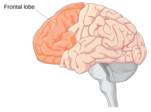
Figure 9.16 Brain growth continues into the early 20s. The development of the frontal lobe, in particular, is important during this stage.
Figure 9.17 Teenage thinking is characterized by the ability to reason logically and solve hypothetical problems such as how to design, plan, and build a structure. (credit: U.S. Army RDECOM)
Rose M. Spielman, William J. Jenkins, and Marilyn D. Lovett, Psychology 2E, OpenStax. (credit: U.S. Army RDECOM) · CC BY-NC-SA 4.0
· Original image ↗ · Access for free ↗Figure 9.18 — 9.3 Stages of Development
Figure 9.18 Physical declines of middle and late adulthood can be minimized with proper exercise, nutrition, and an active lifestyle. (credit: modification of work by Peter Stevens)
Rose M. Spielman, William J. Jenkins, and Marilyn D. Lovett, Psychology 2E, OpenStax. (credit: modification of work by Peter Stevens) · CC BY-NC-SA 4.0
· Original image ↗ · Access for free ↗Figure 9.19 — 9.3 Stages of Development
Figure 9.19 Cognitive activities such as playing mahjong, chess, or other games, can keep you mentally fit. The same is true for solo pastimes like reading and completing crossword puzzles. (credit: Philippe Put)
Rose M. Spielman, William J. Jenkins, and Marilyn D. Lovett, Psychology 2E, OpenStax. (credit: Philippe Put) · CC BY-NC-SA 4.0
· Original image ↗ · Access for free ↗Figure 9.20 — 9.3 Stages of Development
Figure 9.20 Social support is important as we age. (credit: Gabriel Rocha)
Figure 14.7 DNA has (a) a double helix structure and (b) phosphodiester bonds; the dotted lines between Thymine and Adenine and Guanine and Cytosine represent hydrogen bonds. The (c) major and minor grooves are binding sites for DNA binding proteins during processes such as transcription (the copying of RNA from DNA) and replication.
Figure 14.7 DNA has (a) a double helix structure and (b) phosphodiester bonds; the dotted lines between Thymine and Adenine and Guanine and Cytosine represent hydrogen bonds. The (c) major and minor grooves are binding sites for DNA binding proteins during processes such as transcription (the copying of RNA from DNA) and replication.
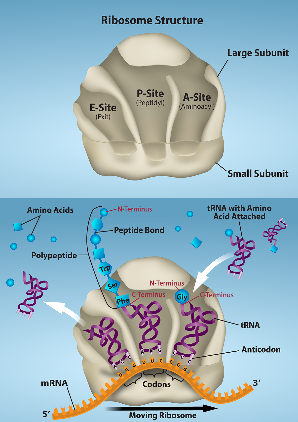
Figure 15.16 The ribosome and its function. The ribosome is responsible for translating the mRNA into protein. A. The ribosome consists of a large and small ribosomal subunit. Assembly of the subunits on the mRNA forms three tRNA binding sites. B. During translation, charged tRNAs enter the Acceptor site, and the anticodon on the tRNA base pairs with the codon in the mRNA. After the incoming amino acid forms a peptide bond with the growing polypeptide chain, the ribosome will move three nucleotides toward the 3’ end of the mRNA. This movement will transfer the tRNA with the growing polypeptide to the Peptidyl-tRNA binding site and allow the empty tRNA to exit at the Exit site. Credit: Rao, A., Ryan, K. and Fletcher, S. Department of Biology, Texas A&M University.
Rao, A., Ryan, K. and Fletcher, S. Department of Biology, Texas A&M University.; Mary Ann Clark, Matthew Douglas, and Jung Choi, Biology 2e, OpenStax. · CC BY-NC-SA 4.0
· Original image ↗ · Access for free ↗
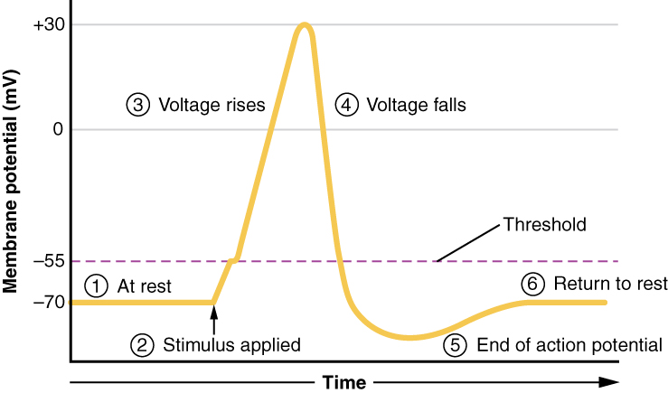
Figure 12.24 Stages of an Action Potential Plotting voltage measured across the cell membrane against time, the events of the action potential can be related to specific changes in the membrane voltage. (1) At rest, the membrane voltage is -70 mV. (2) The membrane begins to depolarize when an external stimulus is applied. (3) The membrane voltage begins a rapid rise toward +30 mV. (4) The membrane voltage starts to return to a negative value. (5) Repolarization continues past the resting membrane voltage, resulting in hyperpolarization. (6) The membrane voltage returns to the resting value shortly after hyperpolarization.
Figure 10.16 The role of normal p53 is to monitor DNA and the supply of oxygen (hypoxia is a condition of reduced oxygen supply). If damage is detected, p53 triggers repair mechanisms. If repairs are unsuccessful, p53 signals apoptosis. A cell with an abnormal p53 protein cannot repair damaged DNA and thus cannot signal apoptosis. Cells with abnormal p53 can become cancerous. (credit: modification of work by Thierry Soussi)
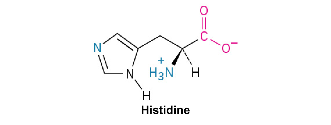
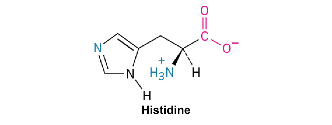
A nitrogen-containing functional group. A free amino acid’s amino group is typically protonated (NH₃⁺) near physiological pH; proline’s ring nitrogen is an exception to this formula.
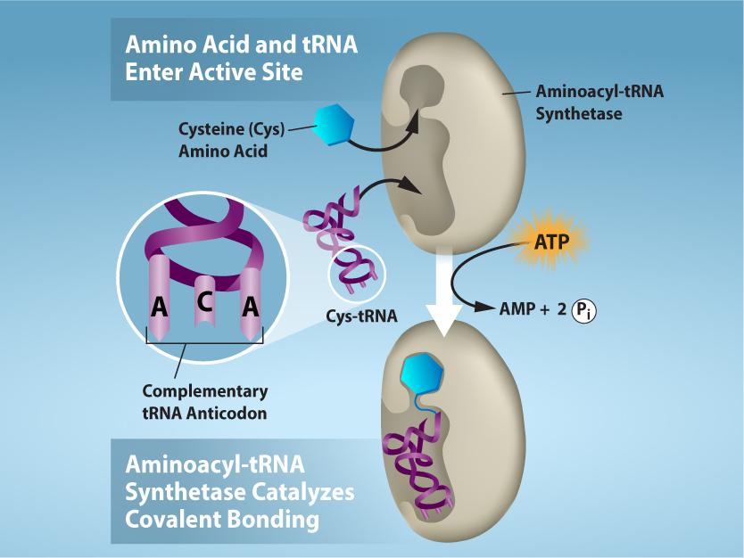
Figure 15.17 Charging of tRNAs with correct amino acids. Aminoacyl-tRNA synthetases catalyze covalent bond formation between the tRNA and the correct amino acid in preparation for translation. Because there are multiple amino acids, there are multiple different tRNA synthetases. All of the synthetases require energy, in the form of ATP, to make sure the correct amino acid is attached to the tRNA with the correct anticodon sequence. Credit: Rao, A., Ryan, K. and Tag, A. Department of Biology, Texas A&M University.
Rao, A., Ryan, K. and Tag, A. Department of Biology, Texas A&M University.; Mary Ann Clark, Matthew Douglas, and Jung Choi, Biology 2e, OpenStax. · CC BY-NC-SA 4.0
· Original image ↗ · Access for free ↗
Figure 15.14 This is a space-filling model of a tRNA molecule that adds the amino acid phenylalanine to a growing polypeptide chain. The anticodon AAG binds the Codon UUC on the mRNA. The amino acid phenylalanine is attached to the other end of the tRNA.
Figure 15.4 This figure shows the genetic code for translating each nucleotide triplet in mRNA into an amino acid or a termination signal in a protein. (credit: modification of work by NIH)
Figure 3.16 (a, b) The corpus callosum connects the left and right hemispheres of the brain. (c) A scientist spreads this dissected sheep brain apart to show the corpus callosum between the hemispheres. (credit c: modification of work by Aaron Bornstein)
Rose M. Spielman, William J. Jenkins, and Marilyn D. Lovett, Psychology 2E, OpenStax. (credit c: modification of work by Aaron Bornstein) · CC BY-NC-SA 4.0
· Original image ↗ · Access for free ↗
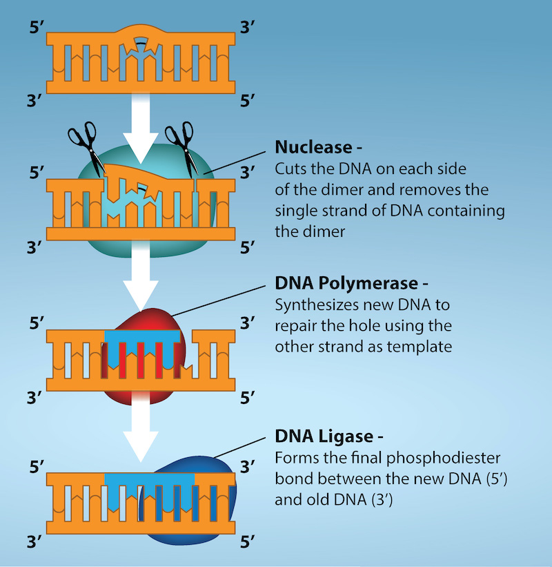
Figure 14.19 Nucleotide excision repairs thymine dimers. When exposed to UV light, thymines lying adjacent to each other can form thymine dimers. In normal cells, they are excised and replaced. Credit: Rao, A., Fletcher, S. and Tag, A. Department of Biology, Texas A&M University.
Figure 14.14 First Components of DNA Replication. As DNA replication begins, DNA Helicase, a large enzyme, separates the two strands of DNA so that they can act as templates for replication. Single-strand binding proteins bind to each strand to stabilize and prevent them from reforming the double helix. Primase, an RNA polymerase, binds to the single stranded DNA and synthesizes a short RNA primer in the 5’ to 3’ direction that is antiparallel to the parental strand. This RNA primer allows for DNA polymerase to begin replicating the DNA. Topoisomerase binds to the double helix upstream of the replication fork to prevent additional coiling by making small cuts in one of the DNA strands.
Credit: Rao, A., Ryan, K. Fletcher, S. and Tag, A. Department of Biology, Texas A&M University.
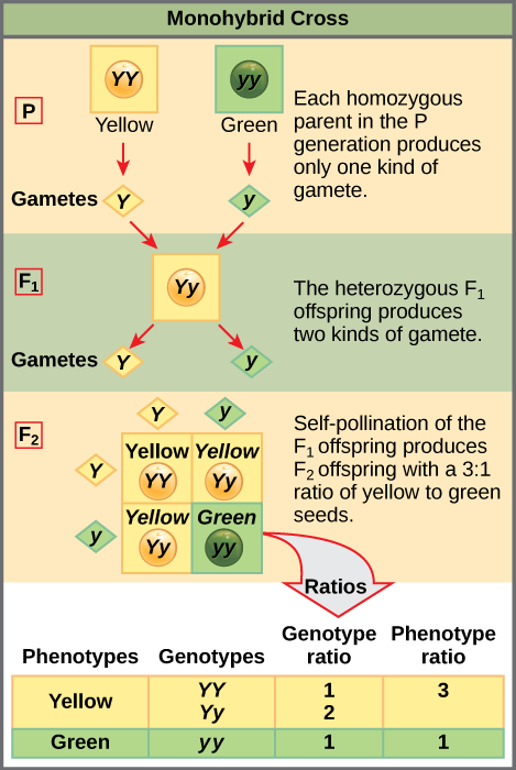
Figure 12.4 In the P generation, pea plants that are true-breeding for the dominant yellow phenotype are crossed with plants with the recessive green phenotype. This cross produces F 1 heterozygotes with a yellow phenotype. Punnett square analysis can be used to predict the genotypes of the F 2 generation.
An electrical property that can be positive or negative. Opposite charges attract and like charges repel; net charge is the sum of all charges on a particle.
Figure 4.18 Membrane and secretory proteins are synthesized in the rough endoplasmic reticulum (RER). The RER also sometimes modifies proteins. In this illustration, a (green) integral membrane protein is modified by attachment of a (purple) carbohydrate in the ER. Vesicles with the integral protein bud from the ER and fuse with the Golgi apparatus' cis face. As the protein passes along the Golgi’s cisternae, the addition of more carbohydrates further modifies it. After its synthesis is complete, it exits as an integral membrane protein of the vesicle that buds from the Golgi’s trans face. When the vesicle fuses with the cell membrane, the protein becomes an integral portion of that cell membrane. (credit: modification of work by Magnus Manske)
Figure 4.18 Membrane and secretory proteins are synthesized in the rough endoplasmic reticulum (RER). The RER also sometimes modifies proteins. In this illustration, a (green) integral membrane protein is modified by attachment of a (purple) carbohydrate in the ER. Vesicles with the integral protein bud from the ER and fuse with the Golgi apparatus' cis face. As the protein passes along the Golgi’s cisternae, the addition of more carbohydrates further modifies it. After its synthesis is complete, it exits as an integral membrane protein of the vesicle that buds from the Golgi’s trans face. When the vesicle fuses with the cell membrane, the protein becomes an integral portion of that cell membrane. (credit: modification of work by Magnus Manske)
Figure 15.10 A generalized promoter of a gene transcribed by RNA polymerase II is shown. Transcription factors recognize the promoter. RNA polymerase II then binds and forms the transcription initiation complex.
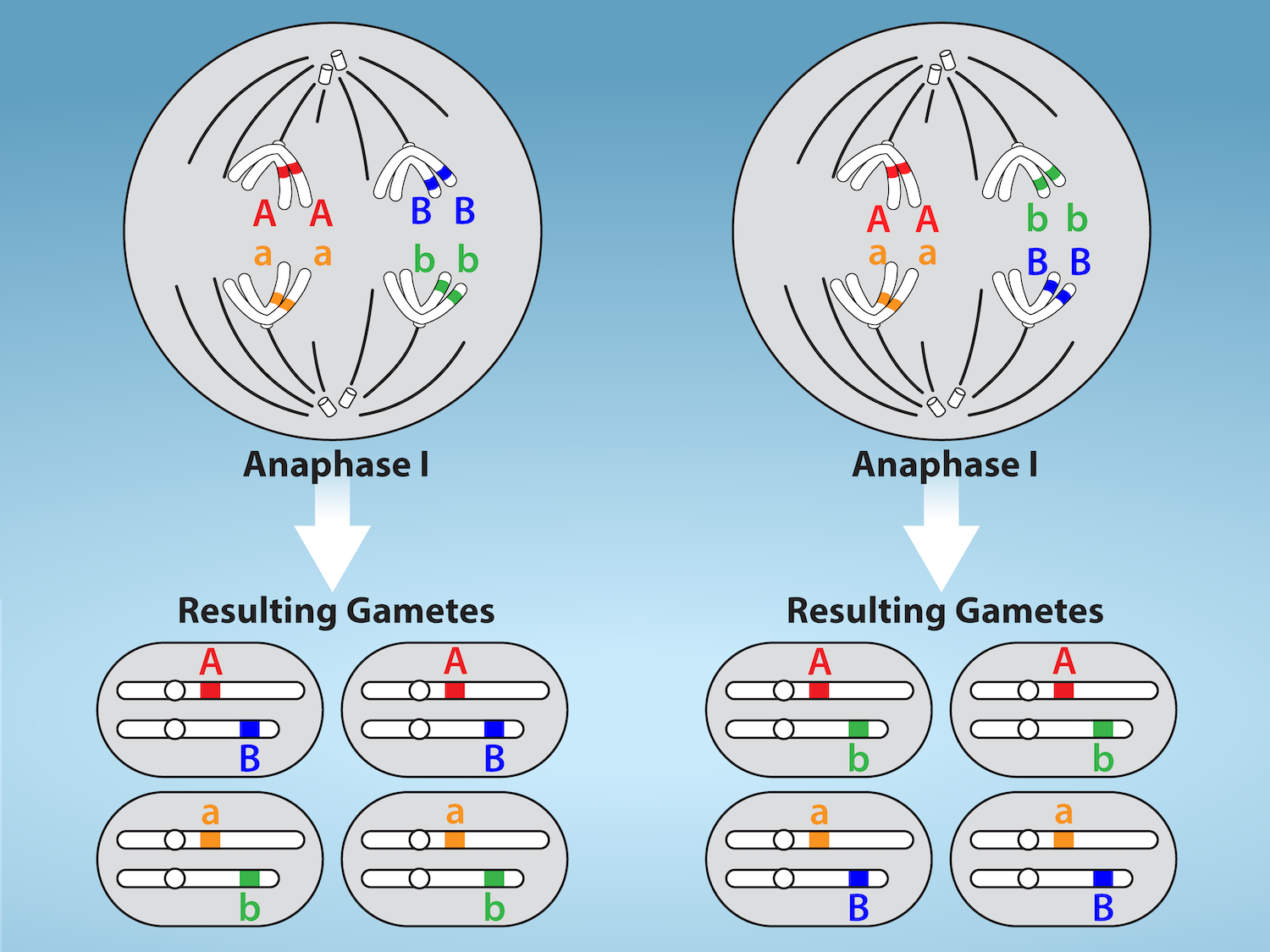
Figure 11.5 Random, independent assortment during metaphase I is demonstrated by considering a cell with a set of two chromosomes (n = 2).
There are two possible homologous chromosome arrangements at the equatorial plane in metaphase I, that are then separated during anaphase I.
The total possible number of different gametes is 2 n , where n equals the number of chromosomes in a set. In this example, there are four possible
genetic combinations for the gametes. With n = 23 in human cells, there are over eight million possible combinations of paternal and maternal
chromosomes. Credit: Rao, A.and Fletcher, S. Department of Biology, Texas A&M University.
Figure 12.4 In the P generation, pea plants that are true-breeding for the dominant yellow phenotype are crossed with plants with the recessive green phenotype. This cross produces F 1 heterozygotes with a yellow phenotype. Punnett square analysis can be used to predict the genotypes of the F 2 generation.
Figure 4.18 Membrane and secretory proteins are synthesized in the rough endoplasmic reticulum (RER). The RER also sometimes modifies proteins. In this illustration, a (green) integral membrane protein is modified by attachment of a (purple) carbohydrate in the ER. Vesicles with the integral protein bud from the ER and fuse with the Golgi apparatus' cis face. As the protein passes along the Golgi’s cisternae, the addition of more carbohydrates further modifies it. After its synthesis is complete, it exits as an integral membrane protein of the vesicle that buds from the Golgi’s trans face. When the vesicle fuses with the cell membrane, the protein becomes an integral portion of that cell membrane. (credit: modification of work by Magnus Manske)
Figure 12.4 In the P generation, pea plants that are true-breeding for the dominant yellow phenotype are crossed with plants with the recessive green phenotype. This cross produces F 1 heterozygotes with a yellow phenotype. Punnett square analysis can be used to predict the genotypes of the F 2 generation.
The exponent needed to obtain a number from a chosen fixed number. For decimal logarithms, log₁₀(100) = 2 because 10² = 100; pH uses the negative decimal logarithm of hydrogen ion activity.
A thin boundary or sheet. Cell membranes are lipid bilayers containing proteins; blotting membranes are synthetic supports that hold proteins or nucleic acids for detection.
Figure 15.11 Eukaryotic pre-mRNA processing. In addition to 5’ Cap and 3’ Poly-A Tail addition, introns must be precisely removed and exons joined to generate a functional mRNA. Nucleotides upstream (towards the 5’cap) of the translation START codon are part of the 5’ untranslated region (5’ UTR). Nucleotides downstream (towards 3’end) of the STOP codon form the 3’ UTR. Both 5’ and 3’ UTRs are important for regulating translation initiation and mRNA stability. Credit: Rao, A., Ryan, K. Fletcher, S. and Tag, A. Department of Biology, Texas A&M University.
Rao, A., Ryan, K. Fletcher, S. and Tag, A. Department of Biology, Texas A&M University.; Mary Ann Clark, Matthew Douglas, and Jung Choi, Biology 2e, OpenStax. · CC BY-NC-SA 4.0
· Original image ↗ · Access for free ↗
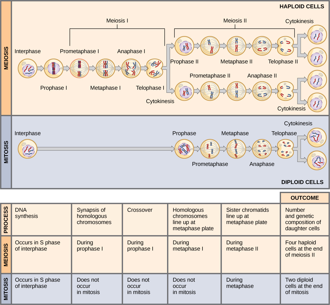
Figure 11.8 Meiosis and mitosis are both preceded by one cycle of DNA replication; however, meiosis includes two nuclear divisions. The four daughter cells resulting from meiosis are haploid and genetically distinct. The daughter cells resulting from mitosis are diploid and identical to the parent cell.
Figure 4.18 Membrane and secretory proteins are synthesized in the rough endoplasmic reticulum (RER). The RER also sometimes modifies proteins. In this illustration, a (green) integral membrane protein is modified by attachment of a (purple) carbohydrate in the ER. Vesicles with the integral protein bud from the ER and fuse with the Golgi apparatus' cis face. As the protein passes along the Golgi’s cisternae, the addition of more carbohydrates further modifies it. After its synthesis is complete, it exits as an integral membrane protein of the vesicle that buds from the Golgi’s trans face. When the vesicle fuses with the cell membrane, the protein becomes an integral portion of that cell membrane. (credit: modification of work by Magnus Manske)
Figure 12.4 In the P generation, pea plants that are true-breeding for the dominant yellow phenotype are crossed with plants with the recessive green phenotype. This cross produces F 1 heterozygotes with a yellow phenotype. Punnett square analysis can be used to predict the genotypes of the F 2 generation.
Figure 14.14 First Components of DNA Replication. As DNA replication begins, DNA Helicase, a large enzyme, separates the two strands of DNA so that they can act as templates for replication. Single-strand binding proteins bind to each strand to stabilize and prevent them from reforming the double helix. Primase, an RNA polymerase, binds to the single stranded DNA and synthesizes a short RNA primer in the 5’ to 3’ direction that is antiparallel to the parental strand. This RNA primer allows for DNA polymerase to begin replicating the DNA. Topoisomerase binds to the double helix upstream of the replication fork to prevent additional coiling by making small cuts in one of the DNA strands.
Credit: Rao, A., Ryan, K. Fletcher, S. and Tag, A. Department of Biology, Texas A&M University.
Figure 12.19 Reaction diagrams for an endothermic process in the absence (red curve) and presence (blue curve) of a catalyst. The catalyzed pathway involves a two-step mechanism (note the presence of two transition states) and an intermediate species (represented by the valley between the two transitions states).
Figure 15.19 Translation Termination is Active. Translation is terminated when a STOP codon is in the A-site of the ribosome. Since there are no tRNAs corresponding to the STOP codons, the Release Factor protein enters and catalyzes the hydrolysis between the last amino acid and its tRNA. This hydrolysis releases the free carboxyl terminus (C-term) of the protein. Additional factors use the energy in GTP hydrolysis to disassemble the large and small ribosomal subunits and mRNA. Credit: Rao, A. and Ryan, K. Department of Biology, Texas A&M University.
Rao, A. and Ryan, K. Department of Biology, Texas A&M University.; Mary Ann Clark, Matthew Douglas, and Jung Choi, Biology 2e, OpenStax. · CC BY-NC-SA 4.0
· Original image ↗ · Access for free ↗
A gene with a readily measured product used to study regulation; reporter output can depend on RNA synthesis, RNA decay, translation, and protein behavior.
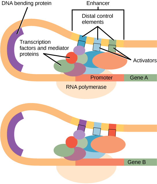
Figure 16.10 Interaction between proteins at the promoter and enhancer sites. An enhancer is a DNA sequence that promotes transcription. Each enhancer is made up of short DNA sequences called distal control elements. Activators bound to the distal control elements interact with mediator proteins and transcription factors. Two different genes may have the same promoter but different distal control elements, enabling differential gene expression.
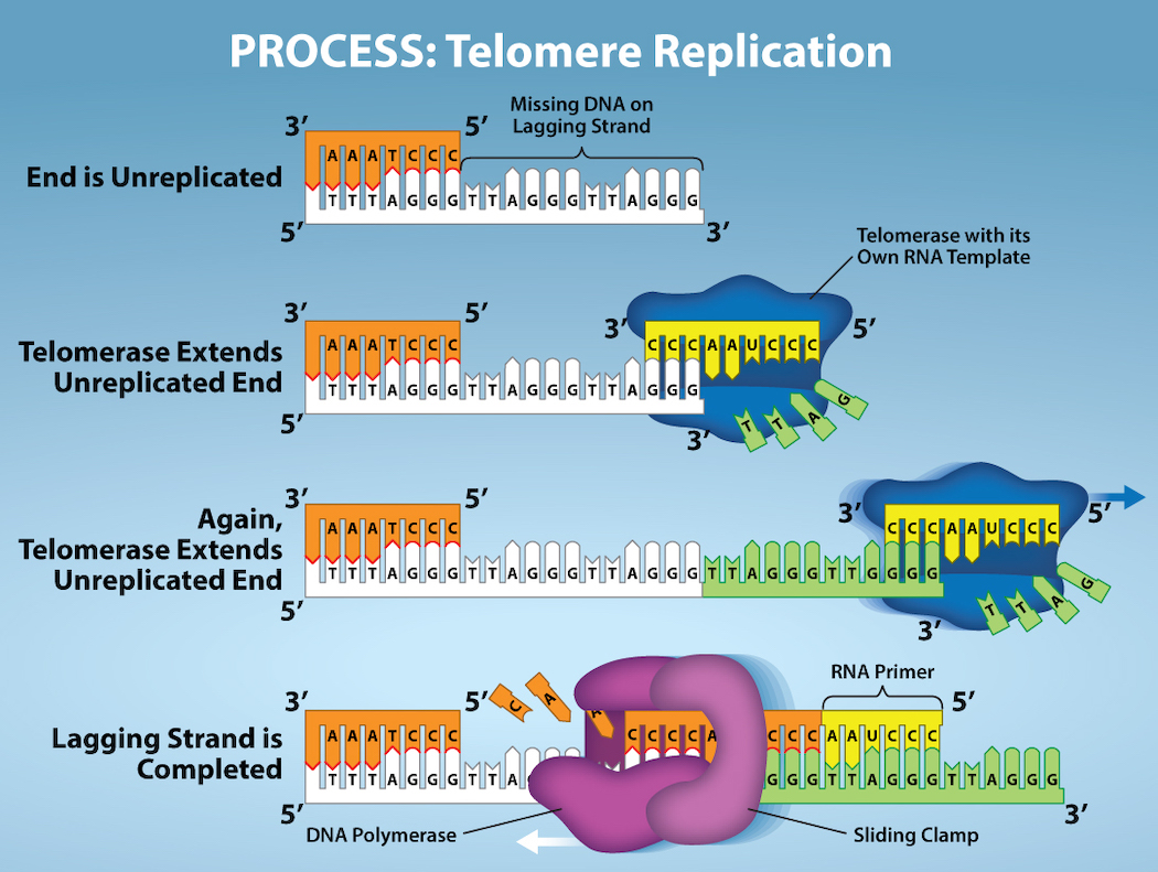
Figure 14.15 The ends of linear chromosomes are maintained by the action of the telomerase enzyme. Credit: Rao, A. and Fletcher, S. Department of Biology, Texas A&M University.
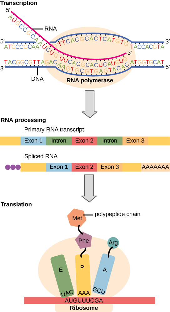
Figure 15.3 Instructions on DNA are transcribed onto messenger RNA. Ribosomes are able to read the genetic information inscribed on a strand of messenger RNA and use this information to string amino acids together into a protein.
Figure 4.18 Membrane and secretory proteins are synthesized in the rough endoplasmic reticulum (RER). The RER also sometimes modifies proteins. In this illustration, a (green) integral membrane protein is modified by attachment of a (purple) carbohydrate in the ER. Vesicles with the integral protein bud from the ER and fuse with the Golgi apparatus' cis face. As the protein passes along the Golgi’s cisternae, the addition of more carbohydrates further modifies it. After its synthesis is complete, it exits as an integral membrane protein of the vesicle that buds from the Golgi’s trans face. When the vesicle fuses with the cell membrane, the protein becomes an integral portion of that cell membrane. (credit: modification of work by Magnus Manske)
Figure 7.45 - Non-homologous end joining (left) versus homologous recombination (right) - Wikipedia
Wikipedia, as credited by Kevin Ahern, Indira Rajagopal, and Taralyn Tan, Biochemistry Free For All, Oregon State University, via Biology LibreTexts. · Source attribution
· Original image ↗ · Access for free ↗
The amount of a substance that can dissolve in a solvent under specified conditions. Soluble means able to dissolve; insoluble means only very slightly soluble in that solvent.
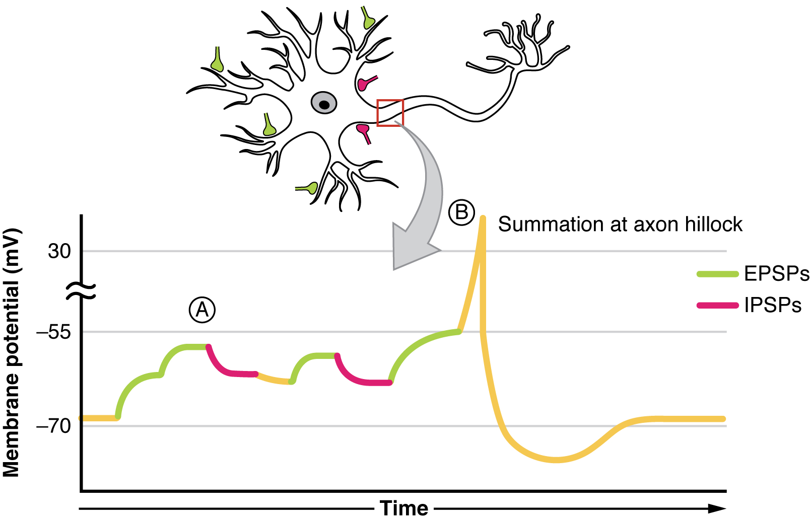
Figure 12.26 Postsynaptic Potential Summation The result of summation of postsynaptic potentials is the overall change in the membrane potential. At point A, several different excitatory postsynaptic potentials add up to a large depolarization. At point B, a mix of excitatory and inhibitory postsynaptic potentials result in a different end result for the membrane potential.
Figure 14.15 The ends of linear chromosomes are maintained by the action of the telomerase enzyme. Credit: Rao, A. and Fletcher, S. Department of Biology, Texas A&M University.
Figure 15.7 Messenger RNA is a copy of protein-coding information in the coding strand of DNA, with the substitution of U in the RNA for T in the coding sequence. However, new RNA nucleotides base pair with the nucleotides of the template strand. RNA is synthesized in its 5'-3' direction, using the enzyme RNA polymerase. As the template is read, the DNA unwinds ahead of the polymerase and then rewinds behind it.
Figure 15.14 This is a space-filling model of a tRNA molecule that adds the amino acid phenylalanine to a growing polypeptide chain. The anticodon AAG binds the Codon UUC on the mRNA. The amino acid phenylalanine is attached to the other end of the tRNA.
A fleeting, high-energy arrangement at a reaction barrier. It is an energy maximum along the reaction path, rather than a stable reaction intermediate.
Figure 12.19 Reaction diagrams for an endothermic process in the absence (red curve) and presence (blue curve) of a catalyst. The catalyzed pathway involves a two-step mechanism (note the presence of two transition states) and an intermediate species (represented by the valley between the two transitions states).
Figure 15.18 Translation begins when an initiator tRNA anticodon recognizes a start codon on mRNA bound to a small ribosomal subunit. The large ribosomal subunit joins the small subunit, and a second tRNA is recruited. As the mRNA moves relative to the ribosome, successive tRNAs move through the ribosome and the polypeptide chain is formed. Entry of a release factor into the A site terminates translation and the components dissociate.
Figure 10.15 Cell Division and Apoptosis. In an adult organism, normal cell division is balanced by apoptosis (programmed cell death) to maintain a constant cell number in homeostasis. Either an increase in cell division or a decrease in apoptosis leads an increase in the number of cells and tumor formation. Credit: Rao, A. and Ryan, K. Department of Biology, Texas A&M University.
Rao, A. and Ryan, K. Department of Biology, Texas A&M University. Mary Ann Clark, Matthew Douglas, and Jung Choi, Biology 2e, OpenStax. · CC BY-NC-SA 4.0
· Original image ↗ · Access for free ↗
Figure 4.18 Membrane and secretory proteins are synthesized in the rough endoplasmic reticulum (RER). The RER also sometimes modifies proteins. In this illustration, a (green) integral membrane protein is modified by attachment of a (purple) carbohydrate in the ER. Vesicles with the integral protein bud from the ER and fuse with the Golgi apparatus' cis face. As the protein passes along the Golgi’s cisternae, the addition of more carbohydrates further modifies it. After its synthesis is complete, it exits as an integral membrane protein of the vesicle that buds from the Golgi’s trans face. When the vesicle fuses with the cell membrane, the protein becomes an integral portion of that cell membrane. (credit: modification of work by Magnus Manske)
No matching terms. Try a different name or abbreviation.
7.2 Language
OpenStax · 7.2 Language ↗ Instructional page including learning objectives, examples, figures, source tables and questions. Site navigation and embedded media players are omitted; source media links remain available.
Learning Objectives
By the end of this section, you will be able to:
Define language and demonstrate familiarity with the components of language
Understand the development of language
Explain the relationship between language and thinking
Language is a communication system that involves using words and systematic rules to organize those words to transmit information from one individual to another. While language is a form of communication, not all communication is language. Many species communicate with one another through their postures, movements, odors, or vocalizations. This communication is crucial for species that need to interact and develop social relationships with their conspecifics. However, many people have asserted that it is language that makes humans unique among all of the animal species (Corballis & Suddendorf, 2007; Tomasello & Rakoczy, 2003). This section will focus on what distinguishes language as a special form of communication, how the use of language develops, and how language affects the way we think.
Components of Language
Language, be it spoken, signed, or written, has specific components: a lexicon and grammar. Lexicon refers to the words of a given language. Thus, lexicon is a language’s vocabulary. Grammar refers to the set of rules that are used to convey meaning through the use of the lexicon (Fernández & Cairns, 2011). For instance, English grammar dictates that most verbs receive an “-ed” at the end to indicate past tense.
Words are formed by combining the various phonemes that make up the language. A phoneme (e.g., the sounds “ah” vs. “eh”) is a basic sound unit of a given language, and different languages have different sets of phonemes. Phonemes are combined to form morphemes, which are the smallest units of language that convey some type of meaning (e.g., “I” is both a phoneme and a morpheme). We use semantics and syntax to construct language. Semantics and syntax are part of a language’s grammar. Semantics refers to the process by which we derive meaning from morphemes and words. Syntax refers to the way words are organized into sentences (Chomsky, 1965; Fernández & Cairns, 2011).
We apply the rules of grammar to organize the lexicon in novel and creative ways, which allow us to communicate information about both concrete and abstract concepts. We can talk about our immediate and observable surroundings as well as the surface of unseen planets. We can share our innermost thoughts, our plans for the future, and debate the value of a college education. We can provide detailed instructions for cooking a meal, fixing a car, or building a fire. Through our use of words and language, we are able to form, organize, and express schemata, and artificial concepts.
Language Development
Given the remarkable complexity of a language, one might expect that mastering a language would be an especially arduous task; indeed, for those of us trying to learn a second language as adults, this might seem to be true. However, young children master language very quickly with relative ease. B. F. Skinner (1957) proposed that language is learned through reinforcement. Noam Chomsky (1965) criticized this behaviorist approach, asserting instead that the mechanisms underlying language acquisition are biologically determined. The use of language develops in the absence of formal instruction and appears to follow a very similar pattern in children from vastly different cultures and backgrounds. It would seem, therefore, that we are born with a biological predisposition to acquire a language (Chomsky, 1965; Fernández & Cairns, 2011). Moreover, it appears that there is a critical period for language acquisition, such that this proficiency at acquiring language is maximal early in life; generally, as people age, the ease with which they acquire and master new languages diminishes (Johnson & Newport, 1989; Lenneberg, 1967; Singleton, 1995).
Children begin to learn about language from a very early age (Table 7.1). In fact, it appears that this is occurring even before we are born. Newborns show preference for their mother’s voice and appear to be able to discriminate between the language spoken by their mother and other languages. Babies are also attuned to the languages being used around them and show preferences for videos of faces that are moving in synchrony with the audio of spoken language versus videos that do not synchronize with the audio (Blossom & Morgan, 2006; Pickens, 1994; Spelke & Cortelyou, 1981).
Stages of Language and Communication Development
Stage
Age
Developmental Language and Communication
1
0–3 months
Reflexive communication
2
3–8 months
Reflexive communication; interest in others
3
8–13 months
Intentional communication; sociability
4
12–18 months
First words
5
18–24 months
Simple sentences of two words
6
2–3 years
Sentences of three or more words
7
3–5 years
Complex sentences; has conversations
Table
7.1
Dig Deeper
The Case of Genie
In the fall of 1970, a social worker in the Los Angeles area found a 13-year-old girl who was being raised in extremely neglectful and abusive conditions. The girl, who came to be known as Genie, had lived most of her life tied to a potty chair or confined to a crib in a small room that was kept closed with the curtains drawn. For a little over a decade, Genie had virtually no social interaction and no access to the outside world. As a result of these conditions, Genie was unable to stand up, chew solid food, or speak (Fromkin, Krashen, Curtiss, Rigler, & Rigler, 1974; Rymer, 1993). The police took Genie into protective custody.
Genie’s abilities improved dramatically following her removal from her abusive environment, and early on, it appeared she was acquiring language—much later than would be predicted by critical period hypotheses that had been posited at the time (Fromkin et al., 1974). Genie managed to amass an impressive vocabulary in a relatively short amount of time. However, she never developed a mastery of the grammatical aspects of language (Curtiss, 1981). Perhaps being deprived of the opportunity to learn language during a critical period impeded Genie’s ability to fully acquire and use language.
You may recall that each language has its own set of phonemes that are used to generate morphemes, words, and so on. Babies can discriminate among the sounds that make up a language (for example, they can tell the difference between the “s” in vision and the “sh” in dishes); early on, they can differentiate between the sounds of all human languages, even those that do not occur in the languages that are used in their environments. However, by the time that they are about 1 year old, they can only discriminate among those phonemes that are used in the language or languages in their environments (Jensen, 2011; Werker & Lalonde, 1988; Werker & Tees, 1984).
After the first few months of life, babies enter what is known as the babbling stage, during which time they tend to produce single syllables that are repeated over and over. As time passes, more variations appear in the syllables that they produce. During this time, it is unlikely that the babies are trying to communicate; they are just as likely to babble when they are alone as when they are with their caregivers (Fernández & Cairns, 2011). Interestingly, babies who are raised in environments in which sign language is used will also begin to show babbling in the gestures of their hands during this stage (Petitto, Holowka, Sergio, Levy, & Ostry, 2004).
Generally, a child’s first word is uttered sometime between the ages of 1 year to 18 months, and for the next few months, the child will remain in the “one word” stage of language development. During this time, children know a number of words, but they only produce one-word utterances. The child’s early vocabulary is limited to familiar objects or events, often nouns. Although children in this stage only make one-word utterances, these words often carry larger meaning (Fernández & Cairns, 2011). So, for example, a child saying “cookie” could be identifying a cookie or asking for a cookie.
As a child’s lexicon grows, they begin to utter simple sentences and to acquire new vocabulary at a very rapid pace. In addition, children begin to demonstrate a clear understanding of the specific rules that apply to their language(s). Even the mistakes that children sometimes make provide evidence of just how much they understand about those rules. This is sometimes seen in the form of overgeneralization. In this context, overgeneralization refers to an extension of a language rule to an exception to the rule. For example, in English, it is usually the case that an “s” is added to the end of a word to indicate plurality. For example, we speak of one dog versus two dogs. Young children will overgeneralize this rule to cases that are exceptions to the “add an s to the end of the word” rule and say things like “those two gooses” or “three mouses.” Clearly, the rules of the language are understood, even if the exceptions to the rules are still being learned (Moskowitz, 1978).
Language and Thought
When we speak one language, we agree that words are representations of ideas, people, places, and events. The given language that children learn is connected to their culture and surroundings. But can words themselves shape the way we think about things? Psychologists have long investigated the question of whether language shapes thoughts and actions, or whether our thoughts and beliefs shape our language. Two researchers, Edward Sapir and Benjamin Lee Whorf, began this investigation in the 1940s. They wanted to understand how the language habits of a community encourage members of that community to interpret language in a particular manner (Sapir, 1941/1964). Sapir and Whorf proposed that language determines thought. For example, in some languages there are many different words for love. However, in English we use the word love for all types of love. Does this affect how we think about love depending on the language that we speak (Whorf, 1956)? Researchers have since identified this view as too absolute, pointing out a lack of empiricism behind what Sapir and Whorf proposed (Abler, 2013; Boroditsky, 2011; van Troyer, 1994). Today, psychologists continue to study and debate the relationship between language and thought.
What Do You Think?
The Meaning of Language
Think about what you know of other languages; perhaps you even speak multiple languages. Imagine for a moment that your closest friend fluently speaks more than one language. Do you think that friend thinks differently, depending on which language is being spoken? You may know a few words that are not translatable from their original language into English. For example, the Portuguese word saudade originated during the 15th century, when Portuguese sailors left home to explore the seas and travel to Africa or Asia. Those left behind described the emptiness and fondness they felt as saudade (Figure 7.6). The word came to express many meanings, including loss, nostalgia, yearning, warm memories, and hope. There is no single word in English that includes all of those emotions in a single description. Do words such as saudade indicate that different languages produce different patterns of thought in people? What do you think??
Figure 7.6 — 7.2 Language
Figure 7.6 These two works of art depict saudade . (a) Saudade de Nápoles , which is translated into “missing Naples,” was painted by Bertha Worms in 1895. (b) Almeida Júnior painted Saudade in 1899.
Language may indeed influence the way that we think, an idea known as linguistic determinism. One recent demonstration of this phenomenon involved differences in the way that English and Mandarin Chinese speakers talk and think about time. English speakers tend to talk about time using terms that describe changes along a horizontal dimension, for example, saying something like “I’m running behind schedule” or “Don’t get ahead of yourself.” While Mandarin Chinese speakers also describe time in horizontal terms, it is not uncommon to also use terms associated with a vertical arrangement. For example, the past might be described as being “up” and the future as being “down.” It turns out that these differences in language translate into differences in performance on cognitive tests designed to measure how quickly an individual can recognize temporal relationships. Specifically, when given a series of tasks with vertical priming, Mandarin Chinese speakers were faster at recognizing temporal relationships between months. Indeed, Boroditsky (2001) sees these results as suggesting that “habits in language encourage habits in thought” (p. 12).
One group of researchers who wanted to investigate how language influences thought compared how English speakers and the Dani people of Papua New Guinea think and speak about color. The Dani have two words for color: one word for light and one word for dark. In contrast, the English language has 11 color words. Researchers hypothesized that the number of color terms could limit the ways that the Dani people conceptualized color. However, the Dani were able to distinguish colors with the same ability as English speakers, despite having fewer words at their disposal (Berlin & Kay, 1969). A recent review of research aimed at determining how language might affect something like color perception suggests that language can influence perceptual phenomena, especially in the left hemisphere of the brain. You may recall from earlier chapters that the left hemisphere is associated with language for most people. However, the right (less linguistic hemisphere) of the brain is less affected by linguistic influences on perception (Regier & Kay, 2009)
3.4 The Brain and Spinal Cord
OpenStax · 3.4 The Brain and Spinal Cord ↗ Instructional page including learning objectives, examples, figures, source tables and questions. Site navigation and embedded media players are omitted; source media links remain available.
Learning Objectives
By the end of this section, you will be able to:
Explain the functions of the spinal cord
Identify the hemispheres and lobes of the brain
Describe the types of techniques available to clinicians and researchers to image or scan the brain
The brain is a remarkably complex organ comprised of billions of interconnected neurons and glia. It is a bilateral, or two-sided, structure that can be separated into distinct lobes. Each lobe is associated with certain types of functions, but, ultimately, all of the areas of the brain interact with one another to provide the foundation for our thoughts and behaviors. In this section, we discuss the overall organization of the brain and the functions associated with different brain areas, beginning with what can be seen as an extension of the brain, the spinal cord.
The Spinal Cord
It can be said that the spinal cord is what connects the brain to the outside world. Because of it, the brain can act. The spinal cord is like a relay station, but a very smart one. It not only routes messages to and from the brain, but it also has its own system of automatic processes, called reflexes.
The top of the spinal cord is a bundle of nerves that merges with the brain stem, where the basic processes of life are controlled, such as breathing and digestion. In the opposite direction, the spinal cord ends just below the ribs—contrary to what we might expect, it does not extend all the way to the base of the spine.
The spinal cord is functionally organized in 30 segments, corresponding with the vertebrae. Each segment is connected to a specific part of the body through the peripheral nervous system. Nerves branch out from the spine at each vertebra. Sensory nerves bring messages in; motor nerves send messages out to the muscles and organs. Messages travel to and from the brain through every segment.
Some sensory messages are immediately acted on by the spinal cord, without any input from the brain. Withdrawal from a hot object and the knee jerk are two examples. When a sensory message meets certain parameters, the spinal cord initiates an automatic reflex. The signal passes from the sensory nerve to a simple processing center, which initiates a motor command. Seconds are saved, because messages don’t have to go to the brain, be processed, and get sent back. In matters of survival, the spinal reflexes allow the body to react extraordinarily fast.
The spinal cord is protected by bony vertebrae and cushioned in cerebrospinal fluid, but injuries still occur. When the spinal cord is damaged in a particular segment, all lower segments are cut off from the brain, causing paralysis. Therefore, the lower on the spine damage occurs, the fewer functions an injured individual will lose.
Neuroplasticity
Bob Woodruff, a reporter for ABC, suffered a traumatic brain injury after a bomb exploded next to the vehicle he was in while covering a news story in Iraq. As a consequence of these injuries, Woodruff experienced many cognitive deficits including difficulties with memory and language. However, over time and with the aid of intensive amounts of cognitive and speech therapy, Woodruff has shown an incredible recovery of function (Fernandez, 2008, October 16).
One of the factors that made this recovery possible was neuroplasticity. Neuroplasticity refers to how the nervous system can change and adapt. Neuroplasticity can occur in a variety of ways including personal experiences, developmental processes, or, as in Woodruff's case, in response to some sort of damage or injury that has occurred. Neuroplasticity can involve creation of new synapses, pruning of synapses that are no longer used, changes in glial cells, and even the birth of new neurons. Because of neuroplasticity, our brains are constantly changing and adapting, and while our nervous system is most plastic when we are very young, as Woodruff's case suggests, it is still capable of remarkable changes later in life.
The Two Hemispheres
The surface of the brain, known as the cerebral cortex, is very uneven, characterized by a distinctive pattern of folds or bumps, known as gyri (singular: gyrus), and grooves, known as sulci (singular: sulcus), shown in Figure 3.15. These gyri and sulci form important landmarks that allow us to separate the brain into functional centers. The most prominent sulcus, known as the longitudinal fissure, is the deep groove that separates the brain into two halves or hemispheres: the left hemisphere and the right hemisphere.
Figure 3.15 — 3.4 The Brain and Spinal Cord
Figure 3.15 The surface of the brain is covered with gyri and sulci. A deep sulcus is called a fissure, such as the longitudinal fissure that divides the brain into left and right hemispheres. (credit: modification of work by Bruce Blaus)
There is evidence of specialization of function—referred to as lateralization—in each hemisphere, mainly regarding differences in language functions. The left hemisphere controls the right half of the body, and the right hemisphere controls the left half of the body. Decades of research on lateralization of function by Michael Gazzaniga and his colleagues suggest that a variety of functions ranging from cause-and-effect reasoning to self-recognition may follow patterns that suggest some degree of hemispheric dominance (Gazzaniga, 2005). For example, the left hemisphere has been shown to be superior for forming associations in memory, selective attention, and positive emotions. The right hemisphere, on the other hand, has been shown to be superior in pitch perception, arousal, and negative emotions (Ehret, 2006). However, it should be pointed out that research on which hemisphere is dominant in a variety of different behaviors has produced inconsistent results, and therefore, it is probably better to think of how the two hemispheres interact to produce a given behavior rather than attributing certain behaviors to one hemisphere versus the other (Banich & Heller, 1998).
The two hemispheres are connected by a thick band of neural fibers known as the corpus callosum, consisting of about 200 million axons. The corpus callosum allows the two hemispheres to communicate with each other and allows for information being processed on one side of the brain to be shared with the other side.
Normally, we are not aware of the different roles that our two hemispheres play in day-to-day functions, but there are people who come to know the capabilities and functions of their two hemispheres quite well. In some cases of severe epilepsy, doctors elect to sever the corpus callosum as a means of controlling the spread of seizures (Figure 3.16). While this is an effective treatment option, it results in individuals who have "split brains." After surgery, these split-brain patients show a variety of interesting behaviors. For instance, a split-brain patient is unable to name a picture that is shown in the patient’s left visual field because the information is only available in the largely nonverbal right hemisphere. However, they are able to recreate the picture with their left hand, which is also controlled by the right hemisphere. When the more verbal left hemisphere sees the picture that the hand drew, the patient is able to name it (assuming the left hemisphere can interpret what was drawn by the left hand).
Figure 3.16 — 3.4 The Brain and Spinal Cord
Figure 3.16 (a, b) The corpus callosum connects the left and right hemispheres of the brain. (c) A scientist spreads this dissected sheep brain apart to show the corpus callosum between the hemispheres. (credit c: modification of work by Aaron Bornstein)
Rose M. Spielman, William J. Jenkins, and Marilyn D. Lovett, Psychology 2E, OpenStax. (credit c: modification of work by Aaron Bornstein) · CC BY-NC-SA 4.0
· Original image ↗ · Access for free ↗
Much of what we know about the functions of different areas of the brain comes from studying changes in the behavior and ability of individuals who have suffered damage to the brain. For example, researchers study the behavioral changes caused by strokes to learn about the functions of specific brain areas. A stroke, caused by an interruption of blood flow to a region in the brain, causes a loss of brain function in the affected region. The damage can be in a small area, and, if it is, this gives researchers the opportunity to link any resulting behavioral changes to a specific area. The types of deficits displayed after a stroke will be largely dependent on where in the brain the damage occurred.
Consider Theona, an intelligent, self-sufficient woman, who is 62 years old. Recently, she suffered a stroke in the front portion of her right hemisphere. As a result, she has great difficulty moving her left leg. (As you learned earlier, the right hemisphere controls the left side of the body; also, the brain’s main motor centers are located at the front of the head, in the frontal lobe.) Theona has also experienced behavioral changes. For example, while in the produce section of the grocery store, she sometimes eats grapes, strawberries, and apples directly from their bins before paying for them. This behavior—which would have been very embarrassing to her before the stroke—is consistent with damage in another region in the frontal lobe—the prefrontal cortex, which is associated with judgment, reasoning, and impulse control.
The two hemispheres of the cerebral cortex are part of the forebrain (Figure 3.17), which is the largest part of the brain. The forebrain contains the cerebral cortex and a number of other structures that lie beneath the cortex (called subcortical structures): thalamus, hypothalamus, pituitary gland, and the limbic system (a collection of structures). The cerebral cortex, which is the outer surface of the brain, is associated with higher level processes such as consciousness, thought, emotion, reasoning, language, and memory. Each cerebral hemisphere can be subdivided into four lobes, each associated with different functions.
Figure 3.17 — 3.4 The Brain and Spinal Cord
Figure 3.17 The brain and its parts can be divided into three main categories: the forebrain, midbrain, and hindbrain.
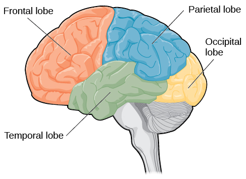
The four lobes of the brain are the frontal, parietal, temporal, and occipital lobes (Figure 3.18). The frontal lobe is located in the forward part of the brain, extending back to a fissure known as the central sulcus. The frontal lobe is involved in reasoning, motor control, emotion, and language. It contains the motor cortex, which is involved in planning and coordinating movement; the prefrontal cortex, which is responsible for higher-level cognitive functioning; and Broca’s area, which is essential for language production.
People who suffer damage to Broca’s area have great difficulty producing language of any form (Figure 3.18). For example, Padma was an electrical engineer who was socially active and a caring, involved parent. About twenty years ago, she was in a car accident and suffered damage to her Broca’s area. She completely lost the ability to speak and form any kind of meaningful language. There is nothing wrong with her mouth or her vocal cords, but she is unable to produce words. She can follow directions but can’t respond verbally, and she can read but no longer write. She can do routine tasks like running to the market to buy milk, but she could not communicate verbally if a situation called for it.
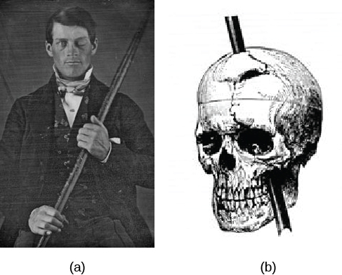
Probably the most famous case of frontal lobe damage is that of a man by the name of Phineas Gage. On September 13, 1848, Gage (age 25) was working as a railroad foreman in Vermont. He and his crew were using an iron rod to tamp explosives down into a blasting hole to remove rock along the railway’s path. Unfortunately, the iron rod created a spark and caused the rod to explode out of the blasting hole, into Gage’s face, and through his skull (Figure 3.19). Although lying in a pool of his own blood with brain matter emerging from his head, Gage was conscious and able to get up, walk, and speak. But in the months following his accident, people noticed that his personality had changed. Many of his friends described him as no longer being himself. Before the accident, it was said that Gage was a well-mannered, soft-spoken man, but he began to behave in odd and inappropriate ways after the accident. Such changes in personality would be consistent with loss of impulse control—a frontal lobe function.
Beyond the damage to the frontal lobe itself, subsequent investigations into the rod's path also identified probable damage to pathways between the frontal lobe and other brain structures, including the limbic system. With connections between the planning functions of the frontal lobe and the emotional processes of the limbic system severed, Gage had difficulty controlling his emotional impulses.
However, there is some evidence suggesting that the dramatic changes in Gage’s personality were exaggerated and embellished. Gage's case occurred in the midst of a 19th century debate over localization—regarding whether certain areas of the brain are associated with particular functions. On the basis of extremely limited information about Gage, the extent of his injury, and his life before and after the accident, scientists tended to find support for their own views, on whichever side of the debate they fell (Macmillan, 1999).
Figure 3.19 — 3.4 The Brain and Spinal Cord
Figure 3.19 (a) Phineas Gage holds the iron rod that penetrated his skull in an 1848 railroad construction accident. (b) Gage’s prefrontal cortex was severely damaged in the left hemisphere. The rod entered Gage’s face on the left side, passed behind his eye, and exited through the top of his skull, before landing about 80 feet away. (credit a: modification of work by Jack and Beverly Wilgus)
Rose M. Spielman, William J. Jenkins, and Marilyn D. Lovett, Psychology 2E, OpenStax. (credit a: modification of work by Jack and Beverly Wilgus) · CC BY-NC-SA 4.0
· Original image ↗ · Access for free ↗
The brain’s parietal lobe is located immediately behind the frontal lobe, and is involved in processing information from the body’s senses. It contains the somatosensory cortex, which is essential for processing sensory information from across the body, such as touch, temperature, and pain. The somatosensory cortex is an area of the brain which processes touch and sensation. The somatosensory cortex is fascinating because each different area of the cortex processes sensations from a different part of your body. Furthermore, the larger the surface area of the specific body part and the greater amount of nerves in that body part, the larger the area dedicated to processing sensation from that body part in the somatosensory cortex. For example, fingers take up a lot more space than toes. As you can notice from (Figure 3.20), the amount of space to process sensation from fingers is much greater than that of toes.
The temporal lobe is located on the side of the head (temporal means “near the temples”), and is associated with hearing, memory, emotion, and some aspects of language. The auditory cortex, the main area responsible for processing auditory information, is located within the temporal lobe. Wernicke’s area, important for speech comprehension, is also located here. Whereas individuals with damage to Broca’s area have difficulty producing language, those with damage to Wernicke’s area can produce sensible language, but they are unable to understand it (Figure 3.21).
Figure 3.21 — 3.4 The Brain and Spinal Cord
Figure 3.21 Damage to either Broca’s area or Wernicke’s area can result in language deficits. The types of deficits are very different, however, depending on which area is affected.
The occipital lobe is located at the very back of the brain, and contains the primary visual cortex, which is responsible for interpreting incoming visual information. The occipital cortex is organized retinotopically, which means there is a close relationship between the position of an object in a person’s visual field and the position of that object’s representation on the cortex. You will learn much more about how visual information is processed in the occipital lobe when you study sensation and perception.
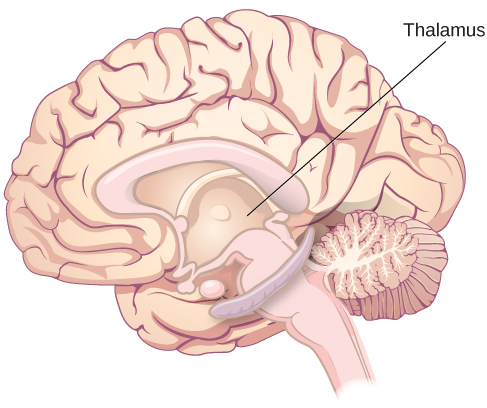
Other areas of the forebrain, located beneath the cerebral cortex, include the thalamus and the limbic system. The thalamus is a sensory relay for the brain. All of our senses, with the exception of smell, are routed through the thalamus before being directed to other areas of the brain for processing (Figure 3.22).
Figure 3.22 — 3.4 The Brain and Spinal Cord
Figure 3.22 The thalamus serves as the relay center of the brain where most senses are routed for processing.
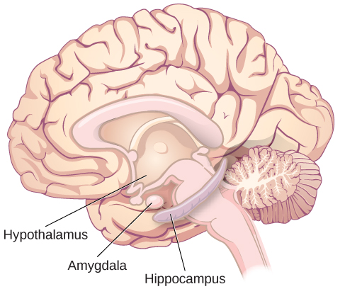
The limbic system is involved in processing both emotion and memory. Interestingly, the sense of smell projects directly to the limbic system; therefore, not surprisingly, smell can evoke emotional responses in ways that other sensory modalities cannot. The limbic system is made up of a number of different structures, but three of the most important are the hippocampus, the amygdala, and the hypothalamus (Figure 3.23). The hippocampus is an essential structure for learning and memory. The amygdala is involved in our experience of emotion and in tying emotional meaning to our memories. The hypothalamus regulates a number of homeostatic processes, including the regulation of body temperature, appetite, and blood pressure. The hypothalamus also serves as an interface between the nervous system and the endocrine system and in the regulation of sexual motivation and behavior.
Figure 3.23 — 3.4 The Brain and Spinal Cord
Figure 3.23 The limbic system is involved in mediating emotional response and memory.
In 1953, Henry Gustav Molaison (H. M.) was a 27-year-old man who experienced severe seizures. In an attempt to control his seizures, H. M. underwent brain surgery to remove hishippocampus and amygdala. Following the surgery, H.M’s seizures became much less severe, but he also suffered some unexpected—and devastating—consequences of the surgery: he lost his ability to form many types of new memories. For example, he was unable to learn new facts, such as who was president of the United States. He was able to learn new skills, but afterward he had no recollection of learning them. For example, while he might learn to use a computer, he would have no conscious memory of ever having used one. He could not remember new faces, and he was unable to remember events, even immediately after they occurred. Researchers were fascinated by his experience, and he is considered one of the most studied cases in medical and psychological history (Hardt, Einarsson, & Nader, 2010; Squire, 2009). Indeed, his case has provided tremendous insight into the role that the hippocampus plays in the consolidation of new learning into explicit memory.
The midbrain is comprised of structures located deep within the brain, between the forebrain and the hindbrain. The reticular formation is centered in the midbrain, but it actually extends up into the forebrain and down into the hindbrain. The reticular formation is important in regulating the sleep/wake cycle, arousal, alertness, and motor activity.
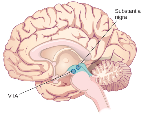
The substantia nigra (Latin for “black substance”) and the ventral tegmental area (VTA) are also located in the midbrain (Figure 3.24). Both regions contain cell bodies that produce the neurotransmitter dopamine, and both are critical for movement. Degeneration of the substantia nigra and VTA is involved in Parkinson’s disease. In addition, these structures are involved in mood, reward, and addiction (Berridge & Robinson, 1998; Gardner, 2011; George, Le Moal, & Koob, 2012).
Figure 3.24 — 3.4 The Brain and Spinal Cord
Figure 3.24 The substantia nigra and ventral tegmental area (VTA) are located in the midbrain.
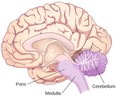
The hindbrain is located at the back of the head and looks like an extension of the spinal cord. It contains the medulla, pons, and cerebellum (Figure 3.25). The medullacontrols the automatic processes of the autonomic nervous system, such as breathing, blood pressure, and heart rate. The word pons literally means “bridge,” and as the name suggests, the pons serves to connect the hindbrain to the rest of the brain. It also is involved in regulating brain activity during sleep. The medulla, pons, and various structures are known as the brainstem, and aspects of the brainstem span both the midbrain and the hindbrain.
Figure 3.25 — 3.4 The Brain and Spinal Cord
Figure 3.25 The pons, medulla, and cerebellum make up the hindbrain.
The cerebellum (Latin for “little brain”) receives messages from muscles, tendons, joints, and structures in our ear to control balance, coordination, movement, and motor skills. The cerebellum is also thought to be an important area for processing some types of memories. In particular, procedural memory, or memory involved in learning and remembering how to perform tasks, is thought to be associated with the cerebellum. Recall that H. M. was unable to form new explicit memories, but he could learn new tasks. This is likely due to the fact that H. M.’s cerebellum remained intact.
What Do You Think?
Brain Dead and on Life Support
What would you do if your spouse or loved one was declared brain dead but their body was being kept alive by medical equipment? What if the loved one was in a persistent vegetative state and remained unconscious but maintained basic functions like breathing? Whose decision should it be to remove a feeding tube? Should medical care costs be a factor?
On February 25, 1990, a Florida woman named Terri Schiavo went into cardiac arrest, apparently triggered by a bulimic episode. She was eventually revived, but her brain had been deprived of oxygen for a long time. Brain scans indicated that there was no activity in her cerebral cortex, and she suffered from severe and permanent cerebral atrophy. Basically, Schiavo was in a vegetative state. Medical professionals determined that she would never again be able to move, talk, or respond in any way. To remain alive, she required a feeding tube, and there was no chance that her situation would ever improve.
On occasion, Schiavo’s eyes would move, and sometimes she would groan. Despite the doctors’ insistence to the contrary, her parents believed that these were signs that she was trying to communicate with them.
After 12 years, Schiavo’s husband argued that his wife would not have wanted to be kept alive with no feelings, sensations, or brain activity. Her parents, however, were very much against removing her feeding tube. Eventually, the case made its way to the courts, both in the state of Florida and at the federal level. By 2005, the courts found in favor of Schiavo’s husband, and the feeding tube was removed on March 18, 2005. Schiavo died 13 days later.
Why did Schiavo’s eyes sometimes move, and why did she groan? Although the parts of her brain that control thought, voluntary movement, and feeling were completely damaged, her brainstem was still intact. Her medulla and pons maintained her breathing and caused involuntary movements of her eyes and the occasional groans. Over the 15-year period that she was on a feeding tube, Schiavo’s medical costs may have topped $7 million (Arnst, 2003).
These questions were brought to popular consciousness decades ago in the case of Terri Schiavo, and they have persisted. In 2013, a 13-year-old girl who suffered complications after tonsil surgery was declared brain dead. There was a battle between her family, who wanted her to remain on life support, and the hospital’s policies regarding persons declared brain dead. In another complicated 2013–14 case in Texas, a pregnant EMT professional declared brain dead was kept alive for weeks, despite her spouse’s directives, which were based on her wishes should this situation arise. In this case, state laws designed to protect an unborn fetus came into consideration until doctors determined the fetus unviable.
Decisions surrounding the medical response to patients declared brain dead are complex. Whether a person is declared brain dead or diagnosed as having a persistent vegetative state, determining death and making appropriate medical care decisions is a complicated process. What do you think about these issues?
Brain Imaging
You have learned how brain injury can provide information about the functions of different parts of the brain. Increasingly, however, we are able to obtain that information using brain imaging techniques on individuals who have not suffered brain injury. In this section, we take a more in-depth look at some of the techniques that are available for imaging the brain, including techniques that rely on radiation, magnetic fields, or electrical activity within the brain.
Techniques Involving Radiation
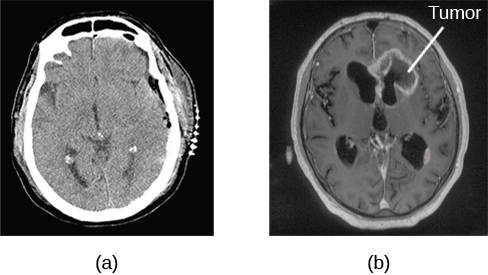
A computerized tomography (CT) scan involves taking a number of x-rays of a particular section of a person’s body or brain (Figure 3.26). The x-rays pass through tissues of different densities at different rates, allowing a computer to construct an overall image of the area of the body being scanned. A CT scan is often used to determine whether someone has a tumor or significant brain atrophy.
Figure 3.26 — 3.4 The Brain and Spinal Cord
Figure 3.26 A CT scan can be used to show brain tumors. (a) The image on the left shows a healthy brain, whereas (b) the image on the right indicates a brain tumor in the left frontal lobe. (credit a: modification of work by "Aceofhearts1968"/Wikimedia Commons; credit b: modification of work by Roland Schmitt et al)
Rose M. Spielman, William J. Jenkins, and Marilyn D. Lovett, Psychology 2E, OpenStax. (credit a: modification of work by "Aceofhearts1968"/Wikimedia Commons; credit b: modification of work by Roland Schmitt et al) · CC BY-NC-SA 4.0
· Original image ↗ · Access for free ↗
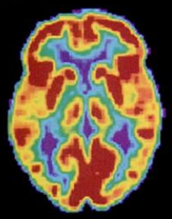
Positron emission tomography (PET) scans create pictures of the living, active brain (Figure 3.27). An individual receiving a PET scan drinks or is injected with a mildly radioactive substance, called a tracer. Once in the bloodstream, the amount of tracer in any given region of the brain can be monitored. As a brain area becomes more active, more blood flows to that area. A computer monitors the movement of the tracer and creates a rough map of active and inactive areas of the brain during a given behavior. PET scans show little detail, are unable to pinpoint events precisely in time, and require that the brain be exposed to radiation; therefore, this technique has been replaced by the fMRI as an alternative diagnostic tool. However, combined with CT, PET technology is still being used in certain contexts. For example, CT/PET scans allow better imaging of the activity of neurotransmitter receptors and open new avenues in schizophrenia research. In this hybrid CT/PET technology, CT contributes clear images of brain structures, while PET shows the brain’s activity.
Figure 3.27 — 3.4 The Brain and Spinal Cord
Figure 3.27 A PET scan is helpful for showing activity in different parts of the brain. (credit: Health and Human Services Department, National Institutes of Health)
Rose M. Spielman, William J. Jenkins, and Marilyn D. Lovett, Psychology 2E, OpenStax. (credit: Health and Human Services Department, National Institutes of Health) · CC BY-NC-SA 4.0
· Original image ↗ · Access for free ↗
Techniques Involving Magnetic Fields
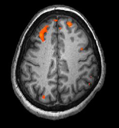
In magnetic resonance imaging (MRI), a person is placed inside a machine that generates a strong magnetic field. The magnetic field causes the hydrogen atoms in the body’s cells to move. When the magnetic field is turned off, the hydrogen atoms emit electromagnetic signals as they return to their original positions. Tissues of different densities give off different signals, which a computer interprets and displays on a monitor. Functional magnetic resonance imaging (fMRI) operates on the same principles, but it shows changes in brain activity over time by tracking blood flow and oxygen levels. The fMRI provides more detailed images of the brain’s structure, as well as better accuracy in time, than is possible in PET scans (Figure 3.28). With their high level of detail, MRI and fMRI are often used to compare the brains of healthy individuals to the brains of individuals diagnosed with psychological disorders. This comparison helps determine what structural and functional differences exist between these populations.
Figure 3.28 — 3.4 The Brain and Spinal Cord
Figure 3.28 An fMRI shows activity in the brain over time. This image represents a single frame from an fMRI. (credit: modification of work by Kim J, Matthews NL, Park S.)
Rose M. Spielman, William J. Jenkins, and Marilyn D. Lovett, Psychology 2E, OpenStax. (credit: modification of work by Kim J, Matthews NL, Park S.) · CC BY-NC-SA 4.0
· Original image ↗ · Access for free ↗
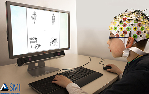
In some situations, it is helpful to gain an understanding of the overall activity of a person’s brain, without needing information on the actual location of the activity. Electroencephalography (EEG) serves this purpose by providing a measure of a brain’s electrical activity. An array of electrodes is placed around a person’s head (Figure 3.29). The signals received by the electrodes result in a printout of the electrical activity of their brain, or brainwaves, showing both the frequency (number of waves per second) and amplitude (height) of the recorded brainwaves, with an accuracy within milliseconds. Such information is especially helpful to researchers studying sleep patterns among individuals with sleep disorders.
Figure 3.29 — 3.4 The Brain and Spinal Cord
Figure 3.29 Using caps with electrodes, modern EEG research can study the precise timing of overall brain activities. (credit: SMI Eye Tracking)
OpenStax · 9.3 Stages of Development ↗ Instructional page including learning objectives, examples, figures, source tables and questions. Site navigation and embedded media players are omitted; source media links remain available.
Learning Objectives
By the end of this section, you will be able to:
Describe the stages of prenatal development and recognize the importance of prenatal care
Appraise physical, cognitive, and emotional development that occurs from infancy through childhood
Compare and contrast physical, cognitive, and emotional development that occurs during adolescence
Examine physical, cognitive, and emotional development that occurs in adulthood
From the moment we are born until the moment we die, we continue to develop.
As discussed at the beginning of this chapter, developmental psychologists often divide our development into three areas: physical development, cognitive development, and psychosocial development. Mirroring Erikson’s stages, lifespan development is divided into different stages that are based on age. We will discuss prenatal, infant, child, adolescent, and adult development.
Prenatal Development
How did you come to be who you are? From beginning as a one-cell structure to your birth, your prenatal development occurred in an orderly and delicate sequence.
There are three stages of prenatal development: germinal, embryonic, and fetal. Let’s take a look at what happens to the developing baby in each of these stages.
Germinal Stage (Weeks 1–2)
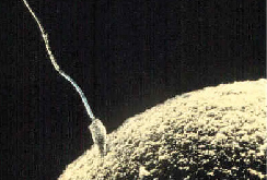
In the discussion of biopsychology earlier in the book, you learned about genetics and DNA. DNA is passed on to the child at the moment of conception. Conception occurs when sperm fertilizes an egg and forms a zygote (Figure 9.7). A zygote begins as a one-cell structure that is created when a sperm and egg merge. The genetic makeup and sex of the zygote are set at this point. During the first week after conception, the zygote divides and multiplies, going from a one-cell structure to two cells, then four cells, then eight cells, and so on. This process of cell division is called mitosis. Mitosis is a fragile process, and fewer than one-half of all zygotes survive beyond the first two weeks (Hall, 2004). After 5 days of mitosis there are 100 cells, and after 9 months there are billions of cells. As the cells divide, they become more specialized, forming different organs and body parts. In the germinal stage, the mass of cells has yet to attach itself to the lining of the uterus. Once it does, the next stage begins.
Figure 9.7 — 9.3 Stages of Development
Figure 9.7 Sperm and ovum fuse at the point of conception.
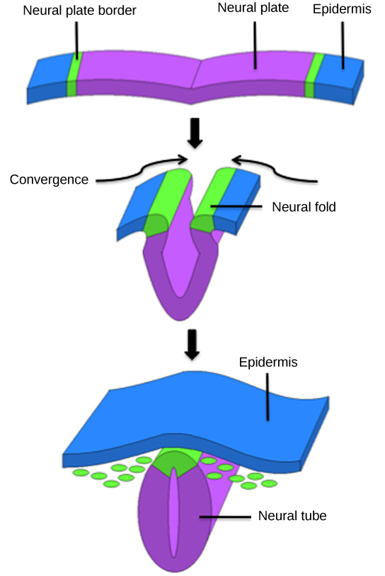
After the zygote divides for about 7–10 days and has 150 cells, it travels down the fallopian tubes and implants itself in the lining of the uterus. Upon implantation, this multi-cellular organism is called an embryo. Now blood vessels grow, forming the placenta. The placenta is a structure connected to the uterus that provides nourishment and oxygen to the developing embryo via the umbilical cord. Basic structures of the embryo start to develop into areas that will become the head, chest, and abdomen. During the embryonic stage, the heart begins to beat and organs form and begin to function. The neural tube forms along the back of the embryo, developing into the spinal cord and brain.
Fetal Stage (Weeks 9–40)
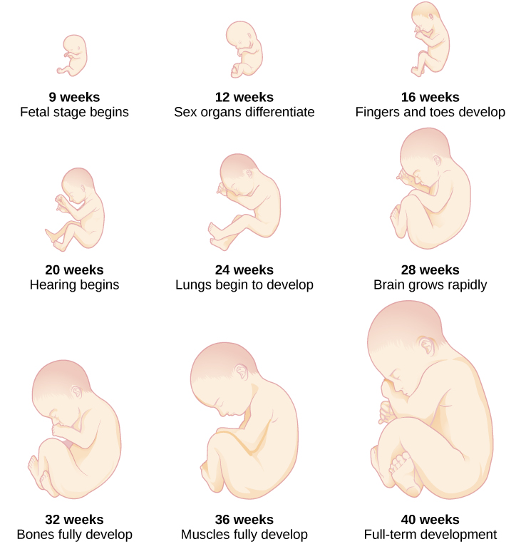
When the organism is about nine weeks old, the embryo is called a fetus. At this stage, the fetus is about the size of a kidney bean and begins to take on the recognizable form of a human being as the “tail” begins to disappear.
From 9–12 weeks, the sex organs begin to differentiate. At about 16 weeks, the fetus is approximately 4.5 inches long. Fingers and toes are fully developed, and fingerprints are visible. By the time the fetus reaches the sixth month of development (24 weeks), it weighs up to 1.4 pounds. Hearing has developed, so the fetus can respond to sounds. The internal organs, such as the lungs, heart, stomach, and intestines, have formed enough that a fetus born prematurely at this point has a chance to survive outside of the womb. Throughout the fetal stage the brain continues to grow and develop, nearly doubling in size from weeks 16 to 28. Around 36 weeks, the fetus is almost ready for birth. It weighs about 6 pounds and is about 18.5 inches long, and by week 37 all of the fetus’s organ systems are developed enough that it could survive outside the uterus without many of the risks associated with premature birth. The fetus continues to gain weight and grow in length until approximately 40 weeks. By then, the fetus has very little room to move around and birth becomes imminent. The progression through the stages is shown in Figure 9.8.
Figure 9.8 — 9.3 Stages of Development
Figure 9.8 During the fetal stage, the baby's brain develops and the body adds size and weight, until the fetus reaches full-term development.
For an amazing look at prenatal development and the process of birth, view the video Life’s Greatest Miracle from Nova and PBS.
Prenatal Influences
During each prenatal stage, genetic and environmental factors can affect development. The developing fetus is completely dependent on the person carrying it. Routine prenatal care, which is medical care during pregnancy that monitors the health of both the pregnant person and the fetus (Figure 9.9), is important because it can reduce the risk of complications during pregnancy (NIH, 2013). In fact, people who are trying to become pregnant or who may become pregnant should discuss pregnancy planning with their doctor. They may be advised, for example, to take a vitamin containing folic acid, which helps prevent certain birth defects, or to monitor aspects of their diet or exercise routines. People can face health risks without prenatal care, which is not provided equitably across populations or locations (Backes and Scrimshaw, 2020).
Figure 9.9 — 9.3 Stages of Development
Figure 9.9 A pregnant person receives an ultrasound as part of their prenatal care. (credit: "MIKI Yoshihito_Flickr"/Flickr)
Recall that when the zygote attaches to the wall of the uterus, the placenta is formed. The placenta provides nourishment and oxygen to the fetus. Most everything a pregnant person ingests, including food, liquid, and even medication, travels through the placenta to the fetus, hence the common phrase “eating for two.” Everything in the environment affects the fetus; if they are exposed to something harmful, the child can show life-long effects.
A teratogen is any environmental agent—biological, chemical, or physical—that causes damage to the developing embryo or fetus. There are different types of teratogens. Alcohol and most drugs cross the placenta and affect the fetus. Alcohol is not safe to drink in any amount during pregnancy. Alcohol use during pregnancy has been found to be the leading preventable cause of intellectual disabilities in children in the United States (Maier & West, 2001). Excessive drinking while pregnant can cause fetal alcohol spectrum disorders with life-long consequences for the child ranging in severity from minor to major (Table 9.3). Fetal alcohol spectrum disorders (FASD) are a collection of birth defects associated with heavy consumption of alcohol during pregnancy. Physically, children with FASD may have a small head size and abnormal facial features. Cognitively, these children may have poor judgment, poor impulse control, higher rates of ADHD, learning issues, and lower IQ scores. These developmental problems and delays persist into adulthood (Streissguth et al., 2004). Based on studies conducted on animals, it also has been suggested that alcohol consumption during pregnancy may predispose offspring to like alcohol (Youngentob et al., 2007).
Fetal Alcohol Syndrome Facial Features
Facial Feature
Potential Effect of Fetal Alcohol Syndrome
Head size
Below-average head circumference
Eyes
Smaller than average eye opening, skin folds at corners of eyes
Nose
Low nasal bridge, short nose
Midface
Smaller than average midface size
Lip and philtrum
Thin upper lip, indistinct philtrum
Table
9.3
Smoking is also considered a teratogen because nicotine travels through the placenta to the fetus. When the pregnant person smokes, the developing baby experiences a reduction in blood oxygen levels. According to the Centers for Disease Control and Prevention (2013), smoking while pregnant can result in premature birth, low-birth-weight infants, stillbirth, and sudden infant death syndrome (SIDS).
Heroin, cocaine, methamphetamine, almost all prescription medicines, and most over-the counter medications are also considered teratogens. Babies born with a heroin addiction need heroin just like an adult with a heroin addiction. The child will need to be gradually weaned from the heroin under medical supervision; otherwise, the child could have seizures and die. Other teratogens include radiation, viruses such as HIV and herpes, and rubella (German measles). People who were raised in the United States are much less likely to be afflicted with rubella because most received childhood immunizations or vaccinations that protect the body from disease.
Each organ of the fetus develops during a specific period in the pregnancy, called the critical or sensitive period (Figure 9.8). For example, research with primate models of FASD has demonstrated that the time during which a developing fetus is exposed to alcohol can dramatically affect the appearance of facial characteristics associated with fetal alcohol syndrome. Specifically, this research suggests that alcohol exposure that is limited to day 19 or 20 of gestation can lead to significant facial abnormalities in the offspring (Ashley, Magnuson, Omnell, & Clarren, 1999). Given regions of the brain also show sensitive periods during which they are most susceptible to the teratogenic effects of alcohol (Tran & Kelly, 2003).
What Do You Think?
Should People Who Use Drugs During Pregnancy Be Arrested and Jailed?
As you now know, people who use drugs or alcohol during pregnancy can cause serious lifelong harm to their child. Some have advocated mandatory screenings for pregnant people who have a history of problematic drug use, and if they continue using, to arrest, prosecute, and incarcerate them (Murphy, 2014). For example, in the 1990s South Carolina implemented such a "fetal protection law" called the Interagency Policy on Management of Substance Abuse During Pregnancy, which had disastrous results.
The Interagency Policy applied to patients attending the obstetrics clinic at MUSC, which primarily serves patients who are indigent or on Medicaid. It did not apply to private obstetrical patients. The policy required patient education about the harmful effects of substance abuse during pregnancy. . . . [A] statement also warned patients that protection of unborn and newborn children from the harms of illegal drug abuse could involve the Charleston police, the Solicitor of the Ninth Judicial Court, and the Protective Services Division of the Department of Social Services (DSS). (Jos, Marshall, & Perlmutter, 1995, pp. 120–121)
As with similar laws, this policy seemed to deter pregnant people from seeking prenatal care, deterred them from seeking other social services, and was applied solely to people with low incomes, resulting in lawsuits. The program was canceled after 5 years, during which 42 women were arrested. A federal agency later determined that the program involved human experimentation without the approval and oversight of an institutional review board (IRB). Many states, including Massachussetts, Florida, California, Illinois, and Utah have "mandatory reporting" laws that require healthcare providers to inform child protective services when they suspect that a fetus or infant is exposed to drugs of abuse. Even though prosecutions are rarely pursued in these states, pregnant people still report reluctance to seek care for either their pregnancy or their substance abuse, and make efforts to hide both from providers (Work, 2023). What were the flaws in these programs, and how could states and healthcare providers improve them? What are the ethical implications of charging pregnant people with child abuse?
Infancy Through Childhood
The average newborn weighs approximately 7.5 pounds. Although small, newborns are not completely helpless because their reflexes and sensory capacities help them interact with the environment from the moment of birth. All healthy babies are born with newborn reflexes: inborn automatic responses to particular forms of stimulation. Reflexes help the newborn survive until it is capable of more complex behaviors—these reflexes are crucial to survival. They are present in babies whose brains are developing normally and usually disappear around 4–5 months old. Let’s take a look at some of these newborn reflexes. The rooting reflex is the newborn’s response to anything that touches their cheek: When you stroke a baby’s cheek, the baby naturally turns the head in that direction and begins to suck. The sucking reflex is the automatic, unlearned, sucking motions that infants do with their mouths. Several other interesting newborn reflexes can be observed. For instance, if you put your finger into a newborn’s hand, you will witness the grasping reflex, in which a baby automatically grasps anything that touches the palms. The Moro reflex is the newborn’s response to the sensation of falling. The baby spreads the arms, pulls them back in, and then (usually) cries. How do you think these reflexes promote survival in the first months of life?
What can young infants see, hear, and smell? Newborn infants’ sensory abilities are significant, but their senses are not yet fully developed. Many of a newborn’s innate preferences facilitate interaction with caregivers and other humans. Although vision is their least developed sense, newborns already show a preference for faces. Babies who are just a few days old also prefer human voices, they will listen to voices longer than sounds that do not involve speech (Vouloumanos & Werker, 2004), and they seem to prefer their mother’s voice over a stranger’s voice (Mills & Melhuish, 1974). In an interesting experiment, 3-week-old babies were given pacifiers that played a recording of the infant’s mother’s voice and of a stranger’s voice. When the infants heard their mother’s voice, they sucked more strongly at the pacifier (Mills & Melhuish, 1974). Newborns also have a strong sense of smell. For instance, newborn babies can distinguish the smell of their own mother from that of others. In a study by MacFarlane (1978), 1-week-old babies who were being breastfed were placed between two gauze pads. One gauze pad was from the bra of a nursing mother who was a stranger, and the other gauze pad was from the bra of the infant’s own mother. More than two-thirds of the week-old babies turned toward the gauze pad with their mother’s scent.
Physical Development
In infancy, toddlerhood, and early childhood, the body’s physical development is rapid (Figure 9.10). On average, newborns weigh between 5 and 10 pounds, and a newborn’s weight typically doubles in six months and triples in one year. By 2 years old the weight will have quadrupled, so we can expect that a 2 year old should weigh between 20 and 40 pounds. The average length of a newborn is 19.5 inches, increasing to 29.5 inches by 12 months and 34.4 inches by 2 years old (WHO Multicentre Growth Reference Study Group, 2006).
Figure 9.10 — 9.3 Stages of Development
Figure 9.10 Children experience rapid physical changes through infancy and early childhood. (credit "left": modification of work by Kerry Ceszyk; credit "middle-left": modification of work by Kristi Fausel; credit "middle-right": modification of work by "devinf"/Flickr; credit "right": modification of work by Rose Spielman)
Rose M. Spielman, William J. Jenkins, and Marilyn D. Lovett, Psychology 2E, OpenStax. (credit "left": modification of work by Kerry Ceszyk; credit "middle-left": modification of work by Kristi Fausel; credit "middle-right": modification of work by "devinf"/Flickr; credit "right": modification of work by Rose Spielman) · CC BY-NC-SA 4.0
· Original image ↗ · Access for free ↗
During infancy and childhood, growth does not occur at a steady rate (Carel, Lahlou, Roger, & Chaussain, 2004). Growth slows between 4 and 6 years old: During this time children gain 5–7 pounds and grow about 2–3 inches per year. Once females reach 8–9 years old, their growth rate outpaces that of males due to a pubertal growth spurt. This growth spurt continues until around 12 years old, coinciding with the start of the menstrual cycle. By 10 years old, the average female weighs 88 pounds, and the average male weighs 85 pounds.
It was previously believed that we are born with all of the brain cells we will ever have. More recent research suggests that neurogenesis (the formation of neurons) can continue through adulthood. However, the vast majority of neural connections and pathways occur during the first few years of a child’s life (National Institute of Neurological Disorders and Stroke, 2019). This period of rapid neural growth is called blooming. Neural pathways continue to develop through puberty. The blooming period of neural growth is then followed by a period of pruning, where neural connections are reduced. It is thought that pruning causes the brain to function more efficiently, allowing for mastery of more complex skills (Hutchinson, 2011). Blooming occurs during the first few years of life, and pruning continues through childhood and into adolescence in various areas of the brain.
The size of our brains increases rapidly. For example, the brain of a 2-year-old is 55% of its adult size, and by 6 years old the brain is about 90% of its adult size (Tanner, 1978). During early childhood (ages 3–6), the frontal lobes grow rapidly. Recalling our discussion of the 4 lobes of the brain earlier in this book, the frontal lobes are associated with planning, reasoning, memory, and impulse control. Therefore, by the time children reach school age, they are developmentally capable of controlling their attention and behavior. Through the elementary school years, the frontal, temporal, occipital, and parietal lobes all grow in size. The brain growth spurts experienced in childhood tend to follow Piaget’s sequence of cognitive development, so that significant changes in neural functioning account for cognitive advances (Kolb & Whishaw, 2009; Overman, Bachevalier, Turner, & Peuster, 1992).
Motor development occurs in an orderly sequence as infants move from reflexive reactions (e.g., sucking and rooting) to more advanced motor functioning. For instance, babies first learn to hold their heads up, then to sit with assistance, and then to sit unassisted, followed later by crawling and then walking.
Motor skills refer to our ability to move our bodies and manipulate objects. Fine motor skills focus on the muscles in our fingers, toes, and eyes, and enable coordination of small actions (e.g., grasping a toy, writing with a pencil, and using a spoon). Gross motor skills focus on large muscle groups that control our arms and legs and involve larger movements (e.g., balancing, running, and jumping).
As motor skills develop, there are certain developmental milestones that young children should achieve (Table 9.4). For each milestone there is an average age, as well as a range of ages in which the milestone should be reached. An example of a developmental milestone is sitting. On average, most babies sit alone at 7 months old. Sitting involves both coordination and muscle strength, and 90% of babies achieve this milestone between 5 and 9 months old. In another example, babies on average are able to hold up their head at 6 weeks old, and 90% of babies achieve this between 3 weeks and 4 months old. If a baby is not holding up their head by 4 months old, they are showing a delay. If the child is displaying delays on several milestones, that is reason for concern, and the parent or caregiver should discuss this with the child’s pediatrician. Some developmental delays can be identified and addressed through early intervention.
Developmental Milestones, Ages 2–5 Years
Age (years)
Physical
Personal/Social
Language
Cognitive
2
Kicks a ball; walks up and down stairs
Plays alongside other children; copies adults
Points to objects when named; puts 2–4 words together in a sentence
Sorts shapes and colors; follows 2-step instructions
3
Climbs and runs; pedals tricycle
Takes turns; expresses many emotions; dresses self
Names familiar things; uses pronouns
Plays make believe; works toys with parts (levers, handles)
4
Catches balls; uses scissors
Prefers social play to solo play; knows likes and interests
Knows songs and rhymes by memory
Names colors and numbers; begins writing letters
5
Hops and swings; uses fork and spoon
Distinguishes real from pretend; likes to please friends
Speaks clearly; uses full sentences
Counts to 10 or higher; prints some letters and copies basic shapes
Table
9.4
Cognitive Development
In addition to rapid physical growth, young children also exhibit significant development of their cognitive abilities. Piaget thought that children’s ability to understand objects—such as learning that a rattle makes a noise when shaken—was a cognitive skill that develops slowly as a child matures and interacts with the environment. Today, developmental psychologists think Piaget was incorrect. Researchers have found that even very young children understand objects and how they work long before they have experience with those objects (Baillargeon, 1987; Baillargeon, Li, Gertner, & Wu, 2011). For example, children as young as 3 months old demonstrated knowledge of the properties of objects that they had only viewed and did not have prior experience with them. In one study, 3-month-old infants were shown a truck rolling down a track and behind a screen. The box, which appeared solid but was actually hollow, was placed next to the track. The truck rolled past the box as would be expected. Then the box was placed on the track to block the path of the truck. When the truck was rolled down the track this time, it continued unimpeded. The infants spent significantly more time looking at this impossible event (Figure 9.11). Baillargeon (1987) concluded that they knew solid objects cannot pass through each other. Baillargeon’s findings suggest that very young children have an understanding of objects and how they work, which Piaget (1954) would have said is beyond their cognitive abilities due to their limited experiences in the world.
Figure 9.11 — 9.3 Stages of Development
Figure 9.11 In Baillargeon’s study, infants observed a truck (a) roll down an unobstructed track, (b) roll down an unobstructed track with an obstruction (box) beside it, and (c) roll down and pass through what appeared to be an obstruction.
Just as there are physical milestones that we expect children to reach, there are also cognitive milestones. It is helpful to be aware of these milestones as children gain new abilities to think, problem solve, and communicate. For example, infants shake their head “no” around 6–9 months, and they respond to verbal requests to do things like “wave bye-bye” or “blow a kiss” around 9–12 months. Remember Piaget’s ideas about object permanence? We can expect children to grasp the concept that objects continue to exist even when they are not in sight by around 8 months old. Because toddlers (i.e., 12–24 months old) have mastered object permanence, they enjoy games like hide and seek, and they realize that when someone leaves the room they will come back (Loop, 2013). Toddlers also point to pictures in books and look in appropriate places when you ask them to find objects.
Preschool-age children (i.e., 3–5 years old) also make steady progress in cognitive development. Not only can they count, name colors, and tell you their name and age, but they can also make some decisions on their own, such as choosing an outfit to wear. Preschool-age children understand basic time concepts and sequencing (e.g., before and after), and they can predict what will happen next in a story. They also begin to enjoy the use of humor in stories. Because they can think symbolically, they enjoy pretend play and inventing elaborate characters and scenarios. One of the most common examples of their cognitive growth is their blossoming curiosity. Preschool-age children love to ask “Why?”
An important cognitive change occurs in children this age. Recall that Piaget described 2–3 year olds as egocentric, meaning that they do not have an awareness of others’ points of view. Between 3 and 5 years old, children come to understand that people have thoughts, feelings, and beliefs that are different from their own. This is known as theory-of-mind (TOM). Children can use this skill to tease others, persuade their parents to purchase a candy bar, or understand why a sibling might be angry. When children develop TOM, they can recognize that others have false beliefs (Dennett, 1987; Callaghan et al., 2005).
Cognitive skills continue to expand in middle and late childhood (6–11 years old). Thought processes become more logical and organized when dealing with concrete information (Figure 9.12). Children at this age understand concepts such as the past, present, and future, giving them the ability to plan and work toward goals. Additionally, they can process complex ideas such as addition and subtraction and cause-and-effect relationships. However, children’s attention spans tend to be very limited until they are around 11 years old. After that point, it begins to improve through adulthood.
Figure 9.12 — 9.3 Stages of Development
Figure 9.12 Because they understand luck and fairness, children in middle and late childhood (6–11 years old) are able to follow rules for games. (credit: Edwin Martinez)
One well-researched aspect of cognitive development is language acquisition. As mentioned earlier, the order in which children learn language structures is consistent across children and cultures (Hatch, 1983). You’ve also learned that some psychological researchers have proposed that children possess a biological predisposition for language acquisition.
Starting before birth, babies begin to develop language and communication skills. At birth, babies apparently recognize their mother’s voice and can discriminate between the language(s) spoken by their mothers and foreign languages, and they show preferences for faces that are moving in synchrony with audible language (Blossom & Morgan, 2006; Pickens, 1994; Spelke & Cortelyou, 1981).
Children communicate information through gesturing long before they speak, and there is some evidence that gesture usage predicts subsequent language development (Iverson & Goldin-Meadow, 2005). In terms of producing spoken language, babies begin to coo almost immediately. Cooing is a one-syllable combination of a consonant and a vowel sound (e.g., coo or ba). Interestingly, babies replicate sounds from their own languages. A baby whose parents speak French will coo in a different tone than a baby whose parents speak Spanish or Urdu. After cooing, the baby starts to babble. Babbling begins with repeating a syllable, such as ma-ma, da-da, or ba-ba. When babies are about 12 months old, we expect them to say their first word for meaning, and to start combining words for meaning at about 18 months.
At about 2 years old, a toddler uses between 50 and 200 words; by 3 years old they have a vocabulary of up to 1,000 words and can speak in sentences. During the early childhood years, children's vocabulary increases at a rapid pace. This is sometimes referred to as the “vocabulary spurt” and has been claimed to involve an expansion in vocabulary at a rate of 10–20 new words per week. Recent research may indicate that while some children experience these spurts, it is far from universal (as discussed in Ganger & Brent, 2004). It has been estimated that 5 year olds understand about 6,000 words, speak 2,000 words, and can define words and question their meanings. They can rhyme and name the days of the week. Seven year olds speak fluently and use slang and clichés (Stork & Widdowson, 1974).
What accounts for such dramatic language learning by children? Behaviorist B. F. Skinner thought that we learn language in response to reinforcement or feedback, such as through parental approval or through being understood. For example, when a two-year-old child asks for juice, he might say, “me juice,” to which his mother might respond by giving him a cup of apple juice. Noam Chomsky (1957) criticized Skinner’s theory and proposed that we are all born with an innate capacity to learn language. Chomsky called this mechanism a language acquisition device (LAD). Who is correct? Both Chomsky and Skinner are right. Remember that we are a product of both nature and nurture. Researchers now believe that language acquisition is partially inborn and partially learned through our interactions with our linguistic environment (Gleitman & Newport, 1995; Stork & Widdowson, 1974).
Psychosocial development occurs as children form relationships, interact with others, and understand and manage their feelings. In social and emotional development, forming healthy attachments is very important and is the major social milestone of infancy. Attachment is a long-standing connection or bond with others. Developmental psychologists are interested in how infants reach this milestone. They ask such questions as: How do parent and infant attachment bonds form? How does neglect affect these bonds? What accounts for children’s attachment differences?
Researchers Harry Harlow, John Bowlby, and Mary Ainsworth conducted studies designed to answer these questions. In the 1950s, Harlow conducted a series of experiments on monkeys. He separated newborn monkeys from their mothers. Each monkey was presented with two surrogate mothers. One surrogate monkey was made out of wire mesh, and she could dispense milk. The other monkey was softer and made from cloth: This monkey did not dispense milk. Research shows that the monkeys preferred the soft, cuddly cloth monkey, even though she did not provide any nourishment. The baby monkeys spent their time clinging to the cloth monkey and only went to the wire monkey when they needed to be fed. Prior to this study, the medical and scientific communities generally thought that babies become attached to the people who provide their nourishment. However, Harlow (1958) concluded that there was more to the mother-child bond than nourishment. Feelings of comfort and security are the critical components to maternal-infant bonding, which leads to healthy psychosocial development.
Link to Learning
Harlow’s studies of monkeys were performed before modern ethics guidelines were in place, and today his experiments are widely considered to be unethical and even cruel. Watch this video of actual footage of Harlow's monkey studies to learn more.
Building on the work of Harlow and others, John Bowlby developed the concept of attachment theory. He defined attachment as the affectional bond or tie that an infant forms with the mother (Bowlby, 1969). An infant must form this bond with a primary caregiver in order to have normal social and emotional development. In addition, Bowlby proposed that this attachment bond is very powerful and continues throughout life. He used the concept of secure base to define a healthy attachment between parent and child (1988). A secure base is a parental presence that gives the child a sense of safety as he explores his surroundings. Bowlby said that two things are needed for a healthy attachment: The caregiver must be responsive to the child’s physical, social, and emotional needs; and the caregiver and child must engage in mutually enjoyable interactions (Bowlby, 1969) (Figure 9.13).
While Bowlby thought attachment was an all-or-nothing process, Mary Ainsworth’s (1970) research showed otherwise. Ainsworth wanted to know if children differ in the ways they bond, and if so, why. To find the answers, she used the Strange Situation procedure to study attachment between mothers and their infants (1970). In the Strange Situation, the mother (or primary caregiver) and the infant (age 12-18 months) are placed in a room together. There are toys in the room, and the caregiver and child spend some time alone in the room. After the child has had time to explore her surroundings, a stranger enters the room. The mother then leaves her baby with the stranger. After a few minutes, she returns to comfort her child.
Based on how the infants/toddlers responded to the separation and reunion, Ainsworth identified three types of parent-child attachments: secure, avoidant, and resistant (Ainsworth & Bell, 1970). A fourth style, known as disorganized attachment, was later described (Main & Solomon, 1990). The most common type of attachment—also considered the healthiest—is called secure attachment (Figure 9.14). In this type of attachment, the toddler prefers his parent over a stranger. The attachment figure is used as a secure base to explore the environment and is sought out in times of stress. Securely attached children were distressed when their caregivers left the room in the Strange Situation experiment, but when their caregivers returned, the securely attached children were happy to see them. Securely attached children have caregivers who are sensitive and responsive to their needs.
Figure 9.14 — 9.3 Stages of Development
Figure 9.14 In secure attachment, the parent provides a secure base for the toddler, allowing him to securely explore his environment. (credit: Kerry Ceszyk)
With avoidant attachment, the child is unresponsive to the parent, does not use the parent as a secure base, and does not care if the parent leaves. The toddler reacts to the parent the same way she reacts to a stranger. When the parent does return, the child is slow to show a positive reaction. Ainsworth theorized that these children were most likely to have a caregiver who was insensitive and inattentive to their needs (Ainsworth, Blehar, Waters, & Wall, 1978).
In cases of resistant attachment, children tend to show clingy behavior, but then they reject the attachment figure’s attempts to interact with them (Ainsworth & Bell, 1970). These children do not explore the toys in the room, as they are too fearful. During separation in the Strange Situation, they became extremely disturbed and angry with the parent. When the parent returns, the children are difficult to comfort. Resistant attachment is the result of the caregivers’ inconsistent level of response to their child.
Finally, children with disorganized attachment behaved oddly in the Strange Situation. They freeze, run around the room in an erratic manner, or try to run away when the caregiver returns (Main & Solomon, 1990). This type of attachment is seen most often in kids who have been abused. Research has shown that abuse disrupts a child’s ability to regulate their emotions.
While Ainsworth’s research has found support in subsequent studies, it has also met criticism. Some researchers have pointed out that a child’s temperament may have a strong influence on attachment (Gervai, 2009; Harris, 2009), and others have noted that attachment varies from culture to culture, a factor not accounted for in Ainsworth’s research (Rothbaum, Weisz, Pott, Miyake, & Morelli, 2000; van Ijzendoorn & Sagi-Schwartz, 2008).
Just as attachment is the main psychosocial milestone of infancy, the primary psychosocial milestone of childhood is the development of a positive sense of self. How does self-awareness develop? Infants don’t have a self-concept, which is an understanding of who they are. If you place a baby in front of a mirror, they will reach out to touch their image, thinking it is another baby. However, by about 18 months a toddler will recognize that the person in the mirror is themself. How do we know this? In a well-known experiment, a researcher placed a red dot of paint on children’s noses before putting them in front of a mirror (Amsterdam, 1972). Commonly known as the mirror test, this behavior is demonstrated by humans and a few other species and is considered evidence of self-recognition (Archer, 1992). At 18 months old they would touch their own noses when they saw the paint, surprised to see a spot on their faces. By 24–36 months old children can name and/or point to themselves in pictures, clearly indicating self-recognition.
Children from 2–4 years old display a great increase in social behavior once they have established a self-concept. They enjoy playing with other children, but they have difficulty sharing their possessions. Also, through play children explore and come to understand their gender roles and can label themselves as a girl or boy (Chick, Heilman-Houser, & Hunter, 2002). By 4 years old, children can cooperate with other children, share when asked, and separate from parents with little anxiety. Children at this age also exhibit autonomy, initiate tasks, and carry out plans. Success in these areas contributes to a positive sense of self. Once children reach 6 years old, they can identify themselves in terms of group memberships: “I’m a first grader!” School-age children compare themselves to their peers and discover that they are competent in some areas and less so in others (recall Erikson’s task of industry versus inferiority). At this age, children recognize their own personality traits as well as some other traits they would like to have. For example, 10-year-old Layla says, “I’m kind of shy. I wish I could be more talkative like my friend Alexa.”
Development of a positive self-concept is important to healthy development. Children with a positive self-concept tend to be more confident, do better in school, act more independently, and are more willing to try new activities (Maccoby, 1980; Ferrer & Fugate, 2003). Formation of a positive self-concept begins in Erikson’s toddlerhood stage, when children establish autonomy and become confident in their abilities. Development of self-concept continues in elementary school, when children compare themselves to others. When the comparison is favorable, children feel a sense of competence and are motivated to work harder and accomplish more. Self-concept is re-evaluated in Erikson’s adolescence stage, as teens form an identity. They internalize the messages they have received regarding their strengths and weaknesses, keeping some messages and rejecting others. Adolescents who have achieved identity formation are capable of contributing positively to society (Erikson, 1968).
Dig Deeper
Phenomenological Variant of Ecological Systems Theory (PVEST)
Kenneth and Mamie Clark were pioneering psychologists responsible for the first psychological study used in a Supreme Court case. Their research with African American children and doll choices was used to highlight the harmful effects of segregation and provided support for the Browns and the NAACP in their lawsuit against the Board of Education. The finding that African American children were more likely to choose a White doll over a Black doll, in both northern and southern states, led them to theorize that the children did not have a healthy concept of themselves (Clark & Clark, 1950).
The Clarks’ research differed from that of Inez Beverly Prosser, who also studied African American children in segregated and integrated schools in Cincinnati. Parents could choose either environment for their children during the 1930s. She found, among other factors, that the self-concept of children at segregated schools was more positive versus those in integrated schools, partly due to teachers’ low expectations. Prosser also noted that the child’s personality should be considered when choosing a segregated school or an integrated school (Benjamin, Henry, & McMahon, 2005).
Later researchers suggested that African American children choosing a doll that did not look like them was not an indication of their self-esteem or their self-image. For instance, Rogers and Meltzoff (2017) found that gender identity was more important than race in their study of diverse children whose average age was about 10 years old. Thus, for children that young, the meaning of race is an evolving process, as opposed to adolescents’ search for identity. The ethnic minority children in the study did view racial identity as important, compared to their White counterparts.
For teenagers who are members of ethnic minority groups, racial/ethnic/cultural identity can be paramount, depending on the family’s processes. Racial socialization involves teaching them the positive aspects of their in-group, usually by caregivers. Most of the students in a study by Neblett, Smalls, Ford, Nguyen, and Sellers (2009) reported having received such messages but a few received no racial socialization messages. They found that these messages played a role in how they felt about their in-group.
Some theories have been developed to explain the behaviors of ethnic minority youth. One such theory is the Phenomenological Variant of Ecological Systems Theory (PVEST), put forth by Margaret Beale Spencer. It is a merging of phenomenology and Bronfenbrenner’s ecological systems theory. A phenomenological approach is based on how a person makes meaning of their experiences. For example, young African American boys have different experiences in educational settings compared to African American girls. Consequently, the meaning they assign to those experiences differs. Bronfenbrenner’s ecological systems theory suggests that development occurs based on interactions among environments such as school, family, and community (Bronfenbrenner, 1977).
The research that Spencer, Dupree, and Hartmann (1997) conducted with African American adolescent boys and girls was explained by PVEST. They found that negative learning attitudes were predicted by unpopularity with peers for girls and boys. Additionally, for boys, more stress predicted a less negative attitude toward learning, possibly due to focus on the school environment instead of on personal issues. This occurred along with perceiving that teachers had positive expectations of African American boys. The researchers surmised that PVEST accounted for how others’ perceptions and their subsequent attitudes were related and worked both ways.
What can parents do to nurture a healthy self-concept? Diana Baumrind (1971, 1991) proposed that parenting style may be a factor. The way we parent is an important factor in a child’s socioemotional growth. Baumrind developed and refined a theory describing four parenting styles: authoritative, authoritarian, permissive, and uninvolved. With the authoritative style, the parent gives reasonable demands and consistent limits, expresses warmth and affection, and listens to the child’s point of view. Parents set rules and explain the reasons behind them. They are also flexible and willing to make exceptions to the rules in certain cases—for example, temporarily relaxing bedtime rules to allow for a nighttime swim during a family vacation. Of the four parenting styles, the authoritative style is the one that is most encouraged in modern American society. American children raised by authoritative parents tend to have high self-esteem and social skills. However, effective parenting styles vary as a function of culture and, as Small (1999) points out, the authoritative style is not necessarily preferred or appropriate in all cultures.
In authoritarian style, the parent places high value on conformity and obedience. The parents are often strict, tightly monitor their children, and express little warmth. In contrast to the authoritative style, authoritarian parents probably would not relax bedtime rules during a vacation because they consider the rules to be set, and they expect obedience. This style can create anxious, withdrawn, and unhappy kids. However, it is important to point out that authoritarian parenting is as beneficial as the authoritative style in some ethnic groups (Russell, Crockett, & Chao, 2010). For instance, first-generation Chinese American children raised by authoritarian parents did just as well in school as their peers who were raised by authoritative parents (Russell et al., 2010).
For parents who employ the permissive style of parenting, the kids run the show and anything goes. Permissive parents make few demands and rarely use punishment. They tend to be very nurturing and loving, and may play the role of friend rather than parent. In terms of our example of vacation bedtimes, permissive parents might not have bedtime rules at all—instead they allow the child to choose their bedtime whether on vacation or not. Not surprisingly, children raised by permissive parents tend to lack self-discipline, and the permissive parenting style is negatively associated with grades (Dornbusch, Ritter, Leiderman, Roberts, & Fraleigh, 1987). The permissive style may also contribute to other risky behaviors such as alcohol abuse (Bahr & Hoffman, 2010), risky sexual behavior especially among female children (Donenberg, Wilson, Emerson, & Bryant, 2002), and increased display of disruptive behaviors by male children (Parent et al., 2011). However, there are some positive outcomes associated with children raised by permissive parents. They tend to have higher self-esteem, better social skills, and report lower levels of depression (Darling, 1999).
With the uninvolved style of parenting, the parents are indifferent, uninvolved, and sometimes referred to as neglectful. They don’t respond to the child’s needs and make relatively few demands. This could be because of severe depression or substance abuse, or other factors such as the parents’ extreme focus on work. These parents may provide for the child’s basic needs, but little else. The children raised in this parenting style are usually emotionally withdrawn, fearful, anxious, perform poorly in school, and are at an increased risk of substance abuse (Darling, 1999).
As you can see, parenting styles influence childhood adjustment, but could a child’s temperament likewise influence parenting? Temperament refers to innate traits that influence how one thinks, behaves, and reacts with the environment. Children with easy temperaments demonstrate positive emotions, adapt well to change, and are capable of regulating their emotions. Conversely, children with difficult temperaments demonstrate negative emotions and have difficulty adapting to change and regulating their emotions. Difficult children are much more likely to challenge parents, teachers, and other caregivers (Thomas, 1984). Therefore, it’s possible that easy children (i.e., social, adaptable, and easy to soothe) tend to elicit warm and responsive parenting, while demanding, irritable, withdrawn children evoke irritation in their parents or cause their parents to withdraw (Sanson & Rothbart, 1995).
Everyday Connection
The Importance of Play and Recess
According to the American Academy of Pediatrics (2007), unstructured play is an integral part of a child’s development. It builds creativity, problem solving skills, and social relationships. Play also allows children to develop a theory-of-mind as they imaginatively take on the perspective of others.
Outdoor play allows children the opportunity to directly experience and sense the world around them. While doing so, they may collect objects that they come across and develop lifelong interests and hobbies. They also benefit from increased exercise, and engaging in outdoor play can actually increase how much they enjoy physical activity. This helps support the development of a healthy heart and brain. Unfortunately, research suggests that today’s children are engaging in less and less outdoor play (Clements, 2004). Perhaps, it is no surprise to learn that lowered levels of physical activity in conjunction with easy access to calorie-dense foods with little nutritional value are contributing to alarming levels of childhood obesity (Karnik & Kanekar, 2012).
Despite the adverse consequences associated with reduced play, some children are over scheduled and have little free time to engage in unstructured play. In addition, some schools have taken away recess time for children in a push for students to do better on standardized tests, and many schools commonly use loss of recess as a form of punishment. Do you agree with these practices? Why or why not?
Adolescence
Adolescence is a socially constructed concept. In pre-industrial society, children were considered adults when they reached physical maturity, but today we have an extended time between childhood and adulthood called adolescence. Adolescence is the period of development that begins at puberty and ends at emerging adulthood, which is discussed later. In the United States, adolescence is seen as a time to develop independence from parents while remaining connected to them (Figure 9.15). The typical age range of adolescence is from 12 to 18 years, and this stage of development also has some predictable physical, cognitive, and psychosocial milestones.
Figure 9.15 — 9.3 Stages of Development
Figure 9.15 Peers are a primary influence on our development in adolescence. (credit: "manseok_Pixabay"/ Pixabay)
As noted above, adolescence begins with puberty. While the sequence of physical changes in puberty is predictable, the onset and pace of puberty vary widely. Several physical changes occur during puberty, such as adrenarche and gonadarche, the maturing of the adrenal glands and sex glands, respectively. Also during this time, primary and secondary sexual characteristics develop and mature. Primary sexual characteristics are organs specifically needed for reproduction, like the uterus and ovaries and testes. Secondary sexual characteristics are physical signs of sexual maturation that do not directly involve sex organs, such as development of breasts and hips, and development of facial hair and a deepened voice. Menarche, the beginning of menstrual periods, usually occurs around 12–13 years old, and spermarche, the first ejaculation, around 13–14 years old.
During puberty, people experience a rapid increase in height (i.e., growth spurt). For females this begins between 8 and 13 years old, with adult height reached between 10 and 16 years old. Males begin their growth spurt slightly later, usually between 10 and 16 years old, and reach their adult height between 13 and 17 years old. Both nature (i.e., genes) and nurture (e.g., nutrition, medications, and medical conditions) can influence height.
Because rates of physical development vary so widely among teenagers, puberty can be a source of pride or embarrassment. Early maturing boys tend to be stronger, taller, and more athletic than their later maturing peers. They are usually more popular, confident, and independent, but they are also at a greater risk for substance abuse and early sexual activity (Flannery, Rowe, & Gulley, 1993; Kaltiala-Heino, Rimpela, Rissanen, & Rantanen, 2001). Early maturing girls may be teased or overtly admired, which can cause them to feel self-conscious about their developing bodies. These girls are at a higher risk for depression, substance abuse, and eating disorders (Ge, Conger, & Elder, 2001; Graber, Lewinsohn, Seeley, & Brooks-Gunn, 1997; Striegel-Moore & Cachelin, 1999). Late blooming boys and girls (i.e., they develop more slowly than their peers) may feel self-conscious about their lack of physical development. Negative feelings are particularly a problem for late maturing boys, who are at a higher risk for depression and conflict with parents (Graber et al., 1997) and more likely to be bullied (Pollack & Shuster, 2000).
Psychologists and clinicians also study the impact of gender identity and the impacts of puberty on transgender and gender-nonconforming youth. This is a relatively recent area of study, and the impacts should not be over-generalized. However, the research seems to indicate that transgender and gender-nonconforming youth who are supported in their identity do not have significantly higher incidences of depression or anxiety symptoms when compared to control groups, while those who are not supported exhibit more symptoms and risk factors (Olson, Durwood, & McLaughlin, 2016). Expression and exploration of gender identity can take many forms and manifest at different times depending on a wide array of factors.
The adolescent brain also remains under development. Up until puberty, brain cells continue to bloom in the frontal region. Adolescents engage in increased risk-taking behaviors and emotional outbursts possibly because the frontal lobes of their brains are still developing (Figure 9.16). Recall that this area is responsible for judgment, impulse control, and planning, and it is still maturing into early adulthood (Casey, Tottenham, Liston, & Durston, 2005).
Figure 9.16 — 9.3 Stages of Development
Figure 9.16 Brain growth continues into the early 20s. The development of the frontal lobe, in particular, is important during this stage.
According to neuroscientist Jay Giedd in the Frontline video “Inside the Teenage Brain” (2013), “It’s sort of unfair to expect [teens] to have adult levels of organizational skills or decision-making before their brains are finished being built.” Watch this segment on “The Wiring of the Adolescent Brain” to find out more about the developing brain during adolescence.
Cognitive Development
More complex thinking abilities emerge during adolescence. Some researchers suggest this is due to increases in processing speed and efficiency rather than as the result of an increase in mental capacity—in other words, due to improvements in existing skills rather than development of new ones (Bjorkland, 1987; Case, 1985). During adolescence, teenagers move beyond concrete thinking and become capable of abstract thought. Recall that Piaget refers to this stage as formal operational thought. Teen thinking is also characterized by the ability to consider multiple points of view, imagine hypothetical situations, debate ideas and opinions (e.g., politics, religion, and justice), and form new ideas (Figure 9.17). In addition, it’s not uncommon for adolescents to question authority or challenge established societal norms.
Cognitive empathy, also known as theory-of-mind (which we discussed earlier with regard to egocentrism), relates to the ability to take the perspective of others and feel concern for others (Shamay-Tsoory, Tomer, & Aharon-Peretz, 2005). Cognitive empathy begins to increase in adolescence and is an important component of social problem solving and conflict avoidance. According to one longitudinal study, levels of cognitive empathy begin rising in girls around 13 years old, and around 15 years old in boys (Van der Graaff et al., 2013). Teens who reported having supportive fathers with whom they could discuss their worries were found to be better able to take the perspective of others (Miklikowska, Duriez, & Soenens, 2011).
Figure 9.17 — 9.3 Stages of Development
Figure 9.17 Teenage thinking is characterized by the ability to reason logically and solve hypothetical problems such as how to design, plan, and build a structure. (credit: U.S. Army RDECOM)
Adolescents continue to refine their sense of self as they relate to others. Erikson referred to the task of the adolescent as one of identity versus role confusion. Thus, in Erikson’s view, an adolescent’s main questions are “Who am I?” and “Who do I want to be?” Some adolescents adopt the values and roles that their parents expect for them. Other teens develop identities that are in opposition to their parents but align with a peer group. This is common as peer relationships become a central focus in adolescents’ lives.
As adolescents work to form their identities, they pull away from their parents, and the peer group becomes very important (Shanahan, McHale, Osgood, & Crouter, 2007). Despite spending less time with their parents, most teens report positive feelings toward them (Moore, Guzman, Hair, Lippman, & Garrett, 2004). Warm and healthy parent-child relationships have been associated with positive child outcomes, such as better grades and fewer school behavior problems, in the United States as well as in other countries (Hair et al., 2005).
It appears that most teens don’t experience adolescent storm and stress to the degree once famously suggested by G. Stanley Hall, a pioneer in the study of adolescent development. Only small numbers of teens have major conflicts with their parents (Steinberg & Morris, 2001), and most disagreements are minor. For example, in a study of over 1,800 parents of adolescents from various cultural and ethnic groups, Barber (1994) found that conflicts occurred over day-to-day issues such as homework, money, curfews, clothing, chores, and friends. These types of arguments tend to decrease as teens develop (Galambos & Almeida, 1992). There is emerging research on the adolescent brain. Galvan, Hare, Voss, Glover and Casey (2007) examined its role in risk-taking behavior. They used fMRI to assess the readings’ relationship to risk-taking, risk perception, and impulsivity. The researchers found that there was no correlation between brain activity in the neural reward center and impulsivity and risk perception. However, activity in that part of the brain was correlated to risk taking. In other words, risk-taking adolescents experienced brain activity in the reward center. The idea that adolescents, however, are more impulsive than other demographics was challenged in their research, which included children and adults.
Emerging Adulthood
The next stage of development is emerging adulthood. This is a relatively newly defined period of lifespan development spanning from 18 years old to the mid-20s, characterized as an in-between time where identity exploration is focused on work and love.
When does a person become an adult? There are many ways to answer this question. In the United States, you are legally considered an adult at 18 years old. But other definitions of adulthood vary widely; in sociology, for example, a person may be considered an adult when they become self-supporting, choose a career, get married, or start a family. The ages at which we achieve these milestones vary from person to person as well as from culture to culture. For example, in the African country of Malawi, 15-year-old Njemile was married at 14 years old and had her first child at 15 years old. In her culture she is considered an adult. Children in Malawi take on adult responsibilities such as marriage and work (e.g., carrying water, tending babies, and working fields) as early as 10 years old. In stark contrast, independence in Western cultures is taking longer and longer, effectively delaying the onset of adult life.
What factors are leading to these changes regarding financial and familial independence? It seems that emerging adulthood is a product of both Western culture and our current times (Arnett, 2000). People in higher income and more industrialized countries are living longer, allowing the freedom to take an extra decade to start a career and family. Changes in the workforce also play a role. For example, 50 years ago, a young adult with a high school diploma could immediately enter the work force and climb the economic ladder. That is no longer the case. Bachelor’s and even graduate degrees are required more and more often—even for entry-level jobs (Arnett, 2000). In addition, many students are taking longer (five or six years) to complete a college degree as a result of working and going to school at the same time. After graduation, many young adults return to the family home because they have difficulty finding a job. The term Boomerang Generation describes recent college graduates for whom lack of adequate employment upon college graduation often leads to a return to the parental home (Davidson, 2014). Changing cultural expectations may be the most important reason for the delay in entering adult roles. Young people are spending more time exploring their options, so they are delaying marriage and work as they change majors and jobs multiple times, putting them on a much later timetable than their parents (Arnett, 2000).
Adulthood
Adulthood begins around 20 years old and has three distinct stages: early, middle, and late. Each stage brings its own set of rewards and challenges.
Physical Development
By the time we reach early adulthood (20 to early 40s), our physical maturation is complete, although our height and weight may increase slightly. In young adulthood, our physical abilities are at their peak, including muscle strength, reaction time, sensory abilities, and cardiac functioning. Most professional athletes are at the top of their game during this stage. People often carry and give birth to children in the young adulthood years, so they may see additional weight gain and breast changes.
Middle adulthood extends from the 40s to the 60s (Figure 9.18). Physical decline is gradual. The skin loses some elasticity, and wrinkles are among the first signs of aging. Visual acuity decreases during this time. Fertility gradually declines with the approach to the onset of menopause, the end of the menstrual cycle, around 50 years old. Middle adulthood tends toward weight gain: in the abdominal area for men and in the hips and thighs for women. Hair begins to thin and turn gray.
Figure 9.18 — 9.3 Stages of Development
Figure 9.18 Physical declines of middle and late adulthood can be minimized with proper exercise, nutrition, and an active lifestyle. (credit: modification of work by Peter Stevens)
Late adulthood is considered to extend from the 60s on. This is the last stage of physical change. The skin continues to lose elasticity, reaction time slows further, and muscle strength diminishes. Smell, taste, hearing, and vision, so sharp in our twenties, decline significantly. The brain may also no longer function at optimal levels, leading to problems like memory loss, dementia, and Alzheimer’s disease in later years.
Because we spend so many years in adulthood (more than any other stage), cognitive changes are numerous. In fact, research suggests that adult cognitive development is a complex, ever changing process that may be even more active than cognitive development in infancy and early childhood (Fischer, Yan, & Stewart, 2003).
Unlike our physical abilities, which peak in our mid-20s and then begin a slow decline, our cognitive abilities remain steady throughout early and middle adulthood. Our crystallized intelligence (information, skills, and strategies we have gathered through a lifetime of experience) tends to hold steady as we age—it may even improve. For example, adults show relatively stable to increasing scores on intelligence tests until their mid-30s to mid-50s (Bayley & Oden, 1955). However, in late adulthood we begin to experience a decline in another area of our cognitive abilities—fluid intelligence (information processing abilities, reasoning, and memory). These processes become slower. How can we delay the onset of cognitive decline? Mental and physical activity seems to play a part (Figure 9.19). Research has found adults who engage in mentally and physically stimulating activities experience less cognitive decline and have a reduced incidence of mild cognitive impairment and dementia (Hertzog, Kramer, Wilson, & Lindenberger, 2009; Larson et al., 2006; Podewils et al., 2005).
Figure 9.19 — 9.3 Stages of Development
Figure 9.19 Cognitive activities such as playing mahjong, chess, or other games, can keep you mentally fit. The same is true for solo pastimes like reading and completing crossword puzzles. (credit: Philippe Put)
Researchers have examined the aging brain by comparing it to brain functioning in younger people. Forstmann and colleagues (2011) compared elderly participants to younger participants, who in the study were asked to report the direction of movement of a set of dots. They were given feedback regarding speed and accuracy. The researchers found that older participants made more errors and were slower due to degeneration of corticostriatal connections. In other words, the decreased ability typically assigned to elderly people may be due to circumstances in the brain beyond their control. Interestingly, other researchers have found similarities in spatial representations when comparing children aged 6–7 to those over the age of 80. Ruggiero, D’Errico, and Iachini (2016) reported that this is due to neurodegeneration in older adults and immature neurology in young children.
Many elderly people experience dementia, changes in the brain that negatively affect cognition. Alzheimer’s disease is one type of dementia, initially studied by medical researcher Solomon Carter Fuller. Alzheimer’s disease has a genetic basis. Plaques in the brain are due to cell death, which then causes those affected with the disease severe forgetfulness. A person can forget how to walk, talk, and eventually eat. The disease can be mitigated by assessing environmental factors (exposure to lead, iron, and zinc increase risk) and nutritional factors (the Mediterranean diet lowers risk) (Arora, Mittal, & Kakkar, 2015). Although there is no cure, there is hope. Cognitive rehabilitation can offset mild cognitive impairment, as it can evolve into dementia. Garcia-Betances, Jimenez-Mixco, Arredondo, and Cabrera-Umpierrez (2015) examined the use of virtual reality as a possible cognitive rehabilitative method. They suggested that virtual reality technology should involve daily living activities, memory, and language, among other considerations.
There are many theories about the social and emotional aspects of aging. Some aspects of healthy aging include activities, social connectedness, and the role of a person’s culture. According to many theorists, including George Vaillant (2002), who studied and analyzed over 50 years of data, we need to have and continue to find meaning throughout our lives. For those in early and middle adulthood, meaning is found through work (Sterns & Huyck, 2001) and family life (Markus, Ryff, Curan, & Palmersheim, 2004). These areas relate to the tasks that Erikson referred to as generativity and intimacy. As mentioned previously, adults tend to define themselves by what they do—their careers. Earnings peak during this time, yet job satisfaction is more closely tied to work that involves contact with other people, is interesting, provides opportunities for advancement, and allows some independence (Mohr & Zoghi, 2006) than it is to salary (Iyengar, Wells, & Schwartz, 2006). How might being unemployed or being in a dead-end job challenge adult well-being?
Positive relationships with significant others in our adult years have been found to contribute to a state of well-being (Ryff & Singer, 2009). Most adults in the United States identify themselves through their relationships with family—particularly with spouses, children, and parents (Markus et al., 2004). While raising children can be stressful, especially when they are young, research suggests that parents reap the rewards down the road, as adult children tend to have a positive effect on parental well-being (Umberson, Pudrovska, & Reczek, 2010). Having a stable marriage has also been found to contribute to well-being throughout adulthood (Vaillant, 2002).
Another aspect of positive aging is believed to be social connectedness and social support. As we get older, socioemotional selectivity theory suggests that our social support and friendships dwindle in number, but remain as close, if not more close than in our earlier years (Carstensen, 1992) (Figure 9.20).
Figure 9.20 — 9.3 Stages of Development
Figure 9.20 Social support is important as we age. (credit: Gabriel Rocha)