Try before revealing the answer. An unfavorable phosphorylation has ΔG = +18 kJ/mol under stated conditions. A mechanistically coupled ATPhydrolysis contributes −30 kJ/mol. What is the net ΔG, and does this determine the rate?
Worked answer
Net ΔG = +18 − 30 = −12 kJ/mol for the combined stoichiometric reaction. The net direction is thermodynamically favorable under those conditions, but its rate still depends on activation barriers and catalyst availability. Merely mixing ATP with a substrate does not establish coupling.
29.1 An Overview of Metabolism and Biochemical Energy — figures and tables
Figure 29.2 — 29.1 An Overview of Metabolism and Biochemical Energy
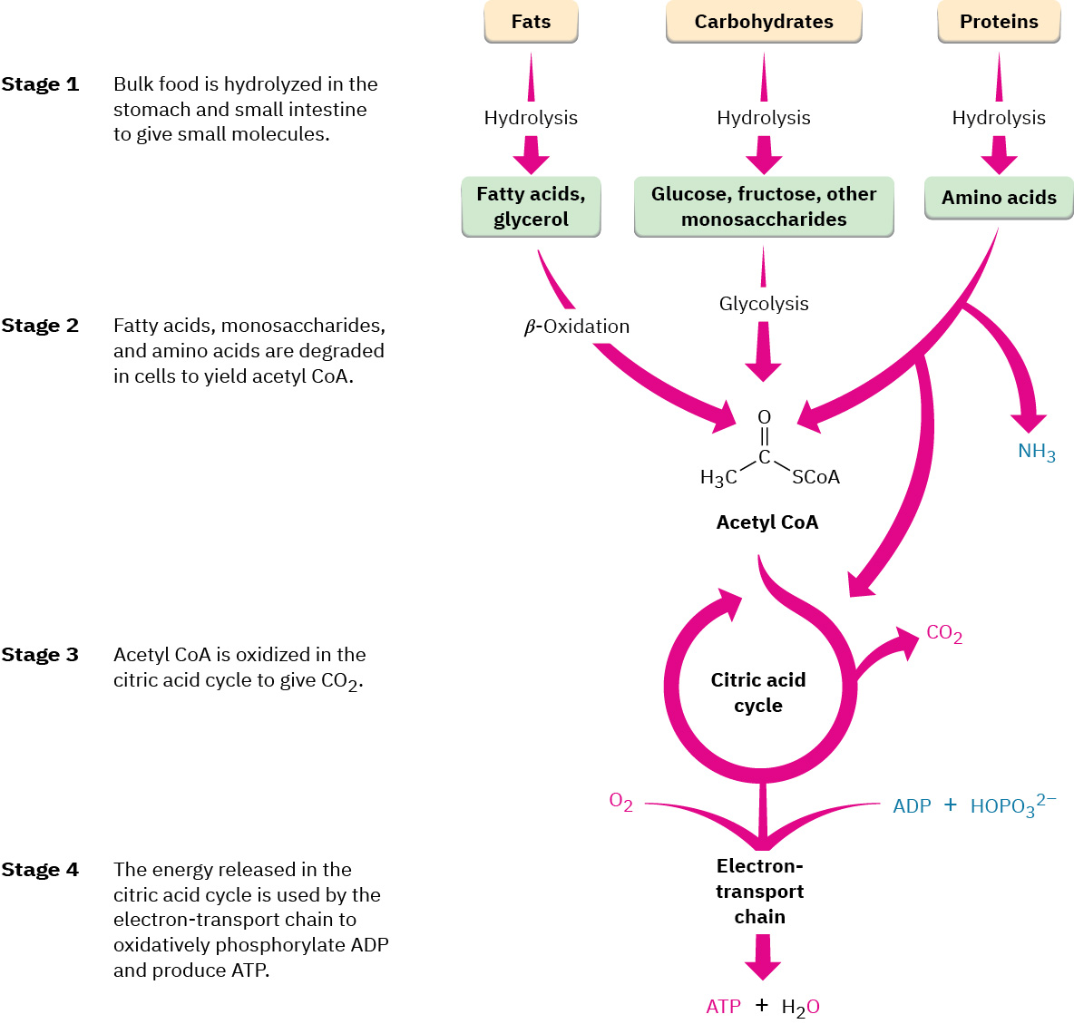
Figure 29.2 An overview of catabolic pathways for the degradation of food and the production of biochemical energy. The ultimate products of food catabolism are CO 2 and H 2 O, with the energy released in the citric acid cycle used to drive the endergonic synthesis of adenosine triphosphate (ATP) from adenosine diphosphate (ADP) plus hydrogen phosphate ion, HOPO 3 2– .
Figure 14.5 The purines have a double ring structure with a six-membered ring fused to a five-membered ring. Pyrimidines are smaller in size; they have a single six-membered ring structure.
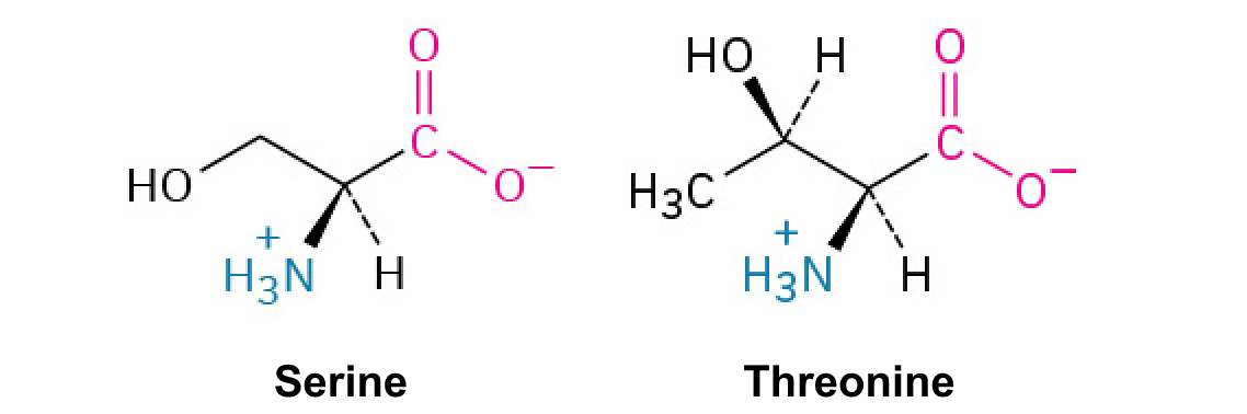
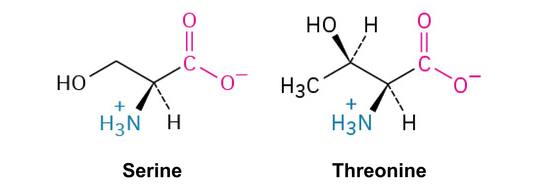
A nitrogen-containing functional group. A free amino acid’s amino group is typically protonated (NH₃⁺) near physiological pH; proline’s ring nitrogen is an exception to this formula.
Figure 15.14 This is a space-filling model of a tRNA molecule that adds the amino acid phenylalanine to a growing polypeptide chain. The anticodon AAG binds the Codon UUC on the mRNA. The amino acid phenylalanine is attached to the other end of the tRNA.
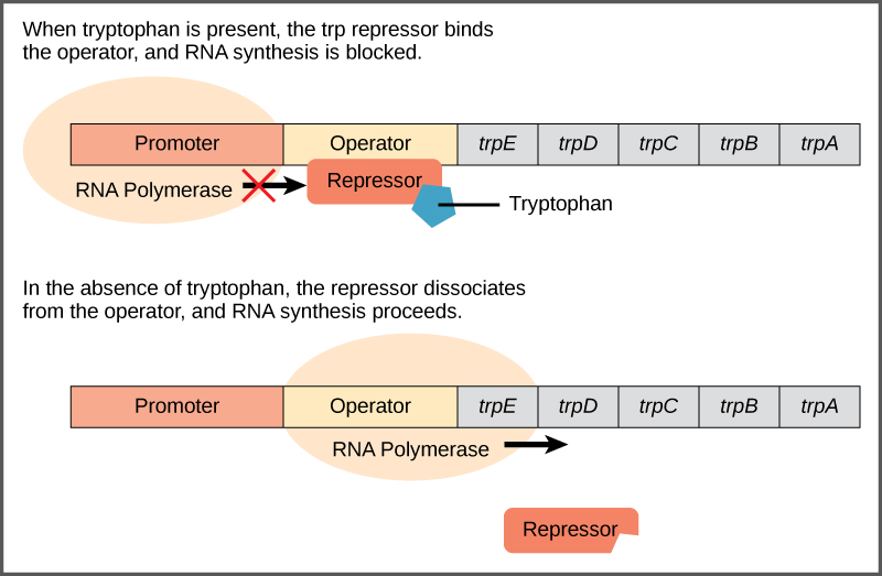
Figure 16.4 The tryptophan operon. The five genes that are needed to synthesize tryptophan in E. coli are located next to each other in the trp operon. When tryptophan is plentiful, two tryptophan molecules bind the repressor protein at the operator sequence. This physically blocks the RNA polymerase from transcribing the tryptophan genes. When tryptophan is absent, the repressor protein does not bind to the operator and the genes are transcribed.
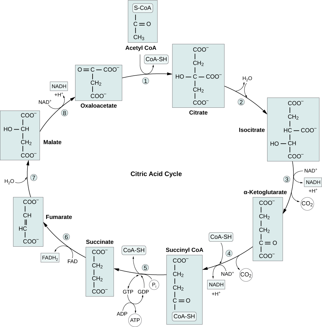
Figure 7.11 In the citric acid cycle, the acetyl group from acetyl CoA is attached to a four-carbon oxaloacetate molecule to form a six-carbon citrate molecule. Through a series of steps, citrate is oxidized, releasing two carbon dioxide molecules for each acetyl group fed into the cycle. In the process, three NAD + molecules are reduced to NADH, one FAD molecule is reduced to FADH 2 , and one ATP or GTP (depending on the cell type) is produced (by substrate-level phosphorylation). Because the final product of the citric acid cycle is also the first reactant, the cycle runs continuously in the presence of sufficient reactants. Credit: Rao, A., Ryan, K., Tag, A., and Fletcher, S. Department of Biology, Texas
A&M University.
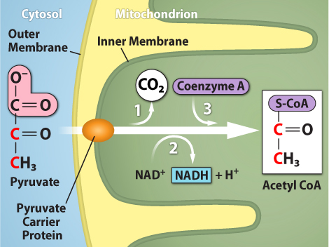
Figure 7.10 Upon entering the mitochondrial matrix, a multienzyme complex converts pyruvate into acetyl CoA. In the process, carbon dioxide is released, and one molecule of NADH is formed. Credit: Rao, A., Ryan, K. and Tag, A. Department of Biology, Texas A&M University.
Figure 15.4 This figure shows the genetic code for translating each nucleotide triplet in mRNA into an amino acid or a termination signal in a protein. (credit: modification of work by NIH)
Figure 4.44 - Kinetic profile of an allosteric enzyme whose activity is controlled by a homotropic effector. Image by Aleia Kim
Kevin Ahern, Indira Rajagopal, and Taralyn Tan, Biochemistry Free For All, Oregon State University, via Biology LibreTexts. Image by Aleia Kim · CC BY-NC-SA 4.0
· Original image ↗ · Access for free ↗
Figure 14.5 The purines have a double ring structure with a six-membered ring fused to a five-membered ring. Pyrimidines are smaller in size; they have a single six-membered ring structure.
An electrical property that can be positive or negative. Opposite charges attract and like charges repel; net charge is the sum of all charges on a particle.
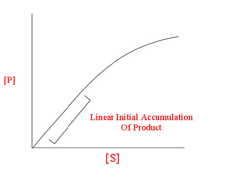
Figure 4.18 - Kinetics of an enzyme obeying Michaelis-Menten kinetics. Image by Aleia Kim
Kevin Ahern, Indira Rajagopal, and Taralyn Tan, Biochemistry Free For All, Oregon State University, via Biology LibreTexts. Image by Aleia Kim · CC BY-NC-SA 4.0
· Original image ↗ · Access for free ↗
Equilibrium
A state where opposing processes occur at equal rates, so macroscopic concentrations or other properties remain constant.
A ratio of product to reactant activities at equilibrium, each raised to its stoichiometric coefficient. At a fixed temperature, a catalyst does not change this ratio.
Figure 15.11 Eukaryotic pre-mRNA processing. In addition to 5’ Cap and 3’ Poly-A Tail addition, introns must be precisely removed and exons joined to generate a functional mRNA. Nucleotides upstream (towards the 5’cap) of the translation START codon are part of the 5’ untranslated region (5’ UTR). Nucleotides downstream (towards 3’end) of the STOP codon form the 3’ UTR. Both 5’ and 3’ UTRs are important for regulating translation initiation and mRNA stability. Credit: Rao, A., Ryan, K. Fletcher, S. and Tag, A. Department of Biology, Texas A&M University.
Rao, A., Ryan, K. Fletcher, S. and Tag, A. Department of Biology, Texas A&M University.; Mary Ann Clark, Matthew Douglas, and Jung Choi, Biology 2e, OpenStax. · CC BY-NC-SA 4.0
· Original image ↗ · Access for free ↗
A thermodynamic quantity used to predict whether a process is favorable at constant temperature and pressure. A negative change favors the forward process.
The change in Gibbs free energy during a process. At constant temperature and pressure, a negative value favors the forward reaction under the stated conditions; it does not specify reaction speed.
Figure 14.5 The purines have a double ring structure with a six-membered ring fused to a five-membered ring. Pyrimidines are smaller in size; they have a single six-membered ring structure.
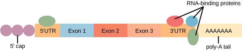
Figure 16.13 RNA-binding proteins. The protein-coding region of this processed mRNA is flanked by 5' and 3' untranslated regions (UTRs). The presence of RNA-binding proteins at the 5' or 3' UTR influences the stability of the RNA molecule.
Figure 4.18 - Kinetics of an enzyme obeying Michaelis-Menten kinetics. Image by Aleia Kim
Kevin Ahern, Indira Rajagopal, and Taralyn Tan, Biochemistry Free For All, Oregon State University, via Biology LibreTexts. Image by Aleia Kim · CC BY-NC-SA 4.0
· Original image ↗ · Access for free ↗
The early rate of product formation, measured before substantial substrate depletion or product accumulation, usually after the brief binding transient.
Figure 4.18 - Kinetics of an enzyme obeying Michaelis-Menten kinetics. Image by Aleia Kim
Kevin Ahern, Indira Rajagopal, and Taralyn Tan, Biochemistry Free For All, Oregon State University, via Biology LibreTexts. Image by Aleia Kim · CC BY-NC-SA 4.0
· Original image ↗ · Access for free ↗
Figure 15.11 Eukaryotic pre-mRNA processing. In addition to 5’ Cap and 3’ Poly-A Tail addition, introns must be precisely removed and exons joined to generate a functional mRNA. Nucleotides upstream (towards the 5’cap) of the translation START codon are part of the 5’ untranslated region (5’ UTR). Nucleotides downstream (towards 3’end) of the STOP codon form the 3’ UTR. Both 5’ and 3’ UTRs are important for regulating translation initiation and mRNA stability. Credit: Rao, A., Ryan, K. Fletcher, S. and Tag, A. Department of Biology, Texas A&M University.
Rao, A., Ryan, K. Fletcher, S. and Tag, A. Department of Biology, Texas A&M University.; Mary Ann Clark, Matthew Douglas, and Jung Choi, Biology 2e, OpenStax. · CC BY-NC-SA 4.0
· Original image ↗ · Access for free ↗
The exponent needed to obtain a number from a chosen fixed number. For decimal logarithms, log₁₀(100) = 2 because 10² = 100; pH uses the negative decimal logarithm of hydrogen ion activity.
Figure 4.18 Membrane and secretory proteins are synthesized in the rough endoplasmic reticulum (RER). The RER also sometimes modifies proteins. In this illustration, a (green) integral membrane protein is modified by attachment of a (purple) carbohydrate in the ER. Vesicles with the integral protein bud from the ER and fuse with the Golgi apparatus' cis face. As the protein passes along the Golgi’s cisternae, the addition of more carbohydrates further modifies it. After its synthesis is complete, it exits as an integral membrane protein of the vesicle that buds from the Golgi’s trans face. When the vesicle fuses with the cell membrane, the protein becomes an integral portion of that cell membrane. (credit: modification of work by Magnus Manske)
A thin boundary or sheet. Cell membranes are lipid bilayers containing proteins; blotting membranes are synthetic supports that hold proteins or nucleic acids for detection.
Figure 15.11 Eukaryotic pre-mRNA processing. In addition to 5’ Cap and 3’ Poly-A Tail addition, introns must be precisely removed and exons joined to generate a functional mRNA. Nucleotides upstream (towards the 5’cap) of the translation START codon are part of the 5’ untranslated region (5’ UTR). Nucleotides downstream (towards 3’end) of the STOP codon form the 3’ UTR. Both 5’ and 3’ UTRs are important for regulating translation initiation and mRNA stability. Credit: Rao, A., Ryan, K. Fletcher, S. and Tag, A. Department of Biology, Texas A&M University.
Rao, A., Ryan, K. Fletcher, S. and Tag, A. Department of Biology, Texas A&M University.; Mary Ann Clark, Matthew Douglas, and Jung Choi, Biology 2e, OpenStax. · CC BY-NC-SA 4.0
· Original image ↗ · Access for free ↗
Figure 4.18 - Kinetics of an enzyme obeying Michaelis-Menten kinetics. Image by Aleia Kim
Kevin Ahern, Indira Rajagopal, and Taralyn Tan, Biochemistry Free For All, Oregon State University, via Biology LibreTexts. Image by Aleia Kim · CC BY-NC-SA 4.0
· Original image ↗ · Access for free ↗
Figure 4.18 Membrane and secretory proteins are synthesized in the rough endoplasmic reticulum (RER). The RER also sometimes modifies proteins. In this illustration, a (green) integral membrane protein is modified by attachment of a (purple) carbohydrate in the ER. Vesicles with the integral protein bud from the ER and fuse with the Golgi apparatus' cis face. As the protein passes along the Golgi’s cisternae, the addition of more carbohydrates further modifies it. After its synthesis is complete, it exits as an integral membrane protein of the vesicle that buds from the Golgi’s trans face. When the vesicle fuses with the cell membrane, the protein becomes an integral portion of that cell membrane. (credit: modification of work by Magnus Manske)
Figure 7.11 In the citric acid cycle, the acetyl group from acetyl CoA is attached to a four-carbon oxaloacetate molecule to form a six-carbon citrate molecule. Through a series of steps, citrate is oxidized, releasing two carbon dioxide molecules for each acetyl group fed into the cycle. In the process, three NAD + molecules are reduced to NADH, one FAD molecule is reduced to FADH 2 , and one ATP or GTP (depending on the cell type) is produced (by substrate-level phosphorylation). Because the final product of the citric acid cycle is also the first reactant, the cycle runs continuously in the presence of sufficient reactants. Credit: Rao, A., Ryan, K., Tag, A., and Fletcher, S. Department of Biology, Texas
A&M University.
Figure 14.5 The purines have a double ring structure with a six-membered ring fused to a five-membered ring. Pyrimidines are smaller in size; they have a single six-membered ring structure.
Figure 14.5 The purines have a double ring structure with a six-membered ring fused to a five-membered ring. Pyrimidines are smaller in size; they have a single six-membered ring structure.
Figure 12.19 Reaction diagrams for an endothermic process in the absence (red curve) and presence (blue curve) of a catalyst. The catalyzed pathway involves a two-step mechanism (note the presence of two transition states) and an intermediate species (represented by the valley between the two transitions states).
Figure 12.19 Reaction diagrams for an endothermic process in the absence (red curve) and presence (blue curve) of a catalyst. The catalyzed pathway involves a two-step mechanism (note the presence of two transition states) and an intermediate species (represented by the valley between the two transitions states).
Figure 15.19 Translation Termination is Active. Translation is terminated when a STOP codon is in the A-site of the ribosome. Since there are no tRNAs corresponding to the STOP codons, the Release Factor protein enters and catalyzes the hydrolysis between the last amino acid and its tRNA. This hydrolysis releases the free carboxyl terminus (C-term) of the protein. Additional factors use the energy in GTP hydrolysis to disassemble the large and small ribosomal subunits and mRNA. Credit: Rao, A. and Ryan, K. Department of Biology, Texas A&M University.
Rao, A. and Ryan, K. Department of Biology, Texas A&M University.; Mary Ann Clark, Matthew Douglas, and Jung Choi, Biology 2e, OpenStax. · CC BY-NC-SA 4.0
· Original image ↗ · Access for free ↗
resonance
Large driven response near a system’s natural frequency.
Figure 15.13 Pre-mRNA splicing involves the precise removal of introns from the primary RNA transcript. The splicing process is catalyzed by protein complexes called spliceosomes that are composed of proteins and RNA molecules called small nuclear RNAs (snRNAs). Spliceosomes recognize sequences at the 5' and 3' end of the intron. Rao, A. and Ryan, K. Department of Biology, Texas A&M University.
Rao, A. and Ryan, K. Department of Biology, Texas A&M University.; Mary Ann Clark, Matthew Douglas, and Jung Choi, Biology 2e, OpenStax. · CC BY-NC-SA 4.0
· Original image ↗ · Access for free ↗
RNA stability
Persistence of an RNA before degradation; greater stability generally means a longer lifetime or half-life under specified conditions.
Figure 16.13 RNA-binding proteins. The protein-coding region of this processed mRNA is flanked by 5' and 3' untranslated regions (UTRs). The presence of RNA-binding proteins at the 5' or 3' UTR influences the stability of the RNA molecule.
Figure 4.44 - Kinetic profile of an allosteric enzyme whose activity is controlled by a homotropic effector. Image by Aleia Kim
Kevin Ahern, Indira Rajagopal, and Taralyn Tan, Biochemistry Free For All, Oregon State University, via Biology LibreTexts. Image by Aleia Kim · CC BY-NC-SA 4.0
· Original image ↗ · Access for free ↗
Figure 7.45 - Non-homologous end joining (left) versus homologous recombination (right) - Wikipedia
Wikipedia, as credited by Kevin Ahern, Indira Rajagopal, and Taralyn Tan, Biochemistry Free For All, Oregon State University, via Biology LibreTexts. · Source attribution
· Original image ↗ · Access for free ↗
The amount of a substance that can dissolve in a solvent under specified conditions. Soluble means able to dissolve; insoluble means only very slightly soluble in that solvent.
Figure 15.4 This figure shows the genetic code for translating each nucleotide triplet in mRNA into an amino acid or a termination signal in a protein. (credit: modification of work by NIH)
Figure 14.5 The purines have a double ring structure with a six-membered ring fused to a five-membered ring. Pyrimidines are smaller in size; they have a single six-membered ring structure.
Figure 15.7 Messenger RNA is a copy of protein-coding information in the coding strand of DNA, with the substitution of U in the RNA for T in the coding sequence. However, new RNA nucleotides base pair with the nucleotides of the template strand. RNA is synthesized in its 5'-3' direction, using the enzyme RNA polymerase. As the template is read, the DNA unwinds ahead of the polymerase and then rewinds behind it.
Figure 15.14 This is a space-filling model of a tRNA molecule that adds the amino acid phenylalanine to a growing polypeptide chain. The anticodon AAG binds the Codon UUC on the mRNA. The amino acid phenylalanine is attached to the other end of the tRNA.
A fleeting, high-energy arrangement at a reaction barrier. It is an energy maximum along the reaction path, rather than a stable reaction intermediate.
Figure 12.19 Reaction diagrams for an endothermic process in the absence (red curve) and presence (blue curve) of a catalyst. The catalyzed pathway involves a two-step mechanism (note the presence of two transition states) and an intermediate species (represented by the valley between the two transitions states).
Figure 15.18 Translation begins when an initiator tRNA anticodon recognizes a start codon on mRNA bound to a small ribosomal subunit. The large ribosomal subunit joins the small subunit, and a second tRNA is recruited. As the mRNA moves relative to the ribosome, successive tRNAs move through the ribosome and the polypeptide chain is formed. Entry of a release factor into the A site terminates translation and the components dissociate.
Figure 4.21 - Kcat (turnover number) values for several enzymes. Image by Aleia Kim
Kevin Ahern, Indira Rajagopal, and Taralyn Tan, Biochemistry Free For All, Oregon State University, via Biology LibreTexts. Image by Aleia Kim · CC BY-NC-SA 4.0
· Original image ↗ · Access for free ↗
Figure 14.5 The purines have a double ring structure with a six-membered ring fused to a five-membered ring. Pyrimidines are smaller in size; they have a single six-membered ring structure.
29.1 • An Overview of Metabolism and Biochemical Energy
The many reactions that occur in the cells of living organisms are collectively called metabolism. The pathways that break down larger molecules into smaller ones are called catabolism, and the pathways that synthesize larger biomolecules from smaller ones are known as anabolism. Catabolic reaction pathways are usually exergonic and release energy, while anabolic pathways are often endergonic and absorb energy. Catabolism can be divided into the four stages shown in Figure 29.2.
Figure 29.2 — 29.1 An Overview of Metabolism and Biochemical Energy
Figure 29.2 An overview of catabolic pathways for the degradation of food and the production of biochemical energy. The ultimate products of food catabolism are CO 2 and H 2 O, with the energy released in the citric acid cycle used to drive the endergonic synthesis of adenosine triphosphate (ATP) from adenosine diphosphate (ADP) plus hydrogen phosphate ion, HOPO 3 2– .
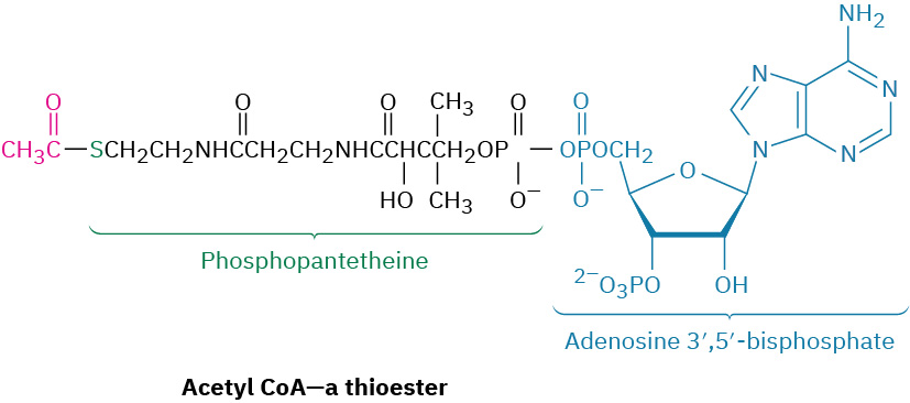
In the first catabolic stage, commonly called digestion, food is broken down in the mouth, stomach, and small intestine by hydrolysis of ester, acetal (glycoside), and amide (peptide) bonds to yield fatty acids, simple sugars, and amino acids. These smaller molecules are then absorbed and further degraded in the second stage of catabolism to yield acetyl groups attached by a thioester bond to the large carriermolecule, coenzyme A. The resultant compound, acetyl coenzyme A (acetyl CoA), is a key substance in the metabolism of food molecules and in many other biological pathways. As noted in Section 21.8, the acetyl group in acetyl CoA is linked to the sulfur atom of phosphopantetheine, which is itself linked to adenosine 3',5'-bisphosphate.
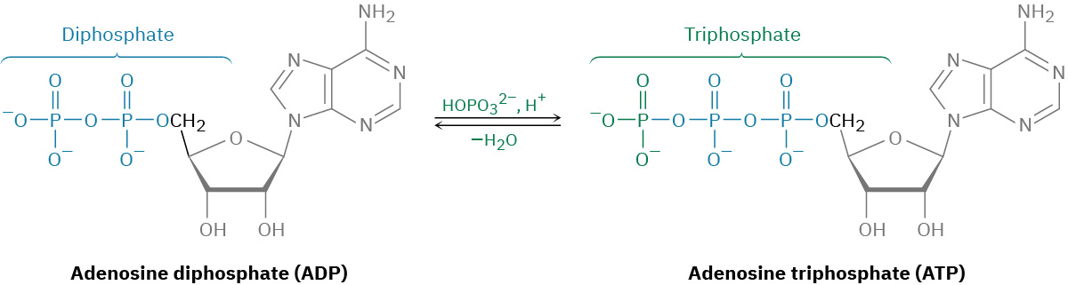
Acetyl groups are oxidized inside cellular mitochondria in the third stage of catabolism, the citric acid cycle, to yield CO2. (We’ll see the details of the process in Section 29.7.) Like most oxidations, this stage releases a large amount of energy, which is used in the fourth stage, the electron-transport chain, to accomplish the endergonicphosphorylation of adenosine diphosphate (ADP) with hydrogen phosphateion (HOPO32–, abbreviated Pi) to give adenosine triphosphate (ATP).
As the final result of food catabolism, ATP has been called the “energy currency” of the cell. Catabolic reactions “buy” ATP with the energy they release to synthesize it from ADP and hydrogen phosphateion. Anabolic reactions then spend the ATP by transferring a phosphate group to another molecule, thereby regenerating ADP. Energy production and use in living organisms thus revolves around the ATP ADP interconversion.
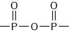
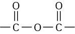
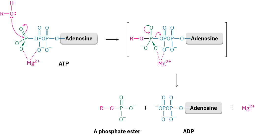
ADP and ATP are both phosphoric acid anhydrides, which contain linkages analogous to the linkage in carboxylic acid anhydrides. Just as carboxylic acid anhydrides react with alcohols by breaking a C–O bond and forming a carboxylic ester, ROCOR' (Section 21.5), phosphoric acid anhydrides react with alcohols by breaking a P–O bond and forming a phosphate ester, ROPO32–. The reaction is, in effect, a nucleophilic acyl substitution at phosphorus. Note that phosphorylationreactions with ATP generally require the presence of a divalent metal cation in the enzyme, usually Mg2+, to form a Lewis acid–base complex with the phosphate oxygen atoms and to neutralize negative charge.
How does the body use ATP? Recall from Section 6.7 that the free-energy change ΔG must be negative and energy must be released for a reaction to be energetically favorable and occur spontaneously. If ΔG is positive, the reaction is energetically unfavorable and the process can’t occur spontaneously.
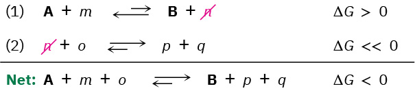
For an energetically unfavorable reaction to occur, it must be “coupled” to an energetically favorable reaction so that the overall free-energy change for the two reactions together is favorable. To understand what it means for reactions to be coupled, imagine that reaction 1 does not occur to any reasonable extent because it has a small equilibrium constant and is energetically unfavorable; that is, the reaction has ΔG > 0.
where A and B are the biochemically “important” substances while m and n are enzymecofactors, H2O, or other small molecules.
Imagine also that productn can react with substance o to yield p and q in a second, highly favorable reaction that has a large equilibrium constant and ΔG << 0.
Taking the two reactions together, they share, or are coupled through, the common intermediate n, which is a product in the first reaction and a reactant in the second. When even a tiny amount of n is formed in reaction 1, it undergoes essentially complete conversion in reaction 2, thereby removing it from the first equilibrium and forcing reaction 1 to continually replenish n until reactantA is gone. That is, the two reactions added together have a favorable ΔG < 0, and we say that the favorable reaction 2 “drives” the unfavorable reaction 1. Because the two reactions are coupled through n, the transformation of A to B becomes favorable.
It’s this ability to drive otherwise unfavorable phosphorylationreactions that makes ATP so useful. The resultant phosphates are much more reactive as leaving groups in nucleophilic substitutions and eliminations than the alcohols they’re derived from and are therefore more chemically useful.