Foundation 7 · 7A · Individual influences on behavior

Neural and influences on behavior

Sources ↗

receive inputs, the cell body integrates them, and an carries to terminals.

Relate , reflex circuits, spinal cord, central and peripheral systems, and neurotransmitters to behavior

receive inputs, the cell body integrates them, and an carries to terminals. At chemical , calcium entry promotes transmitter release; postsynaptic type determines whether the effect is excitatory, inhibitory, or modulatory. is commonly excitatory and inhibitory in the mature brain; acetylcholine, dopamine, serotonin, and norepinephrine have diverse circuit-dependent effects. The comprises brain and spinal cord; peripheral nerves connect it to the body. pathways serve skeletal muscle and sensation, while autonomic pathways regulate organs. A spinal reflex can begin before conscious awareness through sensory , interneurons, and motor .

Source: 3.2 Cells of the Nervous System · 3.3 Parts of the Nervous System · 3.4 The Brain and Spinal Cord · 3.5 The Endocrine System

Compare , , , cortical , and methods for studying brain function

The includes cortex, , , , and limbic-associated structures. circuits support orienting and contain important dopaminergic . structures include medulla for vital autonomic functions, pons for relay and sleep-related functions, and for coordination and motor learning. Frontal, parietal, temporal, and occipital cortices support overlapping functions; is relative specialization, not a division into logical and creative personality types. resolves rapid electrical changes, structural reveals anatomy, and estimates activity indirectly through blood oxygenation. can suggest necessity but may damage multiple systems; stimulation can test causal effects under controlled conditions.

Source: 3.2 Cells of the Nervous System · 3.3 Parts of the Nervous System · 3.4 The Brain and Spinal Cord · 3.5 The Endocrine System

Explain how structures and affect behavior

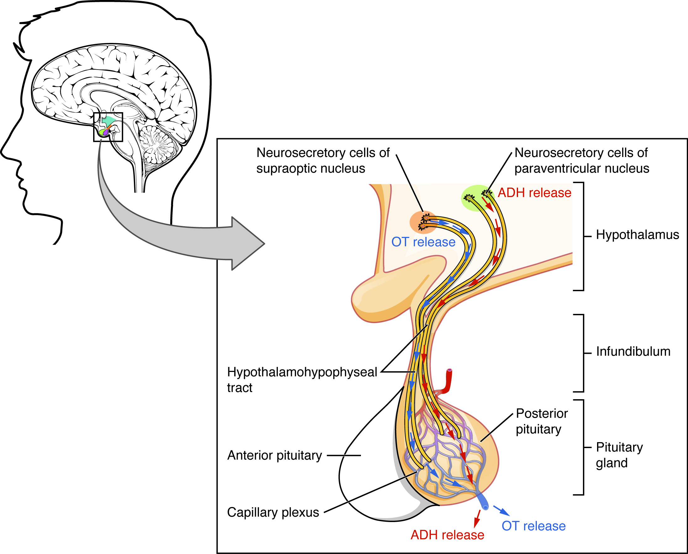
travel through blood and alter cells bearing suitable . The coordinates and autonomic responses; pituitary signals regulate other glands. Thyroid influence activity and development, adrenal support rapid arousal, supports longer stress , and gonadal influence reproductive development and motivation. participates in childbirth, milk ejection, and context-dependent social processes; it is not a universal trust chemical. Hormonal effects depend on dose, distribution, developmental timing, and social context. stabilizes many axes. A –behavior correlation may reflect behavior changing as well as influencing behavior.

Source: 3.2 Cells of the Nervous System · 3.3 Parts of the Nervous System · 3.4 The Brain and Spinal Cord · 3.5 The Endocrine System

Apply the ideas

Worked question. An study finds greater activity in one region during a memory task. Does this establish that the region is necessary for memory, and how could a stronger test be designed?

No. measures a blood-oxygenation correlate of neural activity and can reflect associated attention or effort. A selective or controlled perturbation paired with suitable comparison tasks can test necessity more directly, although network effects still matter. Compare memory accuracy and general task difficulty rather than interpreting any activation difference as a unique memory center.

Source: 3.2 Cells of the Nervous System · 3.3 Parts of the Nervous System · 3.4 The Brain and Spinal Cord · 3.5 The Endocrine System

3.2 Cells of the Nervous System — figures and tables
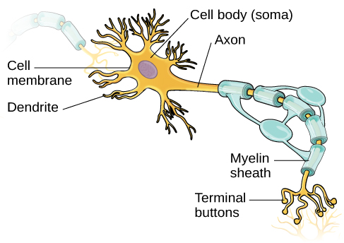
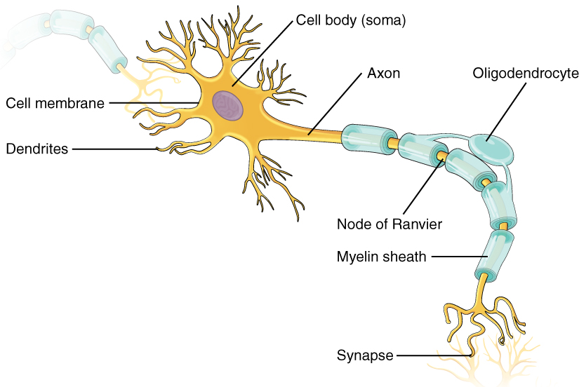
An illustration shows a neuron with labeled parts for the cell membrane, dendrite, cell body, axon, and terminal buttons. A myelin sheath covers part of the neuron.
Figure 3.8 — 3.2 Cells of the Nervous System

Figure 3.8 This illustration shows a prototypical neuron, which is being myelinated by a glial cell.

Rose M. Spielman, William J. Jenkins, and Marilyn D. Lovett, Psychology 2E, OpenStax. · CC BY-NC-SA 4.0 · Original image ↗ · Access for free ↗
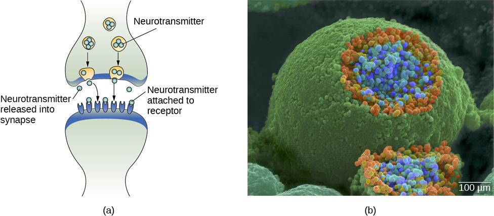
Image (a) shows the synaptic space between two neurons, with neurotransmitters being released into the synapse and attaching to receptors. Image (b) is a micrograph showing a spherical terminal button with part of the exterior removed, revealing a solid interior of small round parts.
Figure 3.9 — 3.2 Cells of the Nervous System

Figure 3.9 (a) The synaptic cleft is the space between the terminal button of one neuron and the dendrite of another neuron. (b) In this pseudo-colored image from a scanning electron microscope, a terminal button (green) has been opened to reveal the synaptic vesicles (orange and blue) inside. Each vesicle contains about 10,000 neurotransmitter molecules. (credit b: modification of work by Tina Carvalho, NIH-NIGMS; scale-bar data from Matt Russell)

Rose M. Spielman, William J. Jenkins, and Marilyn D. Lovett, Psychology 2E, OpenStax. (credit b: modification of work by Tina Carvalho, NIH-NIGMS; scale-bar data from Matt Russell) · CC BY-NC-SA 4.0 · Original image ↗ · Access for free ↗
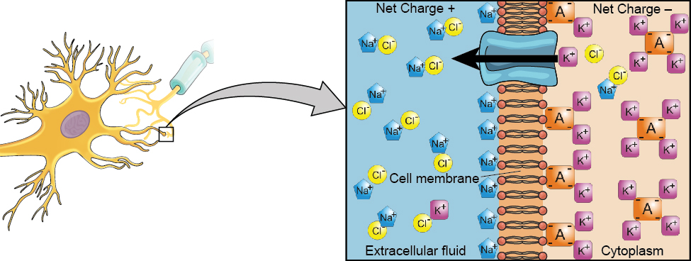
A close-up illustration depicts the difference in charges across the cell membrane, and shows how Na+ and K+ cells concentrate more closely near the membrane.
Figure 3.10 — 3.2 Cells of the Nervous System

Figure 3.10 At resting potential, Na + (blue pentagons) is more highly concentrated outside the cell in the extracellular fluid (shown in blue), whereas K + (purple squares) is more highly concentrated near the membrane in the cytoplasm or intracellular fluid. Other molecules, such as chloride ions (yellow circles) and negatively charged proteins (brown squares), help contribute to a positive net charge in the extracellular fluid and a negative net charge in the intracellular fluid.

Rose M. Spielman, William J. Jenkins, and Marilyn D. Lovett, Psychology 2E, OpenStax. · CC BY-NC-SA 4.0 · Original image ↗ · Access for free ↗
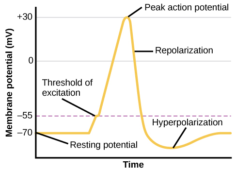
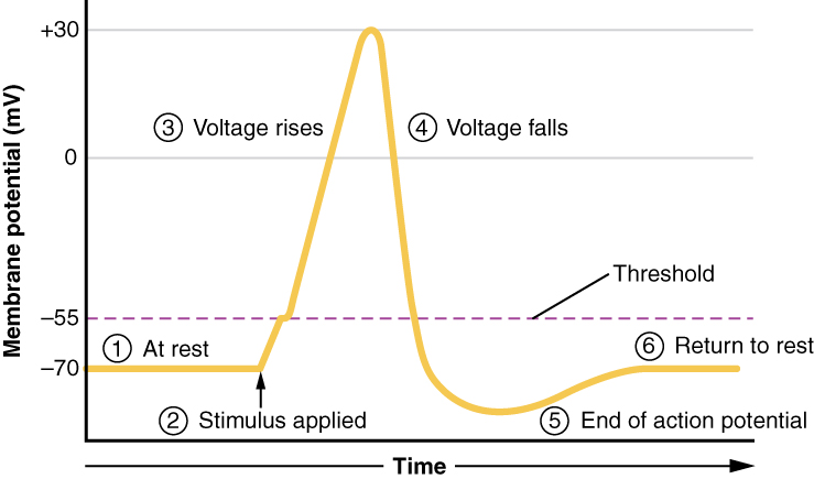
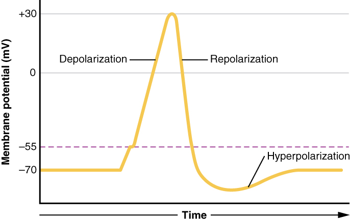
A graph shows the increase, peak, and decrease in membrane potential. The millivolts through the phases are approximately -70mV at resting potential, -55mV at threshold of excitation, 30mV at peak action potential, 5mV at repolarization, and -80mV at hyperpolarization.
Figure 3.11 — 3.2 Cells of the Nervous System

Figure 3.11 During the action potential, the electrical charge across the membrane changes dramatically.

Rose M. Spielman, William J. Jenkins, and Marilyn D. Lovett, Psychology 2E, OpenStax. · CC BY-NC-SA 4.0 · Original image ↗ · Access for free ↗
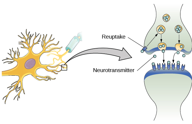
The synaptic space between two neurons is shown. Some neurotransmitters that have been released into the synapse are attaching to receptors while others undergo reuptake into the axon terminal.
Figure 3.12 — 3.2 Cells of the Nervous System

Figure 3.12 Reuptake involves moving a neurotransmitter from the synapse back into the axon terminal from which it was released.

Rose M. Spielman, William J. Jenkins, and Marilyn D. Lovett, Psychology 2E, OpenStax. · CC BY-NC-SA 4.0 · Original image ↗ · Access for free ↗
Neurotransmitter Involved in Potential Effect on Behavior
Acetylcholine Muscle action, memory Increased arousal, enhanced cognition
Beta-endorphin Pain, pleasure Decreased anxiety, decreased tension
Dopamine Mood, sleep, learning Increased pleasure, suppressed appetite
() Brain function, sleep Decreased anxiety, decreased tension
Memory, learning Increased learning, enhanced memory
Norepinephrine Heart, intestines, alertness Increased arousal, suppressed appetite
Serotonin Mood, sleep Modulated mood, suppressed appetite

Table 3.1. Rose M. Spielman, William J. Jenkins, and Marilyn D. Lovett, Psychology 2E, OpenStax.

Source: 3.2 Cells of the Nervous System

3.3 Parts of the Nervous System — figures and tables
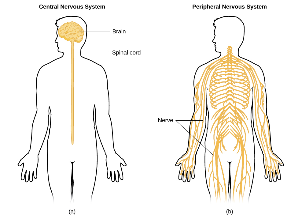
Image (a) shows an outline of a human body with the brain and spinal cord illustrated. Image (b) shows an outline of a human body with a network of nerves depicted.
Figure 3.13 — 3.3 Parts of the Nervous System

Figure 3.13 The nervous system is divided into two major parts: (a) the Central Nervous System and (b) the Peripheral Nervous System.

Rose M. Spielman, William J. Jenkins, and Marilyn D. Lovett, Psychology 2E, OpenStax. · CC BY-NC-SA 4.0 · Original image ↗ · Access for free ↗
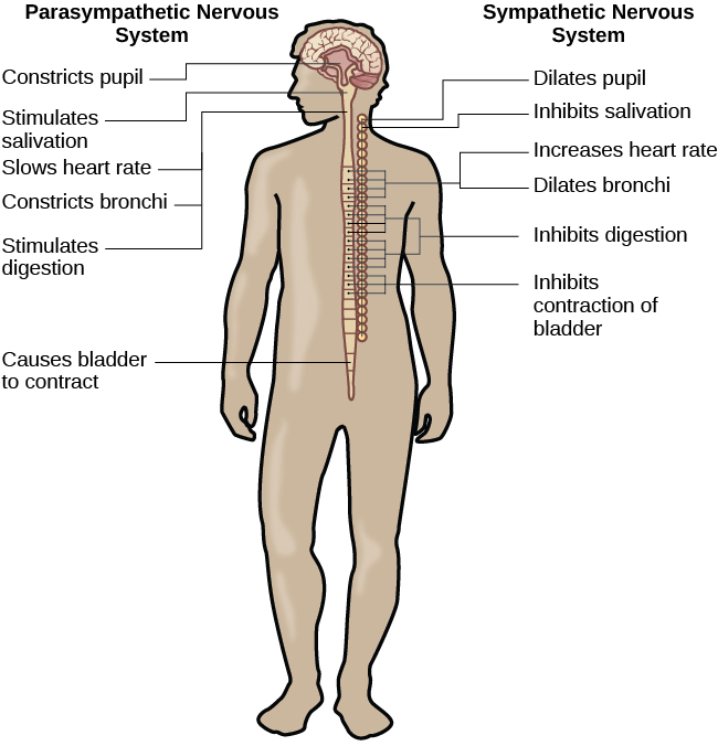
A diagram of a human body lists the different functions of the sympathetic and parasympathetic nervous system. The parasympathetic system can constrict pupils, stimulate salivation, slow heart rate, constrict bronchi, stimulate digestion, stimulate bile secretion, and cause the bladder to contract. The sympathetic nervous system can dilate pupils, inhibit salivation, increase heart rate, dilate bronchi, inhibit digestion, stimulate the breakdown of glycogen, stimulate secretion of adrenaline and noradrenaline, and inhibit contraction of the bladder.
Figure 3.14 — 3.3 Parts of the Nervous System

Figure 3.14 The sympathetic and parasympathetic divisions of the autonomic nervous system have the opposite effects on various systems.

Rose M. Spielman, William J. Jenkins, and Marilyn D. Lovett, Psychology 2E, OpenStax. · CC BY-NC-SA 4.0 · Original image ↗ · Access for free ↗

Source: 3.3 Parts of the Nervous System

3.4 The Brain and Spinal Cord — figures and tables
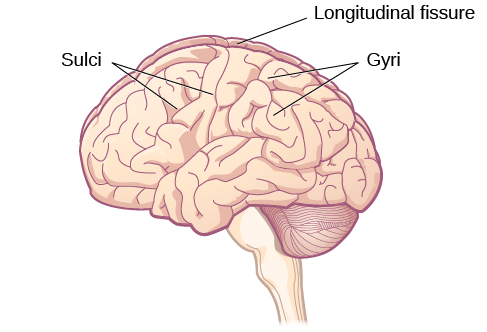
An illustration of the brain’s exterior surface shows the ridges and depressions, and the deep fissure that runs through the center.
Figure 3.15 — 3.4 The Brain and Spinal Cord

Figure 3.15 The surface of the brain is covered with gyri and sulci. A deep sulcus is called a fissure, such as the longitudinal fissure that divides the brain into left and right hemispheres. (credit: modification of work by Bruce Blaus)

Rose M. Spielman, William J. Jenkins, and Marilyn D. Lovett, Psychology 2E, OpenStax. (credit: modification of work by Bruce Blaus) · CC BY-NC-SA 4.0 · Original image ↗ · Access for free ↗
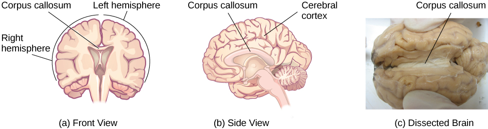
Illustrations (a) and (b) show the corpus callosum’s location in the brain in front and side views. Photograph (c) shows the corpus callosum in a dissected brain.
Figure 3.16 — 3.4 The Brain and Spinal Cord

Figure 3.16 (a, b) The corpus callosum connects the left and right hemispheres of the brain. (c) A scientist spreads this dissected sheep brain apart to show the corpus callosum between the hemispheres. (credit c: modification of work by Aaron Bornstein)

Rose M. Spielman, William J. Jenkins, and Marilyn D. Lovett, Psychology 2E, OpenStax. (credit c: modification of work by Aaron Bornstein) · CC BY-NC-SA 4.0 · Original image ↗ · Access for free ↗
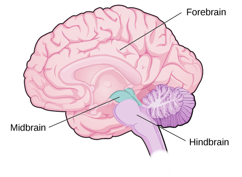
An illustration shows the position and size of the forebrain (the largest portion), midbrain (a small central portion), and hindbrain (a portion in the lower back part of the brain).
Figure 3.17 — 3.4 The Brain and Spinal Cord

Figure 3.17 The brain and its parts can be divided into three main categories: the forebrain, midbrain, and hindbrain.

Rose M. Spielman, William J. Jenkins, and Marilyn D. Lovett, Psychology 2E, OpenStax. · CC BY-NC-SA 4.0 · Original image ↗ · Access for free ↗
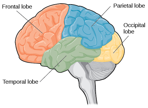
An illustration shows the four lobes of the brain.
Figure 3.18 — 3.4 The Brain and Spinal Cord

Figure 3.18 The lobes of the brain are shown.

Rose M. Spielman, William J. Jenkins, and Marilyn D. Lovett, Psychology 2E, OpenStax. · CC BY-NC-SA 4.0 · Original image ↗ · Access for free ↗
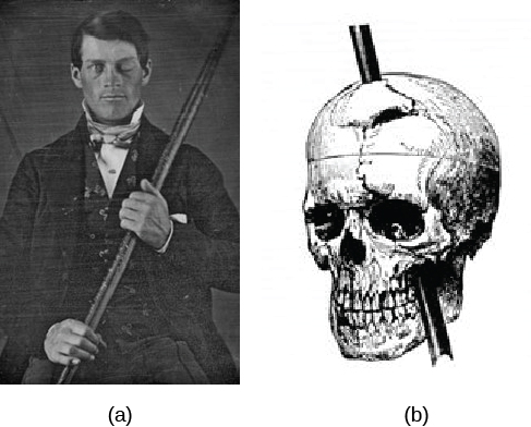
Image (a) is a photograph of Phineas Gage holding a metal rod. Image (b) is an illustration of a skull with a metal rod passing through it from the cheek area to the top of the skull.
Figure 3.19 — 3.4 The Brain and Spinal Cord

Figure 3.19 (a) Phineas Gage holds the iron rod that penetrated his skull in an 1848 railroad construction accident. (b) Gage’s prefrontal cortex was severely damaged in the left hemisphere. The rod entered Gage’s face on the left side, passed behind his eye, and exited through the top of his skull, before landing about 80 feet away. (credit a: modification of work by Jack and Beverly Wilgus)

Rose M. Spielman, William J. Jenkins, and Marilyn D. Lovett, Psychology 2E, OpenStax. (credit a: modification of work by Jack and Beverly Wilgus) · CC BY-NC-SA 4.0 · Original image ↗ · Access for free ↗
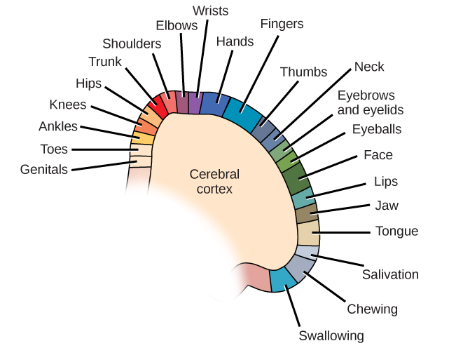
A diagram shows the organization in the somatosensory cortex, with functions for these parts in this proximal sequential order: genitals, toes, ankles, knees, hips, trunk, shoulders, elbows, wrists, hands, fingers, thumbs, neck, eyebrows and eyelids, eyeballs, face, lips, jaw, tongue, salivation, chewing, and swallowing.
Figure 3.20 — 3.4 The Brain and Spinal Cord

Figure 3.20 Spatial relationships in the body are mirrored in the organization of the somatosensory cortex.

Rose M. Spielman, William J. Jenkins, and Marilyn D. Lovett, Psychology 2E, OpenStax. · CC BY-NC-SA 4.0 · Original image ↗ · Access for free ↗
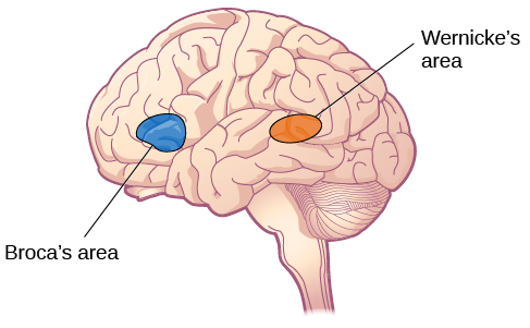
An illustration shows the locations of Broca’s and Wernicke’s areas.
Figure 3.21 — 3.4 The Brain and Spinal Cord

Figure 3.21 Damage to either Broca’s area or Wernicke’s area can result in language deficits. The types of deficits are very different, however, depending on which area is affected.

Rose M. Spielman, William J. Jenkins, and Marilyn D. Lovett, Psychology 2E, OpenStax. · CC BY-NC-SA 4.0 · Original image ↗ · Access for free ↗
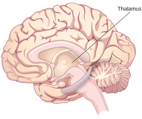
An illustration shows the location of the thalamus in the brain.
Figure 3.22 — 3.4 The Brain and Spinal Cord

Figure 3.22 The thalamus serves as the relay center of the brain where most senses are routed for processing.

Rose M. Spielman, William J. Jenkins, and Marilyn D. Lovett, Psychology 2E, OpenStax. · CC BY-NC-SA 4.0 · Original image ↗ · Access for free ↗
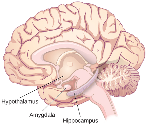
An illustration shows the locations of parts of the brain involved in the limbic system: the hypothalamus, amygdala, and hippocampus.
Figure 3.23 — 3.4 The Brain and Spinal Cord

Figure 3.23 The limbic system is involved in mediating emotional response and memory.

Rose M. Spielman, William J. Jenkins, and Marilyn D. Lovett, Psychology 2E, OpenStax. · CC BY-NC-SA 4.0 · Original image ↗ · Access for free ↗
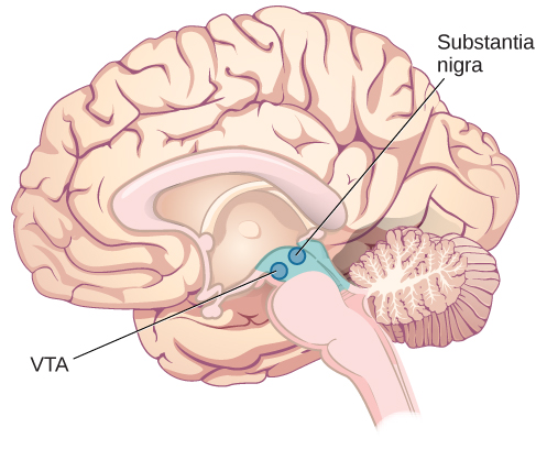
An illustration shows the location of the substantia nigra and VTA in the brain.
Figure 3.24 — 3.4 The Brain and Spinal Cord

Figure 3.24 The substantia nigra and ventral tegmental area (VTA) are located in the midbrain.

Rose M. Spielman, William J. Jenkins, and Marilyn D. Lovett, Psychology 2E, OpenStax. · CC BY-NC-SA 4.0 · Original image ↗ · Access for free ↗
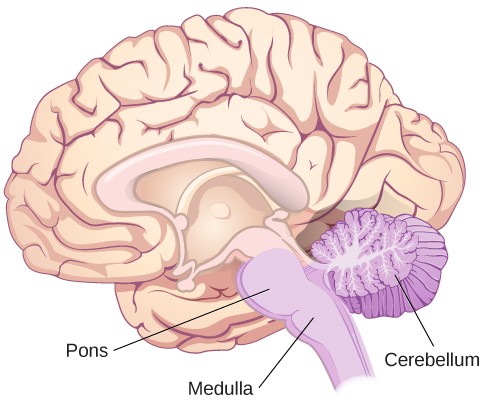
An illustration shows the location of the pons, medulla, and cerebellum.
Figure 3.25 — 3.4 The Brain and Spinal Cord

Figure 3.25 The pons, medulla, and cerebellum make up the hindbrain.

Rose M. Spielman, William J. Jenkins, and Marilyn D. Lovett, Psychology 2E, OpenStax. · CC BY-NC-SA 4.0 · Original image ↗ · Access for free ↗
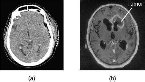
Image (a) shows a brain scan where the brain matter’s appearance is fairly uniform. Image (b) shows a section of the brain that looks different from the surrounding tissue and is labeled “tumor.”
Figure 3.26 — 3.4 The Brain and Spinal Cord

Figure 3.26 A CT scan can be used to show brain tumors. (a) The image on the left shows a healthy brain, whereas (b) the image on the right indicates a brain tumor in the left frontal lobe. (credit a: modification of work by "Aceofhearts1968"/Wikimedia Commons; credit b: modification of work by Roland Schmitt et al)

Rose M. Spielman, William J. Jenkins, and Marilyn D. Lovett, Psychology 2E, OpenStax. (credit a: modification of work by "Aceofhearts1968"/Wikimedia Commons; credit b: modification of work by Roland Schmitt et al) · CC BY-NC-SA 4.0 · Original image ↗ · Access for free ↗
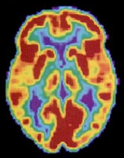
A brain scan shows different parts of the brain in different colors.
Figure 3.27 — 3.4 The Brain and Spinal Cord

Figure 3.27 A PET scan is helpful for showing activity in different parts of the brain. (credit: Health and Human Services Department, National Institutes of Health)

Rose M. Spielman, William J. Jenkins, and Marilyn D. Lovett, Psychology 2E, OpenStax. (credit: Health and Human Services Department, National Institutes of Health) · CC BY-NC-SA 4.0 · Original image ↗ · Access for free ↗
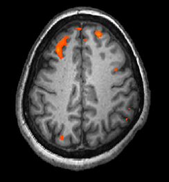
A brain scan shows brain tissue in gray with some small areas highlighted red.
Figure 3.28 — 3.4 The Brain and Spinal Cord

Figure 3.28 An fMRI shows activity in the brain over time. This image represents a single frame from an fMRI. (credit: modification of work by Kim J, Matthews NL, Park S.)

Rose M. Spielman, William J. Jenkins, and Marilyn D. Lovett, Psychology 2E, OpenStax. (credit: modification of work by Kim J, Matthews NL, Park S.) · CC BY-NC-SA 4.0 · Original image ↗ · Access for free ↗
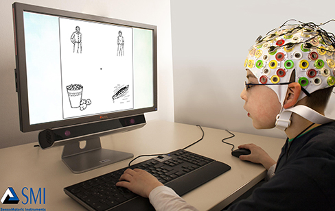
A photograph depicts a person looking at a computer screen and using the keyboard and mouse. The person wears a white cap covered in electrodes and wires.
Figure 3.29 — 3.4 The Brain and Spinal Cord

Figure 3.29 Using caps with electrodes, modern EEG research can study the precise timing of overall brain activities. (credit: SMI Eye Tracking)

Rose M. Spielman, William J. Jenkins, and Marilyn D. Lovett, Psychology 2E, OpenStax. (credit: SMI Eye Tracking) · CC BY-NC-SA 4.0 · Original image ↗ · Access for free ↗

Source: 3.4 The Brain and Spinal Cord

3.5 The System — figures and tables

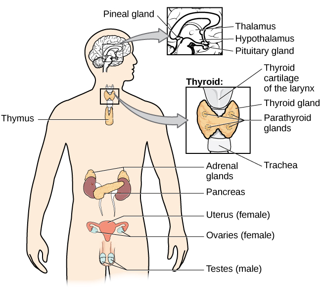
A diagram of the human body illustrates the locations of the thymus, several parts within the brain (pineal gland, thalamus, hypothalamus,  pituitary gland), several parts within the thyroid (cartilage of the larynx, thyroid gland, parathyroid glands, trachea), the adrenal glands, pancreas, uterus (female), ovaries (female), and testes (male).
Figure 3.30 — 3.5 The System

Figure 3.30 The major glands of the endocrine system are shown.

Rose M. Spielman, William J. Jenkins, and Marilyn D. Lovett, Psychology 2E, OpenStax. · CC BY-NC-SA 4.0 · Original image ↗ · Access for free ↗
Gland Associated Function
Pituitary , releasing and inhibiting (such as thyroid stimulating ) Regulate growth, regulate release
Thyroid Thyroxine, triiodothyronine Regulate and appetite
Pineal Regulate some biological rhythms such as sleep cycles
Adrenal Epinephrine, norepinephrine Stress response, increase activities
, Regulate blood sugar levels
Ovaries Estrogen, Mediate sexual motivation and behavior, reproduction
Testes Androgens, such as testosterone Mediate sexual motivation and behavior, reproduction

Table 3.2. Rose M. Spielman, William J. Jenkins, and Marilyn D. Lovett, Psychology 2E, OpenStax.

Source: 3.5 The Endocrine System

254 terms, alphabetically ordered

3′ end · ·

The end of a nucleic- strand associated with the sugar’s 3′ position. extends a strand at a free .

Read explanation · OpenStax ↗
3′ hydroxyl group · · ·

The at the sugar’s 3′ carbon that attacks the incoming ’s during strand extension.

Read explanation · OpenStax ↗
A site · ·

The site that accepts most incoming during elongation and at termination.

Read explanation · OpenStax ↗
Acid · ·

In the Brønsted–Lowry model, a substance that can donate a . Acidic have a higher activity than water.

Read explanation · OpenStax ↗
action potential

A regenerative -voltage change produced by voltage-dependent -channel activity.

Read explanation · OpenStax ↗
Activation energy ·

The energy barrier that must overcome to reach a and undergo .

Read explanation · OpenStax ↗
Active site

The region of an that binds its and carries out .

Read explanation · OpenStax ↗
adaptation

A heritable feature shaped by selection because it improves reproductive success in a relevant environment.

Read explanation · OpenStax ↗
Adenosine triphosphate

An energy-carrying ; its powers many cellular processes, including cycles.

Read explanation · OpenStax ↗
Allele

One version or copy of a at a ; a typical cell carries two copies of an autosomal .

Read explanation · NCBI Bookshelf / Sinauer Associates ↗
Alpha carbon · ·

The central carbon next to the carboxyl carbon. In a standard it binds an , hydrogen, and .

Read explanation · OpenStax ↗
Alpha helix · · · · · ·

A helical stabilized by between a oxygen and an hydrogen four later.

Read explanation · OpenStax ↗
Amide · · ·

A group containing a carbon bonded to nitrogen. are amide linkages.

Read explanation · OpenStax ↗
Amino acid

A building block containing amino and and a characteristic .

Read explanation · OpenStax ↗
Amino acid residue · ·

The part of an incorporated into a chain. Residue can also describe another building block within a larger .

Read explanation · OpenStax ↗
Amino group · · ·

A nitrogen-containing . A free ’s amino group is typically (NH₃⁺) near physiological ; ’s ring nitrogen is an exception to this formula.

Read explanation · OpenStax ↗
Aminoacyl-tRNA · ·

A covalently linked to an at its , ready to deliver that during .

Read explanation · OpenStax ↗
Amphipathic

Having both a region and a region, as in a or .

Read explanation · OpenStax ↗
amplitude

Maximum displacement from an oscillation’s position.

Read explanation · OpenStax ↗
amygdala

Group of temporal-lobe involved in learning and responding to biologically significant stimuli.

Read explanation · OpenStax ↗
Amylose

The largely unbranched component of , made mainly of linked by alpha-1,4 .

Read explanation · OpenStax ↗
Analytical standard ·

A material with a known identity or amount used to compare or calibrate measurements.

Read explanation · Agilent ↗
Anode

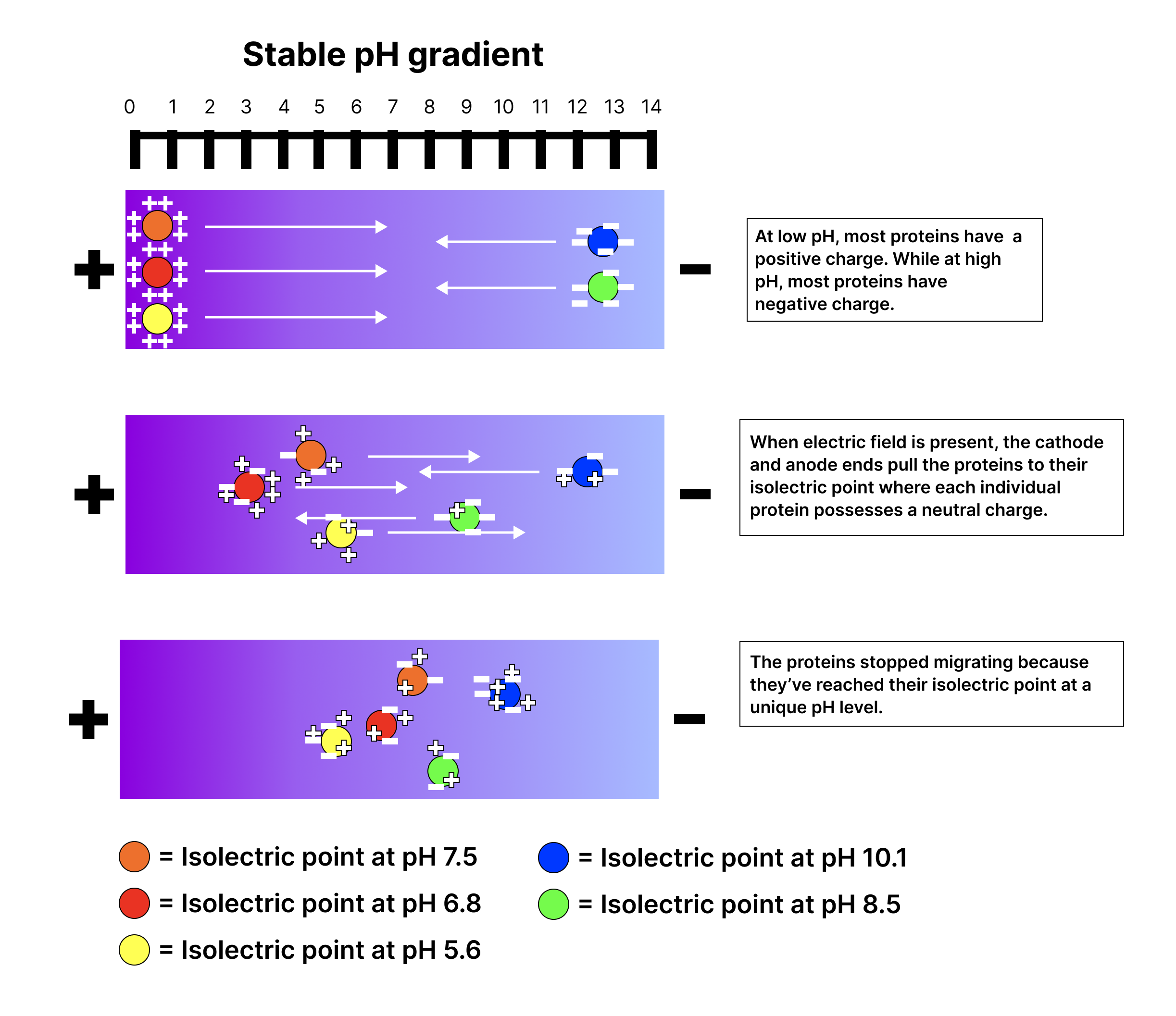
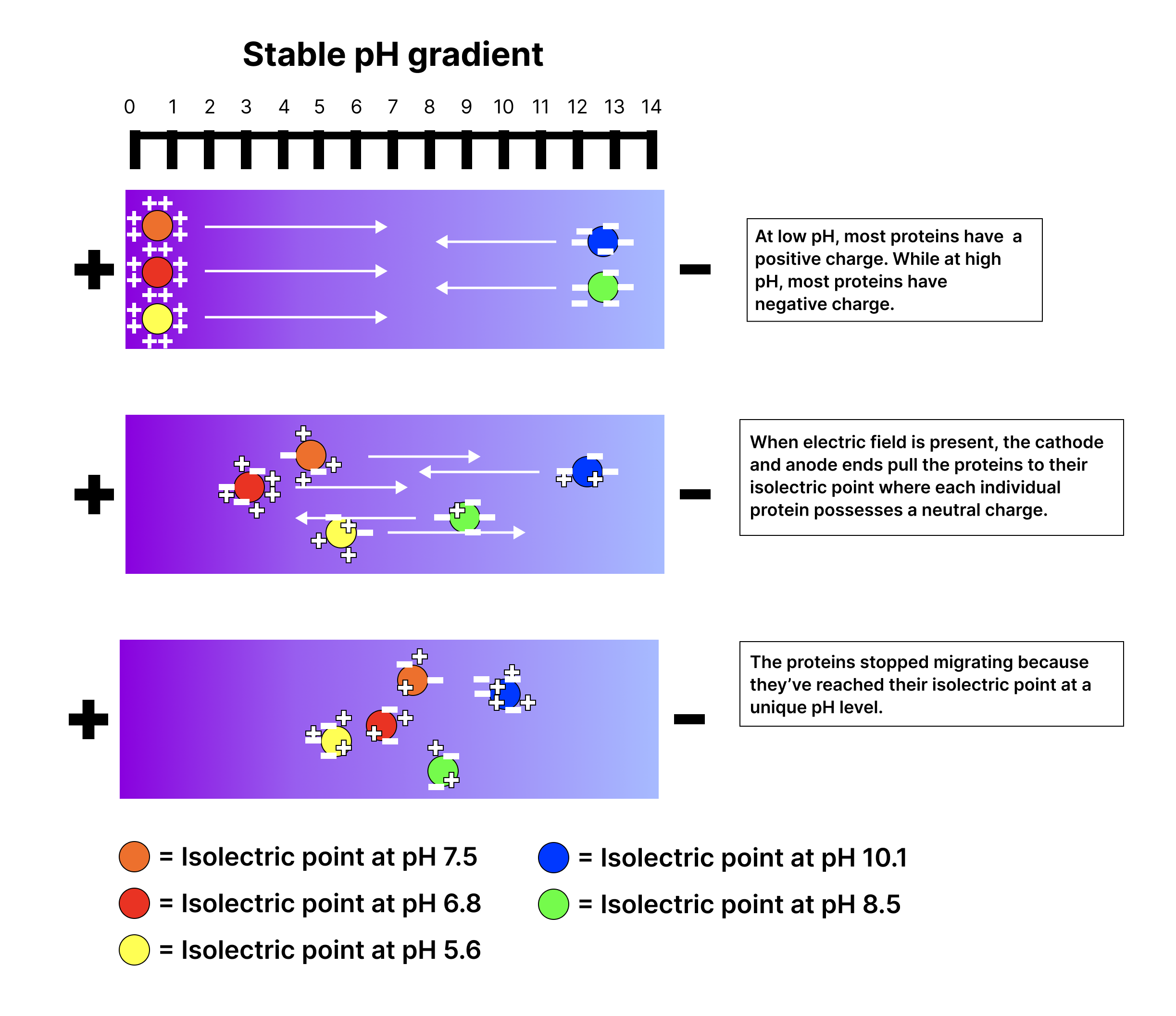
The positive in an apparatus; negatively migrate toward it.

Read explanation · Thermo Fisher ↗
anomeric carbon

The former carbon of a sugar, which becomes the or hemiketal center upon cyclization.

Read explanation · OpenStax ↗
Anticodon

A triplet that pairs to an . The anticodon for 5′-AUG-3′ is aligned 3′-UAC-5′, or written 5′-CAU-3′.

Read explanation · NCBI Bookshelf / Garland Science ↗
Antiparallel

Running in opposite directions, as with two strands or neighboring strands in some .

Read explanation · OpenStax ↗
Aqueous solution ·

A in which water is the .

Read explanation · OpenStax ↗
Aromatic

Containing a cyclic, with . , , and have aromatic .

Read explanation · OpenStax ↗
Atom

The smallest unit of an element that retains its chemical identity; it has a surrounded by .

Read explanation · OpenStax ↗
attachment

Enduring emotional bond and expectations about another’s availability and responsiveness.

Read explanation · OpenStax ↗
autonomic nervous system

Neural pathways regulating visceral organs through sympathetic, parasympathetic, and enteric systems.

Read explanation · OpenStax ↗
axon

Neuronal process that usually conducts toward terminals.

Read explanation · OpenStax ↗
basal ganglia

Interconnected subcortical involved in action selection and learning.

Read explanation · OpenStax ↗
Benzene ring

A six-carbon ring with .

Read explanation · OpenStax ↗
Benzyl group

A attached through a –CH₂– group; part of ’s .

Read explanation · OpenStax ↗
Beta sheet · · · · · ·

A made of aligned segments connected by ; strands may be parallel or .

Read explanation · OpenStax ↗
Biomolecule

A made by or involved in living systems, including , , , and .

Read explanation · OpenStax ↗
Biosynthesis

Formation of biological through cellular , such as synthesis of .

Read explanation · OpenStax ↗
Blotting ·

Transfer of separated or to a so a specific target can be detected.

Read explanation · Thermo Fisher ↗
Brønsted–Lowry base · · · · · · · · · · ·

In the Brønsted–Lowry model, a substance that can accept a . Basic solutions have lower activity than water.

Read explanation · OpenStax ↗
Calibration

Establishing the relation between a measured signal and known values or .

Read explanation · Agilent ↗
Carbohydrate

A class of including sugars and their , used for energy storage, structure, and recognition.

Read explanation · OpenStax ↗
Carbonyl group · ·

A carbon double-bonded to oxygen, C=O; present in and many other .

Read explanation · OpenStax ↗
Carboxyl group · ·

An , COOH. In free it is usually to COO⁻ near physiological .

Read explanation · OpenStax ↗
Catalyst · ·

A substance that speeds a by providing a pathway with lower and is regenerated rather than consumed overall.

Read explanation · OpenStax ↗
catecholamine

A -derived chemical class including dopamine, norepinephrine, and epinephrine.

Read explanation · OpenStax ↗
Cathode

The negative in an apparatus; positively migrate toward it.

Read explanation · Thermo Fisher ↗
Cellulose

A structural in plant cell walls, with beta-1,4 linkages; also used as a support.

Read explanation · OpenStax ↗
central nervous system

Brain and spinal cord.

Read explanation · OpenStax ↗
cerebellum

structure important for coordination, timing, and motor learning.

Read explanation · OpenStax ↗
Chemical compound ·

A substance containing of two or more different chemically combined in a definite composition.

Read explanation · OpenStax ↗
Chemical element

A substance defined by having the same number of , such as carbon or oxygen.

Read explanation · OpenStax ↗
Chemical reaction ·

A process that rearranges and chemical bonds, converting into .

Read explanation · OpenStax ↗
cholesterol

An animal that modulates packing and serves as a precursor for and bile .

Read explanation · OpenStax ↗
Chromatin

The complex of and associated , including , in .

Read explanation · OpenStax ↗
Chromatographic peak · ·

A rise and fall in detector signal as one or more components pass through the detector; a peak does not guarantee a single pure .

Read explanation · Agilent ↗
Chromatography

Separation of mixture components by their different interactions with a and a moving .

Read explanation · Waters ↗
Chromosome ·

An organized with associated that carries . A replicated chromosome has two .

Read explanation · OpenStax ↗
Codon

A sequence of three that specifies an or a stop signal.

Read explanation · OpenStax ↗
Collagen

A major structural in , with a characteristic triple-helical arrangement.

Read explanation · OpenStax ↗
competence

A cell’s capacity to take up extracellular in transformation.

Read explanation · OpenStax ↗
Concentration

The amount of a substance in a specified volume or mass of mixture.

Read explanation · OpenStax ↗
conduction

Thermal energy transfer through microscopic interactions without bulk matter flow.

Read explanation · OpenStax ↗
conductor

Material with that can move readily through it.

Read explanation · OpenStax ↗
Conjugated system ·

Adjacent orbitals that allow to be distributed across several , often through alternating single and multiple bonds.

Read explanation · OpenStax ↗
connective tissue

A tissue category generally characterized by cells distributed within substantial .

Read explanation · OpenStax ↗
Copy number ·

The number of copies of a segment in a defined sample or ; it is distinct from or abundance.

Read explanation · NHGRI / NIH ↗
corpus callosum

Large bundle connecting the cerebral hemispheres.

Read explanation · OpenStax ↗
cortisol

An adrenal-cortex glucocorticoid regulating , stress responses, and immune activity.

Read explanation · OpenStax ↗
Covalent bond · ·

A bond in which share pairs. and are covalent.

Read explanation · OpenStax ↗
CSF

Cerebrospinal fluid surrounding and circulating within the .

Read explanation · OpenStax ↗
Cysteine ·

An with a . Two cysteines can form a .

Read explanation · OpenStax ↗
Cytoplasm

Cell contents inside the plasma and outside the , where one is present.

Read explanation · OpenStax ↗
dendrite

Branching neuronal process receiving many synaptic inputs.

Read explanation · OpenStax ↗
Deoxyribonucleic acid

The that stores genetic information used to specify and sequences.

Read explanation · OpenStax ↗
depolarization

A shift in voltage toward a less negative value.

Read explanation · OpenStax ↗
Deprotonation

Removal of a from a or group; this changes its by minus one.

Read explanation · OpenStax ↗
Detergent

An substance that can associate with both water and material; detergents can disrupt or interactions.

Read explanation · Thermo Fisher ↗
Diploid

Having two sets; comparisons of must account for and sample composition.

Read explanation · NHGRI / NIH ↗
Disulfide bond · · ·

A covalent sulfur–sulfur bond formed by of two , which can stabilize structure.

Read explanation · NCBI Bookshelf ↗
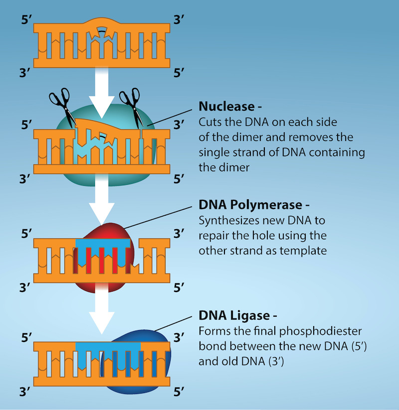
DNA lesion ·

of molecular damage in . A lesion can cause a if it is copied or repaired incorrectly, but damage and sequence change are distinct.

Read explanation · OpenStax ↗
DNA polymerase ·

An that synthesizes 5′ to 3′ using a template and a with a free .

Read explanation · OpenStax ↗
DNA replication

Copying so genetic information can be passed to daughter cells. Each guides a complementary new strand.

Read explanation · OpenStax ↗
dominant

Describing an whose specified is expressed in a ; dominance does not imply greater frequency or .

Read explanation · OpenStax ↗
Electric charge · · ·

An electrical property that can be positive or negative. Opposite charges attract and like charges repel; net charge is the sum of all charges on a particle.

Read explanation · OpenStax ↗
Electric field

A quantity describing the electrical force per unit positive . An applied field drives through a gel during .

Read explanation · Thermo Fisher ↗
Electrode

A that connects an electrical circuit to a or other medium; the and are electrodes.

Read explanation · Thermo Fisher ↗
electroencephalography

Recording of electrical activity from , commonly on the scalp.

Read explanation · OpenStax ↗
Electron

A negatively subatomic particle. Electron sharing and transfer determine much of chemical bonding.

Read explanation · OpenStax ↗
Electron delocalization ·

Distribution of across several rather than confinement to one or bond.

Read explanation · OpenStax ↗
Electronegativity ·

An ’s tendency to attract shared in a chemical bond. Unequal attraction can make a bond .

Read explanation · OpenStax ↗
Electrophoresis

Movement of in an , used to separate through a support such as a gel.

Read explanation · Thermo Fisher ↗
Endocrine signaling ·

Communication in which enter the circulation and act on distant target cells.

Read explanation · OpenStax ↗
Endomembrane system

The interacting and compartments involved in processes such as modification, transport, and .

Read explanation · OpenStax ↗
endometrium

The uterine lining that cycles hormonally and supports implantation.

Read explanation · OpenStax ↗
Endoplasmic reticulum

An interconnected network continuous with the ; it includes regions involved in processing and synthesis.

Read explanation · OpenStax ↗
Enzyme

A biological that speeds a without being consumed. Most enzymes are .

Read explanation · OpenStax ↗
Enzyme inhibitor · ·

A substance that reduces activity. The binding mechanism and rate data determine whether inhibition is competitive, uncompetitive, mixed, or another type.

Read explanation · Biology LibreTexts ↗
Equilibrium

A state where opposing processes occur at equal rates, so macroscopic or other properties remain constant.

Read explanation · OpenStax ↗
Eukaryote ·

An organism whose cells have a -enclosed ; animals, plants, fungi, and protists are eukaryotes.

Read explanation · OpenStax ↗
Experimental control ·

A comparison sample or condition used to distinguish the effect being tested from other influences.

Read explanation · Thermo Fisher ↗
extracellular matrix

The network of and surrounding cells and providing structural and signaling functions.

Read explanation · OpenStax ↗
FACT complex ·

A -associated complex that helps machinery traverse and supports reorganization.

Read explanation · OpenStax ↗
fitness

Relative reproductive contribution of a or organism in a particular environment.

Read explanation · OpenStax ↗
forebrain

Major developmental brain division including cerebral hemispheres, , and .

Read explanation · OpenStax ↗
Fraction · ·

A portion of a sample collected during a separation, such as the fluid leaving a column during a time interval.

Read explanation · Waters ↗
Functional group

A group of whose chemical behavior gives a characteristic and properties.

Read explanation · OpenStax ↗
functional magnetic resonance imaging

Imaging method estimating neural activity indirectly through blood-oxygenation changes.

Read explanation · OpenStax ↗
gamete

A reproductive cell that contributes a during fertilization.

Read explanation · OpenStax ↗
gamma-aminobutyric acid

Major inhibitory neurotransmitter in the mature .

Read explanation · OpenStax ↗
gap junction

A junction containing intercellular channels for and small .

Read explanation · OpenStax ↗
Gene

A sequence that contributes to a functional or , together with the information needed to express it.

Read explanation · OpenStax ↗
Gene expression

Production of a functional from a , including and, for -coding , .

Read explanation · NCBI Bookshelf ↗
Genome

The complete genetic material of an organism or cell.

Read explanation · OpenStax ↗
genotype

The allelic composition of an organism at one or more specified .

Read explanation · OpenStax ↗
Germline · · ·

The cell lineage that produces . A germline mutation can be inherited if the altered contributes to an offspring.

Read explanation · OpenStax ↗
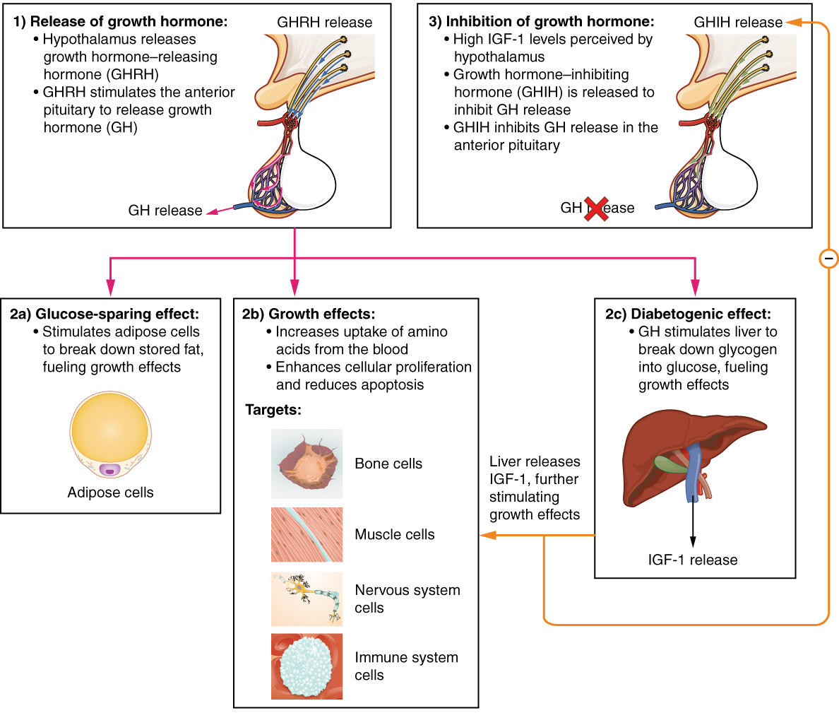
GH

Growth hormone, an anterior pituitary affecting growth and directly and through .

Read explanation · OpenStax ↗
Glucagon

A pancreatic that helps raise , especially by promoting release from the liver.

Read explanation · OpenStax ↗
Glucose

A six-carbon sugar that is a major cellular energy source and a building block of , , and .

Read explanation · OpenStax ↗
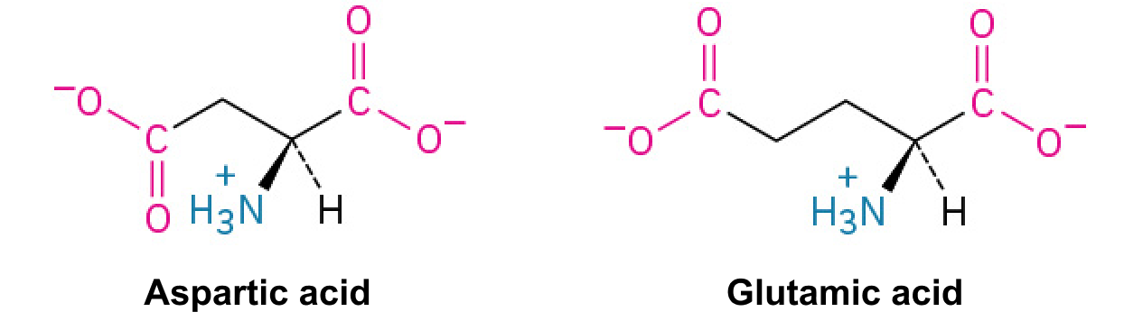
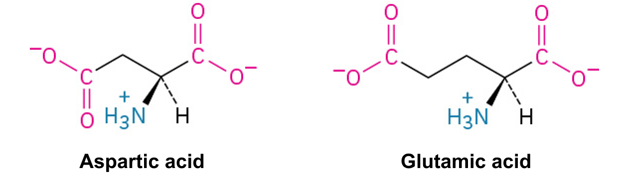
Glutamate · ·

An whose is usually negatively near physiological .

Read explanation · OpenStax ↗
glycogen

A highly branched used for storage in animals.

Read explanation · Biology LibreTexts / Oregon State University ↗
Glycosidic bond

A involving a sugar’s . In a , an N-glycosidic bond links the sugar to the .

Read explanation · Biology LibreTexts / Oregon State University ↗
Golgi apparatus ·

A stack of compartments that modifies and sorts and traveling through the .

Read explanation · OpenStax ↗
Haploid

Having one set of , as in a human .

Read explanation · OpenStax ↗
hemiacetal

Carbon bonded to both and alkoxy groups, usually formed by alcohol addition to a .

Read explanation · OpenStax ↗
heterozygote ·

An individual with two different at a specified .

Read explanation · OpenStax ↗
hindbrain

Developmental brain division including medulla, pons, and .

Read explanation · OpenStax ↗
hippocampus

Medial temporal structure important for forming episodic memories and representing context and space.

Read explanation · OpenStax ↗
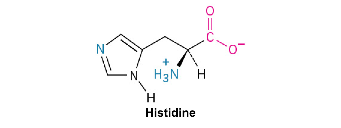
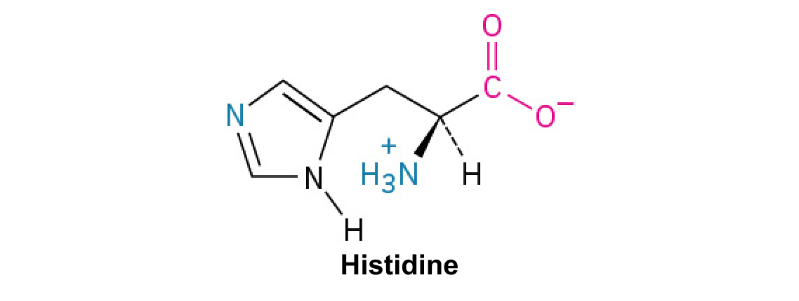
Histidine ·

A with an . Mostly uncharged near physiological , it can accept or donate .

Read explanation · OpenStax ↗
Histone

A -associated rich in . Core histones form the around which nucleosomal wraps.

Read explanation · OpenStax ↗
homeostasis

Dynamic regulation that maintains physiological variables within functional ranges despite disturbances.

Read explanation · OpenStax ↗
Hormone

A chemical messenger released by cells that changes the activity of target cells with matching .

Read explanation · OpenStax ↗
Hydrogen bond ·

An attraction between a hydrogen bonded to an and another . hydrogen bonds stabilize .

Read explanation · OpenStax ↗
Hydrogen ion · ·

A positively hydrogen species. H⁺ denotes a , which is hydrated in water; its activity determines .

Read explanation · OpenStax ↗
Hydrolysis

Breaking a chemical bond by adding water. hydrolysis separates .

Read explanation · OpenStax ↗
Hydrophilic

Interacting favorably with water, typically because a group is or .

Read explanation · OpenStax ↗
Hydrophobic

Interacting poorly with water. often gather in a ’s interior.

Read explanation · OpenStax ↗
Hydroxide ion ·

The negatively OH⁻. Basic contain a higher hydroxide ion activity than water at the same temperature.

Read explanation · OpenStax ↗
Hydroxyl group ·

An oxygen bonded to hydrogen, –OH. Hydroxyl groups can and often increase .

Read explanation · OpenStax ↗
hypothalamus

region coordinating homeostatic, , and autonomic functions.

Read explanation · OpenStax ↗
IGF-1

Insulin-like growth factor 1, a growth-promoting signal whose production is stimulated by .

Read explanation · OpenStax ↗
Imidazole

A five-membered ring containing two nitrogens; ’s contains this ring.

Read explanation · OpenStax ↗
Indole

A fused six-membered and five-membered ring system containing nitrogen; found in .

Read explanation · OpenStax ↗
insulator

Material in which cannot move freely over macroscopic distances.

Read explanation · OpenStax ↗
Insulin

A from that lowers by promoting uptake and storage and reducing production.

Read explanation · OpenStax ↗
Ion · ·

An or with a net because its numbers of and do not balance.

Read explanation · OpenStax ↗
Isotope ·

One of several forms of an element with the same number of but different numbers of neutrons.

Read explanation · OpenStax ↗
lateralization

Relative specialization of the two cerebral hemispheres for particular functions.

Read explanation · OpenStax ↗
linkage

A tendency of nearby on the same to be inherited together.

Read explanation · OpenStax ↗
Lipid

A broad class of largely or , including fats, , and steroids.

Read explanation · OpenStax ↗
Lock-and-key model

A simplified model of specificity in which an ’s is already complementary to the before binding.

Read explanation · OpenStax ↗
locus

The specific position of a or other feature.

Read explanation · OpenStax ↗
Logarithm · · ·

The exponent needed to obtain a number from a chosen fixed number. For decimal logarithms, log₁₀(100) = 2 because 10² = 100; uses the negative decimal logarithm of activity.

Read explanation · OpenStax ↗
lysergic acid diethylamide

Classic hallucinogen whose perceptual effects involve serotonergic signaling.

Read explanation · OpenStax ↗
magnetic field

Vector field that exerts forces on moving and magnetic moments.

Read explanation · OpenStax ↗
magnetic resonance imaging

Imaging method using magnetic fields and radiofrequency signals to reveal tissue structure.

Read explanation · OpenStax ↗
Mass spectrometry ·

Analysis of according to their mass-to- ratios, used to identify and or .

Read explanation · Thermo Fisher ↗
mean arterial pressure

The time-averaged pressure driving systemic circulation over a cardiac cycle.

Read explanation · OpenStax ↗
melatonin

A pineal signaling circadian timing, normally associated with biological night.

Read explanation · OpenStax ↗
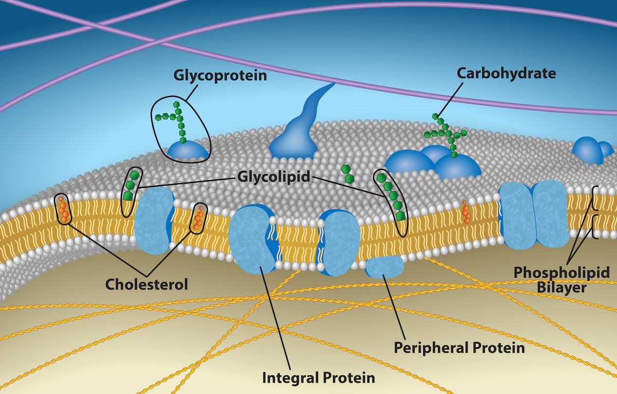
Membrane · · · ·

A thin boundary or sheet. Cell membranes are bilayers containing ; membranes are synthetic supports that hold or for detection.

Read explanation · OpenStax ↗
Messenger RNA ·

An that carries coding information used by to synthesize a .

Read explanation · OpenStax ↗
Metabolism

The chemical that build , break them down, and transform energy in a living system.

Read explanation · OpenStax ↗
midbrain

Brainstem division containing orienting circuits and important motor and neuromodulatory .

Read explanation · OpenStax ↗
Mobile phase

The moving fluid that carries a sample through a system.

Read explanation · Waters ↗
Molecular chaperone · ·

A that assists other in folding or prevents , without specifying their sequence.

Read explanation · NCBI Bookshelf ↗
Molecule

Two or more joined by . A molecule may be electrically or carry a .

Read explanation · OpenStax ↗
Mutation

A change in a sequence; some mutations alter a ’s sequence.

Read explanation · OpenStax ↗
Myelin

A -rich insulating sheath around many nerve axons that supports efficient signal .

Read explanation · NHGRI / NIH ↗
negative feedback

A control arrangement in which a response opposes the initiating deviation.

Read explanation · OpenStax ↗
neuron

An electrically excitable cell specialized for receiving and transmitting information.

Read explanation · OpenStax ↗
Neutral

Having no net . For a , neutral instead means equal and activities; its is about 7 at 25 °C.

Read explanation · OpenStax ↗
Nitrogenous base · · ·

A nitrogen-containing ring component of ; its pairing interactions help specify sequences.

Read explanation · OpenStax ↗
Nonpolar ·

Lacking a substantial separation of . Nonpolar groups generally interact poorly with water.

Read explanation · OpenStax ↗
Nuclear envelope

The double enclosing the and continuous with the .

Read explanation · OpenStax ↗
Nucleic acid

A of , such as or , used in information storage, expression, and other cellular functions.

Read explanation · OpenStax ↗
Nucleosome

The basic unit of : wrapped around a .

Read explanation · OpenStax ↗
Nucleotide

A joined to a sugar and one or more .

Read explanation · OpenStax ↗
Nucleus ·

A -enclosed compartment containing most of a cell’s .

Read explanation · OpenStax ↗
Octamer

A complex made of eight . A core contains eight .

Read explanation · OpenStax ↗
Oxidation ·

Loss of or an increase in oxidation state. Oxidizing two can form a .

Read explanation · OpenStax ↗
oxytocin

A hypothalamic released by the posterior pituitary, promoting uterine contraction and milk ejection.

Read explanation · OpenStax ↗
Pancreas

An organ with digestive and functions; its islets contain cells that secrete and .

Read explanation · OpenStax ↗
Pancreatic beta cell ·

An cell in a pancreatic islet that secretes .

Read explanation · OpenStax ↗
Peptide

A chain of joined by ; the term commonly describes chains shorter than a typical .

Read explanation · OpenStax ↗
Peptide bond ·

A covalent between the carboxyl carbon of one and the amino nitrogen of the next.

Read explanation · OpenStax ↗
Peptide hormone

An amino--based chemical messenger released by ; and are examples.

Read explanation · OpenStax ↗
peripheral nervous system

Nerves and ganglia outside the brain and spinal cord.

Read explanation · OpenStax ↗
pH

A measure of , defined as the negative of activity. Lower pH means greater ; pH influences .

Read explanation · OpenStax ↗
phenotype

A measurable trait produced by interacting with development and environment.

Read explanation · OpenStax ↗
Phenylalanine ·

A with an .

Read explanation · OpenStax ↗
Phosphate group ·

A phosphorus-containing group bonded to oxygens; phosphate groups occur in , , and modified .

Read explanation · OpenStax ↗
Phospholipid

An with a -containing region and tails; a major component of cellular .

Read explanation · OpenStax ↗
ploidy

The number of complete sets in a cell.

Read explanation · OpenStax ↗
Polar

Having an uneven distribution of . Polar groups often interact favorably with water.

Read explanation · OpenStax ↗
Polymer

A large made from many linked molecular building blocks.

Read explanation · OpenStax ↗
Polypeptide

A chain of connected by . A functional can contain one or several such chains.

Read explanation · OpenStax ↗
Porous material · · · ·

A material containing small spaces that can admit of suitable size.

Read explanation · Cytiva ↗
Primer · ·

A short nucleic- strand paired with a template that provides a free for polymerase extension.

Read explanation · OpenStax ↗
procedural memory

Memory expressed through learned skills and procedures.

Read explanation · OpenStax ↗
progesterone

A supporting secretory and pregnancy-associated tissue states.

Read explanation · OpenStax ↗
Proline ·

An whose loops back to the nitrogen, restricting rotation and often creating bends.

Read explanation · OpenStax ↗
Protein

One or more folded chains that perform biological roles such as , transport, signaling, or structural support.

Read explanation · OpenStax ↗
Protein aggregation · ·

Association of into larger clusters, often when exposed surfaces of unfolded stick together.

Read explanation · NCBI Bookshelf ↗
Protein backbone ·

The repeating nitrogen–alpha-carbon– chain of a , excluding its variable .

Read explanation · OpenStax ↗
Proton

A positively subatomic particle. In – chemistry, donating a proton means transferring a .

Read explanation · OpenStax ↗
Protonation

Addition of a to an , , or ; this changes its by plus one.

Read explanation · OpenStax ↗
Quantification · ·

Estimating the amount or of a substance from measured signals and suitable or .

Read explanation · Waters ↗
R group · ·

The variable side chain attached to an ’s . It determines identity, , , and chemical behavior.

Read explanation · OpenStax ↗
Radioisotope · ·

An with an unstable that releases radiation as it decays.

Read explanation · OpenStax ↗
Reactant

A starting substance transformed during a .

Read explanation · OpenStax ↗
Reaction intermediate · ·

A species formed in one step and consumed in another. It occupies a local energy minimum between .

Read explanation · OpenStax ↗
Reaction product ·

A substance formed by a .

Read explanation · OpenStax ↗
Receptor

A that recognizes a particular signal and helps produce a cellular response.

Read explanation · OpenStax ↗
Reduction

Gain of or a decrease in state. Reducing a restores two .

Read explanation · OpenStax ↗
reflex arc

Circuit that links sensory input to a relatively rapid patterned response.

Read explanation · OpenStax ↗
Release factor ·

A that recognizes a stop signal and promotes of the – to release the .

Read explanation · OpenStax ↗
repolarization

Return of voltage toward its resting value after .

Read explanation · OpenStax ↗
Reporter gene · ·

A with a readily measured used to study regulation; reporter output can depend on synthesis, decay, , and behavior.

Read explanation · NCBI Bookshelf / Sinauer Associates ↗
Ribonucleic acid ·

A with roles in , regulation, and ; carries -coding information.

Read explanation · OpenStax ↗
Ribosome ·

A complex of and that translates into a .

Read explanation · OpenStax ↗
scaffolding

Temporary instructional support adjusted to a learner’s developing .

Read explanation · OpenStax ↗
Secondary structure

Local folding, such as and , stabilized mainly by .

Read explanation · OpenStax ↗
Secretory pathway ·

The route through the , , and used to deliver many to the cell surface, outside the cell, or compartments.

Read explanation · OpenStax ↗
selective attention

Prioritization of some information over competing input.

Read explanation · OpenStax ↗
Sister chromatid

One of the two copies of a produced by . An intact sister chromatid can supply a template for accurate repair.

Read explanation · Biology LibreTexts / Oregon State University ↗
sodium–potassium ATPase ·

The -driven pump exporting three Na+ and importing two K+ per cycle.

Read explanation · OpenStax ↗
Solubility ·

The amount of a substance that can dissolve in a under specified conditions. Soluble means able to dissolve; insoluble means only very slightly soluble in that .

Read explanation · OpenStax ↗
Solute

A dissolved component of a .

Read explanation · OpenStax ↗
Solution

A homogeneous mixture in which are dissolved in a .

Read explanation · OpenStax ↗
Solvent

The component of a that dissolves other substances, usually the component present in the greatest amount.

Read explanation · OpenStax ↗
soma

The neuronal cell body containing the and much machinery.

Read explanation · OpenStax ↗
Somatic cell ·

A body cell outside the . A in it can spread to descendant cells without ordinarily being inherited by offspring.

Read explanation · OpenStax ↗
somatic nervous system

Peripheral pathways carrying sensation and motor commands to skeletal muscle.

Read explanation · OpenStax ↗
Starch

A plant storage made of chains; its two main forms are and amylopectin.

Read explanation · OpenStax ↗
Stationary phase

The material that remains fixed in a system.

Read explanation · Waters ↗
steroid hormone

A -derived with fused-ring structure, commonly acting through intracellular .

Read explanation · OpenStax ↗
sterol

A containing fused rings and a ; is a major animal example.

Read explanation · OpenStax ↗
Substrate

A that an binds and chemically transforms.

Read explanation · OpenStax ↗
Subunit ·

An individual chain within a containing multiple chains.

Read explanation · OpenStax ↗
synapse

A specialized connection through which a cell communicates with another electrically or chemically.

Read explanation · OpenStax ↗
Template strand · · ·

A preexisting strand whose sequence guides complementary nucleic- synthesis.

Read explanation · OpenStax ↗
tendon

A -rich structure transmitting muscle force to bone or another .

Read explanation · OpenStax ↗
thalamus

structure containing relays and circuits connecting sensory, motor, and cortical systems.

Read explanation · OpenStax ↗
Thiol group · · · ·

A sulfur bonded to hydrogen, –SH. thiols can form through .

Read explanation · OpenStax ↗
tracking

Placement of students into differentiated educational pathways or ability groups.

Read explanation · OpenStax ↗
Transcription

Synthesis of using a template.

Read explanation · OpenStax ↗
Transfer RNA ·

An adaptor that carries an and pairs its with a on .

Read explanation · OpenStax ↗
Transition state

A fleeting, high-energy arrangement at a barrier. It is an energy maximum along the path, rather than a stable .

Read explanation · OpenStax ↗
Translation ·

Synthesis of a on a using the sequence of in .

Read explanation · OpenStax ↗
Tryptophan ·

An with a bulky ; commonly grouped as .

Read explanation · OpenStax ↗
Tumor · ·

An abnormal mass of tissue; tumors can be benign or malignant.

Read explanation · OpenStax ↗
Tyrosine ·

An with a phenolic . Usually uncharged near physiological .

Read explanation · OpenStax ↗
Vesicle · · ·

A small -enclosed compartment that can transport material between cellular locations.

Read explanation · OpenStax ↗
withdrawal

Symptoms emerging when exposure to an adapted-to substance is reduced or stopped.

Read explanation · OpenStax ↗

3.2 Cells of the Nervous System

OpenStax · 3.2 Cells of the Nervous System ↗
Instructional page including learning objectives, examples, figures, source tables and questions. Site navigation and embedded media players are omitted; source media links remain available.

Learning Objectives

By the end of this section, you will be able to:

  • Identify the basic parts of a
  • Describe how communicate with each other
  • Explain how drugs act as agonists or antagonists for a given neurotransmitter system

Psychologists striving to understand the human mind may study the nervous system. Learning how the body's cells and organs function can help us understand the biological basis of human psychology. The nervous system is composed of two basic cell types: glial cells (also known as glia) and . Glial cells are traditionally thought to play a supportive role to , both physically and metabolically. Glial cells provide on which the nervous system is built, help line up closely with each other to allow neuronal communication, provide insulation to , transport nutrients and waste , and mediate immune responses. For years, researchers believed that there were many more glial cells than ; however, more recent work from Suzanna Herculano-Houzel's laboratory has called this long-standing assumption into question and has provided important evidence that there may be a nearly 1:1 ratio of glia cells to . This is important because it suggests that human brains are more similar to other primate brains than previously thought (Azevedo et al, 2009; Herculano-Houzel, 2012; Herculano-Houzel, 2009). , on the other hand, serve as interconnected information processors that are essential for all of the tasks of the nervous system. This section briefly describes the structure and function of .

Structure

are the central building blocks of the nervous system, approximately 100 billion strong at birth. Like all cells, consist of several different parts, each serving a specialized function (Figure 3.8). A ’s outer surface is made up of a semipermeable . This allows smaller and without an to pass through it, while stopping larger or highly .

An illustration shows a neuron with labeled parts for the cell membrane, dendrite, cell body, axon, and terminal buttons. A myelin sheath covers part of the neuron.
Figure 3.8 — 3.2 Cells of the Nervous System

Figure 3.8 This illustration shows a prototypical neuron, which is being myelinated by a glial cell.

Rose M. Spielman, William J. Jenkins, and Marilyn D. Lovett, Psychology 2E, OpenStax. · CC BY-NC-SA 4.0 · Original image ↗ · Access for free ↗

The of the is located in the , or cell body. The has branching extensions known as . The is a small information processor, and serve as input sites where signals are received from other . These signals are transmitted electrically across the and down a major extension from the known as the , which ends at multiple terminal buttons. The terminal buttons contain synaptic that house neurotransmitters, the chemical messengers of the nervous system.

Axons range in length from a of an inch to several feet. In some axons, glial cells form a fatty substance known as the , which coats the and acts as an , increasing the speed at which the signal travels. The is not continuous and there are small gaps that occur down the length of the . These gaps in the are known as the Nodes of Ranvier. The is crucial for the normal operation of the within the nervous system: the loss of the insulation it provides can be detrimental to normal function. To understand how this works, let’s consider an example. PKU, a genetic disorder discussed earlier, causes a in and abnormalities in white matter cortical and subcortical structures. The disorder is associated with a variety of issues including severe cognitive deficits, exaggerated reflexes, and seizures (Anderson & Leuzzi, 2010; Huttenlocher, 2000). Another disorder, multiple sclerosis (), an autoimmune disorder, involves a large-scale loss of the on axons throughout the nervous system. The resulting interference in the electrical signal prevents the quick transmittal of information by and can lead to a number of symptoms, such as dizziness, fatigue, loss of motor control, and sexual dysfunction. While some treatments may help to modify the course of the disease and manage certain symptoms, there is currently no known cure for multiple sclerosis.

In healthy individuals, the neuronal signal moves rapidly down the to the terminal buttons, where synaptic release neurotransmitters into the synaptic cleft (Figure 3.9). The synaptic cleft is a very small space between two and is an important site where communication between occurs. Once neurotransmitters are released into the synaptic cleft, they travel across it and bind with corresponding on the of an adjacent . , on the cell surface where neurotransmitters attach, vary in shape, with different shapes “matching” different neurotransmitters.

How does a neurotransmitter “know” which to bind to? The neurotransmitter and the have what is referred to as a relationship—specific neurotransmitters fit specific similar to how a key fits a lock. The neurotransmitter binds to any that it fits.

Image (a) shows the synaptic space between two neurons, with neurotransmitters being released into the synapse and attaching to receptors. Image (b) is a micrograph showing a spherical terminal button with part of the exterior removed, revealing a solid interior of small round parts.
Figure 3.9 — 3.2 Cells of the Nervous System

Figure 3.9 (a) The synaptic cleft is the space between the terminal button of one neuron and the dendrite of another neuron. (b) In this pseudo-colored image from a scanning electron microscope, a terminal button (green) has been opened to reveal the synaptic vesicles (orange and blue) inside. Each vesicle contains about 10,000 neurotransmitter molecules. (credit b: modification of work by Tina Carvalho, NIH-NIGMS; scale-bar data from Matt Russell)

Rose M. Spielman, William J. Jenkins, and Marilyn D. Lovett, Psychology 2E, OpenStax. (credit b: modification of work by Tina Carvalho, NIH-NIGMS; scale-bar data from Matt Russell) · CC BY-NC-SA 4.0 · Original image ↗ · Access for free ↗

Neuronal Communication

Now that we have learned about the basic structures of the and the role that these structures play in neuronal communication, let’s take a closer look at the signal itself—how it moves through the and then jumps to the next , where the process is repeated.

We begin at the neuronal . The exists in a fluid environment—it is surrounded by extracellular fluid and contains intracellular fluid (i.e., ). The neuronal keeps these two fluids separate—a critical role because the electrical signal that passes through the depends on the intra- and extracellular fluids being electrically different. This difference in across the , called the potential, provides energy for the signal.

The of the fluids is caused by () dissolved in the fluid. The semipermeable nature of the neuronal somewhat restricts the movement of these , and, as a result, some of the particles tend to become more concentrated either inside or outside the cell.

Between signals, the ’s potential is held in a state of readiness, called the resting potential. Like a rubber band stretched out and waiting to spring into action, line up on either side of the , ready to rush across the when the goes active and the opens its gates. in high- areas are ready to move to low- areas, and positive are ready to move to areas with a negative .

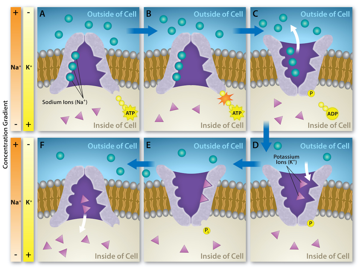
In the resting state, sodium (Na+) is at higher outside the cell, so it will tend to move into the cell. Potassium (K+), on the other hand, is more concentrated inside the cell, and will tend to move out of the cell (Figure 3.10). In addition, the inside of the cell is slightly negatively compared to the outside, due to the activity of the . This pump actively transports three sodium out of the cell for every two potassium in, creating a net negative inside the cell. This provides an additional force on sodium, causing it to move into the cell.

A close-up illustration depicts the difference in charges across the cell membrane, and shows how Na+ and K+ cells concentrate more closely near the membrane.
Figure 3.10 — 3.2 Cells of the Nervous System

Figure 3.10 At resting potential, Na + (blue pentagons) is more highly concentrated outside the cell in the extracellular fluid (shown in blue), whereas K + (purple squares) is more highly concentrated near the membrane in the cytoplasm or intracellular fluid. Other molecules, such as chloride ions (yellow circles) and negatively charged proteins (brown squares), help contribute to a positive net charge in the extracellular fluid and a negative net charge in the intracellular fluid.

Rose M. Spielman, William J. Jenkins, and Marilyn D. Lovett, Psychology 2E, OpenStax. · CC BY-NC-SA 4.0 · Original image ↗ · Access for free ↗

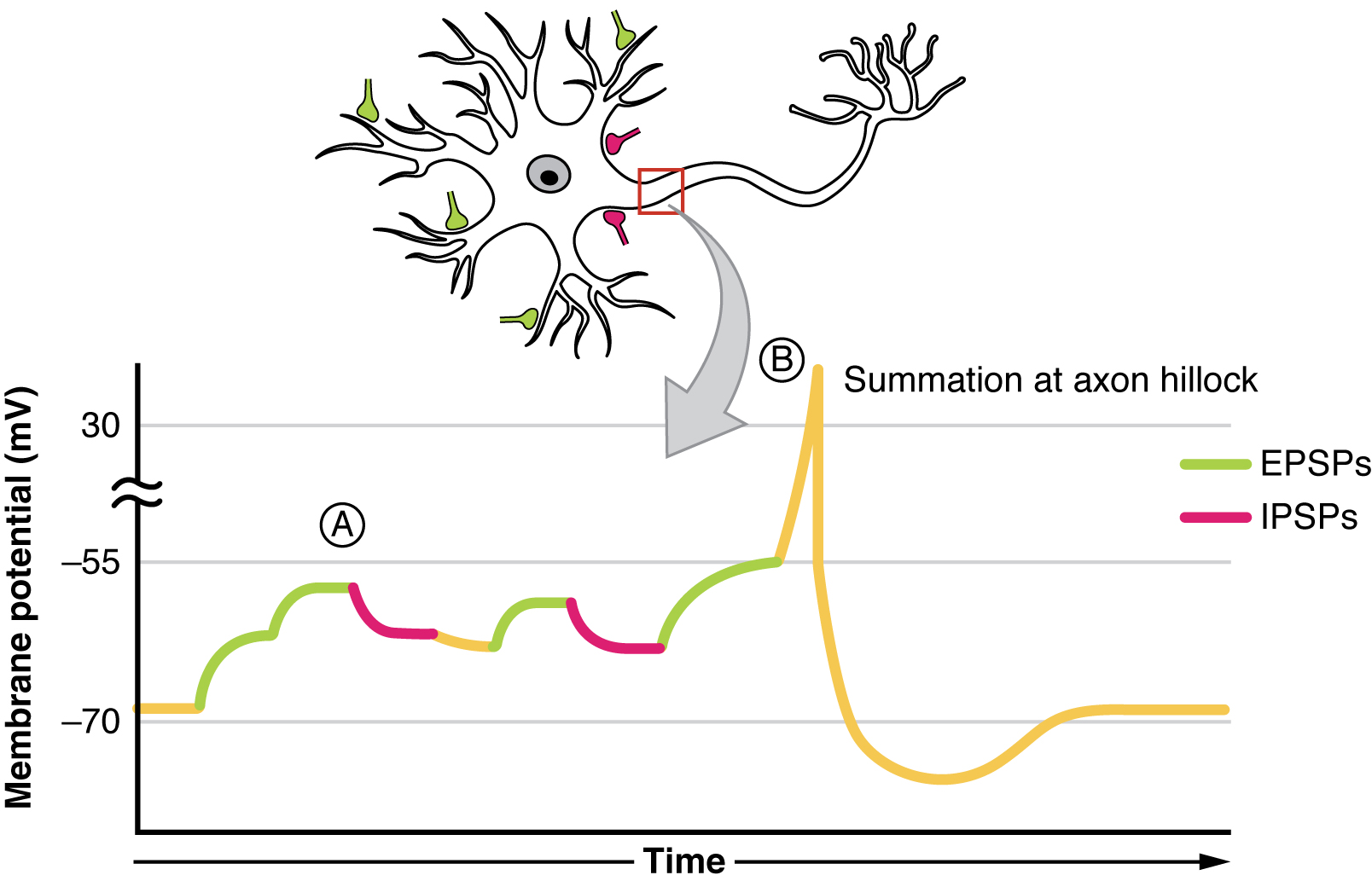
From this resting potential state, the receives a signal and its state changes abruptly (Figure 3.11). When a receives signals at the —due to neurotransmitters from an adjacent binding to its —small , or gates, open on the neuronal , allowing Na+ , propelled by both and differences, to move into the cell. With this influx of positive , the internal of the cell becomes more positive. If that reaches a certain level, called the threshold of excitation, the becomes active and the begins.

Many additional open, causing a massive influx of Na+ and a huge positive spike in the potential, the . At the of the spike, the sodium gates close and the potassium gates open. As positively potassium leave, the cell quickly begins . At first, it hyperpolarizes, becoming slightly more negative than the resting potential, and then it levels off, returning to the resting potential.

A graph shows the increase, peak, and decrease in membrane potential. The millivolts through the phases are approximately -70mV at resting potential, -55mV at threshold of excitation, 30mV at peak action potential, 5mV at repolarization, and -80mV at hyperpolarization.
Figure 3.11 — 3.2 Cells of the Nervous System

Figure 3.11 During the action potential, the electrical charge across the membrane changes dramatically.

Rose M. Spielman, William J. Jenkins, and Marilyn D. Lovett, Psychology 2E, OpenStax. · CC BY-NC-SA 4.0 · Original image ↗ · Access for free ↗

This positive spike constitutes the : the electrical signal that typically moves from the cell body down the to the terminals. The electrical signal moves down the with the impulses jumping in a leapfrog fashion between the Nodes of Ranvier. The Nodes of Ranvier are natural gaps in the . At each point, some of the sodium that enter the cell diffuse to the next section of the , raising the past the threshold of excitation and triggering a new influx of sodium . The moves all the way down the in this fashion until reaching the terminal buttons.

The is an all-or-none phenomenon. In simple terms, this means that an incoming signal from another is either sufficient or insufficient to reach the threshold of excitation. There is no in-between, and there is no turning off an once it starts. Think of it like sending an email or a text message. You can think about sending it all you want, but the message is not sent until you hit the send button. Furthermore, once you send the message, there is no stopping it.

Because it is all or none, the is recreated, or propagated, at its full strength at every point along the . Much like the lit fuse of a firecracker, it does not fade away as it travels down the . It is this all-or-none property that explains the that your brain perceives an injury to a distant body part like your toe as equally painful as one to your nose.

As noted earlier, when the arrives at the terminal button, the synaptic release their neurotransmitters into the synaptic cleft. The neurotransmitters travel across the and bind to on the of the adjacent , and the process repeats itself in the new (assuming the signal is sufficiently strong to trigger an ). Once the signal is delivered, excess neurotransmitters in the synaptic cleft drift away, are broken down into inactive fragments, or are reabsorbed in a process known as reuptake. Reuptake involves the neurotransmitter being pumped back into the that released it, in order to clear the (Figure 3.12). Clearing the serves both to provide a clear “on” and “off” state between signals and to regulate the production of neurotransmitter (full synaptic provide signals that no additional neurotransmitters need to be produced).

The synaptic space between two neurons is shown. Some neurotransmitters that have been released into the synapse are attaching to receptors while others undergo reuptake into the axon terminal.
Figure 3.12 — 3.2 Cells of the Nervous System

Figure 3.12 Reuptake involves moving a neurotransmitter from the synapse back into the axon terminal from which it was released.

Rose M. Spielman, William J. Jenkins, and Marilyn D. Lovett, Psychology 2E, OpenStax. · CC BY-NC-SA 4.0 · Original image ↗ · Access for free ↗

Neuronal communication is often referred to as an electrochemical event. The movement of the down the length of the is an electrical event, and movement of the neurotransmitter across the synaptic space represents the chemical portion of the process. However, there are some specialized connections between that are entirely electrical. In such cases, the are said to communicate via an electrical . In these cases, two physically connect to one another via , which allows the current from one cell to pass into the next. There are far fewer electrical in the brain, but those that do exist are much faster than the chemical that have been described above (Connors & Long, 2004).

Link to Learning

Watch this video about neuronal communication to learn more.

Neurotransmitters and Drugs

There are several different types of neurotransmitters released by different , and we can speak in broad terms about the kinds of functions associated with different neurotransmitters (Table 3.1). Much of what psychologists know about the functions of neurotransmitters comes from research on the effects of drugs in psychological disorders. Psychologists who take a biological perspective and focus on the physiological causes of behavior assert that psychological disorders like depression and schizophrenia are associated with imbalances in one or more neurotransmitter systems. In this perspective, psychotropic medications can help improve the symptoms associated with these disorders. Psychotropic medications are drugs that treat psychiatric symptoms by restoring neurotransmitter balance.

Major Neurotransmitters and How They Affect Behavior
Neurotransmitter Involved in Potential Effect on Behavior
Acetylcholine Muscle action, memory Increased arousal, enhanced cognition
Beta-endorphin Pain, pleasure Decreased anxiety, decreased tension
Dopamine Mood, sleep, learning Increased pleasure, suppressed appetite
() Brain function, sleep Decreased anxiety, decreased tension
Memory, learning Increased learning, enhanced memory
Norepinephrine Heart, intestines, alertness Increased arousal, suppressed appetite
Serotonin Mood, sleep Modulated mood, suppressed appetite
Table 3.1

Psychoactive drugs can act as agonists or antagonists for a given neurotransmitter system. Agonists are chemicals that mimic a neurotransmitter at the site. An antagonist, on the other hand, blocks or impedes the normal activity of a neurotransmitter at the . Agonists and antagonists represent drugs that are prescribed to correct the specific neurotransmitter imbalances underlying a person’s condition. For example, Parkinson's disease, a progressive nervous system disorder, is associated with low levels of dopamine. Therefore, a common treatment strategy for Parkinson's disease involves using dopamine agonists, which mimic the effects of dopamine by binding to dopamine .

Certain symptoms of schizophrenia are associated with overactive dopamine neurotransmission. The antipsychotics used to treat these symptoms are antagonists for dopamine—they block dopamine’s effects by binding its without activating them. Thus, they prevent dopamine released by one from signaling information to adjacent .

In contrast to agonists and antagonists, which both operate by binding to sites, reuptake prevent unused neurotransmitters from being transported back to the . This allows neurotransmitters to remain active in the synaptic cleft for longer durations, increasing their effectiveness. Depression, which has been consistently linked with reduced serotonin levels, is commonly treated with selective serotonin reuptake (SSRIs). By preventing reuptake, SSRIs strengthen the effect of serotonin, giving it more time to interact with serotonin on . Common SSRIs on the market today include Prozac, Paxil, and Zoloft. The drug is structurally very similar to serotonin, and it affects the same and as serotonin. Psychotropic drugs are not instant for people suffering from psychological disorders. Often, an individual must take a drug for several weeks before seeing improvement, and many psychoactive drugs have significant negative side effects. Furthermore, individuals vary dramatically in how they respond to the drugs. To improve chances for success, it is not uncommon for people receiving pharmacotherapy to undergo psychological and/or behavioral therapies as well. Some research suggests that combining drug therapy with other forms of therapy tends to be more effective than any one treatment alone (for one such example, see March et al., 2007).

3.3 Parts of the Nervous System

OpenStax · 3.3 Parts of the Nervous System ↗
Instructional page including learning objectives, examples, figures, source tables and questions. Site navigation and embedded media players are omitted; source media links remain available.

Learning Objectives

By the end of this section, you will be able to:

  • Describe the difference between the central and peripheral nervous systems
  • Explain the difference between the and autonomic nervous systems
  • Differentiate between the sympathetic and parasympathetic divisions of the

The nervous system can be divided into two major subdivisions: the () and the (), shown in Figure 3.13. The is comprised of the brain and spinal cord; the connects the to the rest of the body. In this section, we focus on the ; later, we look at the brain and spinal cord.

Image (a) shows an outline of a human body with the brain and spinal cord illustrated. Image (b) shows an outline of a human body with a network of nerves depicted.
Figure 3.13 — 3.3 Parts of the Nervous System

Figure 3.13 The nervous system is divided into two major parts: (a) the Central Nervous System and (b) the Peripheral Nervous System.

Rose M. Spielman, William J. Jenkins, and Marilyn D. Lovett, Psychology 2E, OpenStax. · CC BY-NC-SA 4.0 · Original image ↗ · Access for free ↗

The is made up of thick bundles of axons, called nerves, carrying messages back and forth between the and the muscles, organs, and senses in the periphery of the body (i.e., everything outside the ). The has two major subdivisions: the and the .

The is associated with activities traditionally thought of as conscious or voluntary. It is involved in the relay of sensory and motor information to and from the ; therefore, it consists of motor and sensory . Motor , carrying instructions from the to the muscles, are efferent fibers (efferent means “moving away from”). Sensory , carrying sensory information to the , are afferent fibers (afferent means “moving toward”). A helpful way to remember this is that efferent = exit and afferent = arrive. Each nerve is basically a bundle of forming a two-way superhighway, containing thousands of axons, both efferent and afferent.

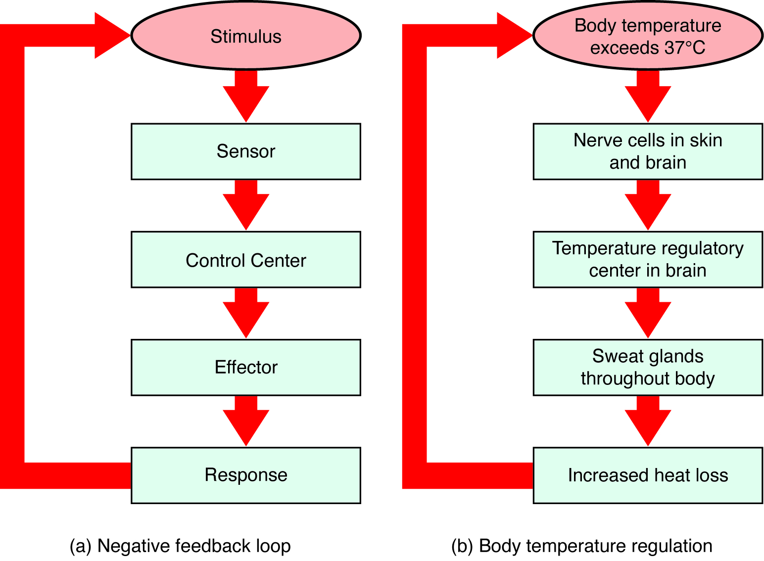
The our internal organs and glands and is generally considered to be outside the realm of voluntary control. It can be further subdivided into the sympathetic and parasympathetic divisions (Figure 3.14). The sympathetic nervous system is involved in preparing the body for stress-related activities; the parasympathetic nervous system is associated with returning the body to routine, day-to-day operations. The two systems have complementary functions, operating in tandem to maintain the body’s . is a state of , or balance, in which biological conditions (such as body temperature) are maintained at optimal levels.

A diagram of a human body lists the different functions of the sympathetic and parasympathetic nervous system. The parasympathetic system can constrict pupils, stimulate salivation, slow heart rate, constrict bronchi, stimulate digestion, stimulate bile secretion, and cause the bladder to contract. The sympathetic nervous system can dilate pupils, inhibit salivation, increase heart rate, dilate bronchi, inhibit digestion, stimulate the breakdown of glycogen, stimulate secretion of adrenaline and noradrenaline, and inhibit contraction of the bladder.
Figure 3.14 — 3.3 Parts of the Nervous System

Figure 3.14 The sympathetic and parasympathetic divisions of the autonomic nervous system have the opposite effects on various systems.

Rose M. Spielman, William J. Jenkins, and Marilyn D. Lovett, Psychology 2E, OpenStax. · CC BY-NC-SA 4.0 · Original image ↗ · Access for free ↗

The sympathetic nervous system is activated when we are faced with stressful or high-arousal situations. The activity of this system was adaptive for our ancestors, increasing their chances of survival. Imagine, for example, that one of our early ancestors, out hunting small game, suddenly disturbs a large bear with her cubs. At that moment, the hunter's body undergoes a series of changes—a direct function of sympathetic activation—preparing them to face the threat. The pupils dilate, the heart rate and blood pressure increase, the bladder relaxes, and the liver releases ; adrenaline surges into the bloodstream. This constellation of physiological changes, known as the fight or flight response, allows the body access to energy reserves and heightened sensory capacity so that it might fight off a threat or run away to safety.

Link to Learning

While it is clear that such a response would be critical for survival for our ancestors, who lived in a world full of real physical threats, many of the high-arousal situations we face in the modern world are more psychological in nature. For example, think about how you feel when you have to stand up and give a presentation in front of a roomful of people, or right before taking a big test. You are in no real physical danger in those situations, and yet you have evolved to respond to a perceived threat with the fight or flight response. This kind of response is not nearly as adaptive in the modern world; in , we suffer negative health consequences when faced constantly with psychological threats that we can neither fight nor flee. Recent research suggests that an increase in susceptibility to heart disease (Chandola, Brunner, & Marmot, 2006) and impaired function of the immune system (Glaser & Kiecolt-Glaser, 2005) are among the many negative consequences of persistent and repeated exposure to stressful situations. Some of this tendency for stress reactivity can be wired by early experiences of trauma.

Once the threat has been resolved, the parasympathetic nervous system takes over and returns bodily functions to a relaxed state. Our hunter’s heart rate and blood pressure return to normal, the pupils constrict, bladder control is restored, and the liver begins to store in the form of for future use. These restorative processes are associated with activation of the parasympathetic nervous system.

3.4 The Brain and Spinal Cord

OpenStax · 3.4 The Brain and Spinal Cord ↗
Instructional page including learning objectives, examples, figures, source tables and questions. Site navigation and embedded media players are omitted; source media links remain available.

Learning Objectives

By the end of this section, you will be able to:

  • Explain the functions of the spinal cord
  • Identify the hemispheres and lobes of the brain
  • Describe the types of techniques available to clinicians and researchers to image or scan the brain

The brain is a remarkably complex organ comprised of billions of interconnected and glia. It is a bilateral, or two-sided, structure that can be separated into distinct lobes. Each lobe is associated with certain types of functions, but, ultimately, all of the areas of the brain interact with one another to provide the foundation for our thoughts and behaviors. In this section, we discuss the overall organization of the brain and the functions associated with different brain areas, beginning with what can be seen as an extension of the brain, the spinal cord.

The Spinal Cord

It can be said that the spinal cord is what connects the brain to the outside world. Because of it, the brain can act. The spinal cord is like a relay station, but a very smart one. It not only routes messages to and from the brain, but it also has its own system of automatic processes, called reflexes.

The top of the spinal cord is a bundle of nerves that merges with the brain stem, where the basic processes of life are controlled, such as breathing and digestion. In the opposite direction, the spinal cord ends just below the ribs—contrary to what we might expect, it does not extend all the way to the of the spine.

The spinal cord is functionally organized in 30 segments, corresponding with the vertebrae. Each segment is connected to a specific part of the body through the . Nerves branch out from the spine at each vertebra. Sensory nerves bring messages in; motor nerves send messages out to the muscles and organs. Messages travel to and from the brain through every segment.

Some sensory messages are immediately acted on by the spinal cord, without any input from the brain. from a hot object and the knee jerk are two examples. When a sensory message meets certain parameters, the spinal cord initiates an automatic reflex. The signal passes from the sensory nerve to a simple processing center, which initiates a motor command. Seconds are saved, because messages don’t have to go to the brain, be processed, and get sent back. In matters of survival, the spinal reflexes allow the body to react extraordinarily fast.

The spinal cord is protected by bony vertebrae and cushioned in , but injuries still occur. When the spinal cord is damaged in a particular segment, all lower segments are cut off from the brain, causing paralysis. Therefore, the lower on the spine damage occurs, the fewer functions an injured individual will lose.

Neuroplasticity

Bob Woodruff, a for ABC, suffered a traumatic brain injury after a bomb exploded next to the vehicle he was in while covering a news story in Iraq. As a consequence of these injuries, Woodruff experienced many cognitive deficits including difficulties with memory and language. However, over time and with the aid of intensive amounts of cognitive and speech therapy, Woodruff has shown an incredible recovery of function (Fernandez, 2008, October 16).

One of the factors that made this recovery possible was neuroplasticity. Neuroplasticity refers to how the nervous system can change and adapt. Neuroplasticity can occur in a variety of ways including personal experiences, developmental processes, or, as in Woodruff's case, in response to some sort of damage or injury that has occurred. Neuroplasticity can involve creation of new , pruning of that are no longer used, changes in glial cells, and even the birth of new . Because of neuroplasticity, our brains are constantly changing and adapting, and while our nervous system is most plastic when we are very young, as Woodruff's case suggests, it is still capable of remarkable changes later in life.

The Two Hemispheres

The surface of the brain, known as the cerebral cortex, is very uneven, characterized by a distinctive pattern of folds or bumps, known as gyri (singular: gyrus), and grooves, known as sulci (singular: sulcus), shown in Figure 3.15. These gyri and sulci form important landmarks that allow us to separate the brain into functional centers. The most prominent sulcus, known as the longitudinal fissure, is the deep groove that separates the brain into two halves or hemispheres: the left hemisphere and the right hemisphere.

An illustration of the brain’s exterior surface shows the ridges and depressions, and the deep fissure that runs through the center.
Figure 3.15 — 3.4 The Brain and Spinal Cord

Figure 3.15 The surface of the brain is covered with gyri and sulci. A deep sulcus is called a fissure, such as the longitudinal fissure that divides the brain into left and right hemispheres. (credit: modification of work by Bruce Blaus)

Rose M. Spielman, William J. Jenkins, and Marilyn D. Lovett, Psychology 2E, OpenStax. (credit: modification of work by Bruce Blaus) · CC BY-NC-SA 4.0 · Original image ↗ · Access for free ↗

There is evidence of specialization of function—referred to as —in each hemisphere, mainly regarding differences in language functions. The left hemisphere the right half of the body, and the right hemisphere the left half of the body. Decades of research on of function by Michael Gazzaniga and colleagues suggest that a variety of functions ranging from cause-and-effect reasoning to self-recognition may follow patterns that suggest some degree of hemispheric dominance (Gazzaniga, 2005). For example, the left hemisphere has been shown to be superior for forming associations in memory, , and positive emotions. The right hemisphere, on the other hand, has been shown to be superior in pitch perception, arousal, and negative emotions (Ehret, 2006). However, it should be pointed out that research on which hemisphere is in a variety of different behaviors has produced inconsistent results, and therefore, it is probably better to think of how the two hemispheres interact to produce a given behavior rather than attributing certain behaviors to one hemisphere versus the other (Banich & Heller, 1998).

The two hemispheres are connected by a thick band of neural fibers known as the , consisting of about 200 million axons. The allows the two hemispheres to communicate with each other and allows for information being processed on one side of the brain to be shared with the other side.

Normally, we are not aware of the different roles that our two hemispheres play in day-to-day functions, but there are people who come to know the capabilities and functions of their two hemispheres quite well. In some cases of severe epilepsy, doctors elect to sever the as a means of controlling the spread of seizures (Figure 3.16). While this is an effective treatment option, it results in individuals who have "split brains." After surgery, these split-brain patients show a variety of interesting behaviors. For instance, a split-brain patient is unable to name a picture that is shown in the patient’s left visual field because the information is only available in the largely nonverbal right hemisphere. However, they are able to recreate the picture with their left hand, which is also controlled by the right hemisphere. When the more verbal left hemisphere sees the picture that the hand drew, the patient is able to name it (assuming the left hemisphere can interpret what was drawn by the left hand).

Illustrations (a) and (b) show the corpus callosum’s location in the brain in front and side views. Photograph (c) shows the corpus callosum in a dissected brain.
Figure 3.16 — 3.4 The Brain and Spinal Cord

Figure 3.16 (a, b) The corpus callosum connects the left and right hemispheres of the brain. (c) A scientist spreads this dissected sheep brain apart to show the corpus callosum between the hemispheres. (credit c: modification of work by Aaron Bornstein)

Rose M. Spielman, William J. Jenkins, and Marilyn D. Lovett, Psychology 2E, OpenStax. (credit c: modification of work by Aaron Bornstein) · CC BY-NC-SA 4.0 · Original image ↗ · Access for free ↗

Much of what we know about the functions of different areas of the brain comes from studying changes in the behavior and ability of individuals who have suffered damage to the brain. For example, researchers study the behavioral changes caused by strokes to learn about the functions of specific brain areas. A stroke, caused by an interruption of blood flow to a region in the brain, causes a loss of brain function in the affected region. The damage can be in a small area, and, if it is, this gives researchers the opportunity to link any resulting behavioral changes to a specific area. The types of deficits displayed after a stroke will be largely dependent on where in the brain the damage occurred.

Consider Theona, an intelligent, self-sufficient woman, who is 62 years old. Recently, she suffered a stroke in the front portion of her right hemisphere. As a result, she has great difficulty moving her left leg. (As you learned earlier, the right hemisphere the left side of the body; also, the brain’s main motor centers are located at the front of the head, in the frontal lobe.) Theona has also experienced behavioral changes. For example, while in the produce section of the grocery store, she sometimes eats grapes, strawberries, and apples directly from their bins before paying for them. This behavior—which would have been very embarrassing to her before the stroke—is consistent with damage in another region in the frontal lobe—the prefrontal cortex, which is associated with judgment, reasoning, and impulse control.

Structures

The two hemispheres of the cerebral cortex are part of the (Figure 3.17), which is the largest part of the brain. The contains the cerebral cortex and a number of other structures that lie beneath the cortex (called subcortical structures): , , pituitary gland, and the limbic system (a collection of structures). The cerebral cortex, which is the outer surface of the brain, is associated with higher level processes such as consciousness, thought, emotion, reasoning, language, and memory. Each cerebral hemisphere can be subdivided into four lobes, each associated with different functions.

An illustration shows the position and size of the forebrain (the largest portion), midbrain (a small central portion), and hindbrain (a portion in the lower back part of the brain).
Figure 3.17 — 3.4 The Brain and Spinal Cord

Figure 3.17 The brain and its parts can be divided into three main categories: the forebrain, midbrain, and hindbrain.

Rose M. Spielman, William J. Jenkins, and Marilyn D. Lovett, Psychology 2E, OpenStax. · CC BY-NC-SA 4.0 · Original image ↗ · Access for free ↗

Lobes of the Brain

The four lobes of the brain are the frontal, parietal, temporal, and occipital lobes (Figure 3.18). The frontal lobe is located in the forward part of the brain, extending back to a fissure known as the central sulcus. The frontal lobe is involved in reasoning, motor control, emotion, and language. It contains the motor cortex, which is involved in planning and coordinating movement; the prefrontal cortex, which is responsible for higher-level cognitive functioning; and Broca’s area, which is essential for language production.

An illustration shows the four lobes of the brain.
Figure 3.18 — 3.4 The Brain and Spinal Cord

Figure 3.18 The lobes of the brain are shown.

Rose M. Spielman, William J. Jenkins, and Marilyn D. Lovett, Psychology 2E, OpenStax. · CC BY-NC-SA 4.0 · Original image ↗ · Access for free ↗

People who suffer damage to Broca’s area have great difficulty producing language of any form (Figure 3.18). For example, Padma was an electrical engineer who was socially active and a caring, involved parent. About twenty years ago, she was in a car accident and suffered damage to her Broca’s area. She completely lost the ability to speak and form any kind of meaningful language. There is nothing wrong with her mouth or her vocal cords, but she is unable to produce words. She can follow directions but can’t respond verbally, and she can read but no longer write. She can do routine tasks like running to the market to buy milk, but she could not communicate verbally if a situation called for it.

Probably the most famous case of frontal lobe damage is that of a man by the name of Phineas Gage. On September 13, 1848, Gage (age 25) was working as a railroad foreman in Vermont. He and crew were using an iron rod to tamp explosives down into a blasting hole to remove rock along the railway’s path. Unfortunately, the iron rod created a spark and caused the rod to explode out of the blasting hole, into Gage’s face, and through skull (Figure 3.19). Although lying in a pool of own blood with brain matter emerging from head, Gage was conscious and able to get up, walk, and speak. But in the months following accident, people noticed that personality had changed. Many of friends described him as no longer being himself. Before the accident, it was said that Gage was a well-mannered, soft-spoken man, but he began to behave in odd and inappropriate ways after the accident. Such changes in personality would be consistent with loss of impulse control—a frontal lobe function.

Beyond the damage to the frontal lobe itself, subsequent investigations into the rod's path also identified probable damage to pathways between the frontal lobe and other brain structures, including the limbic system. With connections between the planning functions of the frontal lobe and the emotional processes of the limbic system severed, Gage had difficulty controlling emotional impulses.

However, there is some evidence suggesting that the dramatic changes in Gage’s personality were exaggerated and embellished. Gage's case occurred in the midst of a 19th century debate over localization—regarding whether certain areas of the brain are associated with particular functions. On the basis of extremely limited information about Gage, the extent of injury, and life before and after the accident, scientists tended to find support for their own views, on whichever side of the debate they fell (Macmillan, 1999).

Image (a) is a photograph of Phineas Gage holding a metal rod. Image (b) is an illustration of a skull with a metal rod passing through it from the cheek area to the top of the skull.
Figure 3.19 — 3.4 The Brain and Spinal Cord

Figure 3.19 (a) Phineas Gage holds the iron rod that penetrated his skull in an 1848 railroad construction accident. (b) Gage’s prefrontal cortex was severely damaged in the left hemisphere. The rod entered Gage’s face on the left side, passed behind his eye, and exited through the top of his skull, before landing about 80 feet away. (credit a: modification of work by Jack and Beverly Wilgus)

Rose M. Spielman, William J. Jenkins, and Marilyn D. Lovett, Psychology 2E, OpenStax. (credit a: modification of work by Jack and Beverly Wilgus) · CC BY-NC-SA 4.0 · Original image ↗ · Access for free ↗

The brain’s parietal lobe is located immediately behind the frontal lobe, and is involved in processing information from the body’s senses. It contains the somatosensory cortex, which is essential for processing sensory information from across the body, such as touch, temperature, and pain. The somatosensory cortex is an area of the brain which processes touch and sensation. The somatosensory cortex is fascinating because each different area of the cortex processes sensations from a different part of your body. Furthermore, the larger the surface area of the specific body part and the greater amount of nerves in that body part, the larger the area dedicated to processing sensation from that body part in the somatosensory cortex. For example, fingers take up a lot more space than toes. As you can notice from (Figure 3.20), the amount of space to process sensation from fingers is much greater than that of toes.

Link to Learning

One fascinating example of neuroplasticity involves reorganization of the somatosensory cortex following limb amputation. Check out this NPR segment about amputees’ experiences of “phantom limbs” following amputation to learn more.

A diagram shows the organization in the somatosensory cortex, with functions for these parts in this proximal sequential order: genitals, toes, ankles, knees, hips, trunk, shoulders, elbows, wrists, hands, fingers, thumbs, neck, eyebrows and eyelids, eyeballs, face, lips, jaw, tongue, salivation, chewing, and swallowing.
Figure 3.20 — 3.4 The Brain and Spinal Cord

Figure 3.20 Spatial relationships in the body are mirrored in the organization of the somatosensory cortex.

Rose M. Spielman, William J. Jenkins, and Marilyn D. Lovett, Psychology 2E, OpenStax. · CC BY-NC-SA 4.0 · Original image ↗ · Access for free ↗

The temporal lobe is located on the side of the head (temporal means “near the temples”), and is associated with hearing, memory, emotion, and some aspects of language. The auditory cortex, the main area responsible for processing auditory information, is located within the temporal lobe. Wernicke’s area, important for speech comprehension, is also located here. Whereas individuals with damage to Broca’s area have difficulty producing language, those with damage to Wernicke’s area can produce sensible language, but they are unable to understand it (Figure 3.21).

An illustration shows the locations of Broca’s and Wernicke’s areas.
Figure 3.21 — 3.4 The Brain and Spinal Cord

Figure 3.21 Damage to either Broca’s area or Wernicke’s area can result in language deficits. The types of deficits are very different, however, depending on which area is affected.

Rose M. Spielman, William J. Jenkins, and Marilyn D. Lovett, Psychology 2E, OpenStax. · CC BY-NC-SA 4.0 · Original image ↗ · Access for free ↗

The occipital lobe is located at the very back of the brain, and contains the primary visual cortex, which is responsible for interpreting incoming visual information. The occipital cortex is organized retinotopically, which means there is a close relationship between the position of an object in a person’s visual field and the position of that object’s representation on the cortex. You will learn much more about how visual information is processed in the occipital lobe when you study sensation and perception.

Other Areas of the

Other areas of the , located beneath the cerebral cortex, include the and the limbic system. The is a sensory relay for the brain. All of our senses, with the exception of smell, are routed through the before being directed to other areas of the brain for processing (Figure 3.22).

An illustration shows the location of the thalamus in the brain.
Figure 3.22 — 3.4 The Brain and Spinal Cord

Figure 3.22 The thalamus serves as the relay center of the brain where most senses are routed for processing.

Rose M. Spielman, William J. Jenkins, and Marilyn D. Lovett, Psychology 2E, OpenStax. · CC BY-NC-SA 4.0 · Original image ↗ · Access for free ↗

The limbic system is involved in processing both emotion and memory. Interestingly, the sense of smell projects directly to the limbic system; therefore, not surprisingly, smell can evoke emotional responses in ways that other sensory modalities cannot. The limbic system is made up of a number of different structures, but three of the most important are the , the , and the (Figure 3.23). The is an essential structure for learning and memory. The is involved in our experience of emotion and in tying emotional meaning to our memories. The regulates a number of homeostatic processes, including the regulation of body temperature, appetite, and blood pressure. The also serves as an interface between the nervous system and the system and in the regulation of sexual motivation and behavior.

An illustration shows the locations of parts of the brain involved in the limbic system: the hypothalamus, amygdala, and hippocampus.
Figure 3.23 — 3.4 The Brain and Spinal Cord

Figure 3.23 The limbic system is involved in mediating emotional response and memory.

Rose M. Spielman, William J. Jenkins, and Marilyn D. Lovett, Psychology 2E, OpenStax. · CC BY-NC-SA 4.0 · Original image ↗ · Access for free ↗

The Case of Henry Molaison (H.M.)

In 1953, Henry Gustav Molaison (H. M.) was a 27-year-old man who experienced severe seizures. In an attempt to control seizures, H. M. underwent brain surgery to remove and . Following the surgery, H.M’s seizures became much less severe, but he also suffered some unexpected—and devastating—consequences of the surgery: he lost ability to form many types of new memories. For example, he was unable to learn new facts, such as who was president of the United States. He was able to learn new skills, but afterward he had no recollection of learning them. For example, while he might learn to use a computer, he would have no conscious memory of ever having used one. He could not remember new faces, and he was unable to remember events, even immediately after they occurred. Researchers were fascinated by experience, and he is considered one of the most studied cases in medical and psychological history (Hardt, Einarsson, & Nader, 2010; Squire, 2009). Indeed, case has provided tremendous insight into the role that the plays in the consolidation of new learning into explicit memory.

Link to Learning

Clive Wearing, an accomplished musician, lost the ability to form new memories when was damaged through illness. Check out the first few minutes of this documentary video about this man and his condition to learn more.

and Structures

The is comprised of structures located deep within the brain, between the and the . The reticular formation is centered in the , but it actually extends up into the and down into the . The reticular formation is important in regulating the sleep/wake cycle, arousal, alertness, and motor activity.

The substantia nigra (Latin for “black substance”) and the ventral tegmental area (VTA) are also located in the (Figure 3.24). Both regions contain cell bodies that produce the neurotransmitter dopamine, and both are critical for movement. Degeneration of the substantia nigra and VTA is involved in Parkinson’s disease. In addition, these structures are involved in mood, reward, and addiction (Berridge & Robinson, 1998; Gardner, 2011; George, Le Moal, & Koob, 2012).

An illustration shows the location of the substantia nigra and VTA in the brain.
Figure 3.24 — 3.4 The Brain and Spinal Cord

Figure 3.24 The substantia nigra and ventral tegmental area (VTA) are located in the midbrain.

Rose M. Spielman, William J. Jenkins, and Marilyn D. Lovett, Psychology 2E, OpenStax. · CC BY-NC-SA 4.0 · Original image ↗ · Access for free ↗

The is located at the back of the head and looks like an extension of the spinal cord. It contains the medulla, pons, and (Figure 3.25). The medulla the automatic processes of the , such as breathing, blood pressure, and heart rate. The word pons literally means “bridge,” and as the name suggests, the pons serves to connect the to the rest of the brain. It also is involved in regulating brain activity during sleep. The medulla, pons, and various structures are known as the brainstem, and aspects of the brainstem span both the and the .

An illustration shows the location of the pons, medulla, and cerebellum.
Figure 3.25 — 3.4 The Brain and Spinal Cord

Figure 3.25 The pons, medulla, and cerebellum make up the hindbrain.

Rose M. Spielman, William J. Jenkins, and Marilyn D. Lovett, Psychology 2E, OpenStax. · CC BY-NC-SA 4.0 · Original image ↗ · Access for free ↗

The (Latin for “little brain”) receives messages from muscles, , joints, and structures in our ear to control balance, coordination, movement, and motor skills. The is also thought to be an important area for processing some types of memories. In particular, , or memory involved in learning and remembering how to perform tasks, is thought to be associated with the . Recall that H. M. was unable to form new explicit memories, but he could learn new tasks. This is likely due to the that H. M.’s remained intact.

What Do You Think?

Brain Dead and on Life Support

What would you do if your spouse or loved one was declared brain dead but their body was being kept alive by medical equipment? What if the loved one was in a persistent vegetative state and remained unconscious but maintained basic functions like breathing? Whose decision should it be to remove a feeding tube? Should medical care costs be a factor?

On February 25, 1990, a Florida woman named Terri Schiavo went into cardiac arrest, apparently triggered by a bulimic episode. She was eventually revived, but her brain had been deprived of oxygen for a long time. Brain scans indicated that there was no activity in her cerebral cortex, and she suffered from severe and permanent cerebral atrophy. Basically, Schiavo was in a vegetative state. Medical professionals determined that she would never again be able to move, talk, or respond in any way. To remain alive, she required a feeding tube, and there was no chance that her situation would ever improve.

On occasion, Schiavo’s eyes would move, and sometimes she would groan. Despite the doctors’ insistence to the contrary, her parents believed that these were signs that she was trying to communicate with them.

After 12 years, Schiavo’s husband argued that wife would not have wanted to be kept alive with no feelings, sensations, or brain activity. Her parents, however, were very much against removing her feeding tube. Eventually, the case made its way to the courts, both in the state of Florida and at the federal level. By 2005, the courts found in favor of Schiavo’s husband, and the feeding tube was removed on March 18, 2005. Schiavo died 13 days later.

Why did Schiavo’s eyes sometimes move, and why did she groan? Although the parts of her brain that control thought, voluntary movement, and feeling were completely damaged, her brainstem was still intact. Her medulla and pons maintained her breathing and caused involuntary movements of her eyes and the occasional groans. Over the 15-year period that she was on a feeding tube, Schiavo’s medical costs may have topped $7 million (Arnst, 2003).

These questions were brought to popular consciousness decades ago in the case of Terri Schiavo, and they have persisted. In 2013, a 13-year-old girl who suffered complications after tonsil surgery was declared brain dead. There was a battle between her family, who wanted her to remain on life support, and the hospital’s policies regarding persons declared brain dead. In another complicated 2013–14 case in Texas, a pregnant EMT professional declared brain dead was kept alive for weeks, despite her spouse’s directives, which were based on her wishes should this situation arise. In this case, state laws designed to protect an unborn fetus came into consideration until doctors determined the fetus unviable.

Decisions surrounding the medical response to patients declared brain dead are complex. Whether a person is declared brain dead or diagnosed as having a persistent vegetative state, determining death and making appropriate medical care decisions is a complicated process. What do you think about these issues?

Brain Imaging

You have learned how brain injury can provide information about the functions of different parts of the brain. Increasingly, however, we are able to obtain that information using brain imaging techniques on individuals who have not suffered brain injury. In this section, we take a more in-depth look at some of the techniques that are available for imaging the brain, including techniques that rely on radiation, magnetic fields, or electrical activity within the brain.

Techniques Involving Radiation

A computerized tomography (CT) scan involves taking a number of x-rays of a particular section of a person’s body or brain (Figure 3.26). The x-rays pass through tissues of different densities at different rates, allowing a computer to construct an overall image of the area of the body being scanned. A CT scan is often used to determine whether someone has a or significant brain atrophy.

Image (a) shows a brain scan where the brain matter’s appearance is fairly uniform. Image (b) shows a section of the brain that looks different from the surrounding tissue and is labeled “tumor.”
Figure 3.26 — 3.4 The Brain and Spinal Cord

Figure 3.26 A CT scan can be used to show brain tumors. (a) The image on the left shows a healthy brain, whereas (b) the image on the right indicates a brain tumor in the left frontal lobe. (credit a: modification of work by "Aceofhearts1968"/Wikimedia Commons; credit b: modification of work by Roland Schmitt et al)

Rose M. Spielman, William J. Jenkins, and Marilyn D. Lovett, Psychology 2E, OpenStax. (credit a: modification of work by "Aceofhearts1968"/Wikimedia Commons; credit b: modification of work by Roland Schmitt et al) · CC BY-NC-SA 4.0 · Original image ↗ · Access for free ↗

Positron emission tomography (PET) scans create pictures of the living, active brain (Figure 3.27). An individual receiving a PET scan drinks or is injected with a mildly substance, called a tracer. Once in the bloodstream, the amount of tracer in any given region of the brain can be monitored. As a brain area becomes more active, more blood flows to that area. A computer monitors the movement of the tracer and creates a rough of active and inactive areas of the brain during a given behavior. PET scans show little detail, are unable to pinpoint events precisely in time, and require that the brain be exposed to radiation; therefore, this technique has been replaced by the as an alternative diagnostic tool. However, combined with CT, PET technology is still being used in certain contexts. For example, CT/PET scans allow better imaging of the activity of neurotransmitter and open new avenues in schizophrenia research. In this hybrid CT/PET technology, CT contributes clear images of brain structures, while PET shows the brain’s activity.

A brain scan shows different parts of the brain in different colors.
Figure 3.27 — 3.4 The Brain and Spinal Cord

Figure 3.27 A PET scan is helpful for showing activity in different parts of the brain. (credit: Health and Human Services Department, National Institutes of Health)

Rose M. Spielman, William J. Jenkins, and Marilyn D. Lovett, Psychology 2E, OpenStax. (credit: Health and Human Services Department, National Institutes of Health) · CC BY-NC-SA 4.0 · Original image ↗ · Access for free ↗

Techniques Involving Magnetic Fields

In (), a person is placed inside a machine that generates a strong . The causes the hydrogen in the body’s cells to move. When the is turned off, the hydrogen emit electromagnetic signals as they return to their original positions. Tissues of different densities give off different signals, which a computer interprets and displays on a monitor. () operates on the same principles, but it shows changes in brain activity over time by blood flow and oxygen levels. The provides more detailed images of the brain’s structure, as well as better accuracy in time, than is possible in PET scans (Figure 3.28). With their high level of detail, and are often used to compare the brains of healthy individuals to the brains of individuals diagnosed with psychological disorders. This comparison helps determine what structural and functional differences exist between these populations.

A brain scan shows brain tissue in gray with some small areas highlighted red.
Figure 3.28 — 3.4 The Brain and Spinal Cord

Figure 3.28 An fMRI shows activity in the brain over time. This image represents a single frame from an fMRI. (credit: modification of work by Kim J, Matthews NL, Park S.)

Rose M. Spielman, William J. Jenkins, and Marilyn D. Lovett, Psychology 2E, OpenStax. (credit: modification of work by Kim J, Matthews NL, Park S.) · CC BY-NC-SA 4.0 · Original image ↗ · Access for free ↗

Link to Learning

Visit this virtual lab about MRI and fMRI to learn more.

Techniques Involving Electrical Activity

In some situations, it is helpful to gain an understanding of the overall activity of a person’s brain, without needing information on the actual location of the activity. () serves this purpose by providing a measure of a brain’s electrical activity. An array of is placed around a person’s head (Figure 3.29). The signals received by the result in a printout of the electrical activity of their brain, or brainwaves, showing both the frequency (number of waves per second) and (height) of the recorded brainwaves, with an accuracy within milliseconds. Such information is especially helpful to researchers studying sleep patterns among individuals with sleep disorders.

A photograph depicts a person looking at a computer screen and using the keyboard and mouse. The person wears a white cap covered in electrodes and wires.
Figure 3.29 — 3.4 The Brain and Spinal Cord

Figure 3.29 Using caps with electrodes, modern EEG research can study the precise timing of overall brain activities. (credit: SMI Eye Tracking)

Rose M. Spielman, William J. Jenkins, and Marilyn D. Lovett, Psychology 2E, OpenStax. (credit: SMI Eye Tracking) · CC BY-NC-SA 4.0 · Original image ↗ · Access for free ↗

3.5 The Endocrine System

OpenStax · 3.5 The Endocrine System ↗
Instructional page including learning objectives, examples, figures, source tables and questions. Site navigation and embedded media players are omitted; source media links remain available.

Learning Objectives

By the end of this section, you will be able to:

The system consists of a series of glands that produce chemical substances known as (Figure 3.30). Like neurotransmitters, are chemical messengers that must bind to a in order to send their signal. However, unlike neurotransmitters, which are released in close proximity to cells with their , are into the bloodstream and travel throughout the body, affecting any cells that contain for them. Thus, whereas neurotransmitters’ effects are localized, the effects of are widespread. Also, are slower to take effect, and tend to be longer lasting.

A diagram of the human body illustrates the locations of the thymus, several parts within the brain (pineal gland, thalamus, hypothalamus,  pituitary gland), several parts within the thyroid (cartilage of the larynx, thyroid gland, parathyroid glands, trachea), the adrenal glands, pancreas, uterus (female), ovaries (female), and testes (male).
Figure 3.30 — 3.5 The System

Figure 3.30 The major glands of the endocrine system are shown.

Rose M. Spielman, William J. Jenkins, and Marilyn D. Lovett, Psychology 2E, OpenStax. · CC BY-NC-SA 4.0 · Original image ↗ · Access for free ↗

are involved in regulating all sorts of bodily functions, and they are ultimately controlled through interactions between the (in the ) and the pituitary gland (in the system). Imbalances in are related to a number of disorders. This section explores some of the major glands that make up the system and the by these glands (Table 3.2).

Major Glands

The pituitary gland descends from the at the of the brain, and acts in close association with it. The pituitary is often referred to as the “master gland” because its messenger control all the other glands in the system, although it mostly carries out instructions from the . In addition to messenger , the pituitary also secretes , endorphins for pain relief, and a number of key that regulate fluid levels in the body.

Located in the neck, the thyroid gland releases that regulate growth, , and appetite. In hyperthyroidism, the thyroid secretes too much of the thyroxine, causing agitation, bulging eyes, and weight loss. One cause of hyperthyroidism is Graves' disease, an autoimmune disease in which one’s own body attacks itself. In hypothyroidism, reduced levels cause sufferers to experience tiredness, and they often complain of feeling cold. Fortunately, thyroid disorders are often treatable with medications that help reestablish a balance in the by the thyroid.

The adrenal glands sit atop our kidneys and secrete involved in the stress response, such as epinephrine (adrenaline) and norepinephrine (noradrenaline). The is an internal organ that secretes that regulate blood sugar levels: and . These pancreatic are essential for maintaining stable levels of blood sugar throughout the day by lowering levels () or raising them (). People who suffer from diabetes do not produce enough ; therefore, they must take medications that stimulate or replace production, and they must closely control the amount of sugars and they consume.
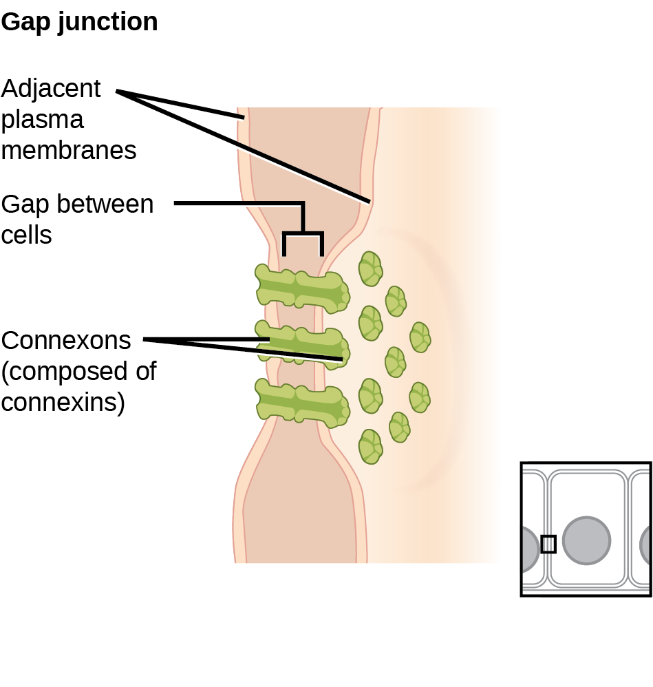
The gonads secrete sexual , which are important in reproduction, and mediate both sexual motivation and behavior. The female gonads are the ovaries; the male gonads are the testes. Ovaries secrete estrogens and , and the testes secrete androgens, such as testosterone.

Major Glands and Associated Functions
Gland Associated Function
Pituitary , releasing and inhibiting (such as thyroid stimulating ) Regulate growth, regulate release
Thyroid Thyroxine, triiodothyronine Regulate and appetite
Pineal Regulate some biological rhythms such as sleep cycles
Adrenal Epinephrine, norepinephrine Stress response, increase activities
, Regulate blood sugar levels
Ovaries Estrogen, Mediate sexual motivation and behavior, reproduction
Testes Androgens, such as testosterone Mediate sexual motivation and behavior, reproduction
Table 3.2

Dig Deeper

Athletes and Anabolic Steroids

Although it is against Federal laws and many professional athletic associations (The National Football League, for example) have banned their use, anabolic steroid drugs continue to be used by amateur and professional athletes. The drugs are believed to enhance athletic performance. Anabolic steroid drugs mimic the effects of the body’s own , like testosterone and its derivatives. These drugs have the potential to provide a competitive edge by increasing muscle mass, strength, and endurance, although not all users may experience these results. Moreover, use of performance-enhancing drugs (PEDs) does not come without risks. Anabolic steroid use has been linked with a wide variety of potentially negative outcomes, ranging in severity from largely cosmetic (acne) to life threatening (heart attack). Furthermore, use of these substances can result in profound changes in mood and can increase aggressive behavior (National Institute on Drug Abuse, 2001).

Baseball player Alex Rodriguez (A-Rod) spent the latter part of playing career at the center of a media storm regarding use of illegal PEDs. Rodriguez excelled while using the drugs; success played a large role in negotiating a contract that made him the highest paid player in professional baseball. A subsequent scandal and suspension tarnished reputation and, according to a statement he made once retired, cost him over $40 million. Even lower-profile athletes, particularly in cycling and Olympic sports, have been revealed as steroid users. What are your thoughts on athletes and doping? Why should the use of PEDs be banned or not banned? What advice would you give an athlete who was considering using PEDs?