Connect tissue composition and remodeling to mechanical support and mineral homeostasis.
Support and movement
An endoskeleton supports the body internally and grows through living-tissue remodeling; an exoskeleton provides external support and often requires molting for growth. Human long bones combine a shaft with expanded ends; compact bone resists loading, and trabecular bone distributes forces with less mass. Marrow spaces support blood-cell production or fat storage. Fibrous and cartilaginous joints generally permit little or no motion, while synovial joints have articular cartilage, fluid, and a capsule. Joint geometry determines allowed movement; greater mobility often trades off with stability.
Source: 6.3 Bone Structure · 6.7 Calcium Homeostasis: Interactions of the Skeletal System and Other Organ Systems · 9.1 Classification of Joints
Build and resorb mineralized matrix
Osteoblasts form osteoid rich in collagen and support mineral deposition; embedded osteocytes sense loading and maintain tissue; osteoclasts resorb bone. Hydroxyapatite provides compressive stiffness while collagen contributes tensile resilience. Mechanical loading influences remodeling, and mineral storage couples skeleton to blood Ca²⁺ regulation. PTH can increase Ca²⁺ availability through coordinated bone, kidney, and vitamin-D effects; vitamin D supports intestinal absorption. Hormone effects depend on pattern and context, so continuous and intermittent exposure need not produce identical net bone changes.
Source: 6.3 Bone Structure · 6.7 Calcium Homeostasis: Interactions of the Skeletal System and Other Organ Systems · 9.1 Classification of Joints
Compare cartilage, tendon, and ligament
Cartilage contains chondrocytes in a hydrated extracellular matrix rich in proteoglycans and collagen; its avascular matrix limits repair. Hyaline cartilage supports smooth joint surfaces, elastic cartilage adds flexibility, and fibrocartilage resists combined tension and compression. Tendons connect muscle to bone and transmit force; ligaments connect bone to bone and stabilize joints. Both contain aligned collagen adapted to tensile load. Their function depends on fiber orientation and attachment, not simply on the presence of the same protein.
Source: 6.3 Bone Structure · 6.7 Calcium Homeostasis: Interactions of the Skeletal System and Other Organ Systems · 9.1 Classification of Joints
Practice and reasoning
Question. Why does removing mineral from bone make it more flexible, while disrupting collagen makes it more brittle? Would a tendon normally connect two bones?
Show reasoning
Mineral supplies much of the compressive rigidity; collagen provides a tough tensile framework. Removing either component weakens a different mechanical contribution. Tendons typically connect muscle to bone, whereas ligaments typically connect bone to bone. Tissue composition and anatomical connection must both be considered.
Source: 6.3 Bone Structure · 6.7 Calcium Homeostasis: Interactions of the Skeletal System and Other Organ Systems · 9.1 Classification of Joints
6.3 Bone Structure — figures and tables
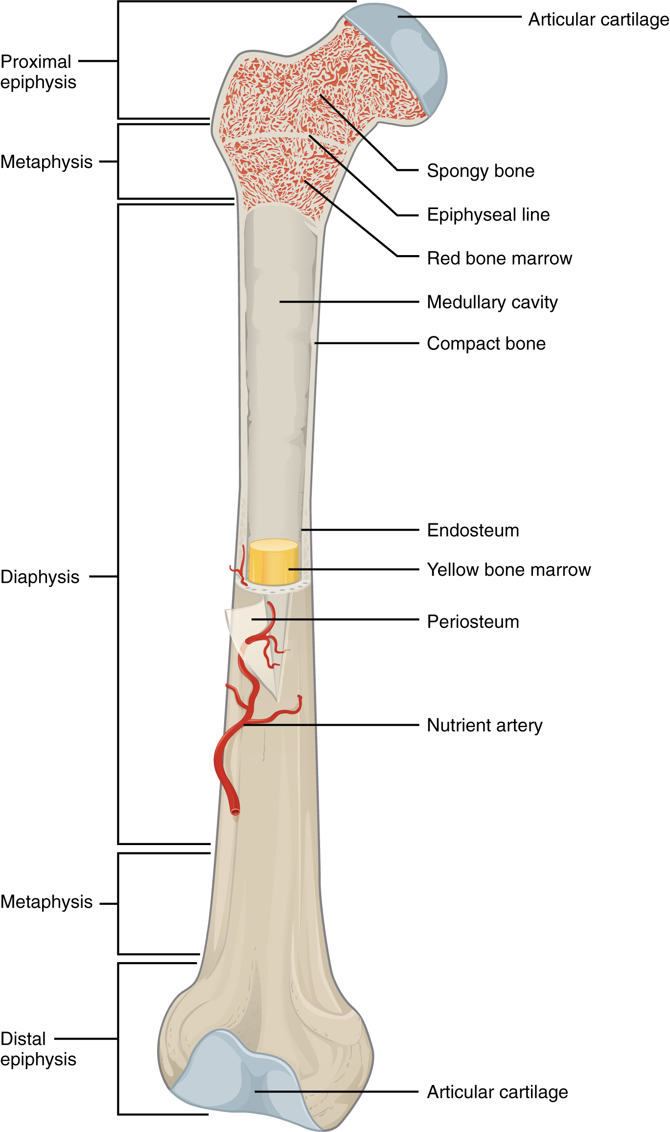
Figure 6.7 Anatomy of a Long Bone A typical long bone shows the gross anatomical characteristics of bone.
J. Gordon Betts and colleagues, Anatomy And Physiology 2E, OpenStax. · CC BY-NC-SA 4.0 · Original image ↗ · Access for free ↗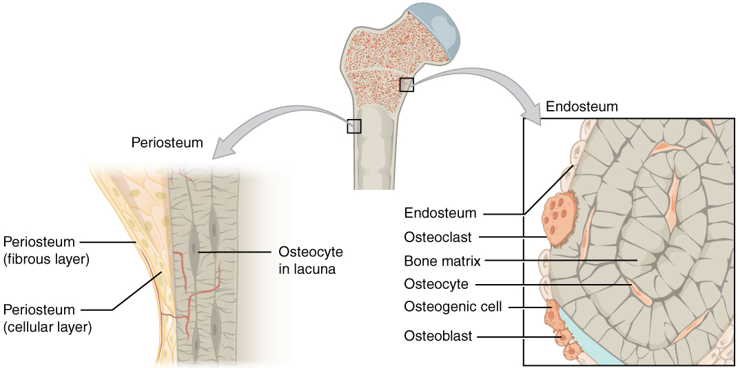
Figure 6.8 Periosteum and Endosteum The periosteum forms the outer surface of bone, and the endosteum lines the medullary cavity.
J. Gordon Betts and colleagues, Anatomy And Physiology 2E, OpenStax. · CC BY-NC-SA 4.0 · Original image ↗ · Access for free ↗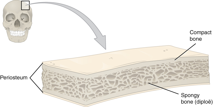
Figure 6.9 Anatomy of a Flat Bone This cross-section of a flat bone shows the spongy bone (diploë) lined on either side by a layer of compact bone.
J. Gordon Betts and colleagues, Anatomy And Physiology 2E, OpenStax. · CC BY-NC-SA 4.0 · Original image ↗ · Access for free ↗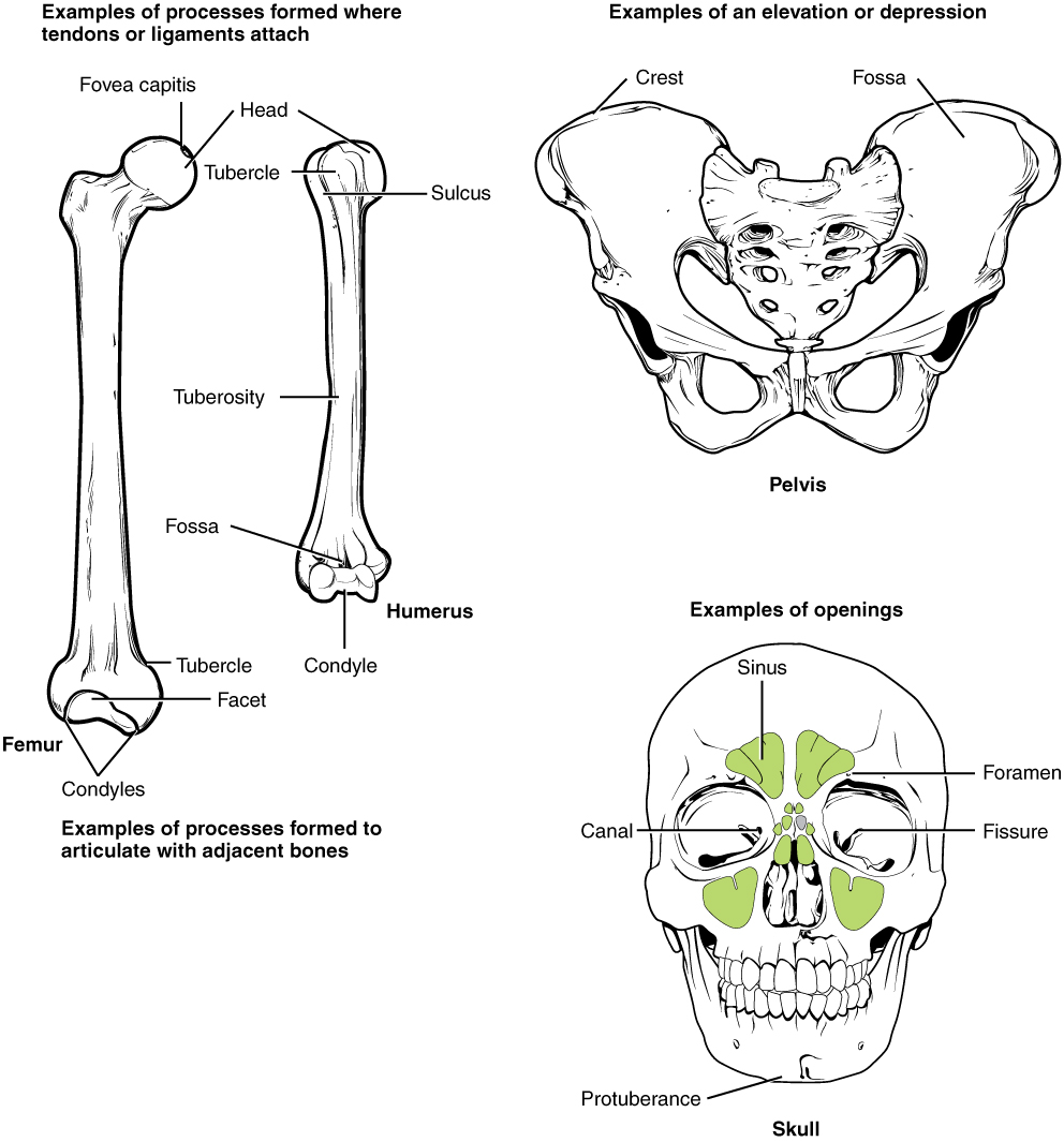
Figure 6.10 Bone Features The surface features of bones depend on their function, location, attachment of ligaments and tendons, or the penetration of blood vessels and nerves.
J. Gordon Betts and colleagues, Anatomy And Physiology 2E, OpenStax. · CC BY-NC-SA 4.0 · Original image ↗ · Access for free ↗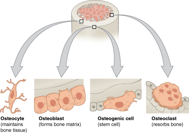
Figure 6.11 Bone Cells Four types of cells are found within bone tissue. Osteogenic cells are undifferentiated and develop into osteoblasts. When osteoblasts get trapped within the calcified matrix, their structure and function changes, and they become osteocytes. Osteoclasts develop from monocytes and macrophages and differ in appearance from other bone cells.
J. Gordon Betts and colleagues, Anatomy And Physiology 2E, OpenStax. · CC BY-NC-SA 4.0 · Original image ↗ · Access for free ↗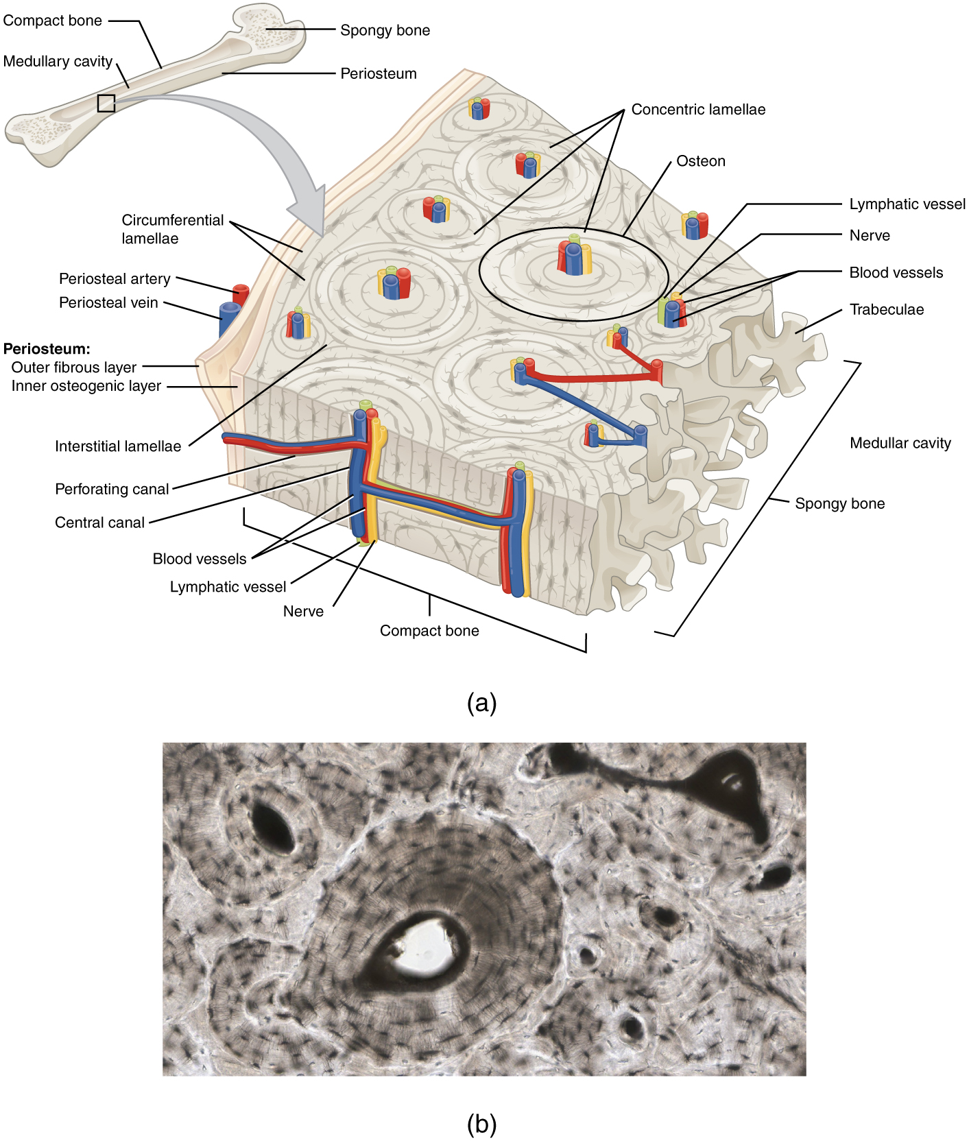
Figure 6.12 Diagram of Compact Bone (a) This cross-sectional view of compact bone shows the basic structural unit, the osteon. (b) In this micrograph of the osteon, you can clearly see the concentric lamellae and central canals. LM × 40. (Micrograph provided by the Regents of University of Michigan Medical School © 2012)
J. Gordon Betts and colleagues, Anatomy And Physiology 2E, OpenStax. · CC BY-NC-SA 4.0 · Original image ↗ · Access for free ↗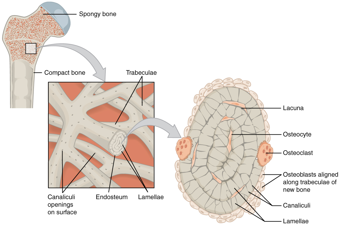
Figure 6.13 Diagram of Spongy Bone Spongy bone is composed of trabeculae that contain the osteocytes. Red marrow fills the spaces in some bones.
J. Gordon Betts and colleagues, Anatomy And Physiology 2E, OpenStax. · CC BY-NC-SA 4.0 · Original image ↗ · Access for free ↗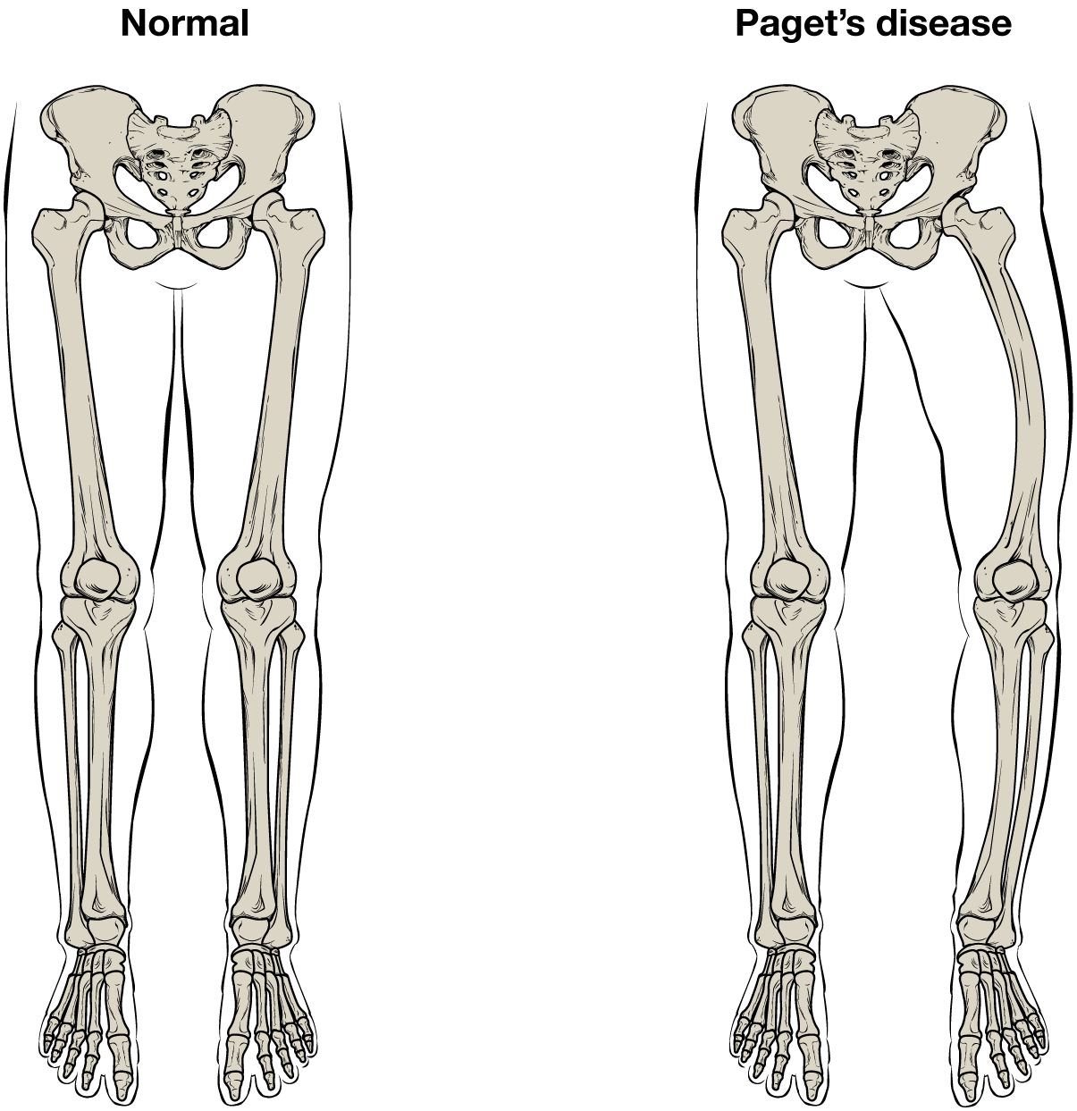
Figure 6.14 Paget's Disease Normal leg bones are relatively straight, but those affected by Paget’s disease are porous and curved.
J. Gordon Betts and colleagues, Anatomy And Physiology 2E, OpenStax. · CC BY-NC-SA 4.0 · Original image ↗ · Access for free ↗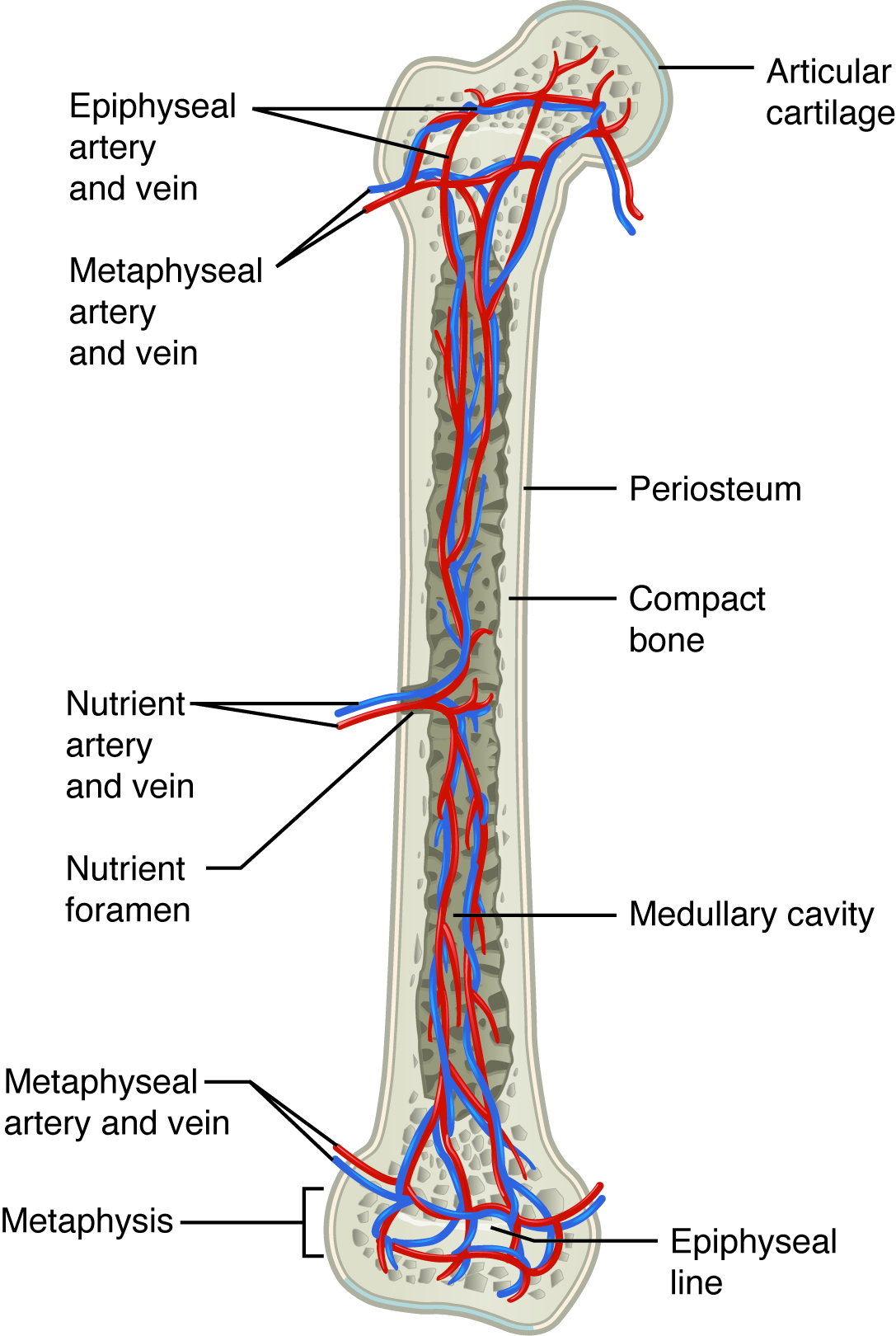
Figure 6.15 Diagram of Blood and Nerve Supply to Bone Blood vessels and nerves enter the bone through the nutrient foramen.
J. Gordon Betts and colleagues, Anatomy And Physiology 2E, OpenStax. · CC BY-NC-SA 4.0 · Original image ↗ · Access for free ↗| Marking | Description | Example |
|---|---|---|
| Articulations | Where two bones meet | Knee joint |
| Head | Prominent rounded surface | Head of femur |
| Facet | Flat surface | Vertebrae |
| Condyle | Rounded surface | Occipital condyles |
| Projections | Raised markings | Spinous process of the vertebrae |
| Protuberance | Protruding | Chin |
| Process | Prominence feature | Transverse process of vertebra |
| Spine | Sharp process | Ischial spine |
| Tubercle | Small, rounded process | Tubercle of humerus |
| Tuberosity | Rough surface | Deltoid tuberosity |
| Line | Slight, elongated ridge | Temporal lines of the parietal bones |
| Crest | Ridge | Iliac crest |
| Holes | Holes and depressions | Foramen (holes through which blood vessels can pass through) |
| Fossa | Elongated basin | Mandibular fossa |
| Fovea | Small pit | Fovea capitis on the head of the femur |
| Sulcus | Groove | Sigmoid sulcus of the temporal bones |
| Canal | Passage in bone | Auditory canal |
| Fissure | Slit through bone | Auricular fissure |
| Foramen | Hole through bone | Foramen magnum in the occipital bone |
| Meatus | Opening into canal | External auditory meatus |
| Sinus | Air-filled space in bone | Nasal sinus |
Table 6.2. J. Gordon Betts and colleagues, Anatomy And Physiology 2E, OpenStax.
| Cell type | Function | Location |
|---|---|---|
| Osteogenic cells | Develop into osteoblasts | Deep layers of the periosteum and the marrow |
| Osteoblasts | Bone formation | Growing portions of bone, including periosteum and endosteum |
| Osteocytes | Maintain mineral concentration of matrix | Entrapped in matrix |
| Osteoclasts | Bone resorption | Bone surfaces and at sites of old, injured, or unneeded bone |
Table 6.3. J. Gordon Betts and colleagues, Anatomy And Physiology 2E, OpenStax.
Source: 6.3 Bone Structure
6.7 Calcium Homeostasis: Interactions of the Skeletal System and Other Organ Systems — figures and tables
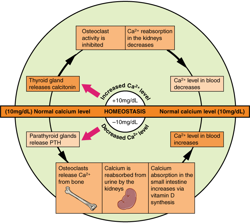
Figure 6.24 Pathways in Calcium Homeostasis The body regulates calcium homeostasis with two pathways; one is signaled to turn on when blood calcium levels drop below normal and one is the pathway that is signaled to turn on when blood calcium levels are elevated.
J. Gordon Betts and colleagues, Anatomy And Physiology 2E, OpenStax. · CC BY-NC-SA 4.0 · Original image ↗ · Access for free ↗Source: 6.7 Calcium Homeostasis: Interactions of the Skeletal System and Other Organ Systems
9.1 Classification of Joints — figures and tables
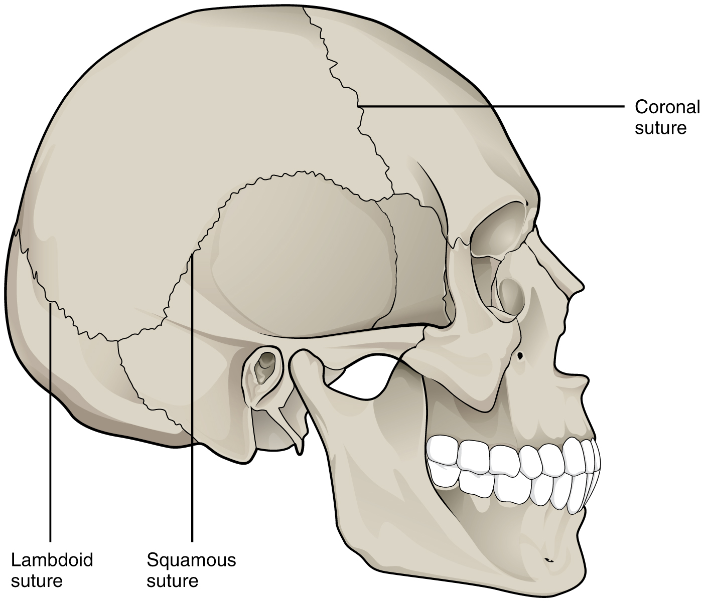
Figure 9.2 Suture Joints of Skull The suture joints of the skull are an example of a synarthrosis, an immobile or essentially immobile joint.
J. Gordon Betts and colleagues, Anatomy And Physiology 2E, OpenStax. · CC BY-NC-SA 4.0 · Original image ↗ · Access for free ↗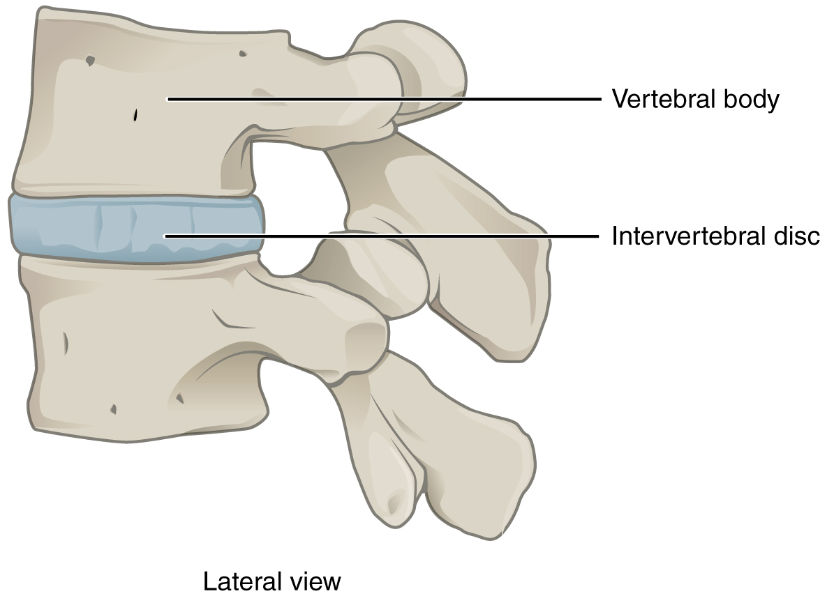
Figure 9.3 Intervertebral Disc An intervertebral disc unites the bodies of adjacent vertebrae within the vertebral column. Each disc allows for limited movement between the vertebrae and thus functionally forms an amphiarthrosis type of joint. Intervertebral discs are made of fibrocartilage and thereby structurally form a symphysis type of cartilaginous joint.
J. Gordon Betts and colleagues, Anatomy And Physiology 2E, OpenStax. · CC BY-NC-SA 4.0 · Original image ↗ · Access for free ↗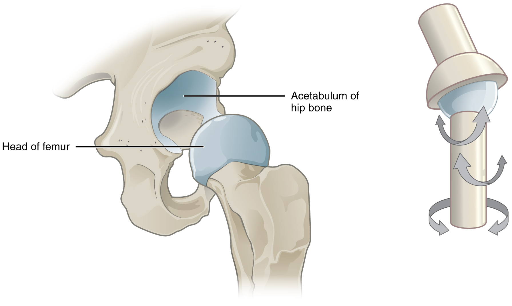
Figure 9.4 Multiaxial Joint A multiaxial joint, such as the hip joint, allows for three types of movement: anterior-posterior, medial-lateral, and rotational.
J. Gordon Betts and colleagues, Anatomy And Physiology 2E, OpenStax. · CC BY-NC-SA 4.0 · Original image ↗ · Access for free ↗Source: 9.1 Classification of Joints