The skin limits water loss, blocks injury, and changes heat exchange through blood flow and evaporation.
Barrier layers and cells
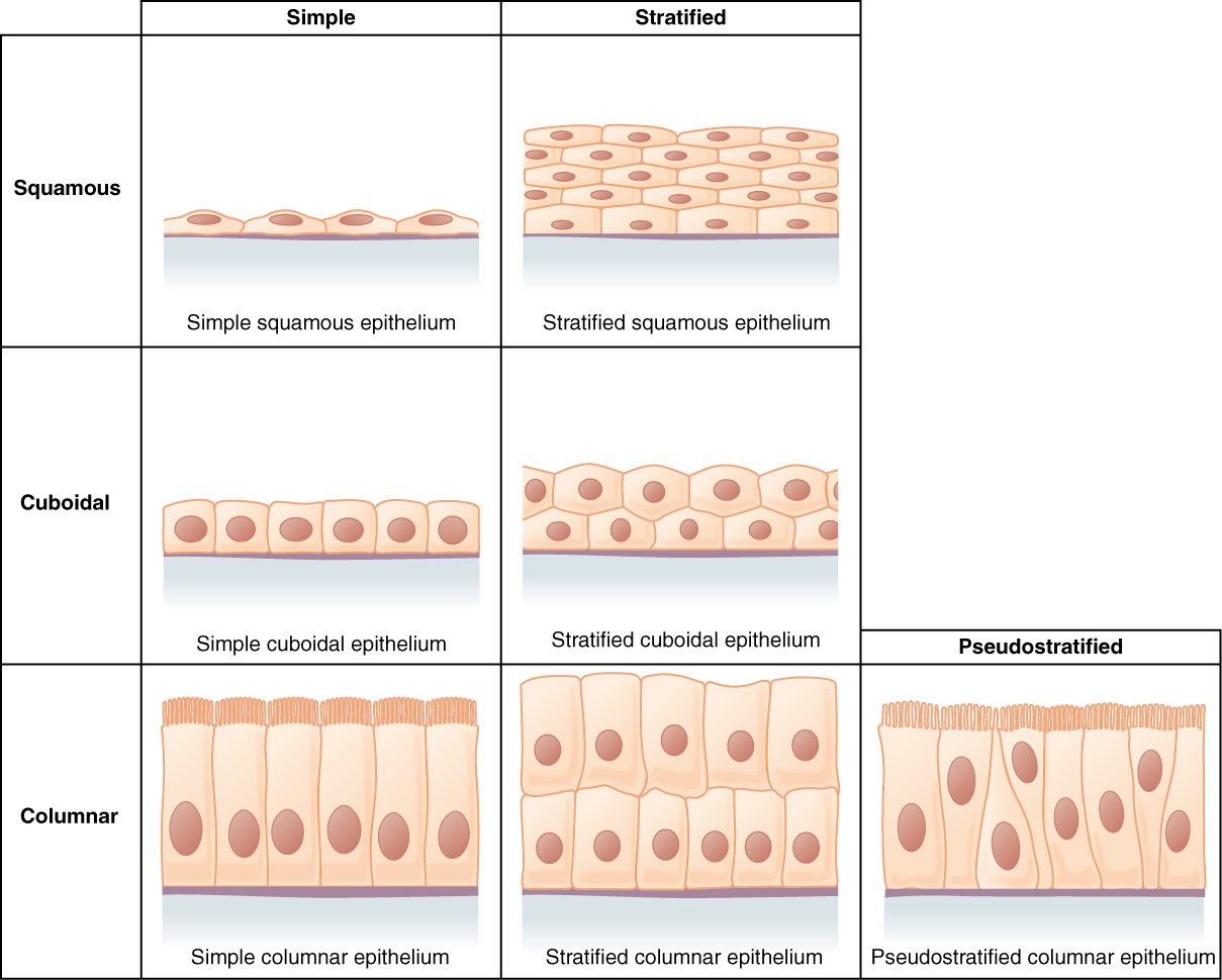
The epidermis is stratified squamous epithelium with keratinocytes that mature into a protective outer layer. Melanocytes supply pigment, epidermal immune cells sample threats, and tactile cells participate in sensation. The dermis contains collagen-rich connective tissue, vessels, nerves, follicles, and glands. The hypodermis beneath the skin contains connective tissue and variable fat. Lipid-rich epidermal barriers reduce water loss, while tight tissue organization limits microbial entry. The epidermis is avascular and obtains nutrients by diffusion from underlying tissue.
Source: 5.1 Layers of the Skin · 5.3 Functions of the Integumentary System
Adjust heat transfer
Dermal vasodilation brings warm blood near the surface, increasing potential heat loss; vasoconstriction limits it. Sweat evaporation removes latent heat, but high humidity reduces its effectiveness. Radiation, convection, and conduction depend on environmental temperature and flow conditions. Subcutaneous fat insulates. Arrector pili muscles raise hairs, a limited thermoregulatory mechanism in humans. Shivering produces heat through skeletal-muscle activity. Sweating without evaporation can lose water without providing the expected cooling.
Source: 5.1 Layers of the Skin · 5.3 Functions of the Integumentary System
Specializations resist abrasion and entry
Keratinized nails protect distal digits and improve manipulation; hair provides localized protection and sensory input; calluses thicken the epidermis in response to repeated mechanical stress. Sebum and skin chemistry contribute to surface conditions, while intact epithelial barriers and resident microbes limit some invaders. Excessive abrasion or burns disrupt both microbial defense and fluid retention. A thicker surface is not universally better: flexibility, sensation, and normal turnover also support tissue function.
Source: 5.1 Layers of the Skin · 5.3 Functions of the Integumentary System
Practice and reasoning
Question. Why can the same sweat production cool poorly on a humid day? What additional danger follows a large loss of epidermal barrier besides infection?
Show reasoning
High humidity reduces the water-vapor gradient, limiting evaporation and its heat removal. Barrier loss also permits excessive water and electrolyte loss and disrupts temperature regulation. Sweat quantity alone is therefore an incomplete measure of cooling effectiveness.
Source: 5.1 Layers of the Skin · 5.3 Functions of the Integumentary System
5.1 Layers of the Skin — figures and tables
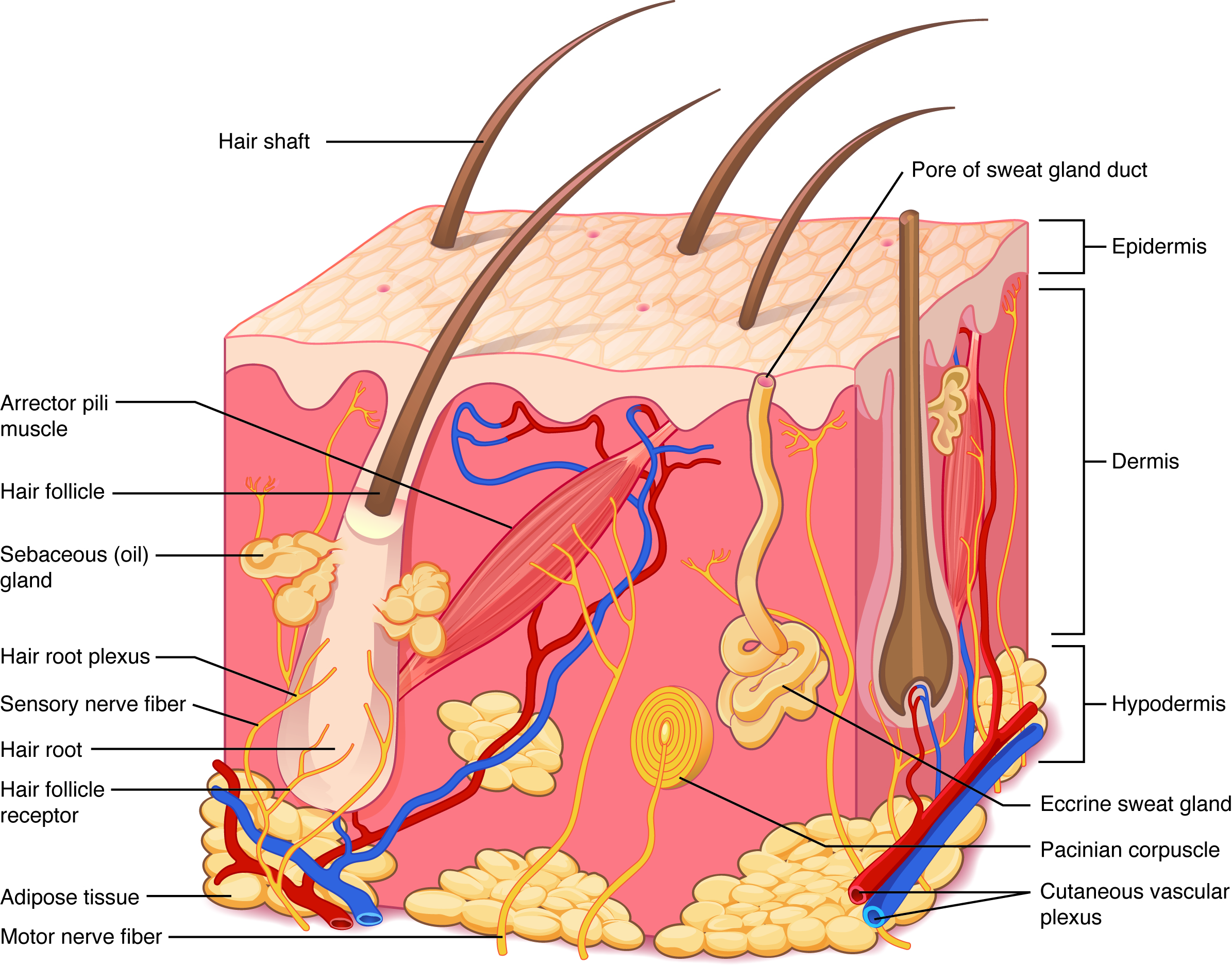
Figure 5.2 Layers of Skin The skin is composed of two main layers: the epidermis, made of closely packed epithelial cells, and the dermis, made of dense, irregular connective tissue that houses blood vessels, hair follicles, sweat glands, and other structures. Beneath the dermis lies the hypodermis, which is composed mainly of loose connective and fatty tissues.
J. Gordon Betts and colleagues, Anatomy And Physiology 2E, OpenStax. · CC BY-NC-SA 4.0 · Original image ↗ · Access for free ↗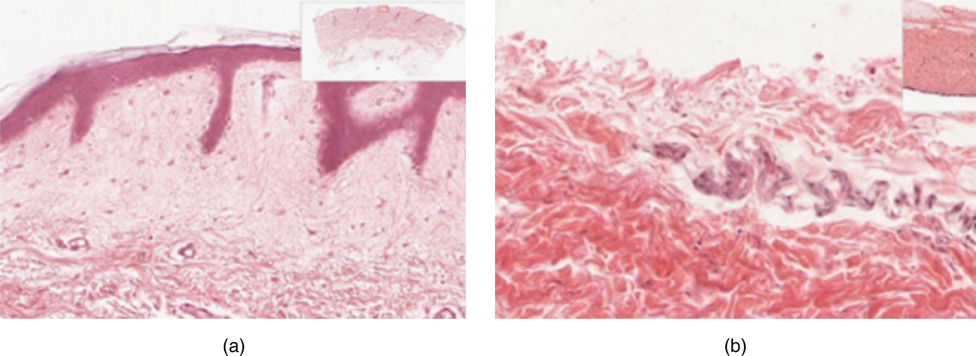
Figure 5.3 Thin Skin versus Thick Skin These slides show cross-sections of the epidermis and dermis of (a) thin and (b) thick skin. Note the significant difference in the thickness of the epithelial layer of the thick skin. From top, LM × 40, LM × 40. (Micrographs provided by the Regents of University of Michigan Medical School © 2012)
J. Gordon Betts and colleagues, Anatomy And Physiology 2E, OpenStax. · CC BY-NC-SA 4.0 · Original image ↗ · Access for free ↗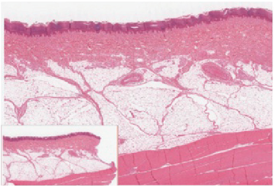
Figure 5.4 Epidermis The epidermis is epithelium composed of multiple layers of cells. The basal layer consists of cuboidal cells, whereas the outer layers are squamous, keratinized cells, so the whole epithelium is often described as being keratinized stratified squamous epithelium. LM × 40. (Micrograph provided by the Regents of University of Michigan Medical School © 2012)
J. Gordon Betts and colleagues, Anatomy And Physiology 2E, OpenStax. · CC BY-NC-SA 4.0 · Original image ↗ · Access for free ↗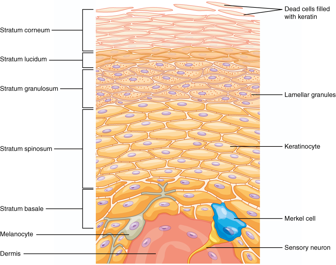
Figure 5.5 Layers of the Epidermis The epidermis of thick skin has five layers: stratum basale, stratum spinosum, stratum granulosum, stratum lucidum, and stratum corneum.
J. Gordon Betts and colleagues, Anatomy And Physiology 2E, OpenStax. · CC BY-NC-SA 4.0 · Original image ↗ · Access for free ↗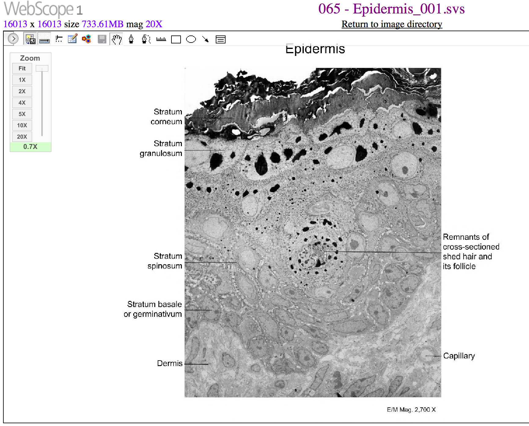
Figure 5.6 Cells of the Epidermis The cells in the different layers of the epidermis originate from basal cells located in the stratum basale, yet the cells of each layer are distinctively different. EM × 2700. (Micrograph provided by the Regents of University of Michigan Medical School © 2012)
J. Gordon Betts and colleagues, Anatomy And Physiology 2E, OpenStax. · CC BY-NC-SA 4.0 · Original image ↗ · Access for free ↗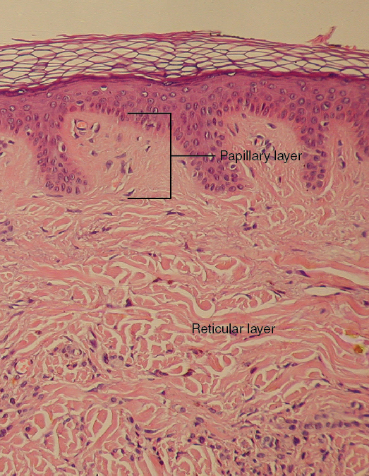
Figure 5.7 Layers of the Dermis This stained slide shows the two components of the dermis—the papillary layer and the reticular layer. Both are made of connective tissue with fibers of collagen extending from one to the other, making the border between the two somewhat indistinct. The dermal papillae extending into the epidermis belong to the papillary layer, whereas the dense collagen fiber bundles below belong to the reticular layer. LM × 10. (credit: modification of work by “kilbad”/Wikimedia Commons)
J. Gordon Betts and colleagues, Anatomy And Physiology 2E, OpenStax. (credit: modification of work by “kilbad”/Wikimedia Commons) · CC BY-NC-SA 4.0 · Original image ↗ · Access for free ↗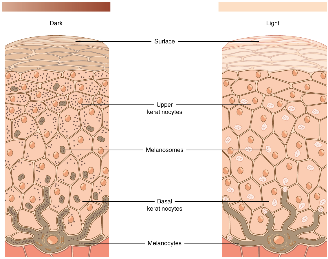
Figure 5.8 Skin Pigmentation The relative coloration of the skin depends on the amount of melanin produced by melanocytes in the stratum basale and taken up by keratinocytes.
J. Gordon Betts and colleagues, Anatomy And Physiology 2E, OpenStax. · CC BY-NC-SA 4.0 · Original image ↗ · Access for free ↗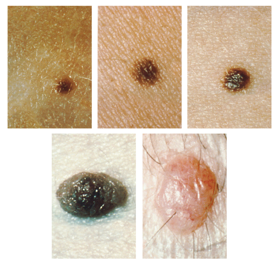
Figure 5.9 Moles Moles range from benign accumulations of melanocytes to melanomas. These structures populate the landscape of our skin. (credit: the National Cancer Institute)
J. Gordon Betts and colleagues, Anatomy And Physiology 2E, OpenStax. (credit: the National Cancer Institute) · CC BY-NC-SA 4.0 · Original image ↗ · Access for free ↗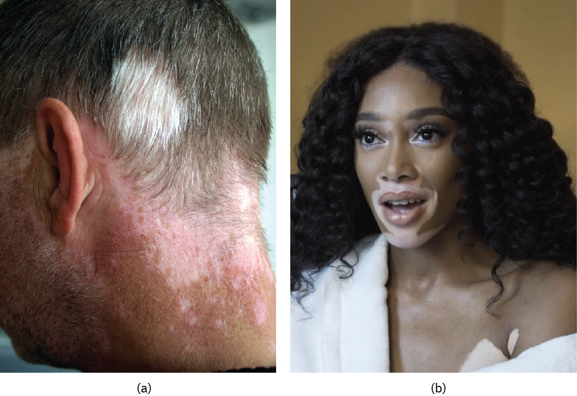
Figure 5.10 Vitiligo Individuals with vitiligo experience depigmentation that results in lighter colored patches of skin. The condition is especially noticeable on darker skin. (credit: (a) Klaus D. Peter (b) Owl Bridge Media / Wikimedia.)
J. Gordon Betts and colleagues, Anatomy And Physiology 2E, OpenStax. (credit: (a) Klaus D. Peter (b) Owl Bridge Media / Wikimedia.) · CC BY-NC-SA 4.0 · Original image ↗ · Access for free ↗Source: 5.1 Layers of the Skin
5.3 Functions of the Integumentary System — figures and tables
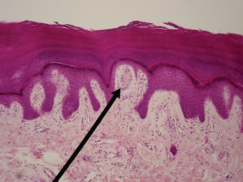
Figure 5.15 Light Micrograph of a Meissner Corpuscle In this micrograph of a skin cross-section, you can see a Meissner corpuscle (arrow), a type of touch receptor located in a dermal papilla adjacent to the basement membrane and stratum basale of the overlying epidermis. LM × 100. (credit: “Wbensmith”/Wikimedia Commons)
J. Gordon Betts and colleagues, Anatomy And Physiology 2E, OpenStax. (credit: “Wbensmith”/Wikimedia Commons) · CC BY-NC-SA 4.0 · Original image ↗ · Access for free ↗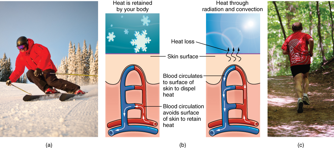
Figure 5.16 Thermoregulation During strenuous physical activities, such as skiing (a) or running (c), the dermal blood vessels dilate and sweat secretion increases (b). These mechanisms prevent the body from overheating. In contrast, the dermal blood vessels constrict to minimize heat loss in response to low temperatures (b). (credit a: “Trysil”/flickr; credit c: Ralph Daily)
J. Gordon Betts and colleagues, Anatomy And Physiology 2E, OpenStax. (credit a: “Trysil”/flickr; credit c: Ralph Daily) · CC BY-NC-SA 4.0 · Original image ↗ · Access for free ↗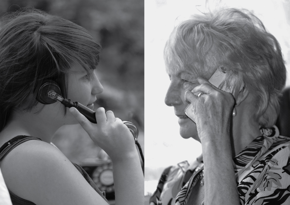
Figure 5.17 Aging Generally, skin, especially on the face and hands, starts to display the first noticeable signs of aging, as it loses its elasticity over time. (credit: Janet Ramsden)
J. Gordon Betts and colleagues, Anatomy And Physiology 2E, OpenStax. (credit: Janet Ramsden) · CC BY-NC-SA 4.0 · Original image ↗ · Access for free ↗