Foundation 3 · 3B · Integrated organ systems

Heart, vessels, and pressure

Sources ↗

Blood flow requires a pressure difference; the heart generates it and the vascular network distributes resistance.

Trace the two circuits

Systemic venous blood enters the right through venae cavae, crosses the to the right , then passes the and arteries to lungs. Pulmonary veins return blood to the left ; the leads to the left , whose output crosses the into systemic arteries. Valves open and close with pressure . The left develops greater pressure for systemic resistance, while the two normally pump the same average flow. Artery and vein refer to direction relative to the heart, not oxygen content.

Source: 19.1 Heart Anatomy · 19.4 Cardiac Physiology · 20.2 Blood Flow, Blood Pressure, and Resistance · 20.3 Capillary Exchange

Structure supports vascular function

Arteries have elastic and muscular walls that tolerate pulsatile pressure. provide major adjustable resistance through smooth-muscle tone. Thin capillaries maximize exchange; their enormous combined cross-sectional area lowers average blood . collect capillary outflow, and compliant veins store blood and return it at low pressure. Venous valves, skeletal-muscle contraction, and breathing assist return. forms a selective barrier and releases regulators such as nitric oxide. Flow = ΔP/R; for , radius has a strong fourth-power influence on resistance.

Source: 19.1 Heart Anatomy · 19.4 Cardiac Physiology · 20.2 Blood Flow, Blood Pressure, and Resistance · 20.3 Capillary Exchange

Capillary exchange and fluid balance

Gases and many diffuse according to ; water movement depends on hydrostatic and osmotic forces and barrier properties. Plasma contribute , while capillary promotes filtration. Modern models emphasize net filtration with lymphatic return rather than obligatory sustained venous-end in every tissue. Increased pressure, loss, increased permeability, or impaired drainage can promote . Exchange of warm blood with tissues also redistributes heat. Plasma volume depends jointly on intake, renal handling, capillary flux, and return.

Source: 19.1 Heart Anatomy · 19.4 Cardiac Physiology · 20.2 Blood Flow, Blood Pressure, and Resistance · 20.3 Capillary Exchange

Relate to arterial pressure

equals heart rate × . depends on filling, , and . is approximately × when right-atrial pressure is small. Systolic pressure during ventricular ejection; diastolic pressure reflects runoff and elastic recoil between beats. feedback changes autonomic output rapidly. –angiotensin– and influence vascular tone and volume over longer times. Increasing heart rate excessively can reduce filling, so output does not rise without limit.

Source: 19.1 Heart Anatomy · 19.4 Cardiac Physiology · 20.2 Blood Flow, Blood Pressure, and Resistance · 20.3 Capillary Exchange

Practice and reasoning

Question. A heart beats 75 times/min and ejects 80 mL/beat. Calculate . What happens to resistance if an ’s radius halves with other laminar-flow factors fixed?

Show reasoning

= 75 × 80 = 6000 mL/min = 6 L/min. Resistance varies as 1/r⁴, so halving radius increases resistance sixteenfold. Actual systemic pressure also depends on parallel vessels, autoregulation, and cardiac responses; a single-vessel calculation does not directly predict whole-body flow.

Source: 19.1 Heart Anatomy · 19.4 Cardiac Physiology · 20.2 Blood Flow, Blood Pressure, and Resistance · 20.3 Capillary Exchange

19.1 Heart Anatomy — figures and tables
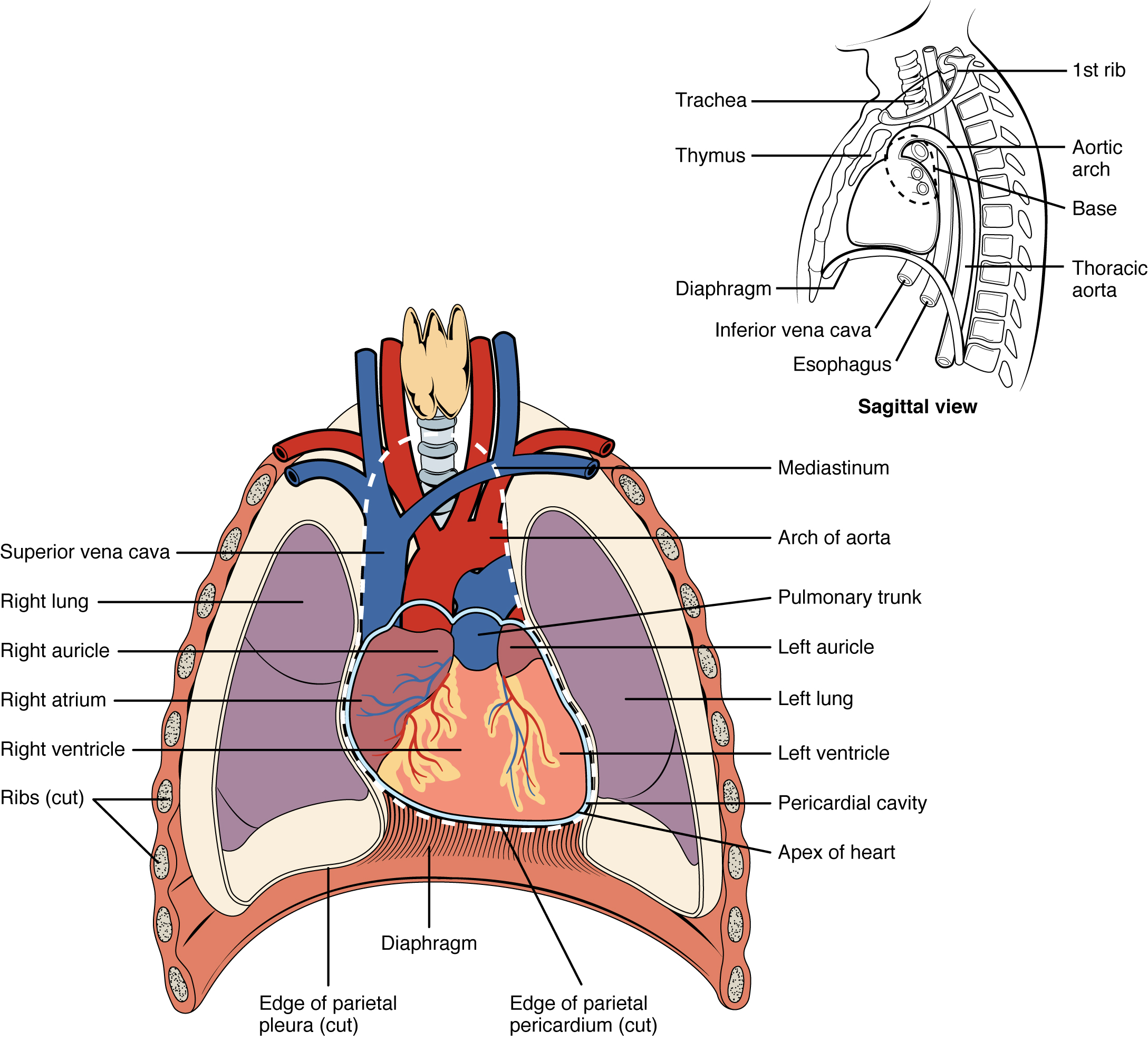
This diagram shows the location of the heart in the thorax.
Figure 19.2 — 19.1 Heart Anatomy

Figure 19.2 Position of the Heart in the Thorax The heart is located within the thoracic cavity, medially between the lungs in the mediastinum. It is about the size of a fist, is broad at the top (called the base), and tapers toward the apex.

J. Gordon Betts and colleagues, Anatomy And Physiology 2E, OpenStax. · CC BY-NC-SA 4.0 · Original image ↗ · Access for free ↗
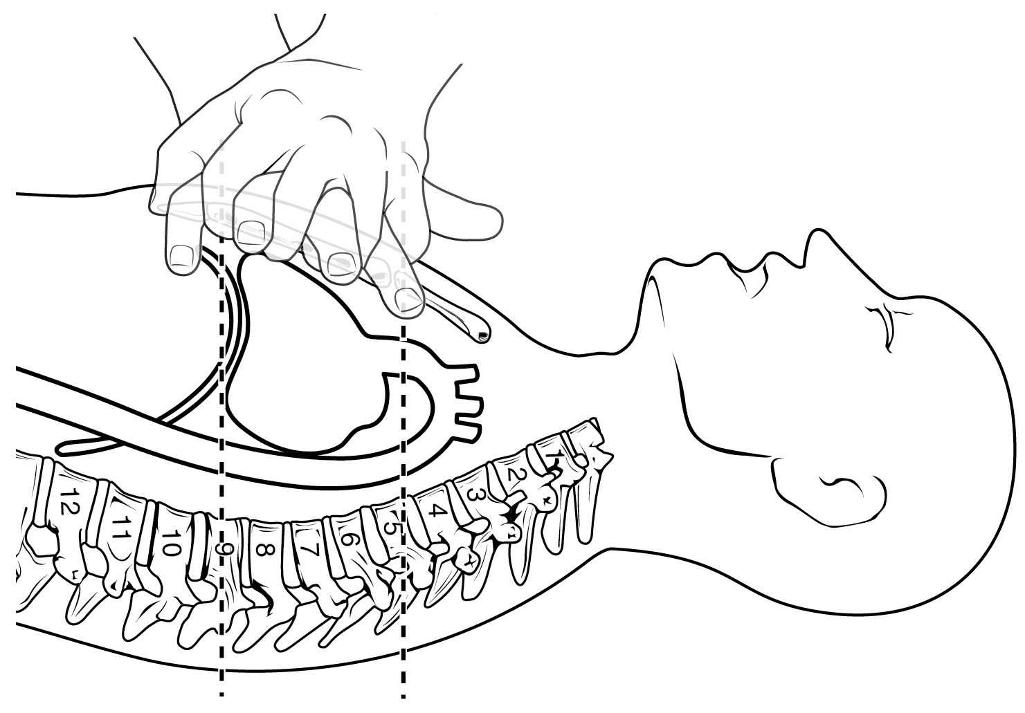
The top panel shows a schematic of a person performing CPR and demarcates the region in the chest where the compression must be performed. The bottom panel shows a photo of a person performing CPR on a dummy.
Figure 19.3 — 19.1 Heart Anatomy

Figure 19.3 CPR Technique If the heart should stop, CPR can maintain the flow of blood until the heart resumes beating. By applying pressure to the sternum, the blood within the heart will be squeezed out of the heart and into the circulation. Proper positioning of the hands on the sternum to perform CPR would be between the lines at T4 and T9.

J. Gordon Betts and colleagues, Anatomy And Physiology 2E, OpenStax. · CC BY-NC-SA 4.0 · Original image ↗ · Access for free ↗
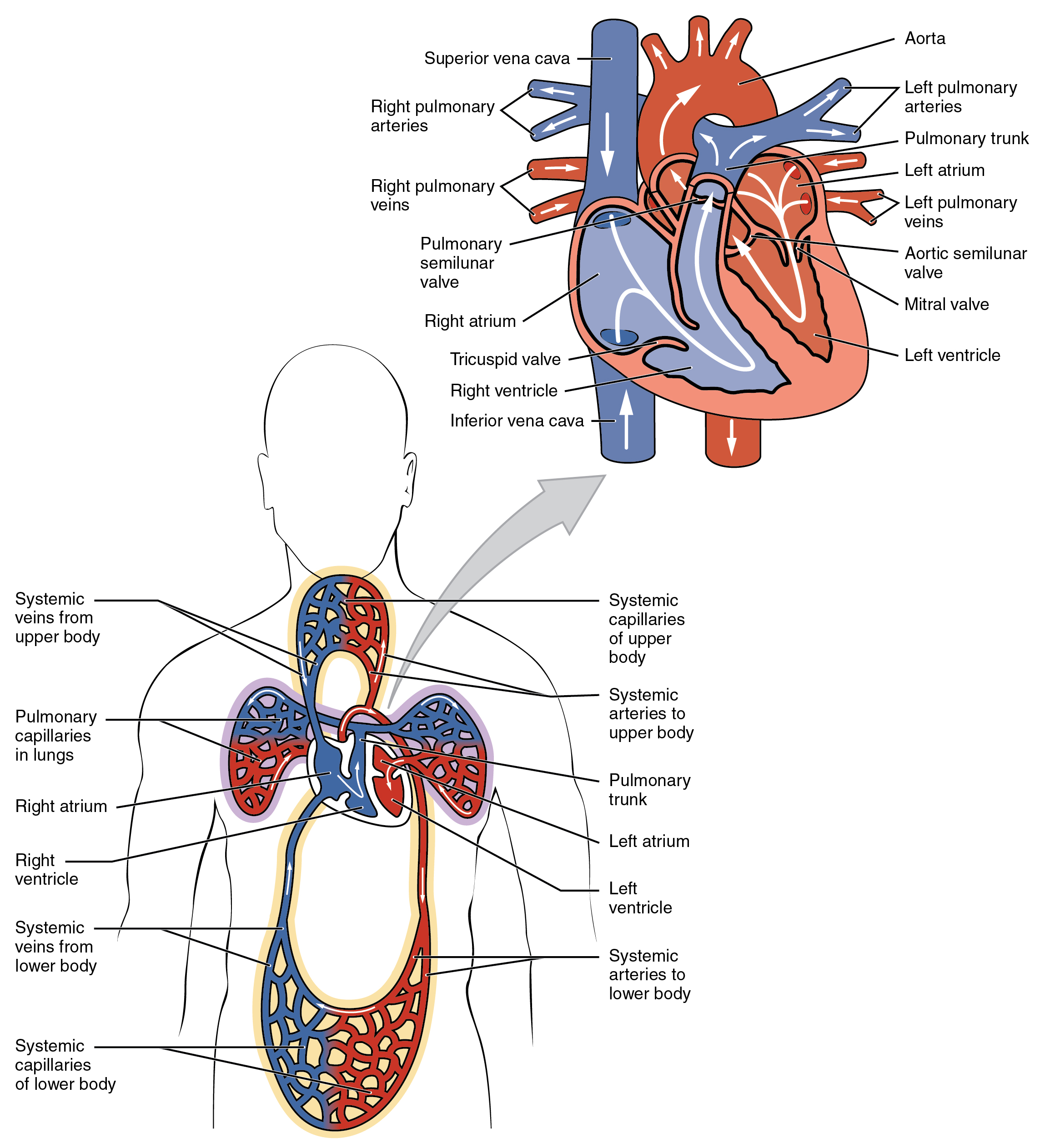
The top panel shows the human heart with the arteries and veins labeled. The bottom panel shows the human circulatory system.
Figure 19.4 — 19.1 Heart Anatomy

Figure 19.4 Dual System of the Human Blood Circulation Blood flows from the right atrium to the right ventricle, where it is pumped into the pulmonary circuit. The blood in the pulmonary artery branches is low in oxygen but relatively high in carbon dioxide. Gas exchange occurs in the pulmonary capillaries (oxygen into the blood, carbon dioxide out), and blood high in oxygen and low in carbon dioxide is returned to the left atrium. From here, blood enters the left ventricle, which pumps it into the systemic circuit. Following exchange in the systemic capillaries (oxygen and nutrients out of the capillaries and carbon dioxide and wastes in), blood returns to the right atrium and the cycle is repeated.

J. Gordon Betts and colleagues, Anatomy And Physiology 2E, OpenStax. · CC BY-NC-SA 4.0 · Original image ↗ · Access for free ↗
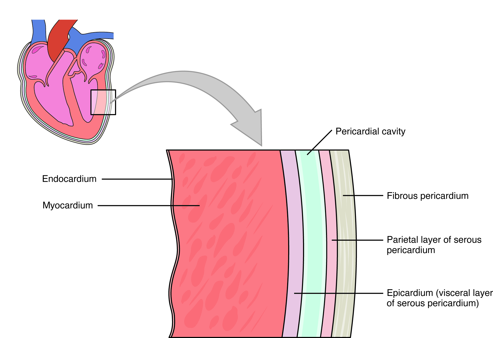
This image shows a magnified view of the structure of the heart wall.
Figure 19.5 — 19.1 Heart Anatomy

Figure 19.5 Pericardial Membranes and Layers of the Heart Wall The pericardial membrane that surrounds the heart consists of three layers and the pericardial cavity. The heart wall also consists of three layers. The pericardial membrane and the heart wall share the epicardium.

J. Gordon Betts and colleagues, Anatomy And Physiology 2E, OpenStax. · CC BY-NC-SA 4.0 · Original image ↗ · Access for free ↗
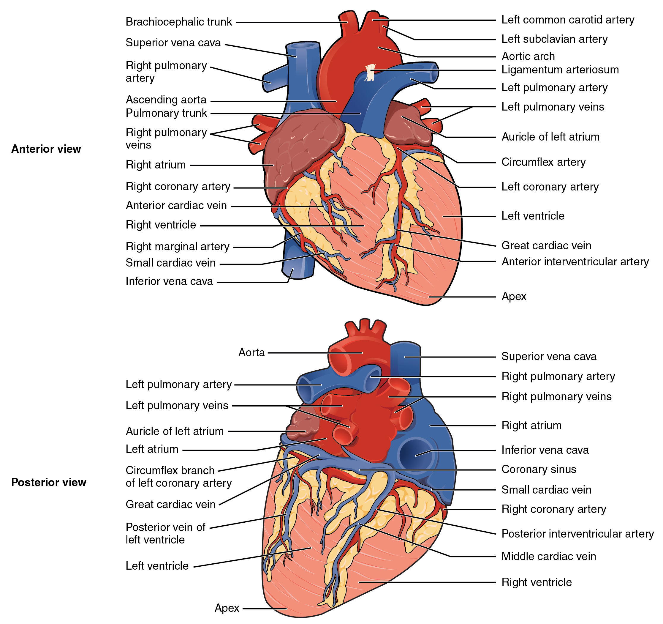
The top panel shows the anterior view of the heart and the bottom panel shows the posterior view of the human heart. In both panels, the main parts of the heart are labeled.
Figure 19.6 — 19.1 Heart Anatomy

Figure 19.6 External Anatomy of the Heart Inside the pericardium, the surface features of the heart are visible.

J. Gordon Betts and colleagues, Anatomy And Physiology 2E, OpenStax. · CC BY-NC-SA 4.0 · Original image ↗ · Access for free ↗
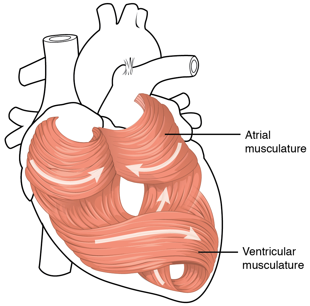
This diagram shows the muscles in the heart.
Figure 19.7 — 19.1 Heart Anatomy

Figure 19.7 Heart Musculature The swirling pattern of cardiac muscle tissue contributes significantly to the heart’s ability to pump blood effectively.

J. Gordon Betts and colleagues, Anatomy And Physiology 2E, OpenStax. · CC BY-NC-SA 4.0 · Original image ↗ · Access for free ↗
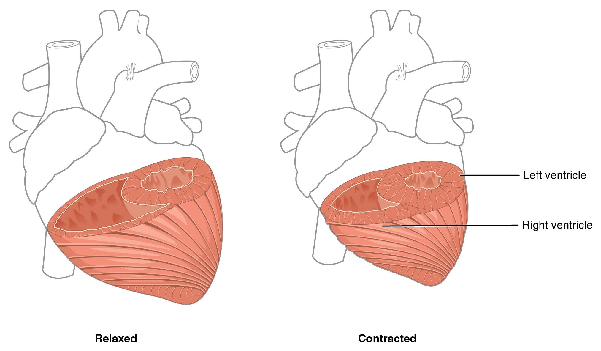
In this figure the left panel shows the muscles of the heart in the relaxed position, and the right panel shows the muscles of the heart in contracted position.
Figure 19.8 — 19.1 Heart Anatomy

Figure 19.8 Differences in Ventricular Muscle Thickness The myocardium in the left ventricle is significantly thicker than that of the right ventricle. Both ventricles pump the same amount of blood, but the left ventricle must generate a much greater pressure to overcome greater resistance in the systemic circuit. The ventricles are shown in both relaxed and contracting states. Note the differences in the relative size of the lumens, the region inside each ventricle where the blood is contained.

J. Gordon Betts and colleagues, Anatomy And Physiology 2E, OpenStax. · CC BY-NC-SA 4.0 · Original image ↗ · Access for free ↗
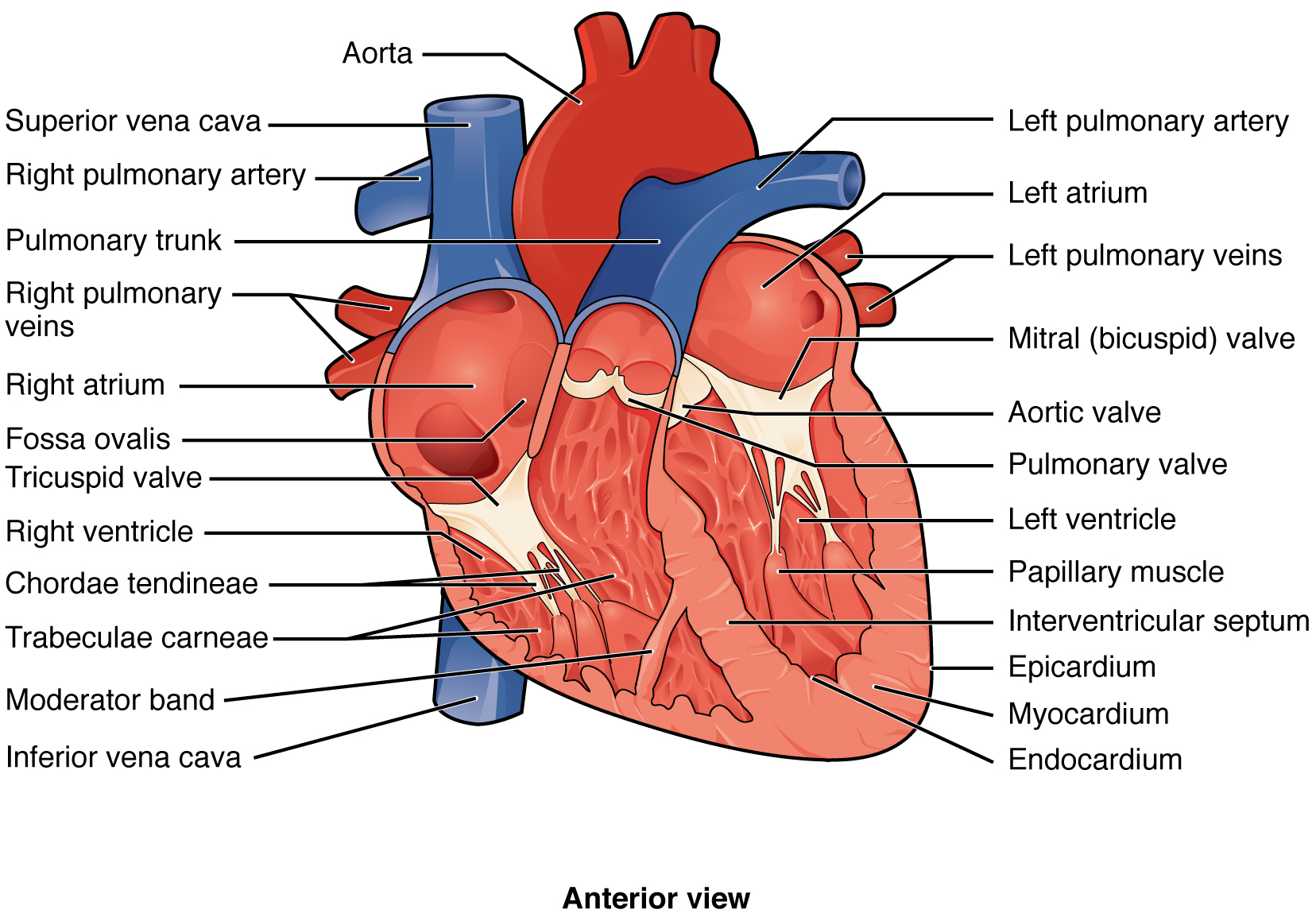
In this figure the top panel shows the image of the heart with the major parts labeled. The bottom left panel shows a photo of the heart with the surface layer peeled off. The images on the bottom right show detailed musculature inside the heart.
Figure 19.9 — 19.1 Heart Anatomy

Figure 19.9 Internal Structures of the Heart This anterior view of the heart shows the four chambers, the major vessels and their early branches, as well as the valves. The presence of the pulmonary trunk and aorta covers the interatrial septum, and the atrioventricular septum is cut away to show the atrioventricular valves.

J. Gordon Betts and colleagues, Anatomy And Physiology 2E, OpenStax. · CC BY-NC-SA 4.0 · Original image ↗ · Access for free ↗
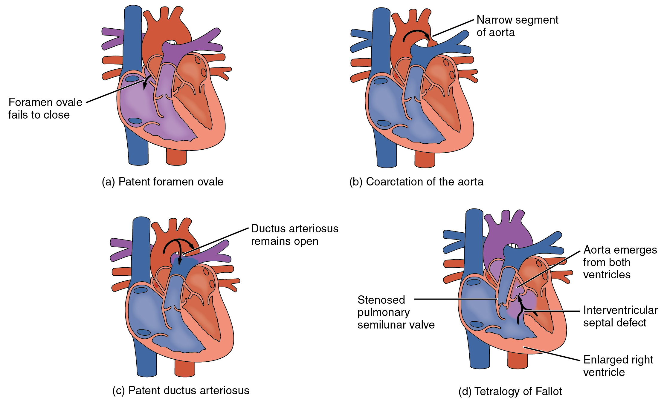
This diagram shows the structure of the heart with different congenital defects. The top left panel shows patent foramen ovale, the top right panel shows coarctation of the aorta, the bottom left panel shows patent ductus ateriosus and the bottom right shows tetralogy of fallot.
Figure 19.10 — 19.1 Heart Anatomy

Figure 19.10 Congenital Heart Defects (a) A patent foramen ovale defect is an abnormal opening in the interatrial septum, or more commonly, a failure of the foramen ovale to close. (b) Coarctation of the aorta is an abnormal narrowing of the aorta. (c) A patent ductus arteriosus is the failure of the ductus arteriosus to close. (d) Tetralogy of Fallot includes an abnormal opening in the interventricular septum.

J. Gordon Betts and colleagues, Anatomy And Physiology 2E, OpenStax. · CC BY-NC-SA 4.0 · Original image ↗ · Access for free ↗
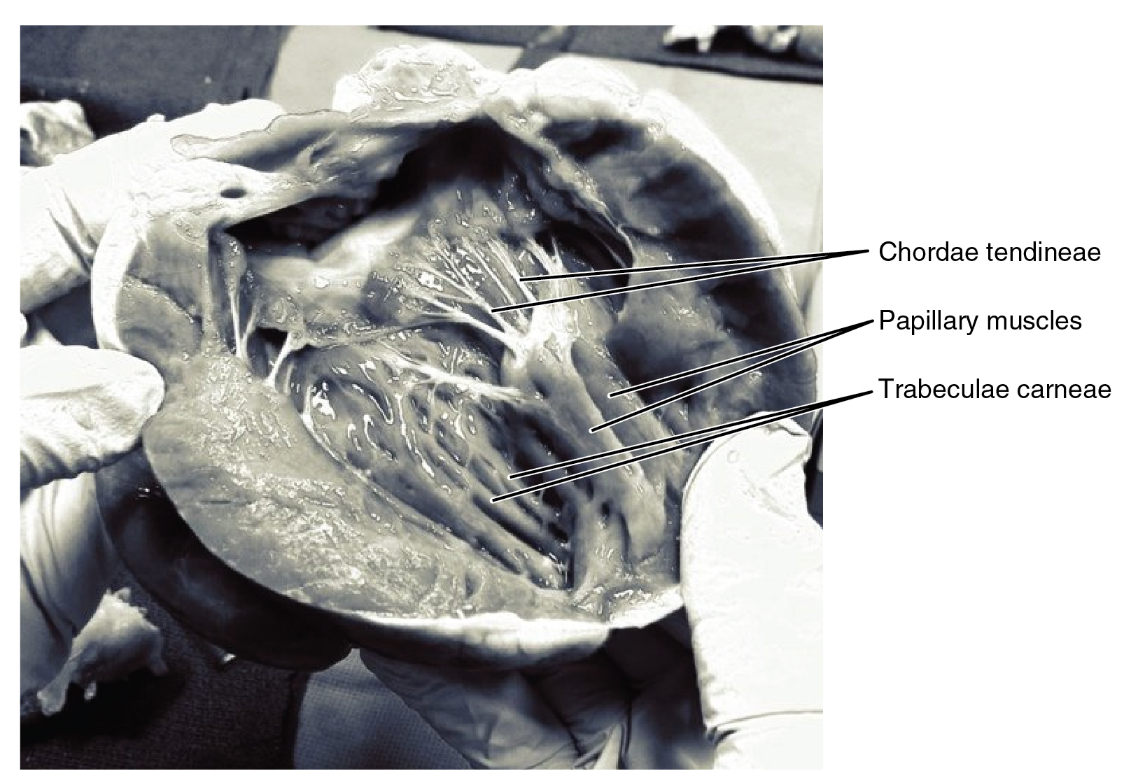
This photo shows the inside of the heart with the main muscles labeled.
Figure 19.11 — 19.1 Heart Anatomy

Figure 19.11 Chordae Tendineae and Papillary Muscles In this frontal section, you can see papillary muscles attached to the tricuspid valve on the right as well as the mitral valve on the left via chordae tendineae. (credit: modification of work by “PV KS”/flickr.com)

J. Gordon Betts and colleagues, Anatomy And Physiology 2E, OpenStax. (credit: modification of work by “PV KS”/flickr.com) · CC BY-NC-SA 4.0 · Original image ↗ · Access for free ↗
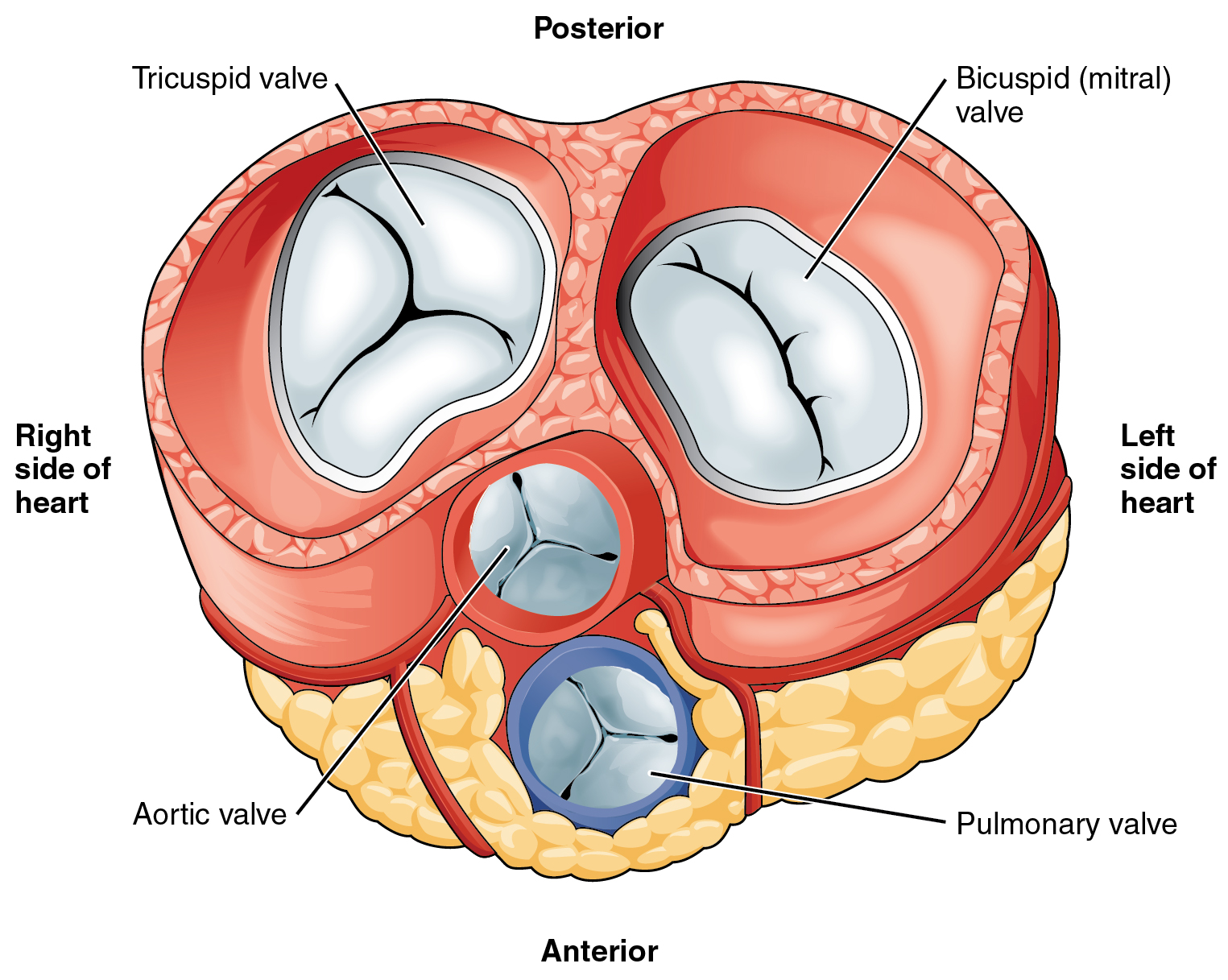
This diagram shows the anterior view of the heart with the different heart valves labeled.
Figure 19.12 — 19.1 Heart Anatomy

Figure 19.12 Heart Valves With the atria and major vessels removed, all four valves are clearly visible, although it is difficult to distinguish the three separate cusps of the tricuspid valve.

J. Gordon Betts and colleagues, Anatomy And Physiology 2E, OpenStax. · CC BY-NC-SA 4.0 · Original image ↗ · Access for free ↗
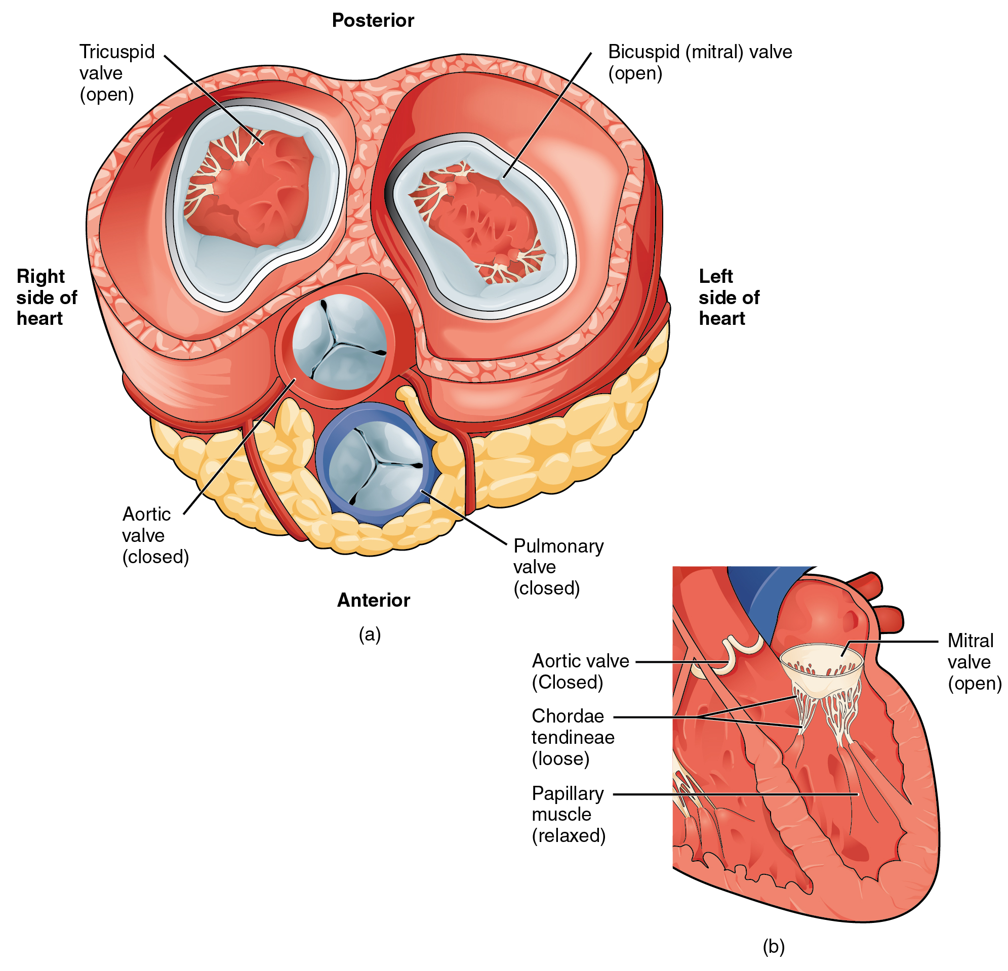
The left panel of this figure shows the anterior view of the heart with the different valves, and the right panel of this figure shows the location of the mitral valve in the open position in the heart.
Figure 19.13 — 19.1 Heart Anatomy

Figure 19.13 Blood Flow from the Left Atrium to the Left Ventricle (a) A transverse section through the heart illustrates the four heart valves. The two atrioventricular valves are open; the two semilunar valves are closed. The atria and vessels have been removed. (b) A frontal section through the heart illustrates blood flow through the mitral valve. When the mitral valve is open, it allows blood to move from the left atrium to the left ventricle. The aortic semilunar valve is closed to prevent backflow of blood from the aorta to the left ventricle.

J. Gordon Betts and colleagues, Anatomy And Physiology 2E, OpenStax. · CC BY-NC-SA 4.0 · Original image ↗ · Access for free ↗
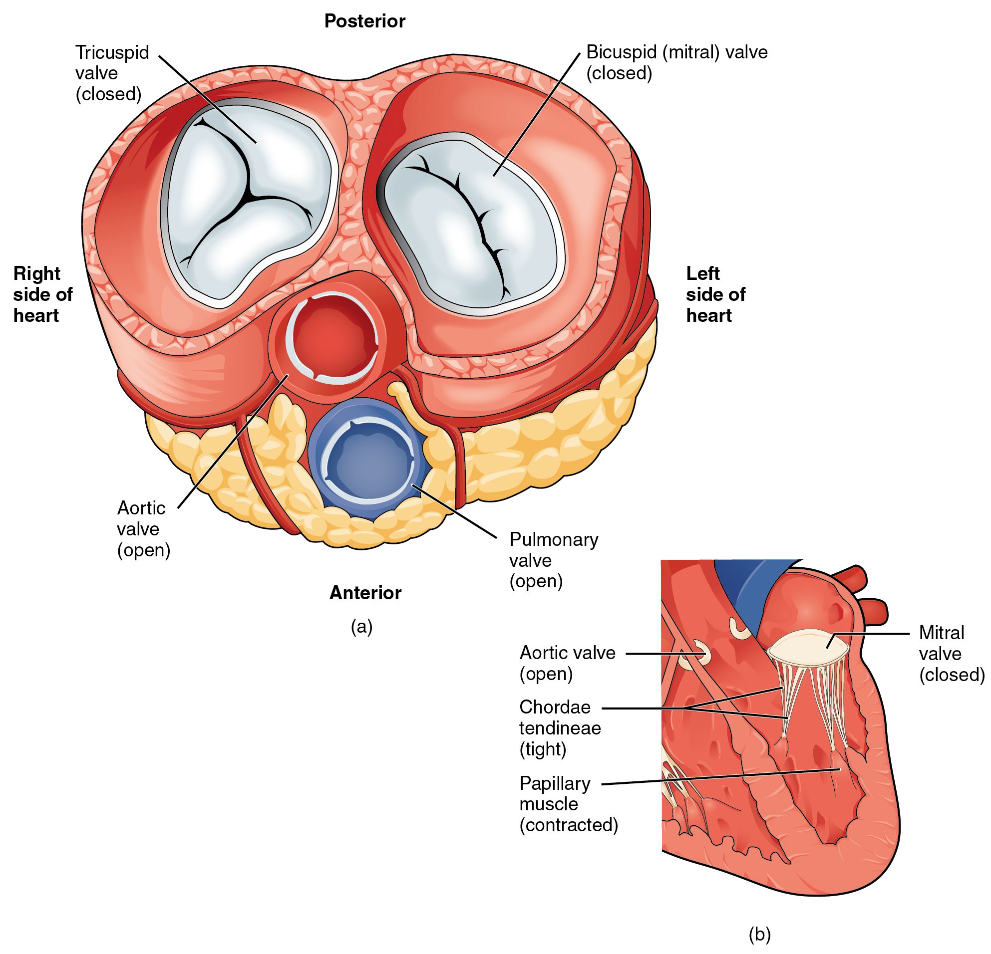
The left panel of this figure shows the anterior view of the heart with the different valves, and the right panel of this figure shows the location of the mitral valve in the closed position in the heart.
Figure 19.14 — 19.1 Heart Anatomy

Figure 19.14 Blood Flow from the Left Ventricle into the Great Vessels (a) A transverse section through the heart illustrates the four heart valves during ventricular contraction. The two atrioventricular valves are closed, but the two semilunar valves are open. The atria and vessels have been removed. (b) A frontal view shows the closed mitral (bicuspid) valve that prevents backflow of blood into the left atrium. The aortic semilunar valve is open to allow blood to be ejected into the aorta.

J. Gordon Betts and colleagues, Anatomy And Physiology 2E, OpenStax. · CC BY-NC-SA 4.0 · Original image ↗ · Access for free ↗
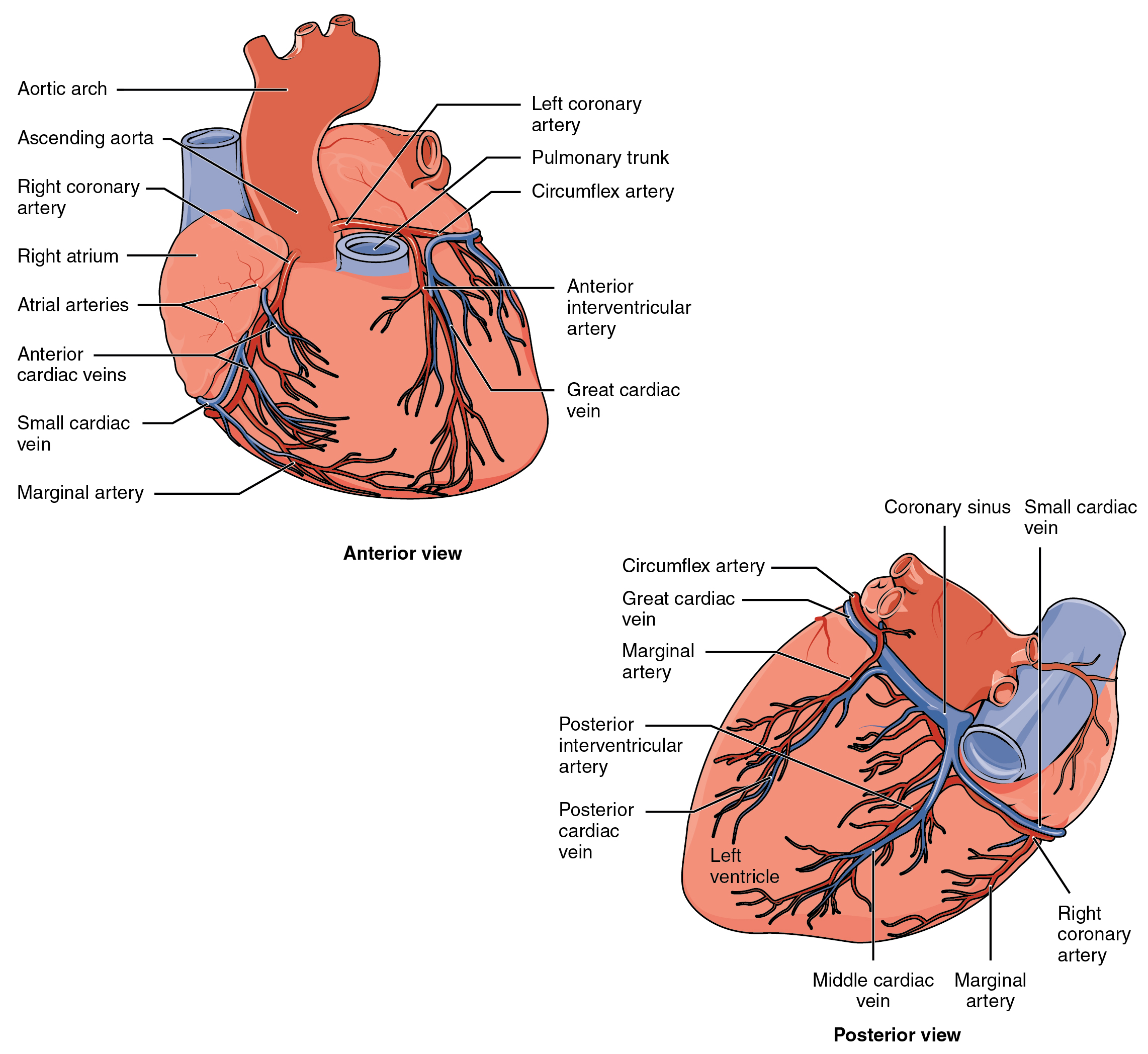
The top panel of this figure shows the anterior view of the heart while the bottom panel shows the posterior view of the heart. The different blood vessels are labeled.
Figure 19.15 — 19.1 Heart Anatomy

Figure 19.15 Coronary Circulation The anterior view of the heart shows the prominent coronary surface vessels. The posterior view of the heart shows the prominent coronary surface vessels.

J. Gordon Betts and colleagues, Anatomy And Physiology 2E, OpenStax. · CC BY-NC-SA 4.0 · Original image ↗ · Access for free ↗
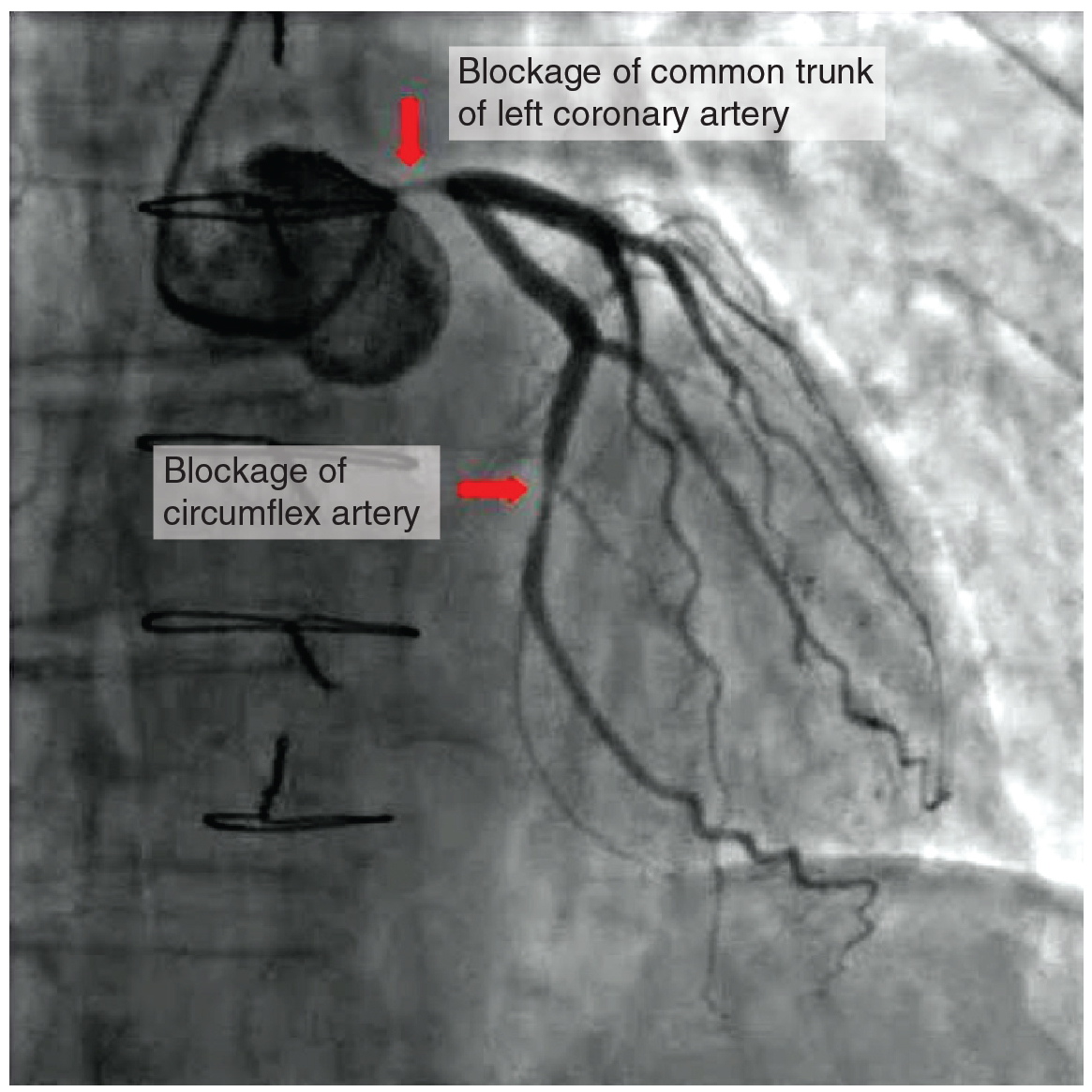
This photo shows a blockage in the coronary artery and in the circumflex artery.
Figure 19.16 — 19.1 Heart Anatomy

Figure 19.16 Atherosclerotic Coronary Arteries In this coronary angiogram (X-ray), the dye makes visible two occluded coronary arteries. Such blockages can lead to decreased blood flow (ischemia) and insufficient oxygen (hypoxia) delivered to the cardiac tissues. If uncorrected, this can lead to cardiac muscle death (myocardial infarction).

J. Gordon Betts and colleagues, Anatomy And Physiology 2E, OpenStax. · CC BY-NC-SA 4.0 · Original image ↗ · Access for free ↗

Source: 19.1 Heart Anatomy

19.4 Cardiac Physiology — figures and tables
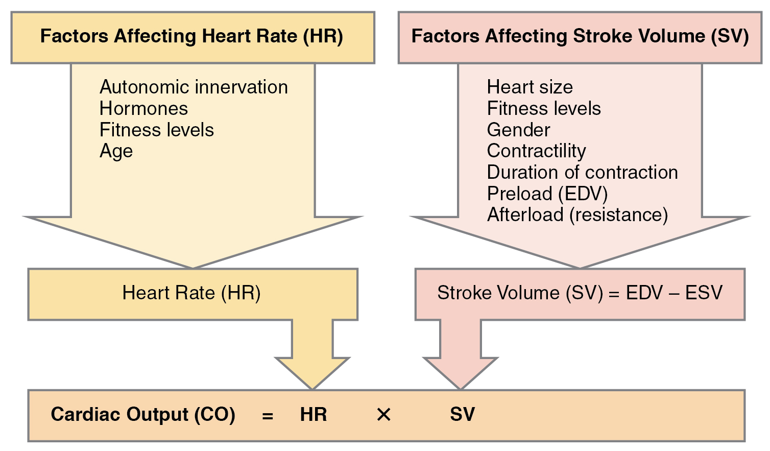
This figure lists the different factors affecting the heart rate and stroke volume. It also shows how they both affect the cardiac output.
Figure 19.31 — 19.4 Cardiac Physiology

Figure 19.31 Major Factors Influencing Cardiac Output Cardiac output is influenced by heart rate and stroke volume, both of which are also variable.

J. Gordon Betts and colleagues, Anatomy And Physiology 2E, OpenStax. · CC BY-NC-SA 4.0 · Original image ↗ · Access for free ↗
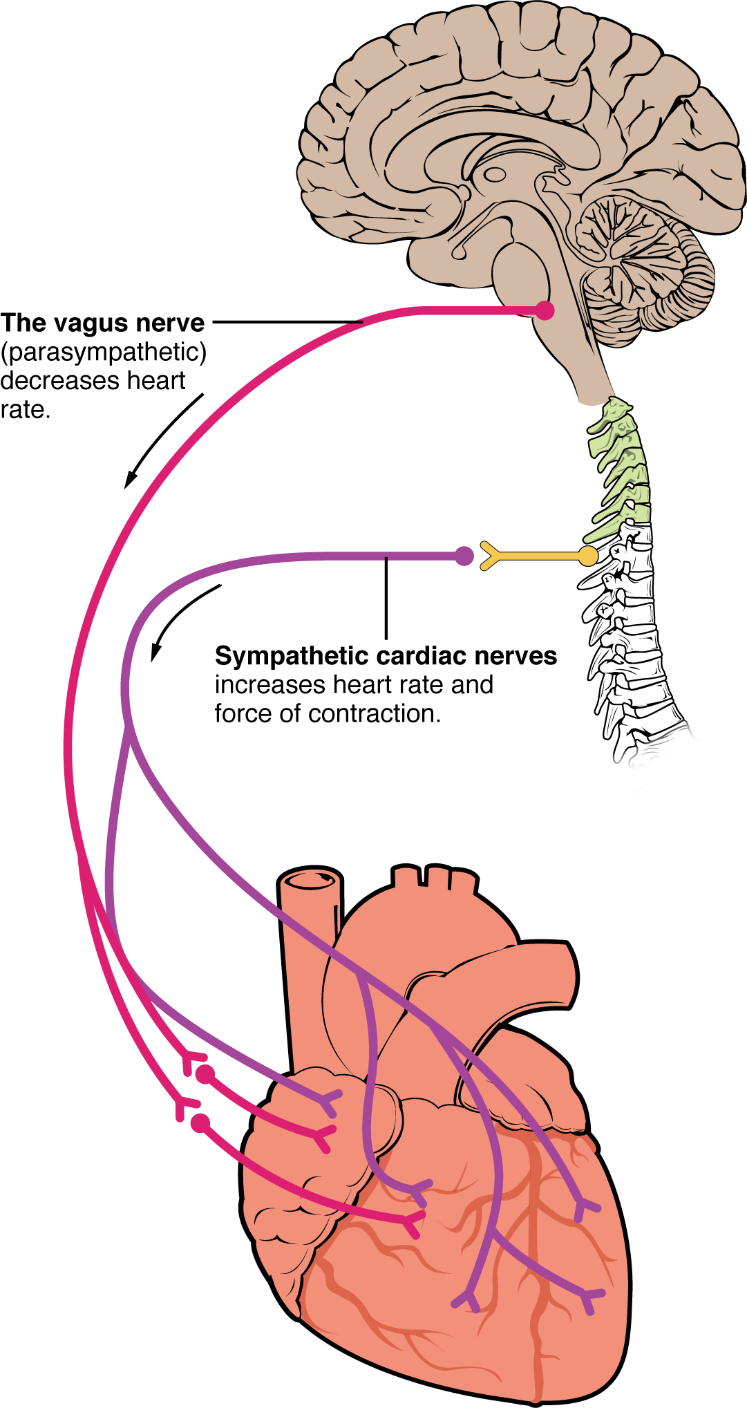
This figure shows the brain and the nerves connecting the brain to the heart.
Figure 19.32 — 19.4 Cardiac Physiology

Figure 19.32 Autonomic Innervation of the Heart Cardioaccelerator and cardioinhibitory areas are components of the paired cardiac centers located in the medulla oblongata of the brain. They innervate the heart via sympathetic cardiac nerves that increase cardiac activity and vagus (parasympathetic) nerves that slow cardiac activity.

J. Gordon Betts and colleagues, Anatomy And Physiology 2E, OpenStax. · CC BY-NC-SA 4.0 · Original image ↗ · Access for free ↗
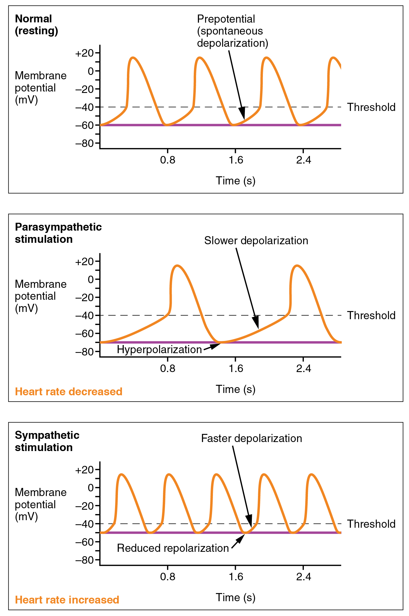
This figure shows three graphs. The top panel shows the normal or resting potential with time. The middle panel shows membrane potential with time in a parasympathetic stimulation where the heart rate is decreased. The bottom panel shows membrane potential with time in a sympathetic stimulation with increased heart rate.
Figure 19.33 — 19.4 Cardiac Physiology

Figure 19.33 Effects of Parasympathetic and Sympathetic Stimulation on Normal Sinus Rhythm The wave of depolarization in a normal sinus rhythm shows a stable resting HR. Following parasympathetic stimulation, HR slows. Following sympathetic stimulation, HR increases.

J. Gordon Betts and colleagues, Anatomy And Physiology 2E, OpenStax. · CC BY-NC-SA 4.0 · Original image ↗ · Access for free ↗
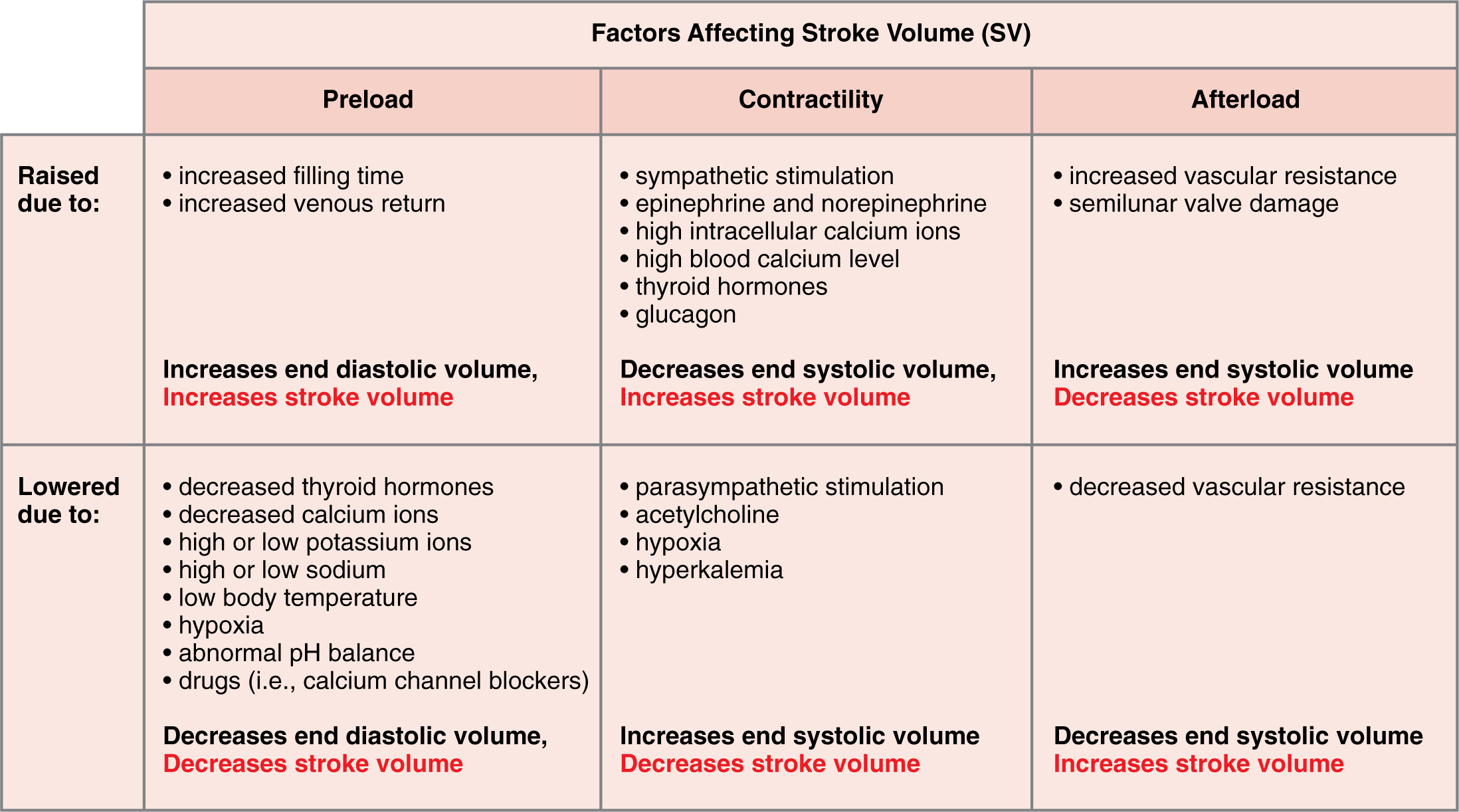
This table describes major factors influencing stroke volume. Preload may be raised due to longer filling time or increased venous return. These factors increase end diastolic volume and increase stroke volume. Preload may be lowered due to decreased thyroid hormones, decreased calcium ions, high or low potassium ions, high or low sodium, low body temperature, hypoxia, abnormal pH balance, or drugs (for example, calcium channel blockers). These factors decrease end diastolic volume and decrease stroke volume. Contractility may be raised due to sympathetic stimulation, epinephrine and norepinephrine, high intracellular calcium ions, high blood calcium level, thyroid hormones, or glucagon. These factors decrease end systolic volume and increase stroke volume. Contractility may be lowered due to parasympathetic stimulation, acetylcholine, hypoxia, or hyperkalemia. These factors increase end systolic volume and decrease stroke volume. Afterload may be raised due to increased vascular resistance or semilunar valve damage. These factors increase end systolic volume and decrease stroke volume. Afterload may be lowered due to decreased vascular resistance. This factor decreases end systolic volume and increases stroke volume.
Figure 19.34 — 19.4 Cardiac Physiology

Figure 19.34 Major Factors Influencing Stroke Volume Multiple factors impact preload, afterload, and contractility, and are the major considerations influencing SV.

J. Gordon Betts and colleagues, Anatomy And Physiology 2E, OpenStax. · CC BY-NC-SA 4.0 · Original image ↗ · Access for free ↗
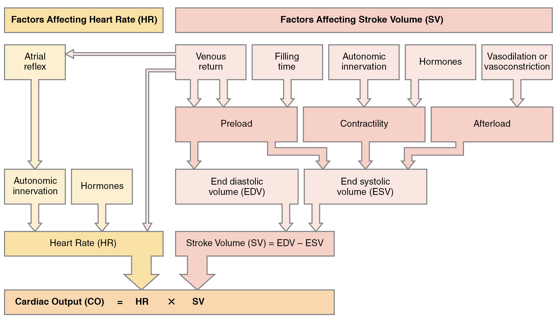
This flowchart lists all the important factors that affect cardiac output.
Figure 19.35 — 19.4 Cardiac Physiology

Figure 19.35 Summary of Major Factors Influencing Cardiac Output The primary factors influencing HR include autonomic innervation plus endocrine control. Not shown are environmental factors, such as electrolytes, metabolic products, and temperature. The primary factors controlling SV include preload, contractility, and afterload. Other factors such as electrolytes may be classified as either positive or negative inotropic agents.

J. Gordon Betts and colleagues, Anatomy And Physiology 2E, OpenStax. · CC BY-NC-SA 4.0 · Original image ↗ · Access for free ↗
Factor Effect
Cardioaccelerator nerves Release of norepinephrine by cardioaccelerator nerves
Proprioreceptors Increased firing rates of proprioreceptors (e.g. during exercise)
sensing decreased levels of O2 or increased levels of H+, CO2 and lactic
Decreased firing rates of (indicating falling blood volume/pressure)
Limbic system Anticipation of physical exercise or strong emotions by the limbic system
Increased epinephrine and norepinephrine release by the adrenal glands
Thyroid Increased T3 and T4 in the blood (released by thyroid)
Calcium Increase in calcium in the blood
Potassium Decrease in potassium in the blood
Sodium Decrease in sodium in the blood
Body temperature Increase in body temperature
Nicotine and caffeine Presence of nicotine, caffeine or other stimulants

Table 19.1. J. Gordon Betts and colleagues, Anatomy And Physiology 2E, OpenStax.

Factor Effect
Cardioinhibitor nerves (vagus) Release of acetylcholine by cardioinhibitor nerves
Proprioreceptors Decreased firing rates of proprioreceptors (e.g. during rest)
sensing increased levels of O2 or decreased levels of H+, CO2 and lactic
Increased firing rates of (indicating rising blood volume/pressure)
Limbic system Anticipation of relaxation by the limbic system
Decreased epinephrine and norepinephrine release by the adrenal glands
Thyroid Decreased T3 and T4 in the blood (released by thyroid)
Calcium Decrease in calcium in the blood
Potassium Increase in potassium in the blood
Sodium Increase in sodium in the blood
Body temperature Decrease in body temperature
Opiates and tranquilizers Presence of opiates (heroin), tranquilizers or other depressants

Table 19.2. J. Gordon Betts and colleagues, Anatomy And Physiology 2E, OpenStax.

(aorta, carotid arteries, venae cavae, and ) (both and in proximity to )
Sensitive to Decreasing stretch Decreasing O2 and increasing CO2, H+, and lactic
Target Parasympathetic stimulation suppressed Sympathetic stimulation increased
Response of heart Increasing heart rate and increasing Increasing heart rate and increasing
Overall effect Increasing blood flow and pressure due to increasing ; restored Increasing blood flow and pressure due to increasing ; restored

Table 19.3. J. Gordon Betts and colleagues, Anatomy And Physiology 2E, OpenStax.

(aorta, carotid arteries, venae cavae, and ) (both and in proximity to )
Sensitive to Increasing stretch Increasing O2 and decreasing CO2, H+, and lactic
Target Parasympathetic stimulation increased Sympathetic stimulation suppressed
Response of heart Decreasing heart rate and decreasing Decreasing heart rate and decreasing
Overall effect Decreasing blood flow and pressure due to decreasing ; restored Decreasing blood flow and pressure due to decreasing ; restored

Table 19.4. J. Gordon Betts and colleagues, Anatomy And Physiology 2E, OpenStax.

Source: 19.4 Cardiac Physiology

20.2 Blood Flow, Blood Pressure, and Resistance — figures and tables
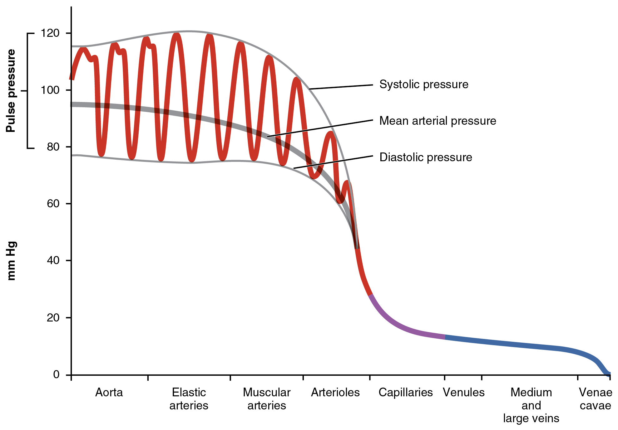
This graph shows the value of pulse pressure in different types of blood vessels.
Figure 20.10 — 20.2 Blood Flow, Blood Pressure, and Resistance

Figure 20.10 Systemic Blood Pressure The graph shows the components of blood pressure throughout the blood vessels, including systolic, diastolic, mean arterial, and pulse pressures.

J. Gordon Betts and colleagues, Anatomy And Physiology 2E, OpenStax. · CC BY-NC-SA 4.0 · Original image ↗ · Access for free ↗
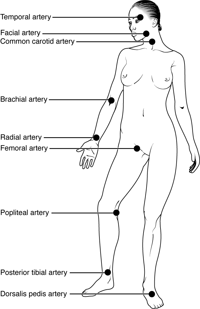
This image shows the pulse points in a woman’s body.
Figure 20.11 — 20.2 Blood Flow, Blood Pressure, and Resistance

Figure 20.11 Pulse Sites The pulse is most readily measured at the radial artery, but can be measured at any of the pulse points shown.

J. Gordon Betts and colleagues, Anatomy And Physiology 2E, OpenStax. · CC BY-NC-SA 4.0 · Original image ↗ · Access for free ↗
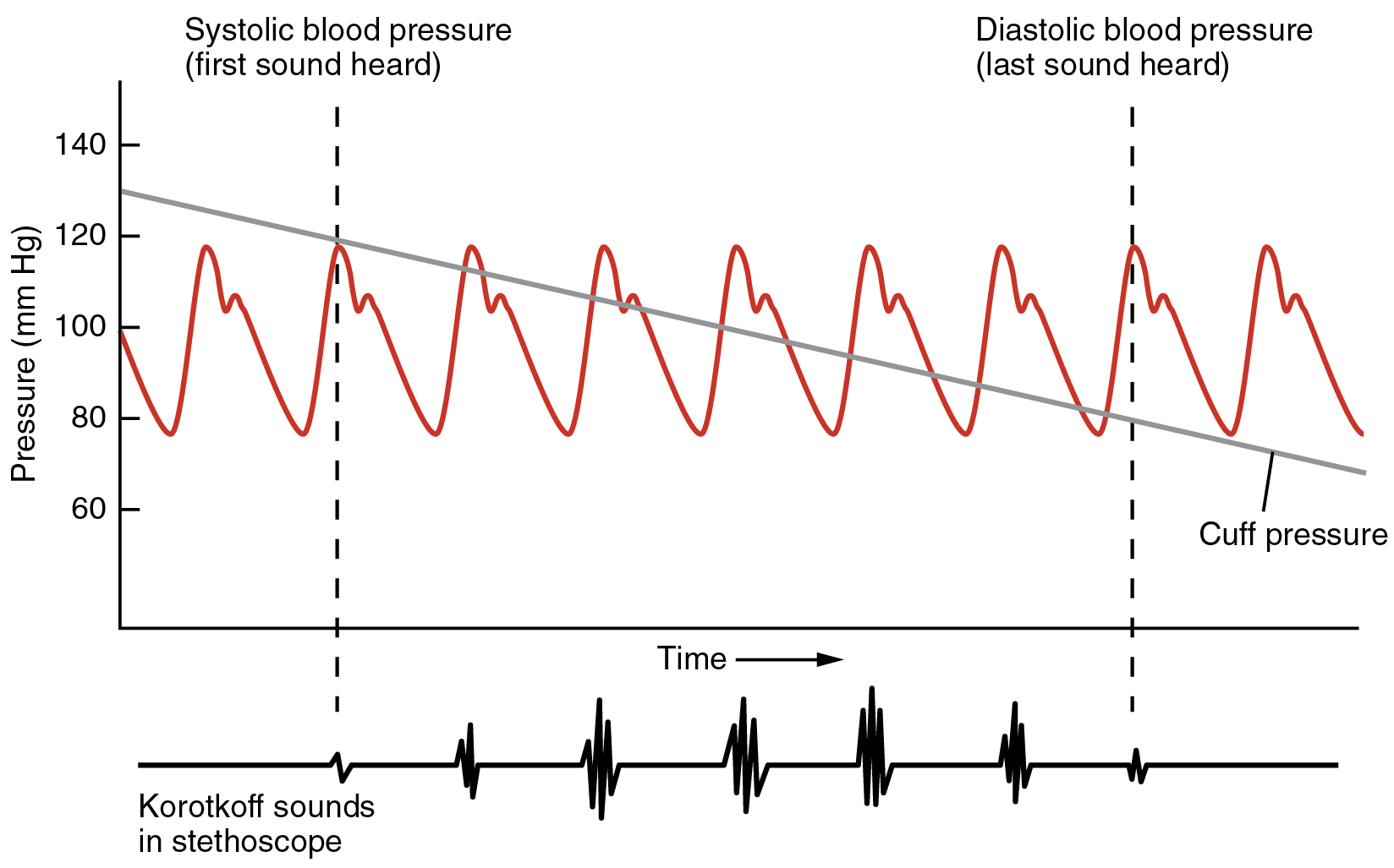
This image shows blood pressure as a function of time.
Figure 20.12 — 20.2 Blood Flow, Blood Pressure, and Resistance

Figure 20.12 Blood Pressure Measurement When pressure in a sphygmomanometer cuff is released, a clinician can hear the Korotkoff sounds. In this graph, a blood pressure tracing is aligned to a measurement of systolic and diastolic pressures.

J. Gordon Betts and colleagues, Anatomy And Physiology 2E, OpenStax. · CC BY-NC-SA 4.0 · Original image ↗ · Access for free ↗
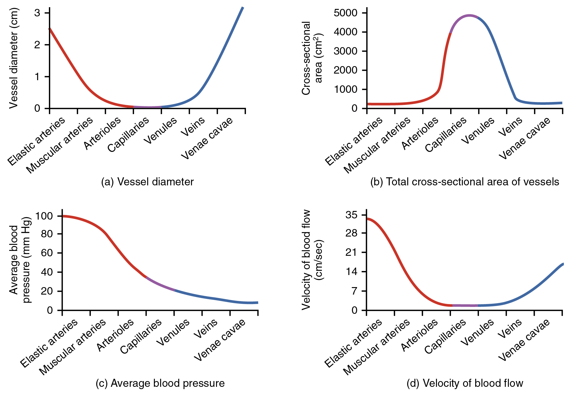
This figure shows four graphs. The top left graph shows the vessel diameter for different types of blood vessels. The top right panel shows cross-sectional area for different blood vessels. The bottom left panel shows the average blood pressure for different blood vessels, and the bottom right panel shows the velocity of blood flow in different blood vessels.
Figure 20.13 — 20.2 Blood Flow, Blood Pressure, and Resistance

Figure 20.13 Relationships among Vessels in the Systemic Circuit The relationships among blood vessels that can be compared include (a) vessel diameter, (b) total cross-sectional area, (c) average blood pressure, and (d) velocity of blood flow.

J. Gordon Betts and colleagues, Anatomy And Physiology 2E, OpenStax. · CC BY-NC-SA 4.0 · Original image ↗ · Access for free ↗
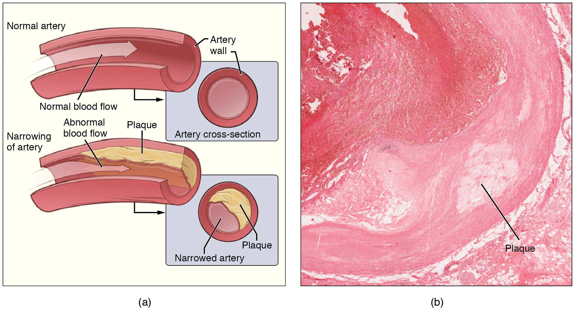
The left panel shows the cross-section of a normal and a narrowed artery. The right panel shows a micrograph of an artery with plaque in it.
Figure 20.14 — 20.2 Blood Flow, Blood Pressure, and Resistance

Figure 20.14 Atherosclerosis (a) Atherosclerosis can result from plaques formed by the buildup of fatty, calcified deposits in an artery. (b) Plaques can also take other forms, as shown in this micrograph of a coronary artery that has a buildup of connective tissue within the artery wall. LM × 40. (Micrograph provided by the Regents of University of Michigan Medical School © 2012)

J. Gordon Betts and colleagues, Anatomy And Physiology 2E, OpenStax. · CC BY-NC-SA 4.0 · Original image ↗ · Access for free ↗
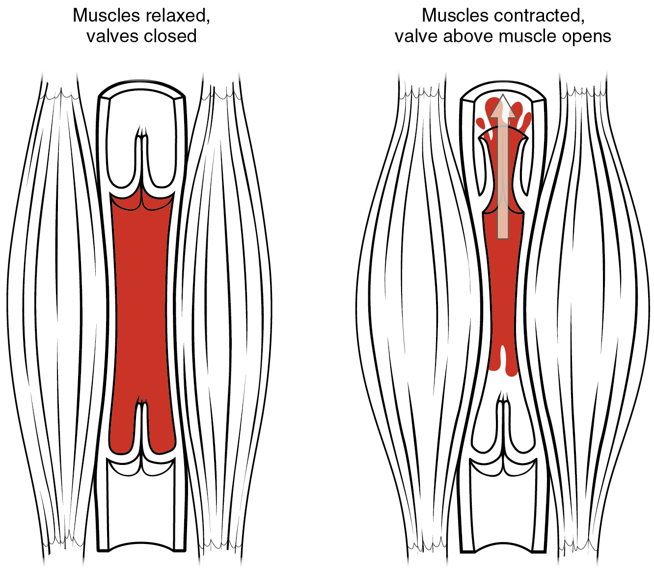
The left panel shows the structure of a skeletal muscle vein pump when the muscle is relaxed, and the right panel shows the structure of a skeletal muscle vein pump when the muscle is contracted.
Figure 20.15 — 20.2 Blood Flow, Blood Pressure, and Resistance

Figure 20.15 Skeletal Muscle Pump The contraction of skeletal muscles surrounding a vein compresses the blood and increases the pressure in that area. This action forces blood closer to the heart where venous pressure is lower. Note the importance of the one-way valves to assure that blood flows only in the proper direction.

J. Gordon Betts and colleagues, Anatomy And Physiology 2E, OpenStax. · CC BY-NC-SA 4.0 · Original image ↗ · Access for free ↗

Source: 20.2 Blood Flow, Blood Pressure, and Resistance

20.3 Capillary Exchange — figures and tables
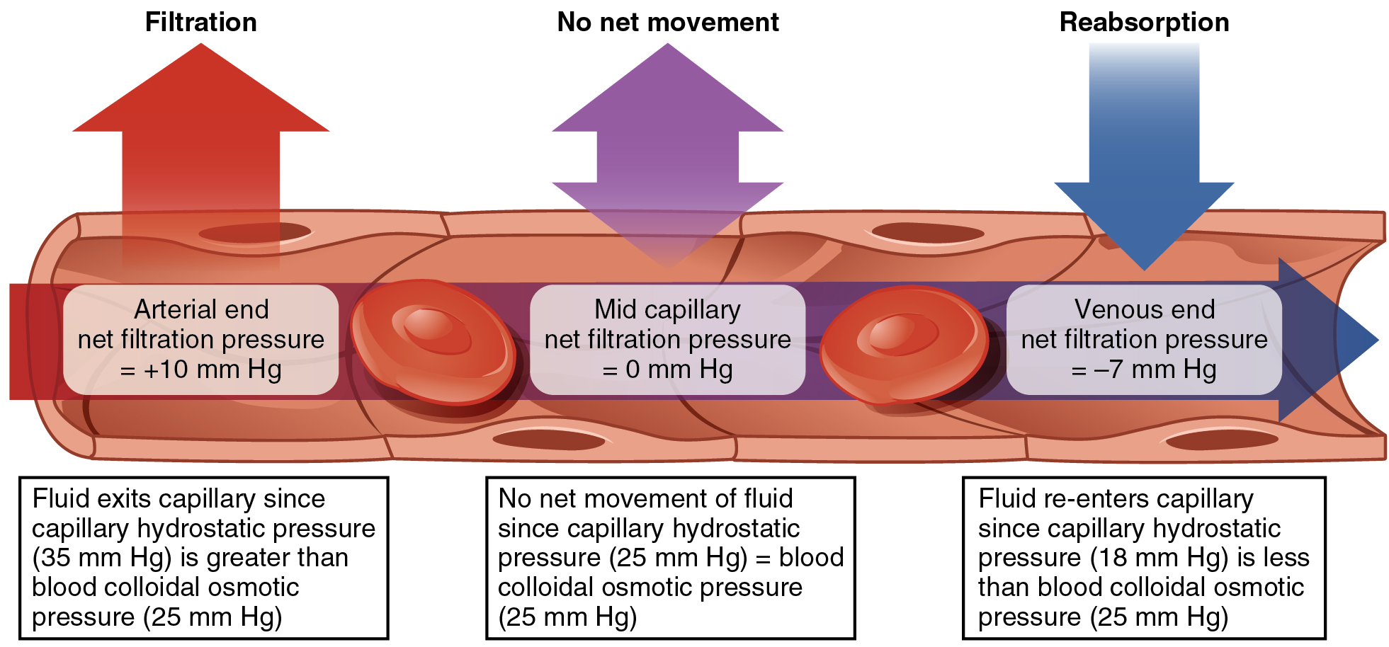
This diagram shows the process of fluid exchange in a capillary from the arterial end to the venous end.
Figure 20.16 — 20.3 Capillary Exchange

Figure 20.16 Capillary Exchange Net filtration occurs near the arterial end of the capillary since capillary hydrostatic pressure (CHP) is greater than blood colloidal osmotic pressure (BCOP). There is no net movement of fluid near the midpoint since CHP = BCOP. Net reabsorption occurs near the venous end since BCOP is greater than CHP.

J. Gordon Betts and colleagues, Anatomy And Physiology 2E, OpenStax. · CC BY-NC-SA 4.0 · Original image ↗ · Access for free ↗

Source: 20.3 Capillary Exchange

327 terms, alphabetically ordered

3′ hydroxyl group · · ·

The at the sugar’s 3′ carbon that attacks the incoming ’s during strand extension.

Read explanation · OpenStax ↗
A band

The sarcomeric region spanning the length of thick filaments.

Read explanation · OpenStax ↗
acceleration

Rate of change of , measured in m/s².

Read explanation · OpenStax ↗
Acid · ·

In the Brønsted–Lowry model, a substance that can donate a . Acidic have a higher activity than water.

Read explanation · OpenStax ↗
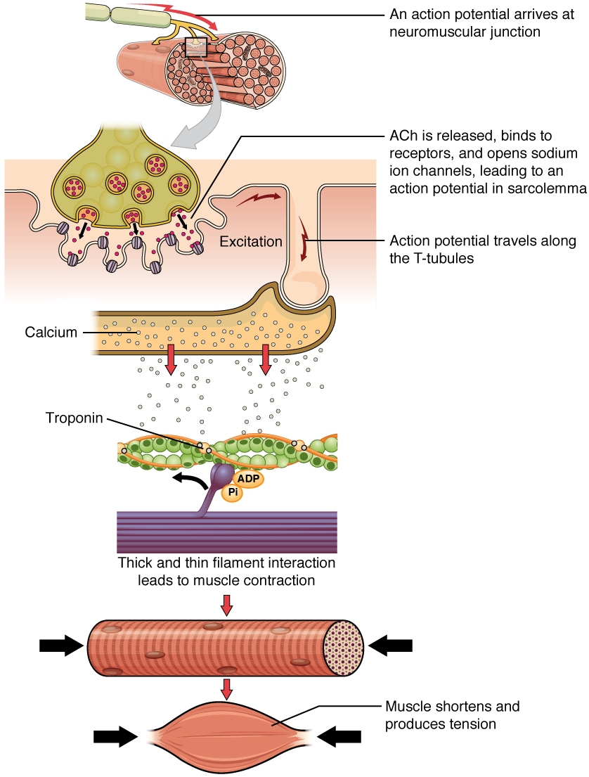
Actin

A that forms cellular filaments and, together with , supports muscle contraction and other movements.

Read explanation · OpenStax ↗
Activation energy ·

The energy barrier that must overcome to reach a and undergo .

Read explanation · OpenStax ↗
Active catalytic-site concentration ·

The total of active sites used to calculate from . It equals active when each has one site.

Read explanation · Biology LibreTexts ↗
Active site

The region of an that binds its and carries out .

Read explanation · OpenStax ↗
Adenosine triphosphate

An energy-carrying ; its powers many cellular processes, including cycles.

Read explanation · OpenStax ↗
ADH ·

Antidiuretic hormone, or vasopressin, synthesized in hypothalamic and released from the posterior pituitary to promote renal water conservation.

Read explanation · OpenStax ↗
afterload

The mechanical load opposing ventricular ejection, influenced by arterial pressure and outflow properties.

Read explanation · OpenStax ↗
aldosterone

An adrenal-cortex mineralocorticoid promoting distal renal sodium and potassium .

Read explanation · OpenStax ↗
Alpha carbon · ·

The central carbon next to the carboxyl carbon. In a standard it binds an , hydrogen, and .

Read explanation · OpenStax ↗
Alpha helix · · · · · ·

A helical stabilized by between a oxygen and an hydrogen four later.

Read explanation · OpenStax ↗
Amide · · ·

A group containing a carbon bonded to nitrogen. are amide linkages.

Read explanation · OpenStax ↗
Amino acid

A building block containing amino and and a characteristic .

Read explanation · OpenStax ↗
Amino acid residue · ·

The part of an incorporated into a chain. Residue can also describe another building block within a larger .

Read explanation · OpenStax ↗
Amino group · · ·

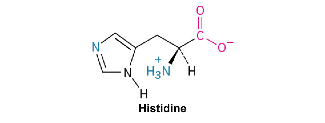
A nitrogen-containing . A free ’s amino group is typically (NH₃⁺) near physiological ; ’s ring nitrogen is an exception to this formula.

Read explanation · OpenStax ↗
Amphipathic

Having both a region and a region, as in a or .

Read explanation · OpenStax ↗
Amylose

The largely unbranched component of , made mainly of linked by alpha-1,4 .

Read explanation · OpenStax ↗
Analytical standard ·

A material with a known identity or amount used to compare or calibrate measurements.

Read explanation · Agilent ↗
anemia

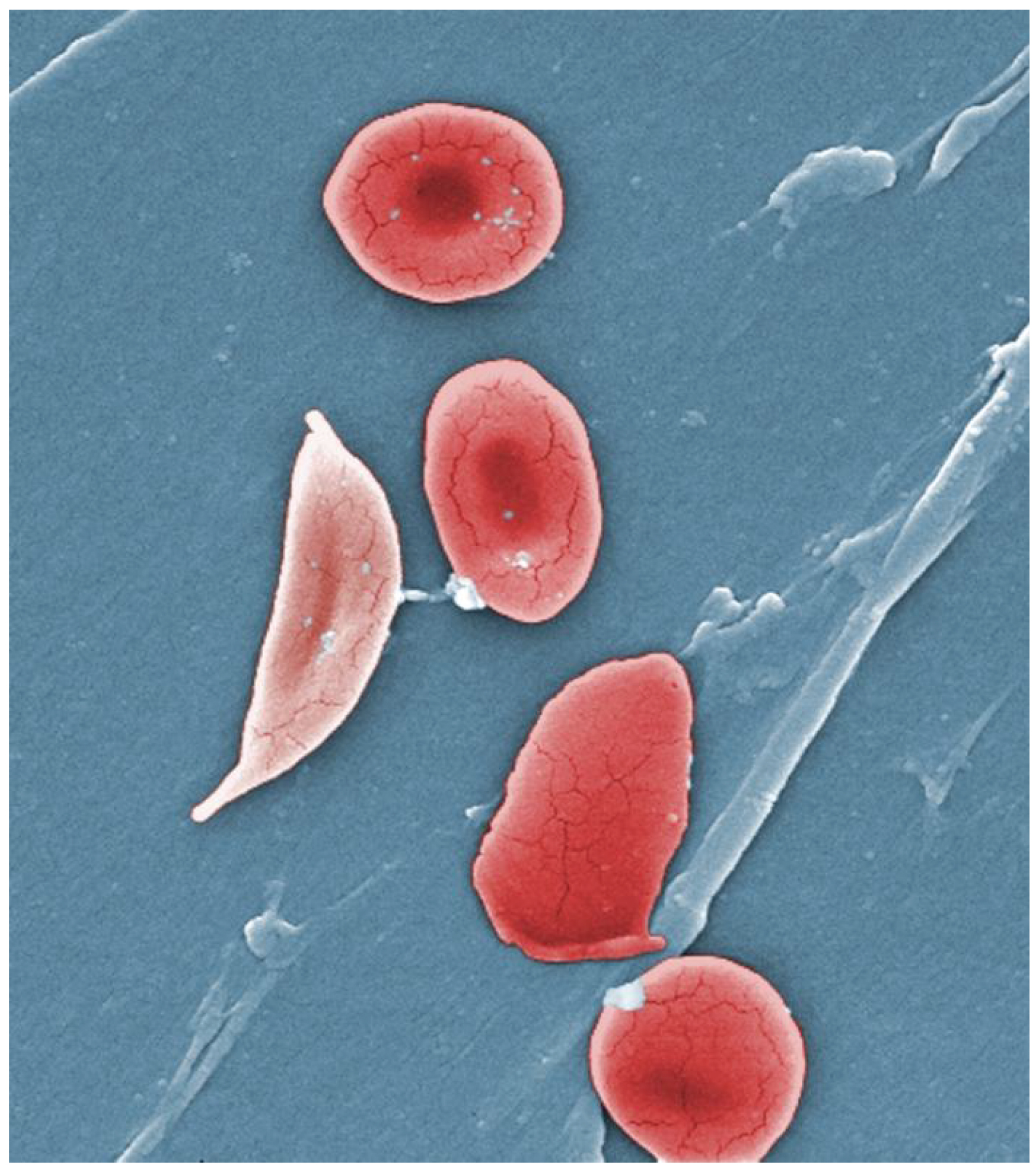
Reduced blood oxygen-carrying capacity associated with low or red-cell mass for the relevant reference context.

Read explanation · OpenStax ↗
anomeric carbon

The former carbon of a sugar, which becomes the or hemiketal center upon cyclization.

Read explanation · OpenStax ↗
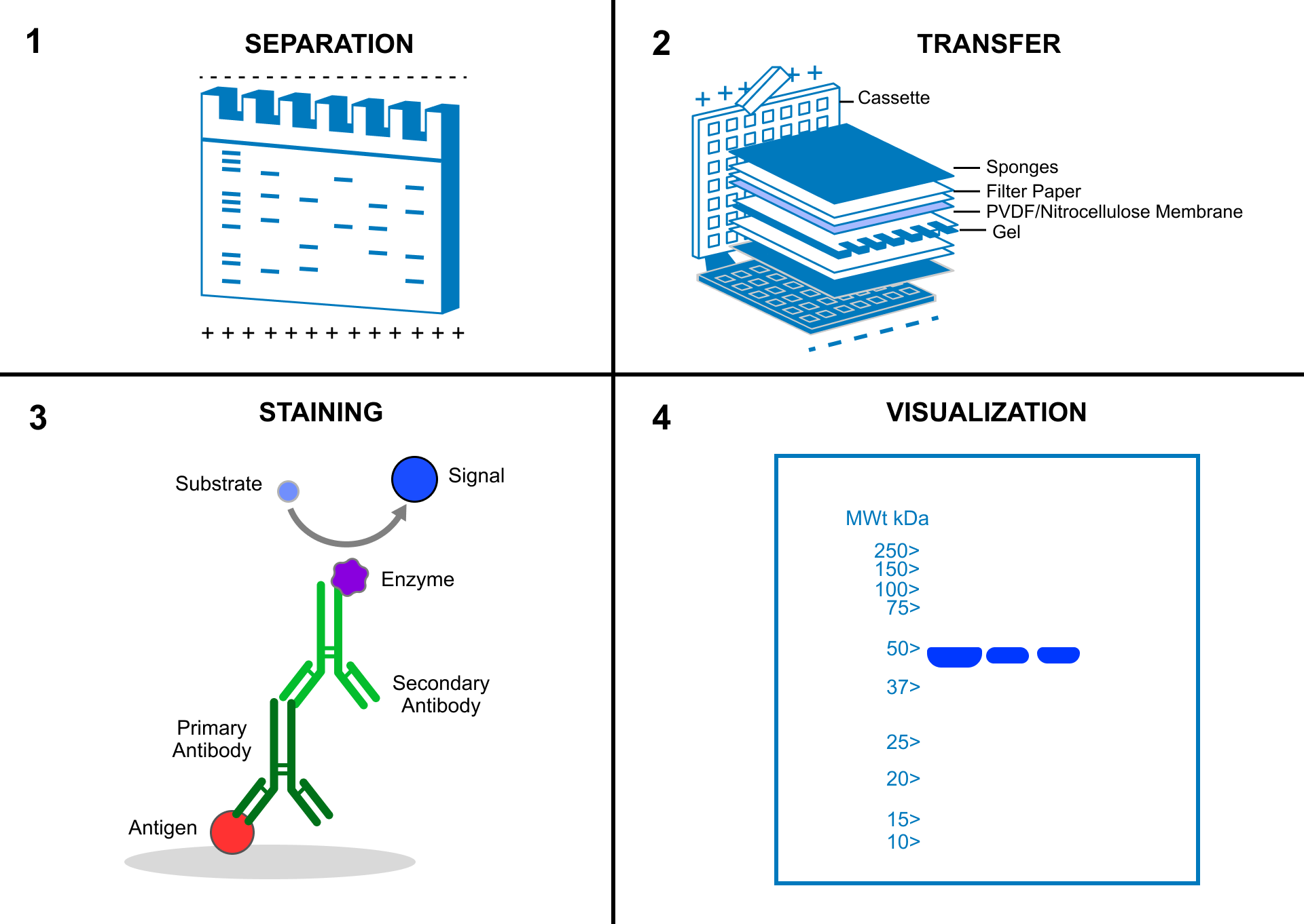
Antibody

An that recognizes a specific region () on an . Antibodies help immune defenses and are used as detection tools in .

Read explanation · OpenStax ↗
Anticodon

A triplet that pairs to an . The anticodon for 5′-AUG-3′ is aligned 3′-UAC-5′, or written 5′-CAU-3′.

Read explanation · NCBI Bookshelf / Garland Science ↗
Antigen

A or molecular structure specifically recognized by an or other adaptive immune .

Read explanation · OpenStax ↗
Antiparallel

Running in opposite directions, as with two strands or neighboring strands in some .

Read explanation · OpenStax ↗
aortic valve

The valve between left and aorta.

Read explanation · OpenStax ↗
apical

Referring to the epithelial surface facing a or external space.

Read explanation · OpenStax ↗
Aqueous solution ·

A in which water is the .

Read explanation · OpenStax ↗
Archaea ·

A of distinct from , with characteristic chemistry and -expression machinery.

Read explanation · OpenStax ↗
Aromatic

Containing a cyclic, with . , , and have aromatic .

Read explanation · OpenStax ↗
arteriole

A small artery whose smooth-muscle tone provides major control of local vascular resistance.

Read explanation · OpenStax ↗
Assay

A procedure used to detect a substance or measure its amount, activity, or another property.

Read explanation · Thermo Fisher ↗
Atom

The smallest unit of an element that retains its chemical identity; it has a surrounded by .

Read explanation · OpenStax ↗
atrium

A cardiac receiving chamber of a .

Read explanation · OpenStax ↗
attachment

Enduring emotional bond and expectations about another’s availability and responsiveness.

Read explanation · OpenStax ↗
autonomic nervous system

Neural pathways regulating visceral organs through sympathetic, parasympathetic, and enteric systems.

Read explanation · OpenStax ↗
Bacteria ·

A of single-celled . Bacterial cells lack a -bound and are distinct from .

Read explanation · OpenStax ↗
baroreceptor

A stretch-sensitive monitoring arterial pressure and participating in autonomic feedback.

Read explanation · OpenStax ↗
Benzene ring

A six-carbon ring with .

Read explanation · OpenStax ↗
Benzyl group

A attached through a –CH₂– group; part of ’s .

Read explanation · OpenStax ↗
Beta sheet · · · · · ·

A made of aligned segments connected by ; strands may be parallel or .

Read explanation · OpenStax ↗
Binding site

A region of a where another interacts selectively.

Read explanation · OpenStax ↗
Biomolecule

A made by or involved in living systems, including , , , and .

Read explanation · OpenStax ↗
Blocking

Covering unoccupied on a or plate to reduce interactions.

Read explanation · Thermo Fisher ↗
Blotting ·

Transfer of separated or to a so a specific target can be detected.

Read explanation · Thermo Fisher ↗
Brønsted–Lowry base · · · · · · · · · · ·

In the Brønsted–Lowry model, a substance that can accept a . Basic solutions have lower activity than water.

Read explanation · OpenStax ↗
Buffer ·

A that resists changes when small amounts of or are added, commonly through a weak and its .

Read explanation · OpenStax ↗
Cancer

A group of diseases involving abnormal cells that can proliferate without normal restraints and invade tissues.

Read explanation · OpenStax ↗
Carbohydrate

A class of including sugars and their , used for energy storage, structure, and recognition.

Read explanation · OpenStax ↗
Carbonyl group · ·

A carbon double-bonded to oxygen, C=O; present in and many other .

Read explanation · OpenStax ↗
Carboxyl group · ·

An , COOH. In free it is usually to COO⁻ near physiological .

Read explanation · OpenStax ↗
cardiac output

The volume pumped by a per time, equal to heart rate multiplied by .

Read explanation · OpenStax ↗
Cardiac ventricle · ·

A cardiac pumping chamber ejecting blood into the pulmonary or systemic circuit.

Read explanation · OpenStax ↗
Catalyst · ·

A substance that speeds a by providing a pathway with lower and is regenerated rather than consumed overall.

Read explanation · OpenStax ↗
catecholamine

A -derived chemical class including dopamine, norepinephrine, and epinephrine.

Read explanation · OpenStax ↗
Cellulose

A structural in plant cell walls, with beta-1,4 linkages; also used as a support.

Read explanation · OpenStax ↗
Centimeter

A unit of length equal to one hundredth of a meter.

Read explanation · NIST ↗
central nervous system

Brain and spinal cord.

Read explanation · OpenStax ↗
Centrifugation ·

Spinning a sample to accelerate separation of particles according to their behavior.

Read explanation · Beckman Coulter ↗
Chemical compound ·

A substance containing of two or more different chemically combined in a definite composition.

Read explanation · OpenStax ↗
Chemical element

A substance defined by having the same number of , such as carbon or oxygen.

Read explanation · OpenStax ↗
Chemical reaction ·

A process that rearranges and chemical bonds, converting into .

Read explanation · OpenStax ↗
chemoreceptor

A sensory responding to chemical conditions, including oxygen, carbon dioxide, or .

Read explanation · OpenStax ↗
cholesterol

An animal that modulates packing and serves as a precursor for and bile .

Read explanation · OpenStax ↗
Chromatin

The complex of and associated , including , in .

Read explanation · OpenStax ↗
Chromatographic peak · ·

A rise and fall in detector signal as one or more components pass through the detector; a peak does not guarantee a single pure .

Read explanation · Agilent ↗
Chromatography

Separation of mixture components by their different interactions with a and a moving .

Read explanation · Waters ↗
Chromosome ·

An organized with associated that carries . A replicated chromosome has two .

Read explanation · OpenStax ↗
Codon

A sequence of three that specifies an or a stop signal.

Read explanation · OpenStax ↗
Collagen

A major structural in , with a characteristic triple-helical arrangement.

Read explanation · OpenStax ↗
Concentration

The amount of a substance in a specified volume or mass of mixture.

Read explanation · OpenStax ↗
conduction

Thermal energy transfer through microscopic interactions without bulk matter flow.

Read explanation · OpenStax ↗
conjugate base

Species formed when an loses one .

Read explanation · OpenStax ↗
Conjugated system ·

Adjacent orbitals that allow to be distributed across several , often through alternating single and multiple bonds.

Read explanation · OpenStax ↗
connective tissue

A tissue category generally characterized by cells distributed within substantial .

Read explanation · OpenStax ↗
contractility

Intrinsic force-generating capacity of cardiac muscle at a given loading condition.

Read explanation · OpenStax ↗
Cooperativity · ·

An interaction in which binding at one site changes binding at other sites. Positive cooperativity can produce a response.

Read explanation · Biology LibreTexts ↗
cortisol

An adrenal-cortex glucocorticoid regulating , stress responses, and immune activity.

Read explanation · OpenStax ↗
Covalent bond · ·

A bond in which share pairs. and are covalent.

Read explanation · OpenStax ↗
Cysteine ·

An with a . Two cysteines can form a .

Read explanation · OpenStax ↗
Cytoplasm

Cell contents inside the plasma and outside the , where one is present.

Read explanation · OpenStax ↗
Cytoskeleton

A network of filaments that supports cell shape, organizes components, and enables movement.

Read explanation · OpenStax ↗
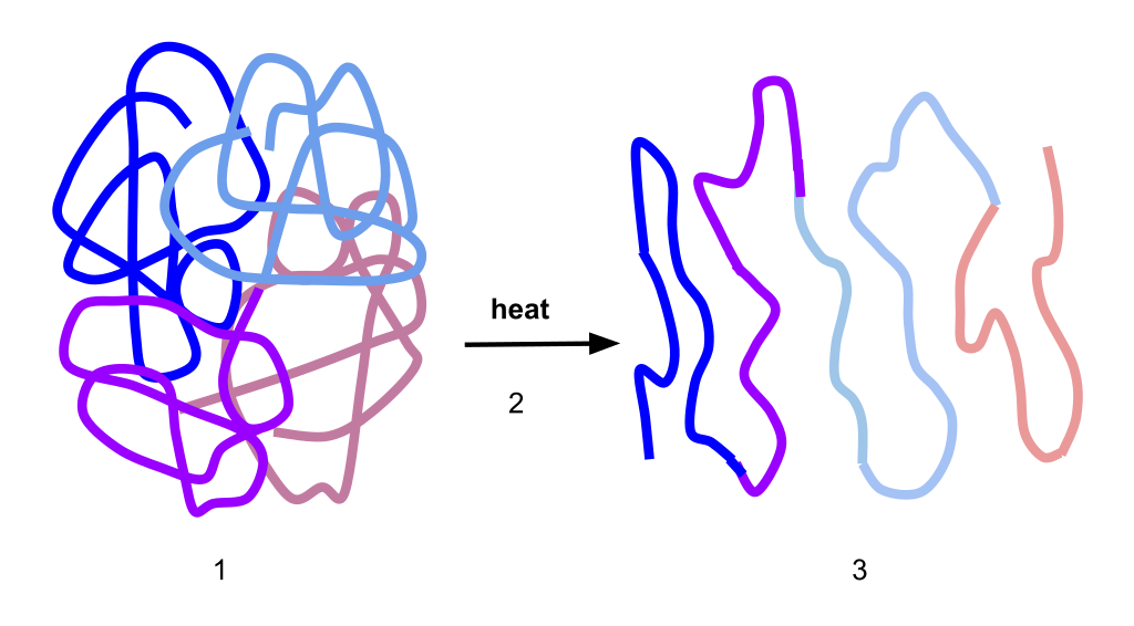
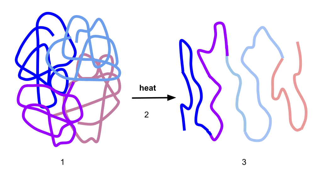
Denaturation · ·

Loss of a ’s , often with . This usually preserves the chain’s .

Read explanation · NCBI Bookshelf ↗
Density gradient

A fluid whose density varies with position, used to improve separation during .

Read explanation · Beckman Coulter ↗
Deoxyribonucleic acid

The that stores genetic information used to specify and sequences.

Read explanation · OpenStax ↗
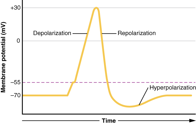
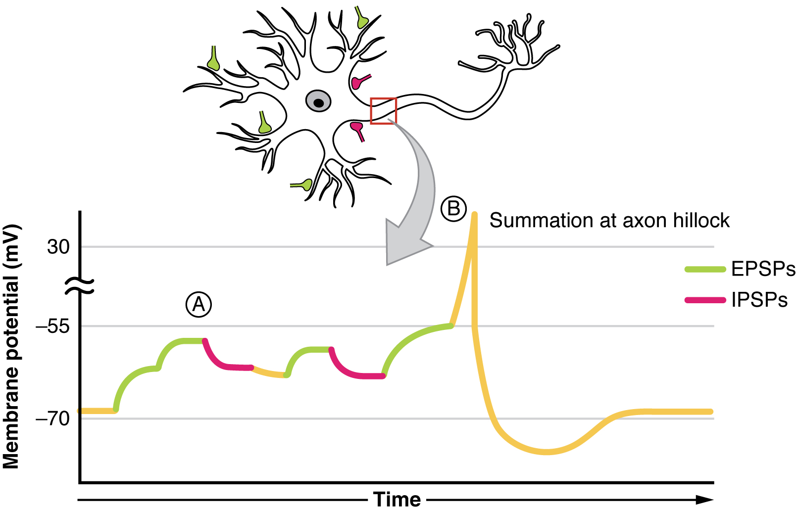
depolarization

A shift in voltage toward a less negative value.

Read explanation · OpenStax ↗
Deprotonation

Removal of a from a or group; this changes its by minus one.

Read explanation · OpenStax ↗
Detergent

An substance that can associate with both water and material; detergents can disrupt or interactions.

Read explanation · Thermo Fisher ↗
diastole

The cardiac phase associated with relaxation and filling, especially ventricular relaxation.

Read explanation · OpenStax ↗
Diffusion

Net spreading of particles down a due to random molecular motion.

Read explanation · OpenStax ↗
Disulfide bond · · ·

A covalent sulfur–sulfur bond formed by of two , which can stabilize structure.

Read explanation · NCBI Bookshelf ↗
DNA ligase · · ·

An that seals a by forming a between an adjacent and 5′ .

Read explanation · OpenStax ↗
DNA nick · · ·

A break in one strand’s , with no missing. can seal a nick with suitable ends.

Read explanation · OpenStax ↗
DNA replication

Copying so genetic information can be passed to daughter cells. Each guides a complementary new strand.

Read explanation · OpenStax ↗
Downstream

Later along a directed sequence or process; in , toward positions following a reference site in the direction of synthesis.

Read explanation · OpenStax ↗
edema

Excess fluid accumulation in interstitial tissue spaces.

Read explanation · OpenStax ↗
eicosanoid

Local signaling derived from a twenty-carbon polyunsaturated .

Read explanation · OpenStax ↗
Electric charge · · ·

An electrical property that can be positive or negative. Opposite charges attract and like charges repel; net charge is the sum of all charges on a particle.

Read explanation · OpenStax ↗
electrochemical gradient

The combined and electrical driving forces acting on an across space or a .

Read explanation · OpenStax ↗
Electron

A negatively subatomic particle. Electron sharing and transfer determine much of chemical bonding.

Read explanation · OpenStax ↗
Electron delocalization ·

Distribution of across several rather than confinement to one or bond.

Read explanation · OpenStax ↗
Electronegativity ·

An ’s tendency to attract shared in a chemical bond. Unequal attraction can make a bond .

Read explanation · OpenStax ↗
Endocrine signaling ·

Communication in which enter the circulation and act on distant target cells.

Read explanation · OpenStax ↗
endocytosis

Uptake of extracellular material or surface through inward budding of .

Read explanation · OpenStax ↗
Endomembrane system

The interacting and compartments involved in processes such as modification, transport, and .

Read explanation · OpenStax ↗
Endoplasmic reticulum

An interconnected network continuous with the ; it includes regions involved in processing and synthesis.

Read explanation · OpenStax ↗
endothelium

The cellular lining of blood and lymphatic vessels, involved in barrier and signaling functions.

Read explanation · OpenStax ↗
Enzyme

A biological that speeds a without being consumed. Most enzymes are .

Read explanation · OpenStax ↗
Enzyme saturation · ·

The high- limit in which sites spend almost all their time processing and the rate approaches .

Read explanation · Biology LibreTexts ↗
Enzyme–substrate complex · · ·

The bound association of an and its during a cycle. Often abbreviated ES in schemes.

Read explanation · OpenStax ↗
epithelium ·

A polarized sheet of closely connected cells that covers surfaces, lines cavities, or forms glands.

Read explanation · OpenStax ↗
Epitope

The particular part of an recognized by an or immune .

Read explanation · OpenStax ↗
Equilibrium

A state where opposing processes occur at equal rates, so macroscopic or other properties remain constant.

Read explanation · OpenStax ↗
Eukaryote ·

An organism whose cells have a -enclosed ; animals, plants, fungi, and protists are eukaryotes.

Read explanation · OpenStax ↗
exocytosis

Fusion of an intracellular with the plasma , releasing contents and adding components.

Read explanation · OpenStax ↗
Exon

A segment retained in a mature spliced . In an exon may contain coding sequence, untranslated sequence, or both.

Read explanation · OpenStax ↗
Experimental control ·

A comparison sample or condition used to distinguish the effect being tested from other influences.

Read explanation · Thermo Fisher ↗
extracellular matrix

The network of and surrounding cells and providing structural and signaling functions.

Read explanation · OpenStax ↗
facilitated diffusion

Passive movement down an through a selective channel or .

Read explanation · OpenStax ↗
FACT complex ·

A -associated complex that helps machinery traverse and supports reorganization.

Read explanation · OpenStax ↗
Fatty acid

A attached to a chain; fatty acids are components of many .

Read explanation · OpenStax ↗
First-order kinetics

A rate dependence proportional to the first power of a . At low , a is approximately first-order in .

Read explanation · Biology LibreTexts ↗
fitness

Relative reproductive contribution of a or organism in a particular environment.

Read explanation · OpenStax ↗
Fraction · ·

A portion of a sample collected during a separation, such as the fluid leaving a column during a time interval.

Read explanation · Waters ↗
Fragmentation ·

Breaking a into smaller and pieces; fragment patterns help identify molecular structure.

Read explanation · Thermo Fisher ↗
Frank–Starling mechanism · ·

The tendency of increased ventricular filling to increase contraction and within physiological limits.

Read explanation · OpenStax ↗
Functional group

A group of whose chemical behavior gives a characteristic and properties.

Read explanation · OpenStax ↗
Gene

A sequence that contributes to a functional or , together with the information needed to express it.

Read explanation · OpenStax ↗
Gene expression

Production of a functional from a , including and, for -coding , .

Read explanation · NCBI Bookshelf ↗
Genome

The complete genetic material of an organism or cell.

Read explanation · OpenStax ↗
genotype

The allelic composition of an organism at one or more specified .

Read explanation · OpenStax ↗
Glucagon

A pancreatic that helps raise , especially by promoting release from the liver.

Read explanation · OpenStax ↗
Glucose

A six-carbon sugar that is a major cellular energy source and a building block of , , and .

Read explanation · OpenStax ↗
Glycerol

A three-carbon alcohol with three ; used in sample to increase density and in some .

Read explanation · OpenStax ↗
glycogen

A highly branched used for storage in animals.

Read explanation · Biology LibreTexts / Oregon State University ↗
Glycosidic bond

A involving a sugar’s . In a , an N-glycosidic bond links the sugar to the .

Read explanation · Biology LibreTexts / Oregon State University ↗
Golgi apparatus ·

A stack of compartments that modifies and sorts and traveling through the .

Read explanation · OpenStax ↗
Gradient

A change in a quantity, such as , , or density, across a distance or over time.

Read explanation · Thermo Fisher ↗
Half-life

The time required for half of a population or amount to remain during decay; a constant half-life is characteristic of decay.

Read explanation · OpenStax ↗
hemiacetal

Carbon bonded to both and alkoxy groups, usually formed by alcohol addition to a .

Read explanation · OpenStax ↗
Hemoglobin

The oxygen-carrying in , composed of four with heme groups.

Read explanation · OpenStax ↗
hemostasis

The coordinated process limiting blood loss after vessel injury while maintaining flow elsewhere.

Read explanation · OpenStax ↗
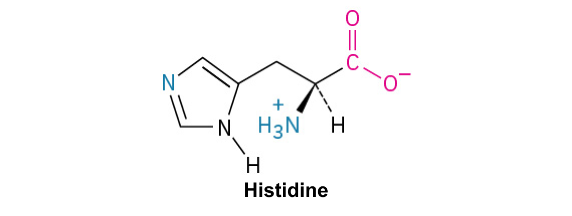
Histidine ·

A with an . Mostly uncharged near physiological , it can accept or donate .

Read explanation · OpenStax ↗
Histone

A -associated rich in . Core histones form the around which nucleosomal wraps.

Read explanation · OpenStax ↗
homeostasis

Dynamic regulation that maintains physiological variables within functional ranges despite disturbances.

Read explanation · OpenStax ↗
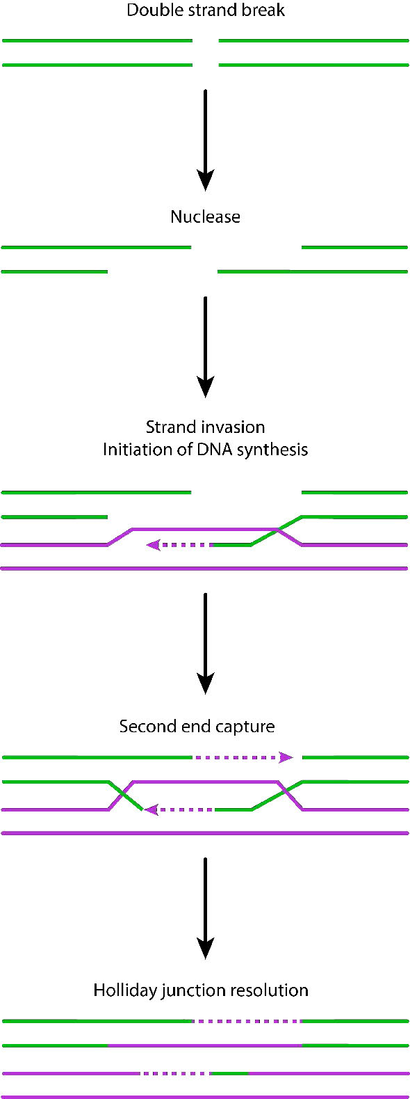
Homologous recombination ·

Exchange or copying between with similar sequences. In break repair, an intact homologous template, usually a , guides restoration.

Read explanation · Biology LibreTexts / Oregon State University ↗
Hormone

A chemical messenger released by cells that changes the activity of target cells with matching .

Read explanation · OpenStax ↗
Hydrocarbon · ·

A made only of carbon and hydrogen. Hydrocarbon regions are generally .

Read explanation · OpenStax ↗
Hydrogen bond ·

An attraction between a hydrogen bonded to an and another . hydrogen bonds stabilize .

Read explanation · OpenStax ↗
Hydrogen ion · ·

A positively hydrogen species. H⁺ denotes a , which is hydrated in water; its activity determines .

Read explanation · OpenStax ↗
Hydrolysis

Breaking a chemical bond by adding water. hydrolysis separates .

Read explanation · OpenStax ↗
Hydrophilic

Interacting favorably with water, typically because a group is or .

Read explanation · OpenStax ↗
Hydrophobic

Interacting poorly with water. often gather in a ’s interior.

Read explanation · OpenStax ↗
hydrostatic pressure

Pressure in a fluid at rest, increasing with depth under gravity.

Read explanation · OpenStax ↗
Hydroxide ion ·

The negatively OH⁻. Basic contain a higher hydroxide ion activity than water at the same temperature.

Read explanation · OpenStax ↗
Hydroxyl group ·

An oxygen bonded to hydrogen, –OH. Hydroxyl groups can and often increase .

Read explanation · OpenStax ↗
Hyperbolic curve ·

A curve that approaches an asymptote. The standard rate curve is a rectangular hyperbola approaching .

Read explanation · Biology LibreTexts ↗
Hypoxia

A state of reduced oxygen availability that can alter cellular stress signaling.

Read explanation · OpenStax ↗
Imidazole

A five-membered ring containing two nitrogens; ’s contains this ring.

Read explanation · OpenStax ↗
Immunoglobulin

An or the -bound form of the same type of -binding .

Read explanation · OpenStax ↗
incidence

Occurrence of new cases in a defined population over a specified period.

Read explanation · OpenStax ↗
Indole

A fused six-membered and five-membered ring system containing nitrogen; found in .

Read explanation · OpenStax ↗
inflammation

A coordinated tissue response recruiting defense and repair mechanisms, often involving increased blood flow and permeability.

Read explanation · OpenStax ↗
Initial reaction rate · · · ·

The early rate of formation, measured before substantial depletion or accumulation, usually after the brief binding transient.

Read explanation · Biology LibreTexts ↗
Insertion

Addition of one or more to a sequence; a coding insertion shifts the frame when its length is not divisible by three.

Read explanation · OpenStax ↗
Insulin

A from that lowers by promoting uptake and storage and reducing production.

Read explanation · OpenStax ↗
interstitial fluid

Fluid occupying extracellular spaces between tissue cells.

Read explanation · OpenStax ↗
Intron ·

A segment removed from a precursor during . Intronic signals or regulatory sequences can affect .

Read explanation · OpenStax ↗
Ion · ·

An or with a net because its numbers of and do not balance.

Read explanation · OpenStax ↗
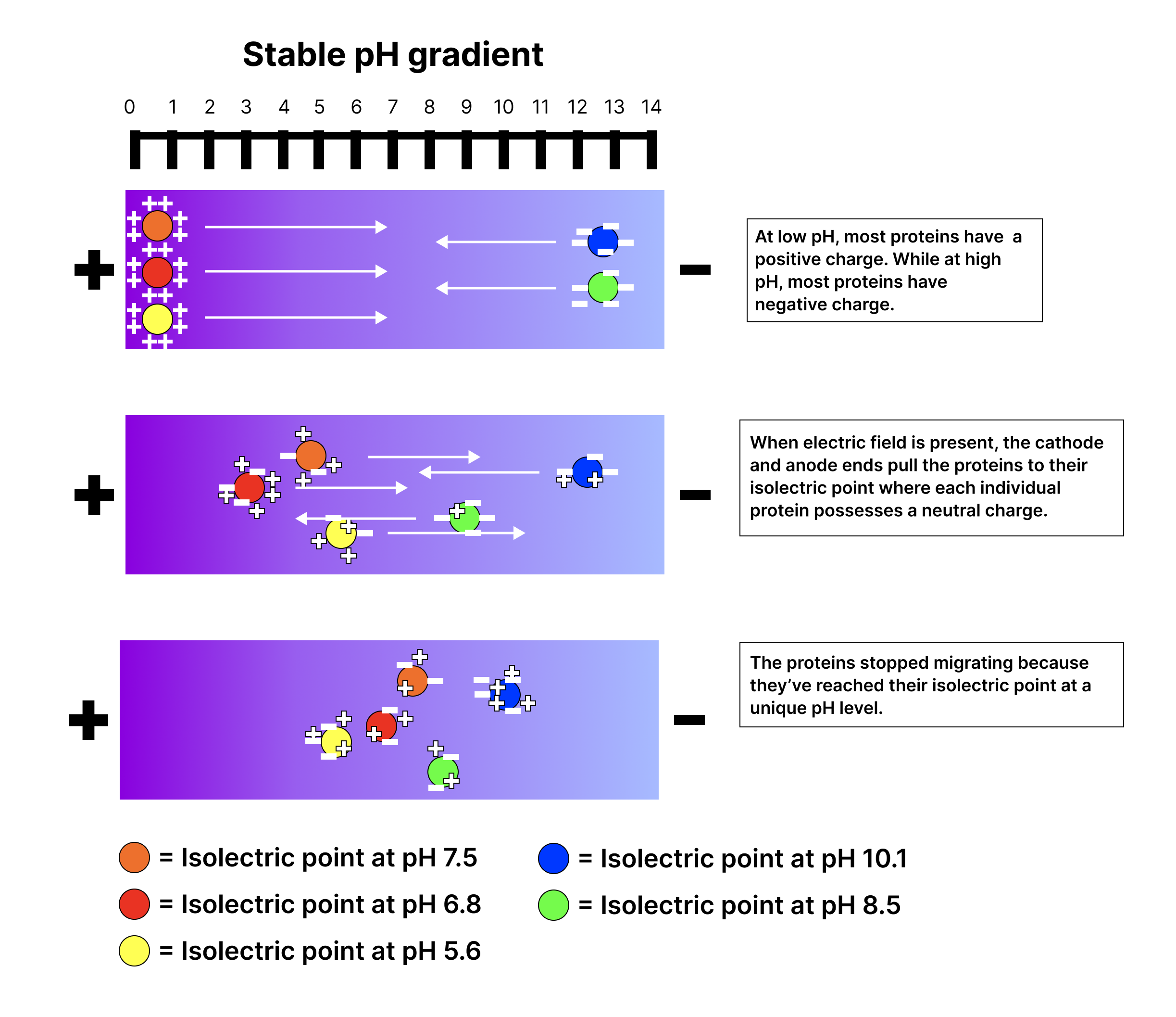
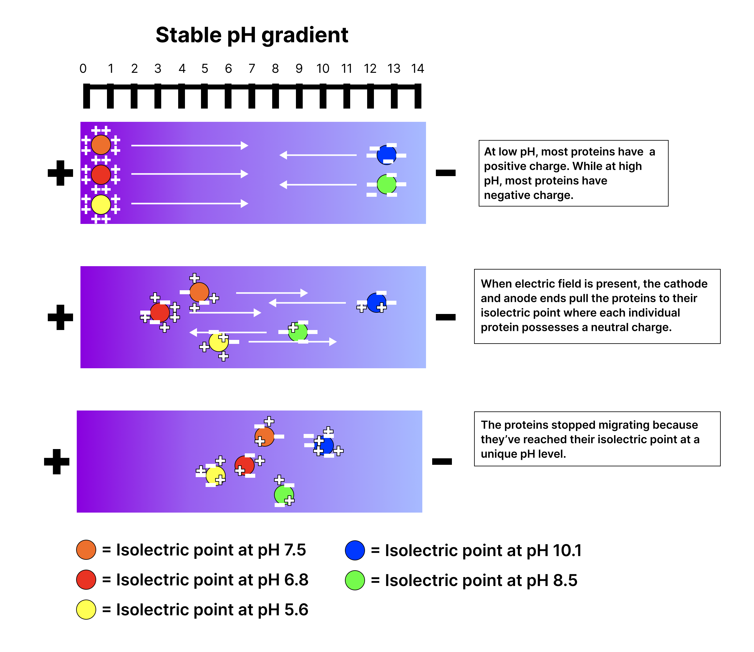
Isoelectric focusing

Separation of in a by their . A stops migrating where it has no net .

Read explanation · Thermo Fisher ↗
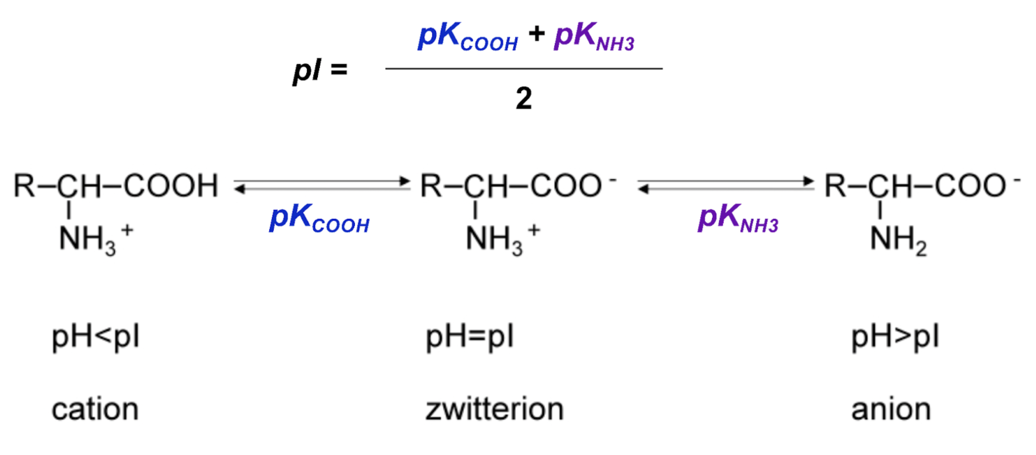
Isoelectric point ·

The at which a has zero net . In , a accumulates where local equals its pI.

Read explanation · OpenStax ↗
Kinase ·

An that transfers a , often from , to a target .

Read explanation · Thermo Fisher ↗
laminar flow

Smooth fluid flow in organized layers.

Read explanation · OpenStax ↗
leukocyte

A white blood cell involved in defense and immune functions.

Read explanation · OpenStax ↗
Ligand

A or that binds to a particular site on another , such as a or metal .

Read explanation · Cytiva ↗
linkage

A tendency of nearby on the same to be inherited together.

Read explanation · OpenStax ↗
Lipid

A broad class of largely or , including fats, , and steroids.

Read explanation · OpenStax ↗
locus

The specific position of a or other feature.

Read explanation · OpenStax ↗
Logarithm · · ·

The exponent needed to obtain a number from a chosen fixed number. For decimal logarithms, log₁₀(100) = 2 because 10² = 100; uses the negative decimal logarithm of activity.

Read explanation · OpenStax ↗
Loss of function

or elimination of a ’s normal activity.

Read explanation · NCBI Bookshelf / Sinauer Associates ↗
Lumen · ·

The interior space of an or tube; the ER lumen is enclosed by the .

Read explanation · OpenStax ↗
lymph

Fluid collected from tissue spaces and transported through lymphatic vessels back toward the bloodstream.

Read explanation · OpenStax ↗
Lysosome

An -enclosed compartment that uses hydrolytic to digest and recycle cellular material.

Read explanation · OpenStax ↗
Macromolecule

A large , such as a , , or polysaccharide.

Read explanation · OpenStax ↗
macrophage

A tissue phagocyte involved in clearance, signaling, and presentation.

Read explanation · OpenStax ↗
magnetic resonance imaging

Imaging method using magnetic fields and radiofrequency signals to reveal tissue structure.

Read explanation · OpenStax ↗
Maximum reaction rate · ·

The limiting as becomes saturating. For a fixed under given conditions, Vmax = times .

Read explanation · Biology LibreTexts ↗
mean arterial pressure

The time-averaged pressure driving systemic circulation over a cardiac cycle.

Read explanation · OpenStax ↗
megakaryocyte

A large bone-marrow cell that generates through .

Read explanation · OpenStax ↗
Membrane · · · ·

A thin boundary or sheet. Cell membranes are bilayers containing ; membranes are synthetic supports that hold or for detection.

Read explanation · OpenStax ↗
Messenger RNA ·

An that carries coding information used by to synthesize a .

Read explanation · OpenStax ↗
Metabolism

The chemical that build , break them down, and transform energy in a living system.

Read explanation · OpenStax ↗
Methyl group ·

A –CH₃ group, consisting of one carbon bonded to three hydrogens.

Read explanation · OpenStax ↗
Michaelis–Menten kinetics · ·

A model with = [S]/(Km + ). Its curve is ; may require a different model.

Read explanation · Biology LibreTexts ↗
Missense mutation ·

A coding-sequence that changes the specified ; its effect depends on the and context.

Read explanation · OpenStax ↗
Mitochondrion ·

A with its own , central to aerobic production and several other processes.

Read explanation · OpenStax ↗
mitral valve

The left atrioventricular valve preventing backflow from left to left .

Read explanation · OpenStax ↗
Mobile phase

The moving fluid that carries a sample through a system.

Read explanation · Waters ↗
Molecular chaperone · ·

A that assists other in folding or prevents , without specifying their sequence.

Read explanation · NCBI Bookshelf ↗
molecular ion

formed from an intact without ; its state must be considered.

Read explanation · OpenStax ↗
Molecule

Two or more joined by . A molecule may be electrically or carry a .

Read explanation · OpenStax ↗
motor protein

that converts chemical energy into movement along a track.

Read explanation · Khan Academy ↗
Mutation

A change in a sequence; some mutations alter a ’s sequence.

Read explanation · OpenStax ↗
Myosin

A that uses to move along filaments.

Read explanation · OpenStax ↗
Native structure · · ·

A ’s functional folded arrangement under its normal conditions.

Read explanation · NCBI Bookshelf ↗
nephron

A kidney functional unit consisting of a filtration apparatus and associated tubular segments.

Read explanation · OpenStax ↗
neuromuscular junction

The connecting a motor to a skeletal-muscle fiber.

Read explanation · OpenStax ↗
neuron

An electrically excitable cell specialized for receiving and transmitting information.

Read explanation · OpenStax ↗
Neutral

Having no net . For a , neutral instead means equal and activities; its is about 7 at 25 °C.

Read explanation · OpenStax ↗
Nitrogenous base · · ·

A nitrogen-containing ring component of ; its pairing interactions help specify sequences.

Read explanation · OpenStax ↗
Nonpolar ·

Lacking a substantial separation of . Nonpolar groups generally interact poorly with water.

Read explanation · OpenStax ↗
Nonsense mutation ·

A that converts an amino- into a premature , potentially truncating the or destabilizing its .

Read explanation · OpenStax ↗
Nonspecific binding · ·

Unwanted binding outside the intended target interaction, often contributing background signal in an .

Read explanation · Thermo Fisher ↗
Nuclear envelope

The double enclosing the and continuous with the .

Read explanation · OpenStax ↗
Nucleic acid

A of , such as or , used in information storage, expression, and other cellular functions.

Read explanation · OpenStax ↗
Nucleosome

The basic unit of : wrapped around a .

Read explanation · OpenStax ↗
Nucleotide

A joined to a sugar and one or more .

Read explanation · OpenStax ↗
Nucleus ·

A -enclosed compartment containing most of a cell’s .

Read explanation · OpenStax ↗
Octamer

A complex made of eight . A core contains eight .

Read explanation · OpenStax ↗
oncotic pressure

The osmotic contribution of and other retained , important in capillary water movement.

Read explanation · OpenStax ↗
Organelle ·

A specialized structure within a cell that carries out particular functions, such as a or .

Read explanation · OpenStax ↗
osmosis

Net water movement across a selectively permeable in response to water chemical-potential differences.

Read explanation · OpenStax ↗
osmotic pressure

The pressure required to prevent net across a under specified conditions.

Read explanation · OpenStax ↗
Oxidation ·

Loss of or an increase in oxidation state. Oxidizing two can form a .

Read explanation · OpenStax ↗
Pancreas

An organ with digestive and functions; its islets contain cells that secrete and .

Read explanation · OpenStax ↗
Pancreatic beta cell ·

An cell in a pancreatic islet that secretes .

Read explanation · OpenStax ↗
Peptide

A chain of joined by ; the term commonly describes chains shorter than a typical .

Read explanation · OpenStax ↗
Peptide bond ·

A covalent between the carboxyl carbon of one and the amino nitrogen of the next.

Read explanation · OpenStax ↗
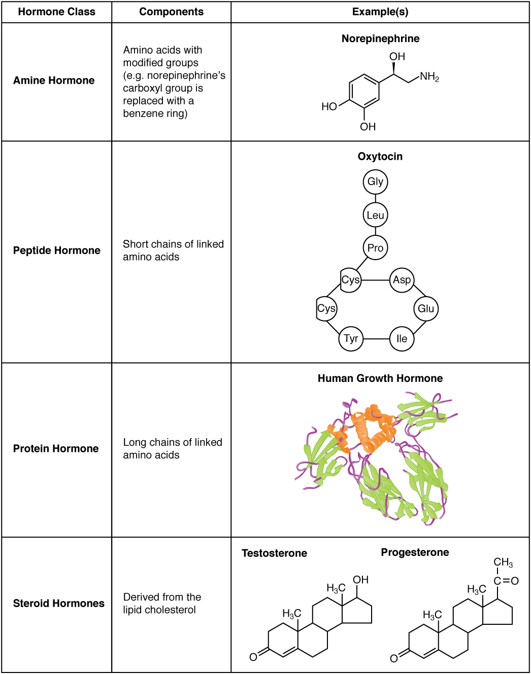
Peptide hormone

An amino--based chemical messenger released by ; and are examples.

Read explanation · OpenStax ↗
pH

A measure of , defined as the negative of activity. Lower pH means greater ; pH influences .

Read explanation · OpenStax ↗
Phenylalanine ·

A with an .

Read explanation · OpenStax ↗
Phosphate group ·

A phosphorus-containing group bonded to oxygens; phosphate groups occur in , , and modified .

Read explanation · OpenStax ↗
phosphocreatine

A creatine reserve that rapidly transfers to ADP during high demand.

Read explanation · OpenStax ↗
Phosphodiester bond · · ·

A in which connects two sugars. In and it links the 3′ oxygen of one sugar to the 5′ oxygen of the next.

Read explanation · OpenStax ↗
Phospholipid

An with a -containing region and tails; a major component of cellular .

Read explanation · OpenStax ↗
Phosphorylation

Addition of a , often to , , or in ; can regulate activity.

Read explanation · Thermo Fisher ↗
platelet

A small -derived blood fragment contributing to .

Read explanation · OpenStax ↗
Polar

Having an uneven distribution of . Polar groups often interact favorably with water.

Read explanation · OpenStax ↗
Polymer

A large made from many linked molecular building blocks.

Read explanation · OpenStax ↗
Polypeptide

A chain of connected by . A functional can contain one or several such chains.

Read explanation · OpenStax ↗
Porous material · · · ·

A material containing small spaces that can admit of suitable size.

Read explanation · Cytiva ↗
prevalence

Proportion or number of existing cases in a defined population and time frame.

Read explanation · OpenStax ↗
Prokaryote ·

An organism whose cells lack a -bound ; and are prokaryotes.

Read explanation · OpenStax ↗
Proline ·

An whose loops back to the nitrogen, restricting rotation and often creating bends.

Read explanation · OpenStax ↗
prostaglandin

signaling formed from arachidonic through cyclooxygenase pathways.

Read explanation · OpenStax ↗
Protease · ·

An that hydrolyzes in or .

Read explanation · OpenStax ↗
Protein

One or more folded chains that perform biological roles such as , transport, signaling, or structural support.

Read explanation · OpenStax ↗
Protein aggregation · ·

Association of into larger clusters, often when exposed surfaces of unfolded stick together.

Read explanation · NCBI Bookshelf ↗
Protein backbone ·

The repeating nitrogen–alpha-carbon– chain of a , excluding its variable .

Read explanation · OpenStax ↗
Protein domain · ·

A region of a that forms a recognizable structural or functional unit.

Read explanation · OpenStax ↗
Proton

A positively subatomic particle. In – chemistry, donating a proton means transferring a .

Read explanation · OpenStax ↗
Protonation

Addition of a to an , , or ; this changes its by plus one.

Read explanation · OpenStax ↗
psychoactive drug

Substance that changes mental processes or behavior through effects on the nervous system.

Read explanation · OpenStax ↗
pulmonary valve

The valve between right and pulmonary outflow.

Read explanation · OpenStax ↗
R group · ·

The variable side chain attached to an ’s . It determines identity, , , and chemical behavior.

Read explanation · OpenStax ↗
Reactant

A starting substance transformed during a .

Read explanation · OpenStax ↗
Reaction intermediate · ·

A species formed in one step and consumed in another. It occupies a local energy minimum between .

Read explanation · OpenStax ↗
Reaction product ·

A substance formed by a .

Read explanation · OpenStax ↗
reaction quotient

-to- activity ratio with stoichiometric exponents at the current composition.

Read explanation · OpenStax ↗
Reaction rate · ·

How quickly are consumed or form, usually expressed as a change per unit time.

Read explanation · OpenStax ↗
Receptor

A that recognizes a particular signal and helps produce a cellular response.

Read explanation · OpenStax ↗
Red blood cell · ·

A blood cell that transports oxygen using ; mature human red blood cells lack a .

Read explanation · OpenStax ↗
Reduction

Gain of or a decrease in state. Reducing a restores two .

Read explanation · OpenStax ↗
Release factor ·

A that recognizes a stop signal and promotes of the – to release the .

Read explanation · OpenStax ↗
renin

A kidney-derived initiating the angiotensin cascade by cleaving angiotensinogen.

Read explanation · OpenStax ↗
repolarization

Return of voltage toward its resting value after .

Read explanation · OpenStax ↗
Ribonucleic acid ·

A with roles in , regulation, and ; carries -coding information.

Read explanation · OpenStax ↗
Ribosome ·

A complex of and that translates into a .

Read explanation · OpenStax ↗
RNA splicing · ·

Removal of and joining of in a precursor .

Read explanation · NCBI Bookshelf / Garland Science ↗
RNA stability

Persistence of an before degradation; greater stability generally means a longer lifetime or under specified conditions.

Read explanation · OpenStax ↗
sarcomere

The repeating contractile unit of striated muscle extending between neighboring .

Read explanation · OpenStax ↗
sarcoplasmic reticulum

Specialized muscle storing and releasing calcium for contraction.

Read explanation · OpenStax ↗
Secondary structure

Local folding, such as and , stabilized mainly by .

Read explanation · OpenStax ↗
Secretory pathway ·

The route through the , , and used to deliver many to the cell surface, outside the cell, or compartments.

Read explanation · OpenStax ↗
Sedimentation ·

Movement of particles through a fluid under gravity or centrifugal .

Read explanation · Beckman Coulter ↗
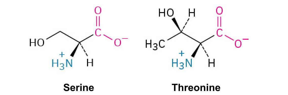
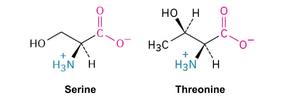
Serine ·

A , uncharged with a -containing .

Read explanation · OpenStax ↗
Sigmoidal curve ·

An S-shaped curve. A sigmoidal rate-versus- curve can indicate rather than simple behavior.

Read explanation · Biology LibreTexts ↗
Sister chromatid

One of the two copies of a produced by . An intact sister chromatid can supply a template for accurate repair.

Read explanation · Biology LibreTexts / Oregon State University ↗
Solubility ·

The amount of a substance that can dissolve in a under specified conditions. Soluble means able to dissolve; insoluble means only very slightly soluble in that .

Read explanation · OpenStax ↗
Solute

A dissolved component of a .

Read explanation · OpenStax ↗
Solution

A homogeneous mixture in which are dissolved in a .

Read explanation · OpenStax ↗
Solvent

The component of a that dissolves other substances, usually the component present in the greatest amount.

Read explanation · OpenStax ↗
squamous

Describing thin, flattened epithelial cells.

Read explanation · OpenStax ↗
Starch

A plant storage made of chains; its two main forms are and amylopectin.

Read explanation · OpenStax ↗
Stationary phase

The material that remains fixed in a system.

Read explanation · Waters ↗
Steady state ·

A condition in which an intermediate remains approximately constant because formation and breakdown rates balance. can still accumulate; this is not .

Read explanation · Biology LibreTexts ↗
steroid hormone

A -derived with fused-ring structure, commonly acting through intracellular .

Read explanation · OpenStax ↗
sterol

A containing fused rings and a ; is a major animal example.

Read explanation · OpenStax ↗
Stop codon · · ·

An that recruits a to end : UAA, UAG, or UGA in the standard code.

Read explanation · OpenStax ↗
stroke volume

The blood volume ejected by a in one beat.

Read explanation · OpenStax ↗
Substitution · ·

Replacement of one by another. In a coding region it can be , , or .

Read explanation · OpenStax ↗
Substrate

A that an binds and chemically transforms.

Read explanation · OpenStax ↗
Substrate concentration

The amount of per volume. Square brackets denote ; [S] is varied to measure an curve.

Read explanation · Biology LibreTexts ↗
Subunit ·

An individual chain within a containing multiple chains.

Read explanation · OpenStax ↗
synapse

A specialized connection through which a cell communicates with another electrically or chemically.

Read explanation · OpenStax ↗
Synonymous mutation · · · · ·

A coding-sequence that leaves the specified unchanged. It can still affect , , or .

Read explanation · OpenStax ↗
systemic vascular resistance

The effective resistance opposing blood flow through the systemic vascular network.

Read explanation · OpenStax ↗
systole

The cardiac phase associated with contraction and ejection, especially ventricular contraction.

Read explanation · OpenStax ↗
Template strand · · ·

A preexisting strand whose sequence guides complementary nucleic- synthesis.

Read explanation · OpenStax ↗
tendon

A -rich structure transmitting muscle force to bone or another .

Read explanation · OpenStax ↗
Thiol group · · · ·

A sulfur bonded to hydrogen, –SH. thiols can form through .

Read explanation · OpenStax ↗
Threonine ·

A , uncharged with a and a on its .

Read explanation · OpenStax ↗
tolerance

Reduced effect of a given drug dose after repeated exposure.

Read explanation · OpenStax ↗
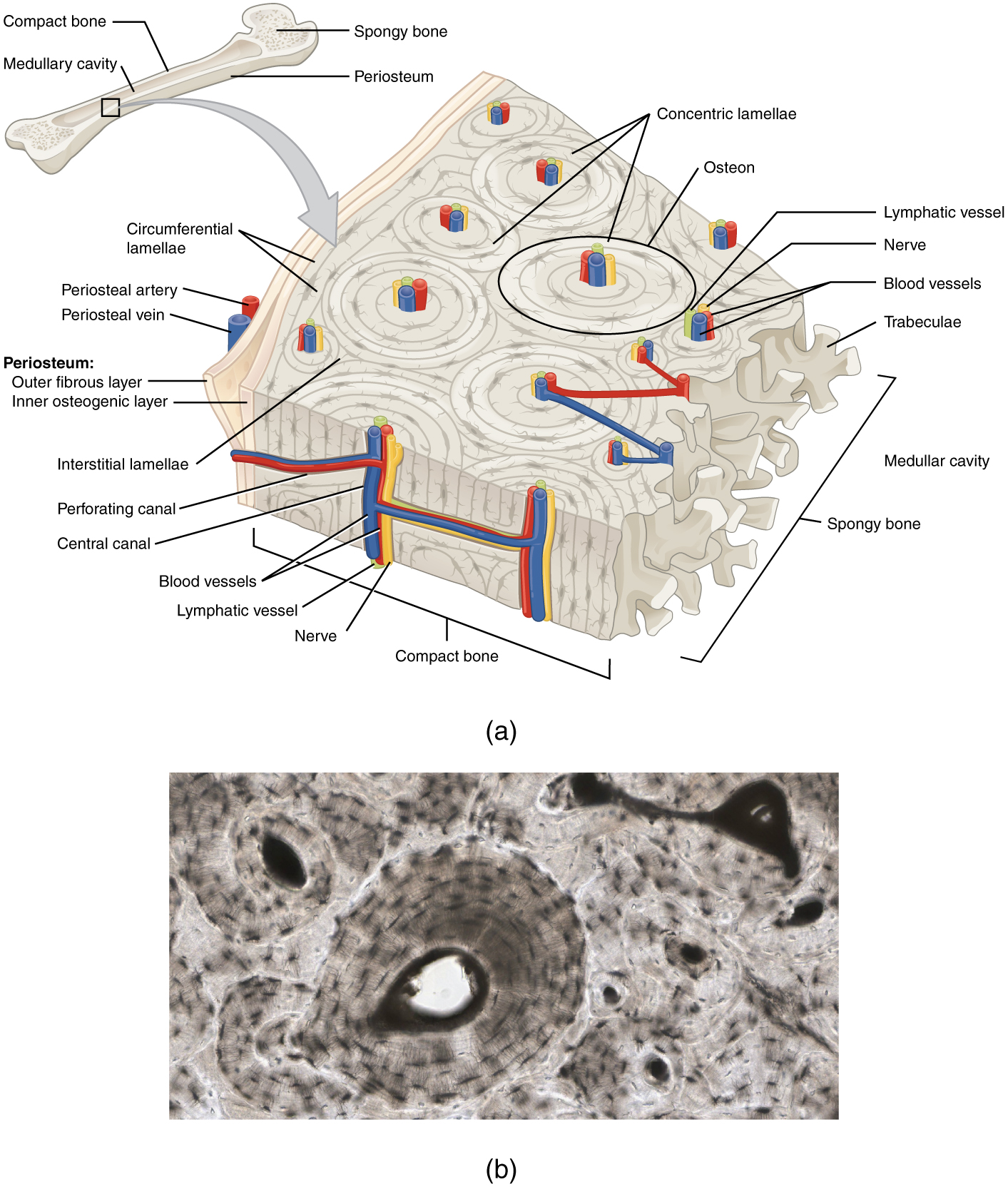
trabecula

A supporting strut or plate of spongy bone.

Read explanation · OpenStax ↗
Transcription

Synthesis of using a template.

Read explanation · OpenStax ↗
Transfer RNA ·

An adaptor that carries an and pairs its with a on .

Read explanation · OpenStax ↗
Transition state

A fleeting, high-energy arrangement at a barrier. It is an energy maximum along the path, rather than a stable .

Read explanation · OpenStax ↗
Translation ·

Synthesis of a on a using the sequence of in .

Read explanation · OpenStax ↗
triacylglycerol · ·

A esterified to three ; a major energy-storage .

Read explanation · Biology LibreTexts / Oregon State University ↗
tricuspid valve

The right atrioventricular valve preventing backflow from right to right .

Read explanation · OpenStax ↗
tropomyosin

A thin-filament regulating access to ’s - in striated muscle.

Read explanation · OpenStax ↗
troponin

A striated-muscle regulatory complex whose calcium-sensitive helps control position.

Read explanation · OpenStax ↗
Tryptophan ·

An with a bulky ; commonly grouped as .

Read explanation · OpenStax ↗
tubular reabsorption

Movement of material from fluid back toward the blood.

Read explanation · OpenStax ↗
turbulence

Irregular flow with eddies and mixing between fluid layers.

Read explanation · OpenStax ↗
Turnover number · · ·

The maximum number of cycles per per unit time, calculated as /. Units are inverse time, commonly s⁻¹.

Read explanation · IUBMB ↗
Tyrosine ·

An with a phenolic . Usually uncharged near physiological .

Read explanation · OpenStax ↗
Upstream

Toward positions preceding a reference site in the direction opposite ; positions before +1 have negative numbering.

Read explanation · OpenStax ↗
vasoconstriction

Narrowing of a blood vessel through smooth-muscle contraction.

Read explanation · OpenStax ↗
vasodilation

Widening of a blood vessel through reduced smooth-muscle tone or other relaxing influences.

Read explanation · OpenStax ↗
velocity

Rate of change of position, including direction.

Read explanation · OpenStax ↗
venule

A small vessel collecting blood from capillary beds.

Read explanation · OpenStax ↗
Vesicle · · ·

A small -enclosed compartment that can transport material between cellular locations.

Read explanation · OpenStax ↗
viscosity

Resistance of a fluid to shear flow, measured in Pa·s.

Read explanation · OpenStax ↗
Z disc

A boundary anchoring thin filaments.

Read explanation · OpenStax ↗

19.1 Heart Anatomy

OpenStax · 19.1 Heart Anatomy ↗
Instructional page including learning objectives, examples, figures, source tables and questions. Site navigation and embedded media players are omitted; source media links remain available.

Learning Objectives

By the end of this section, you will be able to:

  • Describe the location and position of the heart within the body cavity
  • Describe the internal and external anatomy of the heart
  • Identify the tissue layers of the heart
  • Relate the structure of the heart to its function as a pump
  • Compare systemic circulation to pulmonary circulation
  • Identify the veins and arteries of the coronary circulation system
  • Trace the pathway of oxygenated and deoxygenated blood thorough the chambers of the heart

The vital importance of the heart is obvious. If one assumes an average rate of contraction of 75 contractions per minute, a human heart would contract approximately 108,000 times in one day, more than 39 million times in one year, and nearly 3 billion times during a 75-year lifespan. Each of the major pumping chambers of the heart ejects approximately 70 mL blood per contraction in a resting adult. This would be equal to 5.25 liters of fluid per minute and approximately 14,000 liters per day. Over one year, that would equal 10,000,000 liters or 2.6 million gallons of blood sent through roughly 60,000 miles of vessels. In order to understand how that happens, it is necessary to understand the anatomy and physiology of the heart.

Location of the Heart

The human heart is located within the thoracic cavity, medially between the lungs in the space known as the mediastinum. Figure 19.2 shows the position of the heart within the thoracic cavity. Within the mediastinum, the heart is separated from the other mediastinal structures by a tough known as the pericardium, or pericardial sac, and sits in its own space called the pericardial cavity. The dorsal surface of the heart lies near the bodies of the vertebrae, and its anterior surface sits deep to the sternum and costal cartilages. The great veins, the superior and inferior venae cavae, and the great arteries, the aorta and pulmonary trunk, are attached to the superior surface of the heart, called the . The of the heart is located at the level of the third costal cartilage, as seen in Figure 19.2. The inferior tip of the heart, the apex, lies just to the left of the sternum between the junction of the fourth and fifth ribs near their articulation with the costal cartilages. The right side of the heart is deflected anteriorly, and the left side is deflected posteriorly. It is important to remember the position and orientation of the heart when placing a stethoscope on the chest of a patient and listening for heart sounds, and also when looking at images taken from a midsagittal perspective. The slight deviation of the apex to the left is reflected in a depression in the medial surface of the superior lobe of the left lung, called the cardiac notch.

This diagram shows the location of the heart in the thorax.
Figure 19.2 — 19.1 Heart Anatomy

Figure 19.2 Position of the Heart in the Thorax The heart is located within the thoracic cavity, medially between the lungs in the mediastinum. It is about the size of a fist, is broad at the top (called the base), and tapers toward the apex.

J. Gordon Betts and colleagues, Anatomy And Physiology 2E, OpenStax. · CC BY-NC-SA 4.0 · Original image ↗ · Access for free ↗

Everyday Connection

CPR

The position of the heart in the torso between the vertebrae and sternum (see Figure 19.2 for the position of the heart within the thorax) allows for individuals to apply an emergency technique known as cardiopulmonary resuscitation (CPR) if the heart of a patient should stop. By applying pressure with the flat portion of one hand on the sternum in the area between the line at T4 and T9 (Figure 19.3), it is possible to manually compress the blood within the heart enough to push some of the blood within it into the pulmonary and systemic circuits. This is particularly critical for the brain, as irreversible damage and death of occur within minutes of loss of blood flow. Current call for compression of the chest at least 5 deep and at a rate of 100 compressions per minute, a rate equal to the beat in “Staying Alive,” recorded in 1977 by the Bee Gees. If you are unfamiliar with this song, a version is available on www.youtube.com. At this stage, the emphasis is on performing high-quality chest compressions, rather than providing artificial respiration. CPR is generally performed until the patient regains spontaneous contraction or is declared dead by an experienced healthcare professional.

When performed by untrained or overzealous individuals, CPR can result in broken ribs or a broken sternum, and can inflict additional severe damage on the patient. It is also possible, if the hands are placed too low on the sternum, to manually drive the xiphoid process into the liver, a consequence that may prove fatal for the patient. Proper training is essential. This proven life-sustaining technique is so valuable that virtually all medical personnel as well as concerned members of the public should be certified and routinely recertified in its application. CPR courses are offered at a variety of locations, including colleges, hospitals, the American Red Cross, and some commercial companies. They normally include practice of the compression technique on a mannequin.

The top panel shows a schematic of a person performing CPR and demarcates the region in the chest where the compression must be performed. The bottom panel shows a photo of a person performing CPR on a dummy.
Figure 19.3 — 19.1 Heart Anatomy

Figure 19.3 CPR Technique If the heart should stop, CPR can maintain the flow of blood until the heart resumes beating. By applying pressure to the sternum, the blood within the heart will be squeezed out of the heart and into the circulation. Proper positioning of the hands on the sternum to perform CPR would be between the lines at T4 and T9.

J. Gordon Betts and colleagues, Anatomy And Physiology 2E, OpenStax. · CC BY-NC-SA 4.0 · Original image ↗ · Access for free ↗

Interactive Link

Visit the American Heart Association website to help locate a course near your home in the United States. There are also many other national and regional heart associations that offer the same service, depending upon the location.

Shape and Size of the Heart

The shape of the heart is similar to a pinecone, rather broad at the superior surface and tapering to the apex (see Figure 19.2). A typical heart is approximately the size of your fist: 12 (5 in) in length, 8 (3.5 in) wide, and 6 (2.5 in) in thickness. Given the size difference between most members of the sexes, the weight of a female heart is approximately 250–300 grams (9 to 11 ounces), and the weight of a male heart is approximately 300–350 grams (11 to 12 ounces). The heart of a well-trained athlete, especially one specializing in aerobic sports, can be considerably larger than this. Cardiac muscle responds to exercise in a manner similar to that of skeletal muscle. That is, exercise results in the addition of myofilaments that increase the size of the individual cells without increasing their numbers, a concept called hypertrophy. Hearts of athletes can pump blood more effectively at lower rates than those of nonathletes. Enlarged hearts are not always a result of exercise; they can result from pathologies, such as hypertrophic cardiomyopathy. The cause of an abnormally enlarged heart muscle is unknown, but the condition is often undiagnosed and can cause sudden death in apparently otherwise healthy young people.

Chambers and Circulation through the Heart

The human heart consists of four chambers: The left side and the right side each have one and one . Each of the upper chambers, the right (plural = ) and the left , acts as a receiving chamber and contracts to push blood into the lower chambers, the right and the left . The serve as the primary pumping chambers of the heart, propelling blood to the lungs or to the rest of the body.

There are two distinct but linked circuits in the human circulation called the pulmonary and systemic circuits. Although both circuits transport blood and everything it carries, we can initially view the circuits from the point of view of gases. The pulmonary circuit transports blood to and from the lungs, where it picks up oxygen and delivers carbon dioxide for exhalation. The systemic circuit transports oxygenated blood to virtually all of the tissues of the body and returns relatively deoxygenated blood and carbon dioxide to the heart to be sent back to the pulmonary circulation.

The right pumps deoxygenated blood into the pulmonary trunk, which leads toward the lungs and bifurcates into the left and right pulmonary arteries. These vessels in turn branch many times before reaching the pulmonary capillaries, where gas exchange occurs: Carbon dioxide exits the blood and oxygen enters. The pulmonary trunk arteries and their branches are the only arteries in the post-natal body that carry relatively deoxygenated blood. Highly oxygenated blood returning from the pulmonary capillaries in the lungs passes through a series of vessels that join together to form the pulmonary veins—the only post-natal veins in the body that carry highly oxygenated blood. The pulmonary veins conduct blood into the left , which pumps the blood into the left , which in turn pumps oxygenated blood into the aorta and on to the many branches of the systemic circuit. Eventually, these vessels will lead to the systemic capillaries, where exchange with the tissue fluid and cells of the body occurs. In this case, oxygen and nutrients exit the systemic capillaries to be used by the cells in their processes, and carbon dioxide and waste will enter the blood.

The blood exiting the systemic capillaries is lower in oxygen than when it entered. The capillaries will ultimately unite to form , joining to form ever-larger veins, eventually flowing into the two major systemic veins, the superior vena cava and the inferior vena cava, which return blood to the right . The blood in the superior and inferior venae cavae flows into the right , which pumps blood into the right . This process of blood circulation continues as long as the individual remains alive. Understanding the flow of blood through the pulmonary and systemic circuits is critical to all health professions (Figure 19.4).

The top panel shows the human heart with the arteries and veins labeled. The bottom panel shows the human circulatory system.
Figure 19.4 — 19.1 Heart Anatomy

Figure 19.4 Dual System of the Human Blood Circulation Blood flows from the right atrium to the right ventricle, where it is pumped into the pulmonary circuit. The blood in the pulmonary artery branches is low in oxygen but relatively high in carbon dioxide. Gas exchange occurs in the pulmonary capillaries (oxygen into the blood, carbon dioxide out), and blood high in oxygen and low in carbon dioxide is returned to the left atrium. From here, blood enters the left ventricle, which pumps it into the systemic circuit. Following exchange in the systemic capillaries (oxygen and nutrients out of the capillaries and carbon dioxide and wastes in), blood returns to the right atrium and the cycle is repeated.

J. Gordon Betts and colleagues, Anatomy And Physiology 2E, OpenStax. · CC BY-NC-SA 4.0 · Original image ↗ · Access for free ↗

, Surface Features, and Layers

Our exploration of more in-depth heart structures begins by examining the that surrounds the heart, the prominent surface features of the heart, and the layers that form the wall of the heart. Each of these components plays its own unique role in terms of function.

The that directly surrounds the heart and defines the pericardial cavity is called the pericardium or pericardial sac. It also surrounds the “roots” of the major vessels, or the areas of closest proximity to the heart. The pericardium, which literally translates as “around the heart,” consists of two distinct sublayers: the sturdy outer fibrous pericardium and the inner serous pericardium. The fibrous pericardium is made of tough, dense that protects the heart and maintains its position in the thorax. The more delicate serous pericardium consists of two layers: the parietal pericardium, which is fused to the fibrous pericardium, and an inner visceral pericardium, or epicardium, which is fused to the heart and is part of the heart wall. The pericardial cavity, filled with lubricating serous fluid, lies between the epicardium and the pericardium.

In most organs within the body, visceral serous such as the epicardium are microscopic. However, in the case of the heart, it is not a microscopic layer but rather a macroscopic layer, consisting of a simple called a mesothelium, reinforced with loose, irregular, or areolar that attaches to the pericardium. This mesothelium secretes the lubricating serous fluid that fills the pericardial cavity and reduces friction as the heart contracts. Figure 19.5 illustrates the pericardial and the layers of the heart.

This image shows a magnified view of the structure of the heart wall.
Figure 19.5 — 19.1 Heart Anatomy

Figure 19.5 Pericardial Membranes and Layers of the Heart Wall The pericardial membrane that surrounds the heart consists of three layers and the pericardial cavity. The heart wall also consists of three layers. The pericardial membrane and the heart wall share the epicardium.

J. Gordon Betts and colleagues, Anatomy And Physiology 2E, OpenStax. · CC BY-NC-SA 4.0 · Original image ↗ · Access for free ↗

Disorders of the...

Heart: Cardiac Tamponade

If excess fluid builds within the pericardial space, it can lead to a condition called cardiac tamponade, or pericardial tamponade. With each contraction of the heart, more fluid—in most instances, blood—accumulates within the pericardial cavity. In order to fill with blood for the next contraction, the heart must relax. However, the excess fluid in the pericardial cavity puts pressure on the heart and prevents full relaxation, so the chambers within the heart contain slightly less blood as they begin each heart cycle. Over time, less and less blood is ejected from the heart. If the fluid builds up slowly, as in hypothyroidism, the pericardial cavity may be able to expand gradually to accommodate this extra volume. Some cases of fluid in excess of one liter within the pericardial cavity have been reported. Rapid accumulation of as little as 100 mL of fluid following trauma may trigger cardiac tamponade. Other common causes include myocardial rupture, pericarditis, , or even cardiac surgery. Removal of this excess fluid requires of drainage tubes into the pericardial cavity. Premature removal of these drainage tubes, for example, following cardiac surgery, or clot formation within these tubes are causes of this condition. Untreated, cardiac tamponade can lead to death.

Surface Features of the Heart

Inside the pericardium, the surface features of the heart are visible, including the four chambers. There is a superficial leaf-like extension of the near the superior surface of the heart, one on each side, called an auricle—a name that means “ear like”—because its shape resembles the external ear of a human (Figure 19.6). Auricles are relatively thin-walled structures that can fill with blood and empty into the or upper chambers of the heart. You may also hear them referred to as atrial appendages. Also prominent is a series of fat-filled grooves, each of which is known as a sulcus (plural = sulci), along the superior surfaces of the heart. Major coronary blood vessels are located in these sulci. The deep coronary sulcus is located between the and . Located between the left and right are two additional sulci that are not as deep as the coronary sulcus. The anterior interventricular sulcus is visible on the anterior surface of the heart, whereas the posterior interventricular sulcus is visible on the posterior surface of the heart. Figure 19.6 illustrates anterior and posterior views of the surface of the heart.

The top panel shows the anterior view of the heart and the bottom panel shows the posterior view of the human heart. In both panels, the main parts of the heart are labeled.
Figure 19.6 — 19.1 Heart Anatomy

Figure 19.6 External Anatomy of the Heart Inside the pericardium, the surface features of the heart are visible.

J. Gordon Betts and colleagues, Anatomy And Physiology 2E, OpenStax. · CC BY-NC-SA 4.0 · Original image ↗ · Access for free ↗

Layers

The wall of the heart is composed of three layers of unequal thickness. From superficial to deep, these are the epicardium, the myocardium, and the endocardium (see Figure 19.5). The outermost layer of the wall of the heart is also the innermost layer of the pericardium, the epicardium, or the visceral pericardium discussed earlier.

The middle and thickest layer is the myocardium, made largely of cardiac muscle cells. It is built upon a framework of collagenous fibers, plus the blood vessels that supply the myocardium and the nerve fibers that help regulate the heart. It is the contraction of the myocardium that pumps blood through the heart and into the major arteries. The muscle pattern is elegant and complex, as the muscle cells swirl and spiral around the chambers of the heart. They form a figure 8 pattern around the and around the of the great vessels. Deeper ventricular muscles also form a figure 8 around the two and proceed toward the apex. More superficial layers of ventricular muscle wrap around both . This complex swirling pattern allows the heart to pump blood more effectively than a simple linear pattern would. Figure 19.7 illustrates the arrangement of muscle cells.

This diagram shows the muscles in the heart.
Figure 19.7 — 19.1 Heart Anatomy

Figure 19.7 Heart Musculature The swirling pattern of cardiac muscle tissue contributes significantly to the heart’s ability to pump blood effectively.

J. Gordon Betts and colleagues, Anatomy And Physiology 2E, OpenStax. · CC BY-NC-SA 4.0 · Original image ↗ · Access for free ↗

Although the on the right and left sides pump the same amount of blood per contraction, the muscle of the left is much thicker and better developed than that of the right . In order to overcome the high resistance required to pump blood into the long systemic circuit, the left must generate a great amount of pressure. The right does not need to generate as much pressure, since the pulmonary circuit is shorter and provides less resistance. Figure 19.8 illustrates the differences in muscular thickness needed for each of the .

In this figure the left panel shows the muscles of the heart in the relaxed position, and the right panel shows the muscles of the heart in contracted position.
Figure 19.8 — 19.1 Heart Anatomy

Figure 19.8 Differences in Ventricular Muscle Thickness The myocardium in the left ventricle is significantly thicker than that of the right ventricle. Both ventricles pump the same amount of blood, but the left ventricle must generate a much greater pressure to overcome greater resistance in the systemic circuit. The ventricles are shown in both relaxed and contracting states. Note the differences in the relative size of the lumens, the region inside each ventricle where the blood is contained.

J. Gordon Betts and colleagues, Anatomy And Physiology 2E, OpenStax. · CC BY-NC-SA 4.0 · Original image ↗ · Access for free ↗

The innermost layer of the heart wall, the endocardium, is joined to the myocardium with a thin layer of . The endocardium lines the chambers where the blood circulates and covers the heart valves. It is made of simple called , which is continuous with the endothelial lining of the blood vessels (see Figure 19.5).

Once regarded as a simple lining layer, recent evidence indicates that the of the endocardium and the coronary capillaries may play active roles in regulating the contraction of the muscle within the myocardium. The may also regulate the growth patterns of the cardiac muscle cells throughout life, and the endothelins it secretes create an environment in the surrounding tissue fluids that regulates ionic and states of . Endothelins are potent vasoconstrictors and, in a normal individual, establish a homeostatic balance with other vasoconstrictors and vasodilators.

Internal Structure of the Heart

Recall that the heart’s contraction cycle follows a dual pattern of circulation—the pulmonary and systemic circuits—because of the pairs of chambers that pump blood into the circulation. In order to develop a more precise understanding of cardiac function, it is first necessary to explore the internal anatomical structures in more detail.

Septa of the Heart

The word septum is derived from the Latin for “something that encloses;” in this case, a septum (plural = septa) refers to a wall or partition that divides the heart into chambers. The septa are physical extensions of the myocardium lined with endocardium. Located between the two is the interatrial septum. Normally in an adult heart, the interatrial septum bears an oval-shaped depression known as the fossa ovalis, a remnant of an opening in the fetal heart known as the foramen ovale. The foramen ovale allowed blood in the fetal heart to pass directly from the right to the left , allowing some blood to bypass the pulmonary circuit. Within seconds after birth, a flap of tissue known as the septum primum that previously acted as a valve closes the foramen ovale and establishes the typical cardiac circulation pattern.

Between the two is a second septum known as the interventricular septum. Unlike the interatrial septum, the interventricular septum is normally intact after its formation during fetal development. It is substantially thicker than the interatrial septum, since the generate far greater pressure when they contract.

The septum between the and is known as the atrioventricular septum. It is marked by the presence of four openings that allow blood to move from the into the and from the into the pulmonary trunk and aorta. Located in each of these openings between the and is a valve, a specialized structure that ensures one-way flow of blood. The valves between the and are known generically as atrioventricular valves. The valves at the openings that lead to the pulmonary trunk and aorta are known generically as semilunar valves. The interventricular septum is visible in Figure 19.9. In this figure, the atrioventricular septum has been removed to better show the bicuspid and tricuspid valves; the interatrial septum is not visible, since its location is covered by the aorta and pulmonary trunk. Since these openings and valves structurally weaken the atrioventricular septum, the remaining tissue is heavily reinforced with dense called the cardiac skeleton, or skeleton of the heart. It includes four rings that surround the openings between the and , and the openings to the pulmonary trunk and aorta, and serve as the point of for the heart valves. The cardiac skeleton also provides an important boundary in the heart electrical system.

In this figure the top panel shows the image of the heart with the major parts labeled. The bottom left panel shows a photo of the heart with the surface layer peeled off. The images on the bottom right show detailed musculature inside the heart.
Figure 19.9 — 19.1 Heart Anatomy

Figure 19.9 Internal Structures of the Heart This anterior view of the heart shows the four chambers, the major vessels and their early branches, as well as the valves. The presence of the pulmonary trunk and aorta covers the interatrial septum, and the atrioventricular septum is cut away to show the atrioventricular valves.

J. Gordon Betts and colleagues, Anatomy And Physiology 2E, OpenStax. · CC BY-NC-SA 4.0 · Original image ↗ · Access for free ↗

Disorders of the...

Heart: Heart Defects

One very common form of interatrial septum pathology is patent foramen ovale, which occurs when the septum primum does not close at birth, and the fossa ovalis is unable to fuse. The word patent is from the Latin root patens for “open.” It may be benign or asymptomatic, perhaps never being diagnosed, or in extreme cases, it may require surgical repair to close the opening permanently. As much as 20–25 percent of the general population may have a patent foramen ovale, but fortunately, most have the benign, asymptomatic version. Patent foramen ovale is normally detected by auscultation of a heart murmur (an abnormal heart sound) and confirmed by imaging with an echocardiogram. Despite its in the general population, the causes of patent ovale are unknown, and there are no known risk factors. In nonlife-threatening cases, it is better to monitor the condition than to risk heart surgery to repair and seal the opening.

Coarctation of the aorta is a congenital abnormal narrowing of the aorta that is normally located at the of the ligamentum arteriosum, the remnant of the fetal shunt called the ductus arteriosus. If severe, this condition drastically restricts blood flow through the primary systemic artery, which is life threatening. In some individuals, the condition may be fairly benign and not detected until later in life. Detectable symptoms in an infant include difficulty breathing, poor appetite, trouble feeding, or failure to thrive. In older individuals, symptoms include dizziness, fainting, shortness of breath, chest pain, fatigue, headache, and nosebleeds. Treatment involves surgery to resect (remove) the affected region or angioplasty to open the abnormally narrow passageway. Studies have shown that the earlier the surgery is performed, the better the chance of survival.

A patent ductus arteriosus is a congenital condition in which the ductus arteriosus fails to close. The condition may range from severe to benign. Failure of the ductus arteriosus to close results in blood flowing from the higher pressure aorta into the lower pressure pulmonary trunk. This additional fluid moving toward the lungs increases pulmonary pressure and makes respiration difficult. Symptoms include shortness of breath (dyspnea), tachycardia, enlarged heart, a widened pulse pressure, and poor weight gain in infants. Treatments include surgical closure (), manual closure using platinum coils or specialized mesh inserted via the femoral artery or vein, or nonsteroidal anti-inflammatory drugs to block the synthesis of E2, which maintains the vessel in an open position. If untreated, the condition can result in congestive heart failure.

Septal defects are not uncommon in individuals and may be congenital or caused by various disease processes. Tetralogy of Fallot is a congenital condition that may also occur from exposure to unknown environmental factors; it occurs when there is an opening in the interventricular septum caused by blockage of the pulmonary trunk, normally at the pulmonary semilunar valve. This allows blood that is relatively low in oxygen from the right to flow into the left and mix with the blood that is relatively high in oxygen. Symptoms include a distinct heart murmur, low blood oxygen percent , dyspnea or difficulty in breathing, polycythemia, broadening (clubbing) of the fingers and toes, and in children, difficulty in feeding or failure to grow and develop. It is the most common cause of cyanosis following birth. The term “tetralogy” is derived from the four components of the condition, although only three may be present in an individual patient: pulmonary infundibular stenosis (rigidity of the ), overriding aorta (the aorta is shifted above both ), ventricular septal defect (opening), and right ventricular hypertrophy (enlargement of the right ). Other heart defects may also accompany this condition, which is typically confirmed by echocardiography imaging. Tetralogy of Fallot occurs in approximately 400 out of one million live births. Normal treatment involves extensive surgical repair, including the use of stents to redirect blood flow and replacement of valves and patches to repair the septal defect, but the condition has a relatively high mortality. Survival rates are currently 75 percent during the first year of life; 60 percent by 4 years of age; 30 percent by 10 years; and 5 percent by 40 years.

In the case of severe septal defects, including both tetralogy of Fallot and patent foramen ovale, failure of the heart to develop properly can lead to a condition commonly known as a “blue baby.” Regardless of normal skin pigmentation, individuals with this condition have an insufficient supply of oxygenated blood, which leads to cyanosis, a blue or purple coloration of the skin, especially when active.

Septal defects are commonly first detected through auscultation, listening to the chest using a stethoscope. In this case, instead of hearing normal heart sounds attributed to the flow of blood and closing of heart valves, unusual heart sounds may be detected. This is often followed by medical imaging to confirm or rule out a diagnosis. In many cases, treatment may not be needed. Some common congenital heart defects are illustrated in Figure 19.10.

This diagram shows the structure of the heart with different congenital defects. The top left panel shows patent foramen ovale, the top right panel shows coarctation of the aorta, the bottom left panel shows patent ductus ateriosus and the bottom right shows tetralogy of fallot.
Figure 19.10 — 19.1 Heart Anatomy

Figure 19.10 Congenital Heart Defects (a) A patent foramen ovale defect is an abnormal opening in the interatrial septum, or more commonly, a failure of the foramen ovale to close. (b) Coarctation of the aorta is an abnormal narrowing of the aorta. (c) A patent ductus arteriosus is the failure of the ductus arteriosus to close. (d) Tetralogy of Fallot includes an abnormal opening in the interventricular septum.

J. Gordon Betts and colleagues, Anatomy And Physiology 2E, OpenStax. · CC BY-NC-SA 4.0 · Original image ↗ · Access for free ↗

Right

The right serves as the receiving chamber for blood returning to the heart from the systemic circulation. The two major systemic veins, the superior and inferior venae cavae, and the large coronary vein called the coronary sinus that drains the heart myocardium empty into the right . The superior vena cava drains blood from regions superior to the diaphragm: the head, neck, upper limbs, and the thoracic region. It empties into the superior and posterior portions of the right . The inferior vena cava drains blood from areas inferior to the diaphragm: the lower limbs and abdominopelvic region of the body. It, too, empties into the posterior portion of the , but inferior to the opening of the superior vena cava. Immediately superior and slightly medial to the opening of the inferior vena cava on the posterior surface of the is the opening of the coronary sinus. This thin-walled vessel drains most of the coronary veins that return systemic blood from the heart. The majority of the internal heart structures discussed in this and subsequent sections are illustrated in Figure 19.9.

While the bulk of the internal surface of the right is smooth, the depression of the fossa ovalis is medial, and the anterior surface demonstrates prominent ridges of muscle called the pectinate muscles. The right auricle also has pectinate muscles. The left does not have pectinate muscles except in the auricle.

The receive venous blood on a nearly continuous basis, preventing venous flow from stopping while the are contracting. While most ventricular filling occurs while the are relaxed, they do demonstrate a contractile phase and actively pump blood into the just prior to ventricular contraction. The opening between the and is guarded by the .

Right

The right receives blood from the right through the . Each flap of the valve is attached to strong strands of , the chordae tendineae, literally “tendinous cords,” or sometimes more poetically referred to as “heart strings.” There are several chordae tendineae associated with each of the flaps. They are composed of approximately 80 percent collagenous fibers with the remainder consisting of elastic fibers and . They connect each of the flaps to a papillary muscle that extends from the inferior ventricular surface. There are three papillary muscles in the right , called the anterior, posterior, and septal muscles, which correspond to the three sections of the valves.

When the myocardium of the contracts, pressure within the ventricular chamber rises. Blood, like any fluid, flows from higher pressure to lower pressure areas, in this case, toward the pulmonary trunk and the . To prevent any potential backflow, the papillary muscles also contract, generating tension on the chordae tendineae. This prevents the flaps of the valves from being forced into the and regurgitation of the blood back into the during ventricular contraction. Figure 19.11 shows papillary muscles and chordae tendineae attached to the .

This photo shows the inside of the heart with the main muscles labeled.
Figure 19.11 — 19.1 Heart Anatomy

Figure 19.11 Chordae Tendineae and Papillary Muscles In this frontal section, you can see papillary muscles attached to the tricuspid valve on the right as well as the mitral valve on the left via chordae tendineae. (credit: modification of work by “PV KS”/flickr.com)

J. Gordon Betts and colleagues, Anatomy And Physiology 2E, OpenStax. (credit: modification of work by “PV KS”/flickr.com) · CC BY-NC-SA 4.0 · Original image ↗ · Access for free ↗

The walls of the are lined with carneae, ridges of cardiac muscle covered by endocardium. In addition to these muscular ridges, of cardiac muscle, also covered by endocardium, known as the moderator band (see Figure 19.9) reinforces the thin walls of the right and plays a crucial role in cardiac . It arises from the inferior portion of the interventricular septum and crosses the interior space of the right to connect with the inferior papillary muscle.

When the right contracts, it ejects blood into the pulmonary trunk, which branches into the left and right pulmonary arteries that carry it to each lung. The superior surface of the right begins to taper as it approaches the pulmonary trunk. At the of the pulmonary trunk is the pulmonary semilunar valve that prevents backflow from the pulmonary trunk.

Left

After exchange of gases in the pulmonary capillaries, blood returns to the left high in oxygen via one of the four pulmonary veins. While the left does not contain pectinate muscles, it does have an auricle that includes these pectinate ridges. Blood flows nearly continuously from the pulmonary veins back into the , which acts as the receiving chamber, and from here through an opening into the left . Most blood flows passively into the heart while both the and are relaxed, but toward the end of the ventricular relaxation period, the left will contract, pumping blood into the . This atrial contraction accounts for approximately 20 percent of ventricular filling. The opening between the left and is guarded by the .

Left

Recall that, although both sides of the heart will pump the same amount of blood, the muscular layer is much thicker in the left compared to the right (see Figure 19.8). Like the right , the left also has carneae, but there is no moderator band. The is connected to papillary muscles via chordae tendineae. There are two papillary muscles on the left—the anterior and posterior—as opposed to three on the right.

The left is the major pumping chamber for the systemic circuit; it ejects blood into the aorta through the aortic semilunar valve.

Heart Valve Structure and Function

A transverse section through the heart slightly above the level of the atrioventricular septum reveals all four heart valves along the same plane (Figure 19.12). The valves ensure unidirectional blood flow through the heart. Between the right and the right is the right atrioventricular valve, or . It typically consists of three flaps, or leaflets, made of endocardium reinforced with additional . The flaps are connected by chordae tendineae to the papillary muscles, which control the opening and closing of the valves.

This diagram shows the anterior view of the heart with the different heart valves labeled.
Figure 19.12 — 19.1 Heart Anatomy

Figure 19.12 Heart Valves With the atria and major vessels removed, all four valves are clearly visible, although it is difficult to distinguish the three separate cusps of the tricuspid valve.

J. Gordon Betts and colleagues, Anatomy And Physiology 2E, OpenStax. · CC BY-NC-SA 4.0 · Original image ↗ · Access for free ↗

Emerging from the right at the of the pulmonary trunk is the pulmonary semilunar valve, or the ; it is also known as the pulmonic valve or the right semilunar valve. The is comprised of three small flaps of reinforced with . When the relaxes, the pressure differential causes blood to flow back into the from the pulmonary trunk. This flow of blood fills the pocket-like flaps of the , causing the valve to close and producing an audible sound. Unlike the atrioventricular valves, there are no papillary muscles or chordae tendineae associated with the .

Located at the opening between the left and left is the , also called the or the left atrioventricular valve. Structurally, this valve consists of two cusps, compared to the three cusps of the . In a clinical setting, the valve is referred to as the , rather than the . The two cusps of the are attached by chordae tendineae to two papillary muscles that project from the wall of the .

At the of the aorta is the aortic semilunar valve, or the , which prevents backflow from the aorta. It normally is composed of three flaps. When the relaxes and blood attempts to flow back into the from the aorta, blood will fill the cusps of the valve, causing it to close and producing an audible sound.

In Figure 19.13a, the two atrioventricular valves are open and the two semilunar valves are closed. This occurs when both and are relaxed and when the contract to pump blood into the . Figure 19.13b shows a frontal view. Although only the left side of the heart is illustrated, the process is virtually identical on the right.

The left panel of this figure shows the anterior view of the heart with the different valves, and the right panel of this figure shows the location of the mitral valve in the open position in the heart.
Figure 19.13 — 19.1 Heart Anatomy

Figure 19.13 Blood Flow from the Left Atrium to the Left Ventricle (a) A transverse section through the heart illustrates the four heart valves. The two atrioventricular valves are open; the two semilunar valves are closed. The atria and vessels have been removed. (b) A frontal section through the heart illustrates blood flow through the mitral valve. When the mitral valve is open, it allows blood to move from the left atrium to the left ventricle. The aortic semilunar valve is closed to prevent backflow of blood from the aorta to the left ventricle.

J. Gordon Betts and colleagues, Anatomy And Physiology 2E, OpenStax. · CC BY-NC-SA 4.0 · Original image ↗ · Access for free ↗

Figure 19.14a shows the atrioventricular valves closed while the two semilunar valves are open. This occurs when the contract to eject blood into the pulmonary trunk and aorta. Closure of the two atrioventricular valves prevents blood from being forced back into the . This stage can be seen from a frontal view in Figure 19.14b.

The left panel of this figure shows the anterior view of the heart with the different valves, and the right panel of this figure shows the location of the mitral valve in the closed position in the heart.
Figure 19.14 — 19.1 Heart Anatomy

Figure 19.14 Blood Flow from the Left Ventricle into the Great Vessels (a) A transverse section through the heart illustrates the four heart valves during ventricular contraction. The two atrioventricular valves are closed, but the two semilunar valves are open. The atria and vessels have been removed. (b) A frontal view shows the closed mitral (bicuspid) valve that prevents backflow of blood into the left atrium. The aortic semilunar valve is open to allow blood to be ejected into the aorta.

J. Gordon Betts and colleagues, Anatomy And Physiology 2E, OpenStax. · CC BY-NC-SA 4.0 · Original image ↗ · Access for free ↗

When the begin to contract, pressure within the rises and blood flows toward the area of lowest pressure, which is initially in the . This backflow causes the cusps of the tricuspid and mitral (bicuspid) valves to close. These valves are tied down to the papillary muscles by chordae tendineae. During the relaxation phase of the cardiac cycle, the papillary muscles are also relaxed and the tension on the chordae tendineae is slight (see Figure 19.13b). However, as the myocardium of the contracts, so do the papillary muscles. This creates tension on the chordae tendineae (see Figure 19.14b), helping to hold the cusps of the atrioventricular valves in place and preventing them from being blown back into the .

The aortic and pulmonary semilunar valves lack the chordae tendineae and papillary muscles associated with the atrioventricular valves. Instead, they consist of pocket-like folds of endocardium reinforced with additional . When the relax and the change in pressure forces the blood toward the , the blood presses against these cusps and seals the openings.

Interactive Link

Visit this site to observe an echocardiogram of actual heart valves opening and closing. Although much of the heart has been “removed” from this gif loop so the chordae tendineae are not visible, why is their presence more critical for the atrioventricular valves (tricuspid and mitral) than the semilunar (aortic and pulmonary) valves?

Disorders of the...

Heart Valves

When heart valves do not function properly, they are often described as incompetent and result in valvular heart disease, which can range from benign to lethal. Some of these conditions are congenital, that is, the individual was born with the defect, whereas others may be attributed to disease processes or trauma. Some malfunctions are treated with medications, others require surgery, and still others may be mild enough that the condition is merely monitored since treatment might trigger more serious consequences.

Valvular disorders are often caused by carditis, or of the heart. One common trigger for this is rheumatic fever, or scarlet fever, an autoimmune response to the presence of a , Streptococcus pyogenes, normally a disease of childhood.

While any of the heart valves may be involved in valve disorders, mitral regurgitation is the most common, detected in approximately 2 percent of the population, and the pulmonary semilunar valve is the least frequently involved. When a valve malfunctions, the flow of blood to a region will often be disrupted. The resulting inadequate flow of blood to this region will be described in general terms as an insufficiency. The specific type of insufficiency is named for the valve involved: aortic insufficiency, mitral insufficiency, tricuspid insufficiency, or pulmonary insufficiency.

If one of the cusps of the valve is forced backward by the force of the blood, the condition is referred to as a prolapsed valve. Prolapse may occur if the chordae tendineae are damaged or broken, causing the closure mechanism to fail. The failure of the valve to close properly disrupts the normal one-way flow of blood and results in regurgitation, when the blood flows backward from its normal path. Using a stethoscope, the disruption to the normal flow of blood produces a heart murmur.

Stenosis is a condition in which the heart valves become rigid and may calcify over time. The loss of flexibility of the valve interferes with normal function and may cause the heart to work harder to propel blood through the valve, which eventually weakens the heart. Aortic stenosis affects approximately 2 percent of the population over 65 years of age, and the percentage increases to approximately 4 percent in individuals over 85 years. Occasionally, one or more of the chordae tendineae will tear or the papillary muscle itself may die as a component of a myocardial infarction (heart attack). In this case, the patient’s condition will deteriorate dramatically and rapidly, and immediate surgical intervention may be required.

Auscultation, or listening to a patient’s heart sounds, is one of the most useful diagnostic tools, since it is proven, safe, and inexpensive. The term auscultation is derived from the Latin for “to listen,” and the technique has been used for diagnostic purposes as far back as the ancient Egyptians. Valve and septal disorders will trigger abnormal heart sounds. If a valvular disorder is detected or suspected, a test called an echocardiogram, or simply an “echo,” may be ordered. Echocardiograms are sonograms of the heart and can help in the diagnosis of valve disorders as well as a wide variety of heart pathologies.

Interactive Link

Visit this site for audio examples of heart sounds.

Career Connection

Cardiologist

Cardiologists are medical doctors that specialize in the diagnosis and treatment of diseases of the heart. After completing 4 years of medical school, cardiologists complete a three-year residency in internal medicine followed by an additional three or more years in cardiology. Following this 10-year period of medical training and clinical experience, they qualify for a rigorous two-day examination administered by the Board of Internal Medicine that tests their academic training and clinical abilities, including diagnostics and treatment. After successful completion of this examination, a physician becomes a board-certified cardiologist. Some board-certified cardiologists may be invited to become a Fellow of the American College of Cardiology (FACC). This professional recognition is awarded to outstanding physicians based upon merit, including outstanding credentials, achievements, and community contributions to cardiovascular medicine.

Interactive Link

Visit this site to learn more about cardiologists.

Career Connection

Cardiovascular Technologist/Technician

Cardiovascular technologists/technicians are trained professionals who perform a variety of imaging techniques, such as sonograms or echocardiograms, used by physicians to diagnose and treat diseases of the heart. Nearly all of these positions require an associate degree. Growth within the field is fast, projected at 29 percent from 2010 to 2020.

There is a considerable overlap and complementary skills between cardiac technicians and vascular technicians, and so the term cardiovascular technician is often used. Special certifications within the field require documenting appropriate experience and completing additional and often expensive certification examinations. These subspecialties include Certified Rhythm Analysis Technician (CRAT), Certified Cardiographic Technician (CCT), Registered Congenital Cardiac Sonographer (RCCS), Registered Cardiac Electrophysiology Specialist (RCES), Registered Cardiovascular Invasive Specialist (RCIS), Registered Cardiac Sonographer (RCS), Registered Vascular Specialist (RVS), and Registered Phlebology Sonographer (RPhS).

Interactive Link

Visit this site for more information on cardiovascular technologists/technicians.

Coronary Circulation

You will recall that the heart is a remarkable pump composed largely of cardiac muscle cells that are incredibly active throughout life. Like all other cells, a cardiomyocyte requires a reliable supply of oxygen and nutrients, and a way to remove wastes, so it needs a dedicated, complex, and extensive coronary circulation. And because of the critical and nearly ceaseless activity of the heart throughout life, this need for a blood supply is even greater than for a typical cell. However, coronary circulation is not continuous; rather, it cycles, reaching a when the heart muscle is relaxed and nearly ceasing while it is contracting.

Coronary Arteries

Coronary arteries supply blood to the myocardium and other components of the heart. The first portion of the aorta after it arises from the left gives rise to the coronary arteries. There are three dilations in the wall of the aorta just superior to the aortic semilunar valve. Two of these, the left posterior aortic sinus and anterior aortic sinus, give rise to the left and right coronary arteries, respectively. The third sinus, the right posterior aortic sinus, typically does not give rise to a vessel. Coronary vessel branches that remain on the surface of the artery and follow the sulci are called epicardial coronary arteries.

The left coronary artery distributes blood to the left side of the heart, the left and , and the interventricular septum. The circumflex artery arises from the left coronary artery and follows the coronary sulcus to the left. Eventually, it will fuse with the small branches of the right coronary artery. The larger anterior interventricular artery, also known as the left anterior descending artery (LAD), is the second major branch arising from the left coronary artery. It follows the anterior interventricular sulcus around the pulmonary trunk. Along the way it gives rise to numerous smaller branches that interconnect with the branches of the posterior interventricular artery, forming anastomoses. An anastomosis is an area where vessels unite to form interconnections that normally allow blood to circulate to a region even if there may be partial blockage in another branch. The anastomoses in the heart are very small. Therefore, this ability is somewhat restricted in the heart so a coronary artery blockage often results in death of the cells (myocardial infarction) supplied by the particular vessel.

The right coronary artery proceeds along the coronary sulcus and distributes blood to the right , portions of both , and the heart system. Normally, one or more marginal arteries arise from the right coronary artery inferior to the right . The marginal arteries supply blood to the superficial portions of the right . On the posterior surface of the heart, the right coronary artery gives rise to the posterior interventricular artery, also known as the posterior descending artery. It runs along the posterior portion of the interventricular sulcus toward the apex of the heart, giving rise to branches that supply the interventricular septum and portions of both . Figure 19.15 presents views of the coronary circulation from both the anterior and posterior views.

The top panel of this figure shows the anterior view of the heart while the bottom panel shows the posterior view of the heart. The different blood vessels are labeled.
Figure 19.15 — 19.1 Heart Anatomy

Figure 19.15 Coronary Circulation The anterior view of the heart shows the prominent coronary surface vessels. The posterior view of the heart shows the prominent coronary surface vessels.

J. Gordon Betts and colleagues, Anatomy And Physiology 2E, OpenStax. · CC BY-NC-SA 4.0 · Original image ↗ · Access for free ↗

Diseases of the...

Heart: Myocardial Infarction

Myocardial infarction (MI) is the formal term for what is commonly referred to as a heart attack. It normally results from a lack of blood flow (ischemia) and oxygen () to a region of the heart, resulting in death of the cardiac muscle cells. An MI often occurs when a coronary artery is blocked by the buildup of atherosclerotic plaque consisting of , and , and white blood cells, primarily . It can also occur when a portion of an unstable atherosclerotic plaque travels through the coronary arterial system and lodges in one of the smaller vessels. The resulting blockage restricts the flow of blood and oxygen to the myocardium and causes death of the tissue. MIs may be triggered by excessive exercise, in which the partially occluded artery is no longer able to pump sufficient quantities of blood, or severe stress, which may induce spasm of the smooth muscle in the walls of the vessel.

In the case of acute MI, there is often sudden pain beneath the sternum (retrosternal pain) called angina pectoris, often radiating down the left arm in males but not in female patients. Until this anomaly between the sexes was discovered, many female patients suffering MIs were misdiagnosed and sent home. In addition, patients typically present with difficulty breathing and shortness of breath (dyspnea), irregular heartbeat (palpations), nausea and vomiting, sweating (diaphoresis), anxiety, and fainting (syncope), although not all of these symptoms may be present. Many of the symptoms are shared with other medical conditions, including anxiety attacks and simple indigestion, so differential diagnosis is critical. It is estimated that between 22 and 64 percent of MIs present without any symptoms.

An MI can be confirmed by examining the patient’s ECG, which frequently reveals alterations in the ST and components. Some classification schemes of MI are referred to as ST-elevated MI (STEMI) and non-elevated MI (non-STEMI). In addition, echocardiography or cardiac may be employed. Common blood tests indicating an MI include elevated levels of creatine MB (an that catalyzes the conversion of creatine to , consuming ) and cardiac (the regulatory for muscle contraction), both of which are released by damaged cardiac muscle cells.

Immediate treatments for MI are essential and include administering supplemental oxygen, aspirin that helps to break up clots, and nitroglycerine administered sublingually (under the tongue) to facilitate its absorption. Despite its unquestioned success in treatments and use since the 1880s, the mechanism of nitroglycerine is still incompletely understood but is believed to involve the release of nitric oxide, a known vasodilator, and -derived releasing factor, which also relaxes the smooth muscle in the tunica media of coronary vessels. Longer-term treatments include injections of thrombolytic agents such as streptokinase that dissolve the clot, the anticoagulant heparin, balloon angioplasty and stents to open blocked vessels, and bypass surgery to allow blood to pass around the site of blockage. If the damage is extensive, coronary replacement with a donor heart or coronary assist device, a sophisticated mechanical device that supplements the pumping activity of the heart, may be employed. Despite the attention, development of artificial hearts to augment the severely limited supply of heart donors has proven less than satisfactory but will likely improve in the future.

MIs may trigger cardiac arrest, but the two are not . Important risk factors for MI include cardiovascular disease, age, smoking, high blood levels of the low-density lipoprotein (LDL, often referred to as “bad” ), low levels of high-density lipoprotein (HDL, or “good” ), hypertension, diabetes mellitus, obesity, lack of physical exercise, chronic kidney disease, excessive alcohol consumption, and use of illegal drugs.

Coronary Veins

Coronary veins drain the heart and generally parallel the large surface arteries (see Figure 19.15). The great cardiac vein can be seen initially on the surface of the heart following the interventricular sulcus, but it eventually flows along the coronary sulcus into the coronary sinus on the posterior surface. The great cardiac vein initially parallels the anterior interventricular artery and drains the areas supplied by this vessel. It receives several major branches, including the posterior cardiac vein, the middle cardiac vein, and the small cardiac vein. The posterior cardiac vein parallels and drains the areas supplied by the marginal artery branch of the circumflex artery. The middle cardiac vein parallels and drains the areas supplied by the posterior interventricular artery. The small cardiac vein parallels the right coronary artery and drains the blood from the posterior surfaces of the right and . The coronary sinus is a large, thin-walled vein on the posterior surface of the heart lying within the atrioventricular sulcus and emptying directly into the right . The anterior cardiac veins parallel the small cardiac arteries and drain the anterior surface of the right . Unlike these other cardiac veins, it bypasses the coronary sinus and drains directly into the right .

Disorders of the...

Heart: Coronary Artery Disease

Coronary artery disease is the leading cause of death worldwide. It occurs when the buildup of plaque—a fatty material including , , white blood cells, and some smooth muscle cells—within the walls of the arteries obstructs the flow of blood and decreases the flexibility or compliance of the vessels. This condition is called atherosclerosis, a hardening of the arteries that involves the accumulation of plaque. As the coronary blood vessels become occluded, the flow of blood to the tissues will be restricted, a condition called ischemia that causes the cells to receive insufficient amounts of oxygen, called . Figure 19.16 shows the blockage of coronary arteries highlighted by the injection of dye. Some individuals with coronary artery disease report pain radiating from the chest called angina pectoris, but others remain asymptomatic. If untreated, coronary artery disease can lead to MI or a heart attack.

This photo shows a blockage in the coronary artery and in the circumflex artery.
Figure 19.16 — 19.1 Heart Anatomy

Figure 19.16 Atherosclerotic Coronary Arteries In this coronary angiogram (X-ray), the dye makes visible two occluded coronary arteries. Such blockages can lead to decreased blood flow (ischemia) and insufficient oxygen (hypoxia) delivered to the cardiac tissues. If uncorrected, this can lead to cardiac muscle death (myocardial infarction).

J. Gordon Betts and colleagues, Anatomy And Physiology 2E, OpenStax. · CC BY-NC-SA 4.0 · Original image ↗ · Access for free ↗

The disease progresses slowly and often begins in children and can be seen as fatty “streaks” in the vessels. It then gradually progresses throughout life. Well-documented risk factors include smoking, family history, hypertension, obesity, diabetes, high alcohol consumption, lack of exercise, stress, and hyperlipidemia or high circulating levels of in the blood. Treatments may include medication, changes to diet and exercise, angioplasty with a balloon catheter, of a stent, or coronary bypass procedure.

Angioplasty is a procedure in which the occlusion is mechanically widened with a balloon. A specialized catheter with an expandable tip is inserted into a superficial vessel, normally in the leg, and then directed to the site of the occlusion. At this point, the balloon is inflated to compress the plaque material and to open the vessel to increase blood flow. Then, the balloon is deflated and retracted. A stent consisting of a specialized mesh is typically inserted at the site of occlusion to reinforce the weakened and damaged walls. Stent have been routine in cardiology for more than 40 years.

Coronary bypass surgery may also be performed. This surgical procedure grafts a replacement vessel obtained from another, less vital portion of the body to bypass the occluded area. This procedure is clearly effective in treating patients experiencing a MI, but overall does not increase longevity. Nor does it seem advisable in patients with stable although diminished cardiac capacity since frequently loss of mental acuity occurs following the procedure. Long-term changes to behavior, emphasizing diet and exercise plus a medicine regime tailored to lower blood pressure, lower and , and reduce clotting are equally as effective.

19.4 Cardiac Physiology

OpenStax · 19.4 Cardiac Physiology ↗
Instructional page including learning objectives, examples, figures, source tables and questions. Site navigation and embedded media players are omitted; source media links remain available.

Learning Objectives

By the end of this section, you will be able to:

  • Relate heart rate to
  • Describe the effect of exercise on heart rate
  • Identify cardiovascular centers and cardiac reflexes that regulate heart function
  • Describe factors affecting heart rate
  • Distinguish between positive and negative factors that affect heart
  • Summarize factors affecting and
  • Describe the cardiac response to variations in blood flow and pressure

The autorhythmicity inherent in cardiac cells keeps the heart beating at a regular pace; however, the heart is regulated by and responds to outside influences as well. Neural and are vital to the regulation of cardiac function. In addition, the heart is sensitive to several environmental factors, including electrolytes.

Resting

(CO) is a measurement of the amount of blood pumped by each in one minute. To calculate this value, multiply (SV), the amount of blood pumped by each , by heart rate (), in contractions per minute (or beats per minute, bpm). It can be represented mathematically by the following equation:

CO = × SV

SV is normally measured using an echocardiogram to record EDV and ESV, and calculating the difference: SV = EDV – ESV. SV can also be measured using a specialized catheter, but this is an invasive procedure and far more dangerous to the patient. A mean SV for a resting 70-kg (150-lb) individual would be approximately 70 mL. There are several important variables, including size of the heart, physical and mental condition of the individual, sex, , duration of contraction, preload or EDV, and or resistance. Normal range for SV would be 55–100 mL. An average resting would be approximately 75 bpm but could range from 60–100 in some individuals.

Using these numbers, the mean CO is 5.25 L/min, with a range of 4.0–8.0 L/min. Remember, however, that these numbers refer to CO from each separately, not the total for the heart. Factors influencing CO are summarized in Figure 19.31.

This figure lists the different factors affecting the heart rate and stroke volume. It also shows how they both affect the cardiac output.
Figure 19.31 — 19.4 Cardiac Physiology

Figure 19.31 Major Factors Influencing Cardiac Output Cardiac output is influenced by heart rate and stroke volume, both of which are also variable.

J. Gordon Betts and colleagues, Anatomy And Physiology 2E, OpenStax. · CC BY-NC-SA 4.0 · Original image ↗ · Access for free ↗

SVs are also used to calculate ejection , which is the portion of the blood that is pumped or ejected from the heart with each contraction. To calculate ejection , SV is divided by EDV. Despite the name, the ejection is normally expressed as a percentage. Ejection range from approximately 55–70 percent, with a mean of 58 percent.

Exercise and Maximum

In healthy young individuals, may increase to 150 bpm during exercise. SV can also increase from 70 to approximately 130 mL due to increased strength of contraction. This would increase CO to approximately 19.5 L/min, 4–5 times the resting rate. Top cardiovascular athletes can achieve even higher levels. At their performance, they may increase resting CO by 7–8 times.

Since the heart is a muscle, exercising it increases its efficiency. The difference between maximum and resting CO is known as the cardiac reserve. It measures the residual capacity of the heart to pump blood.

Heart Rates

HRs vary considerably, not only with exercise and levels, but also with age. Newborn resting HRs may be 120 bpm. gradually decreases until young adulthood and then gradually increases again with age.

Maximum HRs are normally in the range of 200–220 bpm, although there are some extreme cases in which they may reach higher levels. As one ages, the ability to generate maximum rates decreases. This may be estimated by taking the maximal value of 220 bpm and subtracting the individual’s age. So a 40-year-old individual would be expected to hit a maximum rate of approximately 180, and a 60-year-old person would achieve a of 160.

Disorders of the...

Heart: Abnormal Heart Rates

For an adult, normal resting will be in the range of 60–100 bpm. Bradycardia is the condition in which resting rate drops below 60 bpm, and tachycardia is the condition in which the resting rate is above 100 bpm. Trained athletes typically have very low HRs. If the patient is not exhibiting other symptoms, such as weakness, fatigue, dizziness, fainting, chest discomfort, palpitations, or respiratory distress, bradycardia is not considered clinically significant. However, if any of these symptoms are present, they may indicate that the heart is not providing sufficient oxygenated blood to the tissues. The term relative bradycardia may be used with a patient who has a in the normal range but is still suffering from these symptoms. Most patients remain asymptomatic as long as the remains above 50 bpm.

Bradycardia may be caused by either inherent factors or causes external to the heart. While the condition may be inherited, typically it is acquired in older individuals. Inherent causes include abnormalities in either the SA or AV node. If the condition is serious, a pacemaker may be required. Other causes include ischemia to the heart muscle or diseases of the heart vessels or valves. External causes include disorders, pathologies of the system often involving the thyroid, electrolyte imbalances, neurological disorders including inappropriate autonomic responses, autoimmune pathologies, over-prescription of beta blocker drugs that reduce , recreational drug use, or even prolonged bed rest. Treatment relies upon establishing the underlying cause of the disorder and may necessitate supplemental oxygen.

Tachycardia is not normal in a resting patient but may be detected in pregnant people or individuals experiencing extreme stress. In the latter case, it would likely be triggered by stimulation from the limbic system or disorders of the . In some cases, tachycardia may involve only the . Some individuals may remain asymptomatic, but when present, symptoms may include dizziness, shortness of breath, lightheadedness, rapid pulse, heart palpations, chest pain, or fainting (syncope). While tachycardia is defined as a above 100 bpm, there is considerable variation among people. Further, the normal resting HRs of children are often above 100 bpm, but this is not considered to be tachycardia. Many causes of tachycardia may be benign, but the condition may also be correlated with fever, , , hyperthyroidism, hypersecretion of , some cardiomyopathies, some disorders of the valves, and acute exposure to radiation. Elevated rates in an exercising or resting patient are normal and expected. Resting rate should always be taken after recovery from exercise. Treatment depends upon the underlying cause but may include medications, implantable cardioverter defibrillators, ablation, or surgery.

Correlation Between Heart Rates and

Initially, physiological conditions that cause to increase also trigger an increase in SV. During exercise, the rate of blood returning to the heart increases. However as the rises, there is less time spent in and consequently less time for the to fill with blood. Even though there is less filling time, SV will initially remain high. However, as continues to increase, SV gradually decreases due to decreased filling time. CO will initially stabilize as the increasing compensates for the decreasing SV, but at very high rates, CO will eventually decrease as increasing rates are no longer able to compensate for the decreasing SV. Consider this phenomenon in a healthy young individual. Initially, as increases from resting to approximately 120 bpm, CO will rise. As increases from 120 to 160 bpm, CO remains stable, since the increase in rate is offset by decreasing ventricular filling time and, consequently, SV. As continues to rise above 160 bpm, CO actually decreases as SV falls faster than increases. So although aerobic exercises are critical to maintain the health of the heart, individuals are cautioned to monitor their to ensure they stay within the target heart rate range of between 120 and 160 bpm, so CO is maintained. The target is loosely defined as the range in which both the heart and lungs receive the maximum benefit from the aerobic workout and is dependent upon age.

Cardiovascular Centers

Nervous control over is centralized within the two paired cardiovascular centers of the medulla oblongata (Figure 19.32). The cardioaccelerator regions stimulate activity via sympathetic stimulation of the cardioaccelerator nerves, and the cardioinhibitory centers decrease heart activity via parasympathetic stimulation as one component of the vagus nerve, cranial nerve X. During rest, both centers provide slight stimulation to the heart, contributing to autonomic tone. This is a similar concept to tone in skeletal muscles. Normally, vagal stimulation predominates as, left unregulated, the SA node would initiate a sinus rhythm of approximately 100 bpm.

Both sympathetic and parasympathetic stimulations flow through a paired complex network of nerve fibers known as the cardiac plexus near the of the heart. The cardioaccelerator center also sends additional fibers, forming the cardiac nerves via sympathetic ganglia (the cervical ganglia plus superior thoracic ganglia T1–T4) to both the SA and AV nodes, plus additional fibers to the and . The are more richly innervated by sympathetic fibers than parasympathetic fibers. Sympathetic stimulation causes the release of the neurotransmitter norepinephrine (NE) at the of the cardiac nerves. NE shortens the period, thus speeding the rate of and contraction, which results in an increase in . It opens chemical- or -gated sodium and calcium channels, allowing an influx of positively .

NE binds to the beta-1 . Some cardiac medications (for example, beta blockers) work by these , thereby slowing and are one possible treatment for hypertension. Overprescription of these drugs may lead to bradycardia and even stoppage of the heart.

This figure shows the brain and the nerves connecting the brain to the heart.
Figure 19.32 — 19.4 Cardiac Physiology

Figure 19.32 Autonomic Innervation of the Heart Cardioaccelerator and cardioinhibitory areas are components of the paired cardiac centers located in the medulla oblongata of the brain. They innervate the heart via sympathetic cardiac nerves that increase cardiac activity and vagus (parasympathetic) nerves that slow cardiac activity.

J. Gordon Betts and colleagues, Anatomy And Physiology 2E, OpenStax. · CC BY-NC-SA 4.0 · Original image ↗ · Access for free ↗

Parasympathetic stimulation originates from the cardioinhibitory region with impulses traveling via the vagus nerve (cranial nerve X). The vagus nerve sends branches to both the SA and AV nodes, and to portions of both the and . Parasympathetic stimulation releases the neurotransmitter acetylcholine (ACh) at the . ACh slows by opening chemical- or -gated potassium channels to slow the rate of spontaneous , which extends and increases the time before the next spontaneous occurs. Without any nervous stimulation, the SA node would establish a sinus rhythm of approximately 100 bpm. Since resting rates are considerably less than this, it becomes evident that parasympathetic stimulation normally slows . This is similar to an individual driving a car with one foot on the brake pedal. To speed up, one need merely remove one’s foot from the break and let the engine increase speed. In the case of the heart, decreasing parasympathetic stimulation decreases the release of ACh, which allows to increase up to approximately 100 bpm. Any increases beyond this rate would require sympathetic stimulation. Figure 19.33 illustrates the effects of parasympathetic and sympathetic stimulation on the normal sinus rhythm.

This figure shows three graphs. The top panel shows the normal or resting potential with time. The middle panel shows membrane potential with time in a parasympathetic stimulation where the heart rate is decreased. The bottom panel shows membrane potential with time in a sympathetic stimulation with increased heart rate.
Figure 19.33 — 19.4 Cardiac Physiology

Figure 19.33 Effects of Parasympathetic and Sympathetic Stimulation on Normal Sinus Rhythm The wave of depolarization in a normal sinus rhythm shows a stable resting HR. Following parasympathetic stimulation, HR slows. Following sympathetic stimulation, HR increases.

J. Gordon Betts and colleagues, Anatomy And Physiology 2E, OpenStax. · CC BY-NC-SA 4.0 · Original image ↗ · Access for free ↗

Input to the Cardiovascular Center

The cardiovascular center receives input from a series of visceral with impulses traveling through visceral sensory fibers within the vagus and sympathetic nerves via the cardiac plexus. Among these are various proprioreceptors, , and , plus stimuli from the limbic system. Collectively, these inputs normally enable the cardiovascular centers to regulate heart function precisely, a process known as cardiac reflexes. Increased physical activity results in increased rates of firing by various proprioreceptors located in muscles, joint capsules, and . Any such increase in physical activity would logically warrant increased blood flow. The cardiac centers monitor these increased rates of firing, and suppress parasympathetic stimulation and increase sympathetic stimulation as needed in order to increase blood flow.

Similarly, are stretch located in the aortic sinus, carotid bodies, the venae cavae, and other locations, including pulmonary vessels and the right side of the heart itself. Rates of firing from the represent blood pressure, level of physical activity, and the relative distribution of blood. The cardiac centers monitor firing to maintain cardiac , a mechanism called the reflex. With increased pressure and stretch, the rate of firing increases, and the cardiac centers decrease sympathetic stimulation and increase parasympathetic stimulation. As pressure and stretch decrease, the rate of firing decreases, and the cardiac centers increase sympathetic stimulation and decrease parasympathetic stimulation.

There is a similar reflex, called the atrial reflex or Bainbridge reflex, associated with varying rates of blood flow to the . Increased venous return stretches the walls of the where specialized are located. However, as the atrial increase their rate of firing and as they stretch due to the increased blood pressure, the cardiac center responds by increasing sympathetic stimulation and inhibiting parasympathetic stimulation to increase . The opposite is also true.

Increased byproducts associated with increased activity, such as carbon dioxide, , and lactic , plus falling oxygen levels, are detected by a suite of innervated by the glossopharyngeal and vagus nerves. These provide feedback to the cardiovascular centers about the need for increased or decreased blood flow, based on the relative levels of these substances.

The limbic system can also significantly impact related to emotional state. During periods of stress, it is not unusual to identify higher than normal HRs, often accompanied by a surge in the stress . Individuals experiencing extreme anxiety may manifest panic attacks with symptoms that resemble those of heart attacks. These events are typically transient and treatable. Meditation techniques have been developed to ease anxiety and have been shown to lower effectively. Doing simple deep and slow breathing exercises with one’s eyes closed can also significantly reduce this anxiety and .

Disorders of the...

Heart: Broken Heart Syndrome

Extreme stress from such life events as the death of a loved one, an emotional break up, loss of income, or foreclosure of a home may lead to a condition commonly referred to as broken heart syndrome. This condition may also be called Takotsubo cardiomyopathy, transient ballooning syndrome, ballooning cardiomyopathy, stress-induced cardiomyopathy, Gebrochenes-Herz syndrome, and stress cardiomyopathy. The recognized effects on the heart include congestive heart failure due to a profound weakening of the myocardium not related to lack of oxygen. This may lead to acute heart failure, lethal arrhythmias, or even the rupture of a . The exact etiology is not known, but several factors have been suggested, including transient vasospasm, dysfunction of the cardiac capillaries, or thickening of the myocardium—particularly in the left —that may lead to the critical circulation of blood to this region. While many patients survive the initial acute event with treatment to restore normal function, there is a strong correlation with death. Careful statistical analysis by the Jaap Spreeuw and Iqbal Owadally of Bayes Business School in London, revealed that within one year of the death of a loved one, women are more than twice as likely to die and men are six times as likely to die as would otherwise be expected. The same study indicated that longer relationships also had a greater of a person dying soon after the death of their spouse.

Other Factors Influencing Heart Rate

Using a combination of autorhythmicity and innervation, the cardiovascular center is able to provide relatively precise control over . However, there are a number of other factors that have an impact on as well, including epinephrine, NE, and thyroid ; levels of various including calcium, potassium, and sodium; body temperature; ; and balance (Table 19.1 and Table 19.2). After reading this section, the importance of maintaining should become even more apparent.

Major Factors Increasing Heart Rate and Force of Contraction
Factor Effect
Cardioaccelerator nerves Release of norepinephrine by cardioaccelerator nerves
Proprioreceptors Increased firing rates of proprioreceptors (e.g. during exercise)
sensing decreased levels of O2 or increased levels of H+, CO2 and lactic
Decreased firing rates of (indicating falling blood volume/pressure)
Limbic system Anticipation of physical exercise or strong emotions by the limbic system
Increased epinephrine and norepinephrine release by the adrenal glands
Thyroid Increased T3 and T4 in the blood (released by thyroid)
Calcium Increase in calcium in the blood
Potassium Decrease in potassium in the blood
Sodium Decrease in sodium in the blood
Body temperature Increase in body temperature
Nicotine and caffeine Presence of nicotine, caffeine or other stimulants
Table 19.1
Factors Decreasing Heart Rate and Force of Contraction
Factor Effect
Cardioinhibitor nerves (vagus) Release of acetylcholine by cardioinhibitor nerves
Proprioreceptors Decreased firing rates of proprioreceptors (e.g. during rest)
sensing increased levels of O2 or decreased levels of H+, CO2 and lactic
Increased firing rates of (indicating rising blood volume/pressure)
Limbic system Anticipation of relaxation by the limbic system
Decreased epinephrine and norepinephrine release by the adrenal glands
Thyroid Decreased T3 and T4 in the blood (released by thyroid)
Calcium Decrease in calcium in the blood
Potassium Increase in potassium in the blood
Sodium Increase in sodium in the blood
Body temperature Decrease in body temperature
Opiates and tranquilizers Presence of opiates (heroin), tranquilizers or other depressants
Table 19.2

Epinephrine and Norepinephrine

The , epinephrine and NE, by the adrenal medulla form one component of the extended fight-or-flight mechanism. The other component is sympathetic stimulation. Epinephrine and NE have similar effects: binding to the beta-1 , and opening sodium and calcium chemical- or -gated channels. The rate of is increased by this additional influx of positively , so the threshold is reached more quickly and the period of is shortened. However, massive releases of these coupled with sympathetic stimulation may actually lead to arrhythmias. There is no parasympathetic stimulation to the adrenal medulla.

Thyroid

In general, increased levels of thyroid , or thyroxin, increase cardiac rate and . The impact of thyroid is typically of a much longer duration than that of the . The physiologically active form of thyroid , T3 or triiodothyronine, has been shown to directly enter cardiomyocytes and alter activity at the level of the . It also impacts the beta adrenergic response similar to epinephrine and NE described above. Excessive levels of thyroxin may trigger tachycardia.

Calcium

Calcium levels have great impacts upon both and ; as the levels of calcium increase, so do and . High levels of calcium (hypercalcemia) may be implicated in a short QT interval and a widened T wave in the ECG. The QT interval represents the time from the start of to of the , and includes the period of ventricular . Extremely high levels of calcium may induce cardiac arrest. Drugs known as calcium channel blockers slow by binding to these channels and or slowing the inward movement of calcium .

Caffeine and Nicotine

Caffeine and nicotine are not found naturally within the body. Both of these nonregulated drugs have an excitatory effect on of in general and have a stimulatory effect on the cardiac centers specifically, causing an increase in . Caffeine works by increasing the rates of at the SA node, whereas nicotine stimulates the activity of the sympathetic that deliver impulses to the heart.

Although it is the world’s most widely consumed , caffeine is legal and not regulated. While precise quantities have not been established, “normal” consumption is not considered harmful to most people, although it may cause disruptions to sleep and acts as a diuretic. Its consumption by pregnant people is cautioned against, although no evidence of negative effects has been confirmed. and even physical and mental addiction to the drug result in individuals who routinely consume the substance.

Nicotine, too, is a stimulant and produces addiction. While legal and nonregulated, concerns about nicotine’s safety and documented links to respiratory and cardiac disease have resulted in warning labels on cigarette packages.

Factors Decreasing Heart Rate

can be slowed when a person experiences altered sodium and potassium levels, , acidosis, alkalosis, and hypothermia (see Table 19.1). The relationship between electrolytes and is complex, but maintaining electrolyte balance is critical to the normal wave of . Of the two , potassium has the greater clinical significance. Initially, both hyponatremia (low sodium levels) and hypernatremia (high sodium levels) may lead to tachycardia. Severely high hypernatremia may lead to fibrillation, which may cause CO to cease. Severe hyponatremia leads to both bradycardia and other arrhythmias. Hypokalemia (low potassium levels) also leads to arrhythmias, whereas hyperkalemia (high potassium levels) causes the heart to become weak and flaccid, and ultimately to fail.

Acidosis is a condition in which excess are present, and the patient’s blood expresses a low value. Alkalosis is a condition in which there are too few , and the patient’s blood has an elevated . Normal blood falls in the range of 7.35–7.45, so a number lower than this range represents acidosis and a higher number represents alkalosis. Recall that are the regulators or of virtually all biochemical ; they are sensitive to and will change shape slightly with values outside their normal range. These variations in and accompanying slight physical changes to the on the decrease the rate of formation of the , subsequently decreasing the rate of many enzymatic , which can have complex effects on . Severe changes in will lead to of the .

The last variable is body temperature. Elevated body temperature is called hyperthermia, and suppressed body temperature is called hypothermia. Slight hyperthermia results in increasing and strength of contraction. Hypothermia slows the rate and strength of heart contractions. This distinct slowing of the heart is one component of the larger diving reflex that diverts blood to essential organs while submerged. If sufficiently chilled, the heart will stop beating, a technique that may be employed during open heart surgery. In this case, the patient’s blood is normally diverted to an artificial heart-lung machine to maintain the body’s blood supply and gas exchange until the surgery is complete, and sinus rhythm can be restored. Excessive hyperthermia and hypothermia will both result in death, as drive the body systems to cease normal function, beginning with the .

Many of the same factors that regulate also impact cardiac function by altering SV. While a number of variables are involved, SV is ultimately dependent upon the difference between EDV and ESV. The three primary factors to consider are preload, or the stretch on the prior to contraction; the , or the force or strength of the contraction itself; and , the force the must generate to pump blood against the resistance in the vessels. These factors are summarized in Table 19.1 and Table 19.2.

Preload

Preload is another way of expressing EDV. Therefore, the greater the EDV is, the greater the preload is. One of the primary factors to consider is filling time, or the duration of ventricular during which filling occurs. The more rapidly the heart contracts, the shorter the filling time becomes, and the lower the EDV and preload are. This effect can be partially overcome by increasing the second variable, , and raising SV, but over time, the heart is unable to compensate for decreased filling time, and preload also decreases.

With increasing ventricular filling, both EDV or preload increase, and the cardiac muscle itself is stretched to a greater degree. At rest, there is little stretch of the ventricular muscle, and the remain short. With increased ventricular filling, the ventricular muscle is increasingly stretched and the length increases. As the reach their optimal lengths, they will contract more powerfully, because more of the heads can bind to the on the thin filaments, forming cross bridges and increasing the strength of contraction and SV. If this process were to continue and the stretched beyond their optimal lengths, the force of contraction would decrease. However, due to the physical constraints of the location of the heart, this excessive stretch is not a concern.

The relationship between ventricular stretch and contraction has been stated in the well-known or simply Starling’s Law of the Heart. This principle states that, within physiological limits, the force of heart contraction is directly proportional to the initial length of the muscle fiber. This means that the greater the stretch of the ventricular muscle (within limits), the more powerful the contraction is, which in turn increases SV. Therefore, by increasing preload, you increase the second variable, .

Otto Frank (1865–1944) was a German physiologist; among many published works are detailed studies of this important heart relationship. Ernest Starling (1866–1927) was an important English physiologist who also studied the heart. Although they worked largely independently, their combined efforts and similar conclusions have been recognized in the name “.”

Any sympathetic stimulation to the venous system will increase venous return to the heart, which contributes to ventricular filling, and EDV and preload. While much of the ventricular filling occurs while both and are in , the contraction of the , the atrial kick, plays a crucial role by providing the last 20–30 percent of ventricular filling.

It is virtually impossible to consider preload or ESV without including an early mention of the concept of . Indeed, the two parameters are intimately linked. refers to the force of the contraction of the heart muscle, which SV, and is the primary parameter for impacting ESV. The more forceful the contraction is, the greater the SV and smaller the ESV are. Less forceful contractions result in smaller SVs and larger ESVs. Factors that increase are described as positive inotropic factors, and those that decrease are described as negative inotropic factors (ino- = “fiber;” -tropic = “turning toward”).

Not surprisingly, sympathetic stimulation is a positive inotrope, whereas parasympathetic stimulation is a negative inotrope. Sympathetic stimulation triggers the release of NE at the from the cardiac nerves and also stimulates the adrenal cortex to secrete epinephrine and NE. In addition to their stimulatory effects on , they also bind to both alpha and beta on the cardiac muscle to increase rate and the force of contraction. This combination of actions has the net effect of increasing SV and leaving a smaller residual ESV in the . In comparison, parasympathetic stimulation releases ACh at the from the vagus nerve. The hyperpolarizes and inhibits contraction to decrease the strength of contraction and SV, and to raise ESV. Since parasympathetic fibers are more widespread in the than in the , the primary site of action is in the upper chambers. Parasympathetic stimulation in the decreases the atrial kick and reduces EDV, which decreases ventricular stretch and preload, thereby further limiting the force of ventricular contraction. Stronger parasympathetic stimulation also directly decreases the force of contraction of the .

Several synthetic drugs, including dopamine and isoproterenol, have been developed that mimic the effects of epinephrine and NE by stimulating the influx of calcium from the extracellular fluid. Higher of intracellular calcium increase the strength of contraction. Excess calcium (hypercalcemia) also acts as a positive inotropic agent. The drug digitalis lowers and increases the strength of the contraction, acting as a positive inotropic agent by the sequestering of calcium into the . This leads to higher intracellular calcium levels and greater strength of contraction. In addition to the from the adrenal medulla, other also demonstrate positive inotropic effects. These include thyroid and from the .

Negative inotropic agents include , acidosis, hyperkalemia, and a variety of synthetic drugs. These include numerous beta blockers and calcium channel blockers. Early beta blocker drugs include propranolol and pronethalol, and are credited with revolutionizing treatment of cardiac patients experiencing angina pectoris. There is also a large class of dihydropyridine, phenylalkylamine, and benzothiazepine calcium channel blockers that may be administered decreasing the strength of contraction and SV.

refers to the tension that the must develop to pump blood effectively against the resistance in the vascular system. Any condition that increases resistance requires a greater to force open the semilunar valves and pump the blood. Damage to the valves, such as stenosis, which makes them harder to open will also increase . Any decrease in resistance decreases the . Figure 19.34 summarizes the major factors influencing SV, Figure 19.35 summarizes the major factors influencing CO, and Table 19.3 and Table 19.4 summarize cardiac responses to increased and decreased blood flow and pressure in order to restore .

This table describes major factors influencing stroke volume. Preload may be raised due to longer filling time or increased venous return. These factors increase end diastolic volume and increase stroke volume. Preload may be lowered due to decreased thyroid hormones, decreased calcium ions, high or low potassium ions, high or low sodium, low body temperature, hypoxia, abnormal pH balance, or drugs (for example, calcium channel blockers). These factors decrease end diastolic volume and decrease stroke volume. Contractility may be raised due to sympathetic stimulation, epinephrine and norepinephrine, high intracellular calcium ions, high blood calcium level, thyroid hormones, or glucagon. These factors decrease end systolic volume and increase stroke volume. Contractility may be lowered due to parasympathetic stimulation, acetylcholine, hypoxia, or hyperkalemia. These factors increase end systolic volume and decrease stroke volume. Afterload may be raised due to increased vascular resistance or semilunar valve damage. These factors increase end systolic volume and decrease stroke volume. Afterload may be lowered due to decreased vascular resistance. This factor decreases end systolic volume and increases stroke volume.
Figure 19.34 — 19.4 Cardiac Physiology

Figure 19.34 Major Factors Influencing Stroke Volume Multiple factors impact preload, afterload, and contractility, and are the major considerations influencing SV.

J. Gordon Betts and colleagues, Anatomy And Physiology 2E, OpenStax. · CC BY-NC-SA 4.0 · Original image ↗ · Access for free ↗
This flowchart lists all the important factors that affect cardiac output.
Figure 19.35 — 19.4 Cardiac Physiology

Figure 19.35 Summary of Major Factors Influencing Cardiac Output The primary factors influencing HR include autonomic innervation plus endocrine control. Not shown are environmental factors, such as electrolytes, metabolic products, and temperature. The primary factors controlling SV include preload, contractility, and afterload. Other factors such as electrolytes may be classified as either positive or negative inotropic agents.

J. Gordon Betts and colleagues, Anatomy And Physiology 2E, OpenStax. · CC BY-NC-SA 4.0 · Original image ↗ · Access for free ↗
Cardiac Response to Decreasing Blood Flow and Pressure Due to Decreasing
(aorta, carotid arteries, venae cavae, and ) (both and in proximity to )
Sensitive to Decreasing stretch Decreasing O2 and increasing CO2, H+, and lactic
Target Parasympathetic stimulation suppressed Sympathetic stimulation increased
Response of heart Increasing heart rate and increasing Increasing heart rate and increasing
Overall effect Increasing blood flow and pressure due to increasing ; restored Increasing blood flow and pressure due to increasing ; restored
Table 19.3
Cardiac Response to Increasing Blood Flow and Pressure Due to Increasing
(aorta, carotid arteries, venae cavae, and ) (both and in proximity to )
Sensitive to Increasing stretch Increasing O2 and decreasing CO2, H+, and lactic
Target Parasympathetic stimulation increased Sympathetic stimulation suppressed
Response of heart Decreasing heart rate and decreasing Decreasing heart rate and decreasing
Overall effect Decreasing blood flow and pressure due to decreasing ; restored Decreasing blood flow and pressure due to decreasing ; restored
Table 19.4

20.2 Blood Flow, Blood Pressure, and Resistance

OpenStax · 20.2 Blood Flow, Blood Pressure, and Resistance ↗
Instructional page including learning objectives, examples, figures, source tables and questions. Site navigation and embedded media players are omitted; source media links remain available.

Learning Objectives

By the end of this section, you will be able to:

  • Distinguish between systolic pressure, diastolic pressure, pulse pressure, and
  • Describe the clinical measurement of pulse and blood pressure
  • Identify and discuss five variables affecting arterial blood flow and blood pressure
  • Discuss several factors affecting blood flow in the venous system

Blood flow refers to the movement of blood through a vessel, tissue, or organ, and is usually expressed in terms of volume of blood per unit of time. It is initiated by the contraction of the of the heart. Ventricular contraction ejects blood into the major arteries, resulting in flow from regions of higher pressure to regions of lower pressure, as blood encounters smaller arteries and , then capillaries, then the and veins of the venous system. This section discusses a number of critical variables that contribute to blood flow throughout the body. It also discusses the factors that impede or slow blood flow, a phenomenon known as resistance.

As noted earlier, is the force exerted by a fluid due to gravitational pull, usually against the wall of the container in which it is located. One form of is blood pressure, the force exerted by blood upon the walls of the blood vessels or the chambers of the heart. Blood pressure may be measured in capillaries and veins, as well as the vessels of the pulmonary circulation; however, the term blood pressure without any specific descriptors typically refers to systemic arterial blood pressure—that is, the pressure of blood flowing in the arteries of the systemic circulation. In clinical practice, this pressure is measured in mm Hg and is usually obtained using the brachial artery of the arm.

Components of Arterial Blood Pressure

Arterial blood pressure in the larger vessels consists of several distinct components (Figure 20.10): systolic and diastolic pressures, pulse pressure, and .

Systolic and Diastolic Pressures

When systemic arterial blood pressure is measured, it is recorded as a ratio of two numbers (e.g., 120/80 is a normal adult blood pressure), expressed as systolic pressure over diastolic pressure. The systolic pressure is the higher value (typically around 120 mm Hg) and reflects the arterial pressure resulting from the ejection of blood during ventricular contraction, or . The diastolic pressure is the lower value (usually about 80 mm Hg) and represents the arterial pressure of blood during ventricular relaxation, or .

This graph shows the value of pulse pressure in different types of blood vessels.
Figure 20.10 — 20.2 Blood Flow, Blood Pressure, and Resistance

Figure 20.10 Systemic Blood Pressure The graph shows the components of blood pressure throughout the blood vessels, including systolic, diastolic, mean arterial, and pulse pressures.

J. Gordon Betts and colleagues, Anatomy And Physiology 2E, OpenStax. · CC BY-NC-SA 4.0 · Original image ↗ · Access for free ↗

Pulse Pressure

As shown in Figure 20.10, the difference between the systolic pressure and the diastolic pressure is the pulse pressure. For example, an individual with a systolic pressure of 120 mm Hg and a diastolic pressure of 80 mm Hg would have a pulse pressure of 40 mmHg.

Generally, a pulse pressure should be at least 25 percent of the systolic pressure. A pulse pressure below this level is described as low or narrow. This may occur, for example, in patients with a low , which may be seen in congestive heart failure, stenosis of the , or significant blood loss following trauma. In contrast, a high or wide pulse pressure is common in healthy people following strenuous exercise, when their resting pulse pressure of 30–40 mm Hg may increase temporarily to 100 mm Hg as increases. A persistently high pulse pressure at or above 100 mm Hg may indicate excessive resistance in the arteries and can be caused by a variety of disorders. Chronic high resting pulse pressures can degrade the heart, brain, and kidneys, and warrant medical treatment.

() represents the “average” pressure of blood in the arteries, that is, the average force driving blood into vessels that serve the tissues. Mean is a statistical concept and is calculated by taking the sum of the values divided by the number of values. Although complicated to measure directly and complicated to calculate, can be approximated by adding the diastolic pressure to one-third of the pulse pressure or systolic pressure minus the diastolic pressure:

MAP = diastolic BP + (systolic BP-diastolic BP)3orMAP = diastolic BP +  (pulse pressure)3

In Figure 20.10, this value is approximately 80 + (120 − 80) / 3, or 93.33. Normally, the falls within the range of 70–110 mm Hg. If the value falls below 60 mm Hg for an extended time, blood pressure will not be high enough to ensure circulation to and through the tissues, which results in ischemia, or insufficient blood flow. A condition called , inadequate oxygenation of tissues, commonly accompanies ischemia. The term hypoxemia refers to low levels of oxygen in systemic arterial blood. are especially sensitive to and may die or be damaged if blood flow and oxygen supplies are not quickly restored.

Pulse

After blood is ejected from the heart, elastic fibers in the arteries help maintain a high-pressure as they expand to accommodate the blood, then recoil. This expansion and recoiling effect, known as the pulse, can be palpated manually or measured electronically. Although the effect diminishes over distance from the heart, of the systolic and diastolic components of the pulse are still evident down to the level of the .

Because pulse indicates heart rate, it is measured clinically to provide clues to a patient’s state of health. It is recorded as beats per minute. Both the rate and the strength of the pulse are important clinically. A high or irregular pulse rate can be caused by physical activity or other temporary factors, but it may also indicate a heart condition. The pulse strength indicates the strength of ventricular contraction and . If the pulse is strong, then systolic pressure is high. If it is weak, systolic pressure has fallen, and medical intervention may be warranted.

Pulse can be palpated manually by placing the tips of the fingers across an artery that runs close to the body surface and pressing lightly. While this procedure is normally performed using the radial artery in the wrist or the common carotid artery in the neck, any superficial artery that can be palpated may be used (Figure 20.11). Common sites to find a pulse include temporal and facial arteries in the head, brachial arteries in the upper arm, femoral arteries in the thigh, popliteal arteries behind the knees, posterior tibial arteries near the medial tarsal regions, and dorsalis pedis arteries in the feet. A variety of commercial electronic devices are also available to measure pulse.

This image shows the pulse points in a woman’s body.
Figure 20.11 — 20.2 Blood Flow, Blood Pressure, and Resistance

Figure 20.11 Pulse Sites The pulse is most readily measured at the radial artery, but can be measured at any of the pulse points shown.

J. Gordon Betts and colleagues, Anatomy And Physiology 2E, OpenStax. · CC BY-NC-SA 4.0 · Original image ↗ · Access for free ↗

Measurement of Blood Pressure

Blood pressure is one of the critical parameters measured on virtually every patient in every healthcare setting. The technique used today was developed more than 100 years ago by a pioneering Russian physician, Dr. Nikolai Korotkoff. Turbulent blood flow through the vessels can be heard as a soft ticking while measuring blood pressure; these sounds are known as Korotkoff sounds. The technique of measuring blood pressure requires the use of a sphygmomanometer (a blood pressure cuff attached to a measuring device) and a stethoscope. The technique is as follows:

  • The clinician wraps an inflatable cuff tightly around the patient’s arm at about the level of the heart.
  • The clinician squeezes a rubber pump to inject air into the cuff, raising pressure around the artery and temporarily cutting off blood flow into the patient’s arm.
  • The clinician places the stethoscope on the patient’s antecubital region and, while gradually allowing air within the cuff to escape, listens for the Korotkoff sounds.

Although there are five recognized Korotkoff sounds, only two are normally recorded. Initially, no sounds are heard since there is no blood flow through the vessels, but as air pressure drops, the cuff relaxes, and blood flow returns to the arm. As shown in Figure 20.12, the first sound heard through the stethoscope—the first Korotkoff sound—indicates systolic pressure. As more air is released from the cuff, blood is able to flow freely through the brachial artery and all sounds disappear. The point at which the last sound is heard is recorded as the patient’s diastolic pressure.

This image shows blood pressure as a function of time.
Figure 20.12 — 20.2 Blood Flow, Blood Pressure, and Resistance

Figure 20.12 Blood Pressure Measurement When pressure in a sphygmomanometer cuff is released, a clinician can hear the Korotkoff sounds. In this graph, a blood pressure tracing is aligned to a measurement of systolic and diastolic pressures.

J. Gordon Betts and colleagues, Anatomy And Physiology 2E, OpenStax. · CC BY-NC-SA 4.0 · Original image ↗ · Access for free ↗

The majority of hospitals and clinics have automated equipment for measuring blood pressure that work on the same principles. An even more recent innovation is a small instrument that wraps around a patient’s wrist. The patient then holds the wrist over the heart while the device measures blood flow and records pressure.

Variables Affecting Blood Flow and Blood Pressure

Five variables influence blood flow and blood pressure:

Recall that blood moves from higher pressure to lower pressure. It is pumped from the heart into the arteries at high pressure. If you increase pressure in the arteries (), and cardiac function does not compensate, blood flow will actually decrease. In the venous system, the opposite relationship is true. Increased pressure in the veins does not decrease flow as it does in arteries, but actually increases flow. Since pressure in the veins is normally relatively low, for blood to flow back into the heart, the pressure in the during atrial must be even lower. It normally approaches zero, except when the contract (see Figure 20.10).

is the measurement of blood flow from the heart through the , and is usually measured in liters per minute. Any factor that causes to increase, by elevating heart rate or or both, will elevate blood pressure and promote blood flow. These factors include sympathetic stimulation, the epinephrine and norepinephrine, thyroid , and increased calcium levels. Conversely, any factor that decreases , by decreasing heart rate or or both, will decrease arterial pressure and blood flow. These factors include parasympathetic stimulation, elevated or decreased potassium levels, decreased calcium levels, anoxia, and acidosis.

Compliance

Compliance is the ability of any compartment to expand to accommodate increased content. A metal pipe, for example, is not compliant, whereas a balloon is. The greater the compliance of an artery, the more effectively it is able to expand to accommodate surges in blood flow without increased resistance or blood pressure. Veins are more compliant than arteries and can expand to hold more blood. When vascular disease causes stiffening of arteries, compliance is reduced and resistance to blood flow is increased. The result is more , higher pressure within the vessel, and reduced blood flow. This increases the work of the heart.

A Mathematical Approach to Factors Affecting Blood Flow

Jean Louis Marie Poiseuille was a French physician and physiologist who devised a mathematical equation describing blood flow and its relationship to known parameters. The same equation also applies to engineering studies of the flow of fluids. Although understanding the math behind the relationships among the factors affecting blood flow is not necessary to understand blood flow, it can help solidify an understanding of their relationships. Please note that even if the equation looks intimidating, breaking it down into its components and following the relationships will make these relationships clearer, even if you are weak in math. Focus on the three critical variables: radius (r), vessel length (λ), and (η).

Poiseuille’s equation:

Blood flow =  π ΔP r 4 8ηλ
  • π is the Greek letter , used to represent the mathematical constant that is the ratio of a circle’s circumference to its diameter. It may commonly be represented as 3.14, although the actual number extends to infinity.
  • ΔP represents the difference in pressure.
  • r4 is the radius (one-half of the diameter) of the vessel to the fourth power.
  • η is the Greek letter eta and represents the of the blood.
  • λ is the Greek letter lambda and represents the length of a blood vessel.

One of several things this equation allows us to do is calculate the resistance in the vascular system. Normally this value is extremely difficult to measure, but it can be calculated from this known relationship:

Blood flow =  ΔP Resistance

If we rearrange this slightly,

Resistance =  ΔP Blood flow

Then by substituting Pouseille’s equation for blood flow:

Resistance = 8ηλ πr 4

By examining this equation, you can see that there are only three variables: , vessel length, and radius, since 8 and π are both constants. The important thing to remember is this: Two of these variables, and vessel length, will change slowly in the body. Only one of these factors, the radius, can be changed rapidly by and , thus dramatically impacting resistance and flow. Further, small changes in the radius will greatly affect flow, since it is raised to the fourth power in the equation.

We have briefly considered how and blood volume impact blood flow and pressure; the next step is to see how the other variables (contraction, vessel length, and ) articulate with Pouseille’s equation and what they can teach us about the impact on blood flow.

Blood Volume

The relationship between blood volume, blood pressure, and blood flow is intuitively obvious. Water may merely trickle along a creek bed in a dry season, but rush quickly and under great pressure after a heavy rain. Similarly, as blood volume decreases, pressure and flow decrease. As blood volume increases, pressure and flow increase.

Under normal circumstances, blood volume varies little. Low blood volume, called hypovolemia, may be caused by bleeding, dehydration, vomiting, severe burns, or some medications used to treat hypertension. It is important to recognize that other regulatory mechanisms in the body are so effective at maintaining blood pressure that an individual may be asymptomatic until 10–20 percent of the blood volume has been lost. Treatment typically includes intravenous fluid replacement.

Hypervolemia, excessive fluid volume, may be caused by retention of water and sodium, as seen in patients with heart failure, liver cirrhosis, some forms of kidney disease, hyperaldosteronism, and some glucocorticoid steroid treatments. Restoring in these patients depends upon reversing the condition that triggered the hypervolemia.

Blood

is the thickness of fluids that affects their ability to flow. Clean water, for example, is less viscous than mud. The of blood is directly proportional to resistance and inversely proportional to flow; therefore, any condition that causes to increase will also increase resistance and decrease flow. For example, imagine sipping milk, then a milkshake, through the same size straw. You experience more resistance and therefore less flow from the milkshake. Conversely, any condition that causes to decrease (such as when the milkshake melts) will decrease resistance and increase flow.

Normally the of blood does not change over short periods of time. The two primary determinants of blood are the formed and plasma . Since the vast majority of formed are , any condition affecting erythropoiesis, such as polycythemia or , can alter . Since most plasma are produced by the liver, any condition affecting liver function can also change the slightly and therefore alter blood flow. Liver abnormalities such as hepatitis, cirrhosis, alcohol damage, and drug toxicities result in decreased levels of plasma , which decrease blood . While and are normally a small component of the formed , there are some rare conditions in which severe overproduction can impact as well.

Vessel Length and Diameter

The length of a vessel is directly proportional to its resistance: the longer the vessel, the greater the resistance and the lower the flow. As with blood volume, this makes intuitive sense, since the increased surface area of the vessel will impede the flow of blood. Likewise, if the vessel is shortened, the resistance will decrease and flow will increase.

The length of our blood vessels increases throughout childhood as we grow, of course, but is unchanging in adults under normal physiological circumstances. Further, the distribution of vessels is not the same in all tissues. Adipose tissue does not have an extensive vascular supply. One pound of adipose tissue contains approximately 200 miles of vessels, whereas skeletal muscle contains more than twice that. Overall, vessels decrease in length only during loss of mass or amputation. An individual weighing 150 pounds has approximately 60,000 miles of vessels in the body. Gaining about 10 pounds adds from 2000 to 4000 miles of vessels, depending upon the nature of the gained tissue. One of the great benefits of weight is the reduced stress to the heart, which does not have to overcome the resistance of as many miles of vessels.

In contrast to length, the diameter of blood vessels changes throughout the body, according to the type of vessel, as we discussed earlier. The diameter of any given vessel may also change frequently throughout the day in response to neural and chemical signals that trigger and . The vascular tone of the vessel is the contractile state of the smooth muscle and the primary determinant of diameter, and thus of resistance and flow. The effect of vessel diameter on resistance is inverse: Given the same volume of blood, an increased diameter means there is less blood contacting the vessel wall, thus lower friction and lower resistance, subsequently increasing flow. A decreased diameter means more of the blood contacts the vessel wall, and resistance increases, subsequently decreasing flow.

The influence of diameter on resistance is dramatic: A slight increase or decrease in diameter causes a huge decrease or increase in resistance. This is because resistance is inversely proportional to the radius of the blood vessel (one-half of the vessel’s diameter) raised to the fourth power (R = 1/r4). This means, for example, that if an artery or constricts to one-half of its original radius, the resistance to flow will increase 16 times. And if an artery or dilates to twice its initial radius, then resistance in the vessel will decrease to 1/16 of its original value and flow will increase 16 times.

The Roles of Vessel Diameter and Total Area in Blood Flow and Blood Pressure

Recall that we classified as resistance vessels, because given their small , they dramatically slow the flow of blood from arteries. In , are the site of greatest resistance in the entire vascular network. This may seem surprising, given that capillaries have a smaller size. How can this phenomenon be explained?

Figure 20.13 compares vessel diameter, total cross-sectional area, average blood pressure, and blood through the systemic vessels. Notice in parts (a) and (b) that the total cross-sectional area of the body’s capillary beds is far greater than any other type of vessel. Although the diameter of an individual capillary is significantly smaller than the diameter of an , there are vastly more capillaries in the body than there are other types of blood vessels. Part (c) shows that blood pressure drops unevenly as blood travels from arteries to , capillaries, , and veins, and encounters greater resistance. However, the site of the most precipitous drop, and the site of greatest resistance, is the . This explains why and of play more significant roles in regulating blood pressure than do the and of other vessels.

Part (d) shows that the (speed) of blood flow decreases dramatically as the blood moves from arteries to to capillaries. This slow flow rate allows more time for exchange processes to occur. As blood flows through the veins, the rate of increases, as blood is returned to the heart.

This figure shows four graphs. The top left graph shows the vessel diameter for different types of blood vessels. The top right panel shows cross-sectional area for different blood vessels. The bottom left panel shows the average blood pressure for different blood vessels, and the bottom right panel shows the velocity of blood flow in different blood vessels.
Figure 20.13 — 20.2 Blood Flow, Blood Pressure, and Resistance

Figure 20.13 Relationships among Vessels in the Systemic Circuit The relationships among blood vessels that can be compared include (a) vessel diameter, (b) total cross-sectional area, (c) average blood pressure, and (d) velocity of blood flow.

J. Gordon Betts and colleagues, Anatomy And Physiology 2E, OpenStax. · CC BY-NC-SA 4.0 · Original image ↗ · Access for free ↗

Disorders of the...

Cardiovascular System: Arteriosclerosis

Compliance allows an artery to expand when blood is pumped through it from the heart, and then to recoil after the surge has passed. This helps promote blood flow. In arteriosclerosis, compliance is reduced, and pressure and resistance within the vessel increase. This is a leading cause of hypertension and coronary heart disease, as it causes the heart to work harder to generate a pressure great enough to overcome the resistance.

Arteriosclerosis begins with injury to the of an artery, which may be caused by irritation from high , infection, tobacco use, excessive blood , and other factors. Artery walls that are constantly stressed by blood flowing at high pressure are also more likely to be injured—which means that hypertension can promote arteriosclerosis, as well as result from it.

Recall that tissue injury causes . As spreads into the artery wall, it weakens and scars it, leaving it stiff (sclerotic). As a result, compliance is reduced. Moreover, circulating and can seep between the damaged lining cells and become trapped within the artery wall, where they are frequently joined by , calcium, and cellular debris. Eventually, this buildup, called plaque, can narrow arteries enough to impair blood flow. The term for this condition, atherosclerosis (athero- = “porridge”) describes the mealy deposits (Figure 20.14).

The left panel shows the cross-section of a normal and a narrowed artery. The right panel shows a micrograph of an artery with plaque in it.
Figure 20.14 — 20.2 Blood Flow, Blood Pressure, and Resistance

Figure 20.14 Atherosclerosis (a) Atherosclerosis can result from plaques formed by the buildup of fatty, calcified deposits in an artery. (b) Plaques can also take other forms, as shown in this micrograph of a coronary artery that has a buildup of connective tissue within the artery wall. LM × 40. (Micrograph provided by the Regents of University of Michigan Medical School © 2012)

J. Gordon Betts and colleagues, Anatomy And Physiology 2E, OpenStax. · CC BY-NC-SA 4.0 · Original image ↗ · Access for free ↗

Sometimes a plaque can rupture, causing microscopic tears in the artery wall that allow blood to leak into the tissue on the other side. When this happens, rush to the site to clot the blood. This clot can further obstruct the artery and—if it occurs in a coronary or cerebral artery—cause a sudden heart attack or stroke. Alternatively, plaque can break off and travel through the bloodstream as an embolus until it blocks a more distant, smaller artery.

Even without total blockage, vessel narrowing leads to ischemia—reduced blood flow—to the tissue region “” of the narrowed vessel. Ischemia in turn leads to —decreased supply of oxygen to the tissues. involving cardiac muscle or brain tissue can lead to cell death and severe impairment of brain or heart function.

A major risk factor for both arteriosclerosis and atherosclerosis is advanced age, as the conditions tend to progress over time. Arteriosclerosis is normally defined as the more generalized loss of compliance, “hardening of the arteries,” whereas atherosclerosis is a more specific term for the build-up of plaque in the walls of the vessel and is a specific type of arteriosclerosis. There is also a distinct genetic component, and pre-existing hypertension and/or diabetes also greatly increase the risk. However, obesity, poor nutrition, lack of physical activity, and tobacco use all are major risk factors.

Treatment includes lifestyle changes, such as weight loss, smoking cessation, regular exercise, and adoption of a diet low in sodium and saturated fats. Medications to reduce and blood pressure may be prescribed. For blocked coronary arteries, surgery is warranted. In angioplasty, a catheter is inserted into the vessel at the point of narrowing, and a second catheter with a balloon-like tip is inflated to widen the opening. To prevent subsequent collapse of the vessel, a small mesh tube called a stent is often inserted. In an endarterectomy, plaque is surgically removed from the walls of a vessel. This operation is typically performed on the carotid arteries of the neck, which are a prime source of oxygenated blood for the brain. In a coronary bypass procedure, a non-vital superficial vessel from another part of the body (often the great saphenous vein) or a synthetic vessel is inserted to create a path around the blocked area of a coronary artery.

Venous System

The pumping action of the heart propels the blood into the arteries, from an area of higher pressure toward an area of lower pressure. If blood is to flow from the veins back into the heart, the pressure in the veins must be greater than the pressure in the of the heart. Two factors help maintain this pressure between the veins and the heart. First, the pressure in the during is very low, often approaching zero when the are relaxed (atrial ). Second, two physiologic “pumps” increase pressure in the venous system. The use of the term “pump” implies a physical device that speeds flow. These physiological pumps are less obvious.

Skeletal Muscle Pump

In many body regions, the pressure within the veins can be increased by the contraction of the surrounding skeletal muscle. This mechanism, known as the skeletal muscle pump (Figure 20.15), helps the lower-pressure veins counteract the force of gravity, increasing pressure to move blood back to the heart. As leg muscles contract, for example during walking or running, they exert pressure on nearby veins with their numerous one-way valves. This increased pressure causes blood to flow upward, opening valves superior to the contracting muscles so blood flows through. Simultaneously, valves inferior to the contracting muscles close; thus, blood should not seep back downward toward the feet. Military recruits are trained to flex their legs slightly while standing at attention for prolonged periods. Failure to do so may allow blood to pool in the lower limbs rather than returning to the heart. Consequently, the brain will not receive enough oxygenated blood, and the individual may lose consciousness.

The left panel shows the structure of a skeletal muscle vein pump when the muscle is relaxed, and the right panel shows the structure of a skeletal muscle vein pump when the muscle is contracted.
Figure 20.15 — 20.2 Blood Flow, Blood Pressure, and Resistance

Figure 20.15 Skeletal Muscle Pump The contraction of skeletal muscles surrounding a vein compresses the blood and increases the pressure in that area. This action forces blood closer to the heart where venous pressure is lower. Note the importance of the one-way valves to assure that blood flows only in the proper direction.

J. Gordon Betts and colleagues, Anatomy And Physiology 2E, OpenStax. · CC BY-NC-SA 4.0 · Original image ↗ · Access for free ↗

Respiratory Pump

The respiratory pump aids blood flow through the veins of the thorax and abdomen. During inhalation, the volume of the thorax increases, largely through the contraction of the diaphragm, which moves downward and compresses the abdominal cavity. The elevation of the chest caused by the contraction of the external intercostal muscles also contributes to the increased volume of the thorax. The volume increase causes air pressure within the thorax to decrease, allowing us to inhale. Additionally, as air pressure within the thorax drops, blood pressure in the thoracic veins also decreases, falling below the pressure in the abdominal veins. This causes blood to flow along its pressure from veins outside the thorax, where pressure is higher, into the thoracic region, where pressure is now lower. This in turn promotes the return of blood from the thoracic veins to the . During exhalation, when air pressure increases within the thoracic cavity, pressure in the thoracic veins increases, speeding blood flow into the heart while valves in the veins prevent blood from flowing backward from the thoracic and abdominal veins.

Pressure Relationships in the Venous System

Although vessel diameter increases from the smaller to the larger veins and eventually to the venae cavae (singular = vena cava), the total cross-sectional area actually decreases (see Figure 20.13a and b). The individual veins are larger in diameter than the , but their total number is much lower, so their total cross-sectional area is also lower.

Also notice that, as blood moves from to veins, the average blood pressure drops (see Figure 20.13c), but the blood actually increases (see Figure 20.13d). This pressure drives blood back toward the heart. Again, the presence of one-way valves and the skeletal muscle and respiratory pumps contribute to this increased flow. Since approximately 64 percent of the total blood volume resides in systemic veins, any action that increases the flow of blood through the veins will increase venous return to the heart. Maintaining vascular tone within the veins prevents the veins from merely distending, dampening the flow of blood, and as you will see, actually enhances the flow.

The Role of Venoconstriction in Resistance, Blood Pressure, and Flow

As previously discussed, of an artery or decreases the radius, increasing resistance and pressure, but decreasing flow. Venoconstriction, on the other hand, has a very different outcome. The walls of veins are thin but irregular; thus, when the smooth muscle in those walls constricts, the becomes more rounded. The more rounded the , the less surface area the blood encounters, and the less resistance the vessel offers. increases pressure within a vein as it does in an artery, but in veins, the increased pressure increases flow. Recall that the pressure in the , into which the venous blood will flow, is very low, approaching zero for at least part of the relaxation phase of the cardiac cycle. Thus, venoconstriction increases the return of blood to the heart. Another way of stating this is that venoconstriction increases the preload or stretch of the cardiac muscle and increases contraction.

20.3 Capillary Exchange

OpenStax · 20.3 Capillary Exchange ↗

Instructional page including learning objectives, examples, figures, source tables and questions. Site navigation and embedded media players are omitted; source media links remain available.

Learning Objectives

By the end of this section, you will be able to:

  • Identify the primary mechanisms of capillary exchange
  • Distinguish between capillary and blood , explaining the contribution of each to net filtration pressure
  • Compare filtration and
  • Explain the fate of fluid that is not reabsorbed from the tissues into the vascular capillaries

The primary purpose of the cardiovascular system is to circulate gases, nutrients, wastes, and other substances to and from the cells of the body. Small , such as gases, , and - , can diffuse directly through the of the endothelial cells of the capillary wall. , , and —including sodium, potassium, calcium, and chloride—use transporters to move through specific channels in the by . , , and larger may also leave the blood through intercellular clefts. Larger can pass through the of fenestrated capillaries, and even large plasma can pass through the great gaps in the sinusoids. Some large in blood plasma can move into and out of the endothelial cells packaged within by and . Water moves by .

Bulk Flow

The mass movement of fluids into and out of capillary beds requires a transport mechanism far more efficient than mere . This movement, often referred to as bulk flow, involves two pressure-driven mechanisms: Volumes of fluid move from an area of higher pressure in a capillary bed to an area of lower pressure in the tissues via filtration. In contrast, the movement of fluid from an area of higher pressure in the tissues into an area of lower pressure in the capillaries is . Two types of pressure interact to drive each of these movements: and .

The primary force driving fluid transport between the capillaries and tissues is , which can be defined as the pressure of any fluid enclosed in a space. Blood is the force exerted by the blood confined within blood vessels or heart chambers. Even more specifically, the pressure exerted by blood against the wall of a capillary is called capillary (CHP), and is the same as capillary blood pressure. CHP is the force that drives fluid out of capillaries and into the tissues.

As fluid exits a capillary and moves into tissues, the in the correspondingly rises. This opposing is called the (IFHP). Generally, the CHP originating from the arterial pathways is considerably higher than the IFHP, because lymphatic vessels are continually absorbing excess fluid from the tissues. Thus, fluid generally moves out of the capillary and into the . This process is called filtration.

The net pressure that drives —the movement of fluid from the back into the capillaries—is called (sometimes referred to as ). Whereas forces fluid out of the capillary, draws fluid back in. is determined by osmotic , that is, the difference in the -to-water in the blood and tissue fluid. A region higher in (and lower in water ) draws water across a semipermeable from a region higher in water (and lower in ).

As we discuss in blood and tissue fluid, it is important to recognize that the formed of blood do not contribute to osmotic . Rather, it is the plasma that play the key role. also move across the capillary wall according to their , but overall, the should be similar and not have a significant impact on . Because of their large size and chemical structure, plasma are not truly , that is, they do not dissolve but are dispersed or suspended in their fluid medium, forming a colloid rather than a .

The pressure created by the of colloidal in the blood is called the blood colloidal (BCOP). Its effect on capillary exchange accounts for the of water. The plasma suspended in blood cannot move across the semipermeable capillary , and so they remain in the plasma. As a result, blood has a higher colloidal and lower water than tissue fluid. It therefore attracts water. We can also say that the BCOP is higher than the colloidal (IFCOP), which is always very low because contains few . Thus, water is drawn from the tissue fluid back into the capillary, carrying dissolved with it. This difference in colloidal accounts for .

Interaction of Hydrostatic and Osmotic Pressures

The normal unit used to express pressures within the cardiovascular system is millimeters of mercury (mm Hg). When blood leaving an first enters a capillary bed, the CHP is quite high—about 35 mm Hg. Gradually, this initial CHP declines as the blood moves through the capillary so that by the time the blood has reached the venous end, the CHP has dropped to approximately 18 mm Hg. In comparison, the plasma remain suspended in the blood, so the BCOP remains fairly constant at about 25 mm Hg throughout the length of the capillary and considerably above the in the .

The net filtration pressure (NFP) represents the interaction of the hydrostatic and osmotic pressures, driving fluid out of the capillary. It is equal to the difference between the CHP and the BCOP. Since filtration is, by definition, the movement of fluid out of the capillary, when is occurring, the NFP is a negative number.

NFP changes at different points in a capillary bed (Figure 20.16). Close to the arterial end of the capillary, it is approximately 10 mm Hg, because the CHP of 35 mm Hg minus the BCOP of 25 mm Hg equals 10 mm Hg. Recall that the hydrostatic and osmotic pressures of the are essentially negligible. Thus, the NFP of 10 mm Hg drives a net movement of fluid out of the capillary at the arterial end. At approximately the middle of the capillary, the CHP is about the same as the BCOP of 25 mm Hg, so the NFP drops to zero. At this point, there is no net change of volume: Fluid moves out of the capillary at the same rate as it moves into the capillary. Near the venous end of the capillary, the CHP has dwindled to about 18 mm Hg due to loss of fluid. Because the BCOP remains steady at 25 mm Hg, water is drawn into the capillary, that is, occurs. Another way of expressing this is to say that at the venous end of the capillary, there is an NFP of −7 mm Hg.

This diagram shows the process of fluid exchange in a capillary from the arterial end to the venous end.
Figure 20.16 — 20.3 Capillary Exchange
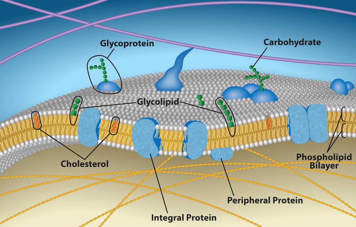
Figure 20.16 Capillary Exchange Net filtration occurs near the arterial end of the capillary since capillary hydrostatic pressure (CHP) is greater than blood colloidal osmotic pressure (BCOP). There is no net movement of fluid near the midpoint since CHP = BCOP. Net reabsorption occurs near the venous end since BCOP is greater than CHP.

J. Gordon Betts and colleagues, Anatomy And Physiology 2E, OpenStax. · CC BY-NC-SA 4.0 · Original image ↗ · Access for free ↗


The Role of Lymphatic Capillaries

Since overall CHP is higher than BCOP, it is inevitable that more net fluid will exit the capillary through filtration at the arterial end than enters through at the venous end. Considering all capillaries over the course of a day, this can be quite a substantial amount of fluid: Approximately 24 liters per day are filtered, whereas 20.4 liters are reabsorbed. This excess fluid is picked up by capillaries of the lymphatic system. These extremely thin-walled vessels have copious numbers of valves that ensure unidirectional flow through ever-larger lymphatic vessels that eventually drain into the subclavian veins in the neck. An important function of the lymphatic system is to return the fluid () to the blood. may be thought of as recycled blood plasma. (Seek additional content for more detail on the lymphatic system.)

Interactive Link

Watch this video to explore capillaries and how they function in the body. Capillaries are never more than 100 micrometers away. What is the main component of ?