Foundation 3 · 3B · Integrated organ systems

and immune defense

Sources ↗

Innate defenses act rapidly; adaptive responses select -specific clones and can build memory.

returns material to circulation

Lymphatic capillaries collect excess and escaped , then return them to veins through larger vessels and ducts. One-way valves and external compression support flow. Intestinal transport absorbed -rich . sample , while the samples blood and removes aged . Bone marrow supports blood-cell production and development; the supports T-cell maturation. Lymphatic obstruction can cause -rich even when arterial circulation remains intact.

Source: 21.1 Anatomy of the Lymphatic and Immune Systems · 21.2 Barrier Defenses and the Innate Immune Response · 21.3 The Adaptive Immune Response: T lymphocytes and Their Functional Types · 21.4 The Adaptive Immune Response: B-lymphocytes and Antibodies

Barriers and pattern recognition

Skin, mucus, environments, and antimicrobial limit entry. Innate recognize common microbial patterns or damage signals. and engulf targets; connect innate recognition to adaptive activation. can promote , , and damage. respond to altered recognition patterns, including reduced inhibitory signals. Innate responses act rapidly without requiring expansion of a newly selected -specific clone. increases recruitment and vascular permeability but can damage tissue if excessive or misdirected.

Source: 21.1 Anatomy of the Lymphatic and Immune Systems · 21.2 Barrier Defenses and the Innate Immune Response · 21.3 The Adaptive Immune Response: T lymphocytes and Their Functional Types · 21.4 The Adaptive Immune Response: B-lymphocytes and Antibodies

presentation and

A binds intact . T-cell typically recognize displayed by : presents mainly intracellular to T cells; on professional -presenting cells presents mainly endocytosed to helper T cells. Activation requires contextual signals as well as recognition. Selected clones proliferate into and memory cells. Cytotoxic T cells kill infected targets; helper subsets coordinate other immune cells. Recognition specificity comes from diversity before exposure, not from an instructing a new sequence.

Source: 21.1 Anatomy of the Lymphatic and Immune Systems · 21.2 Barrier Defenses and the Innate Immune Response · 21.3 The Adaptive Immune Response: T lymphocytes and Their Functional Types · 21.4 The Adaptive Immune Response: B-lymphocytes and Antibodies

and immune memory

Activated can become -secreting or memory cells. neutralize toxins or entry, opsonize targets, and activate through suitable . Variable regions bind ; constant regions determine behavior. changes , and selects improved binding. Primary responses take time; memory supports faster and often stronger subsequent responses. Vaccination aims to establish protective memory without the full risk of natural disease. Passive transfer gives immediate temporary protection without necessarily generating recipient memory.

Source: 21.1 Anatomy of the Lymphatic and Immune Systems · 21.2 Barrier Defenses and the Innate Immune Response · 21.3 The Adaptive Immune Response: T lymphocytes and Their Functional Types · 21.4 The Adaptive Immune Response: B-lymphocytes and Antibodies

Recognize self without attacking it

removes or alters many strongly self-reactive developing lymphocytes. Peripheral mechanisms include lack of , , regulatory T-cell activity, and . is an active collection of processes rather than an absolute inability to bind self. arises when self-directed responses escape regulation and cause damage. concerns inadequate defense, and allergy concerns harmful responses to otherwise relatively harmless external . A detectable self-reactive alone is not equivalent to a complete disease mechanism.

Source: 21.1 Anatomy of the Lymphatic and Immune Systems · 21.2 Barrier Defenses and the Innate Immune Response · 21.3 The Adaptive Immune Response: T lymphocytes and Their Functional Types · 21.4 The Adaptive Immune Response: B-lymphocytes and Antibodies

Practice and reasoning

Question. A virus reduces display on infected cells. Which conventional adaptive recognition pathway becomes harder, and which innate cell can respond to reduced inhibitory signaling? Why does passive not guarantee lasting immunity?

Show reasoning

T-cell recognition of – becomes harder. may respond when inhibitory self signals fall, depending on the balance of activating signals. Transferred eventually decay; they do not by themselves establish the recipient’s -specific memory-cell population.

Source: 21.1 Anatomy of the Lymphatic and Immune Systems · 21.2 Barrier Defenses and the Innate Immune Response · 21.3 The Adaptive Immune Response: T lymphocytes and Their Functional Types · 21.4 The Adaptive Immune Response: B-lymphocytes and Antibodies

21.1 Anatomy of the Lymphatic and Immune Systems — figures and tables
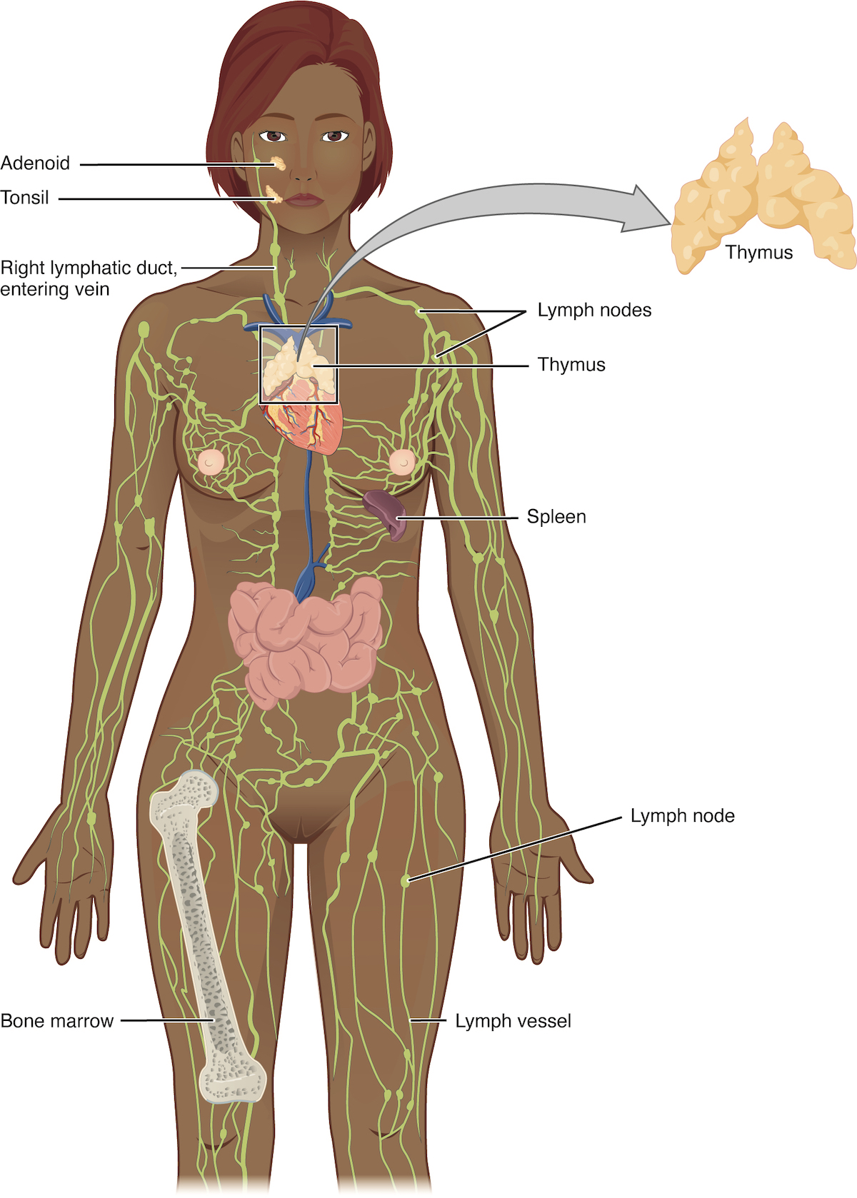
The left panel shows a female human body, and the entire lymphatic system is shown. The right panel shows magnified images of the thymus and the lymph node. All the major parts in the lymphatic system are labeled.
Figure 21.2 — 21.1 Anatomy of the Lymphatic and Immune Systems

Figure 21.2 Anatomy of the Lymphatic System Lymphatic vessels in the arms and legs convey lymph to the larger lymphatic vessels in the torso.

J. Gordon Betts and colleagues, Anatomy And Physiology 2E, OpenStax. · CC BY-NC-SA 4.0 · Original image ↗ · Access for free ↗
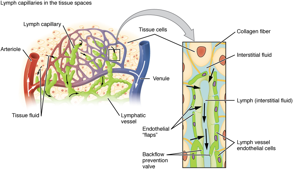
This image shows the lymph capillaries in the tissue spaces, and a magnified image shows the interstitial fluid and the lymph vessels. The major parts are labeled.
Figure 21.3 — 21.1 Anatomy of the Lymphatic and Immune Systems

Figure 21.3 Lymphatic Capillaries Lymphatic capillaries are interlaced with the arterioles and venules of the cardiovascular system. Collagen fibers anchor a lymphatic capillary in the tissue (inset). Interstitial fluid slips through spaces between the overlapping endothelial cells that compose the lymphatic capillary.

J. Gordon Betts and colleagues, Anatomy And Physiology 2E, OpenStax. · CC BY-NC-SA 4.0 · Original image ↗ · Access for free ↗
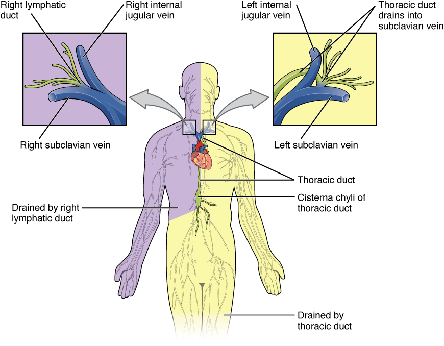
This figure shows the lymphatic trunks and the duct system in the human body. Callouts to the left and right show the magnified views of the left and right jugular vein respectively.
Figure 21.4 — 21.1 Anatomy of the Lymphatic and Immune Systems

Figure 21.4 Major Trunks and Ducts of the Lymphatic System The thoracic duct drains a much larger portion of the body than does the right lymphatic duct.

J. Gordon Betts and colleagues, Anatomy And Physiology 2E, OpenStax. · CC BY-NC-SA 4.0 · Original image ↗ · Access for free ↗
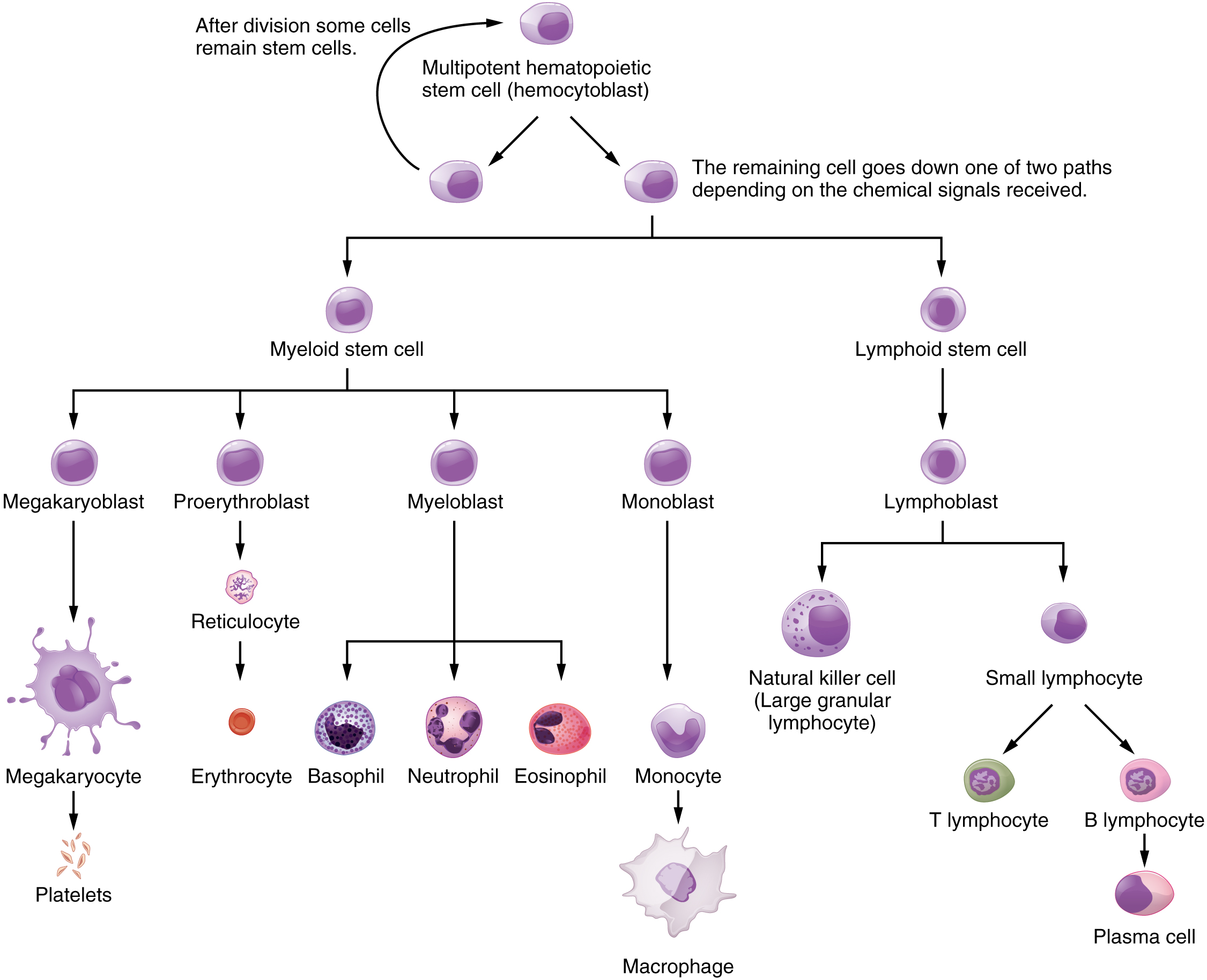
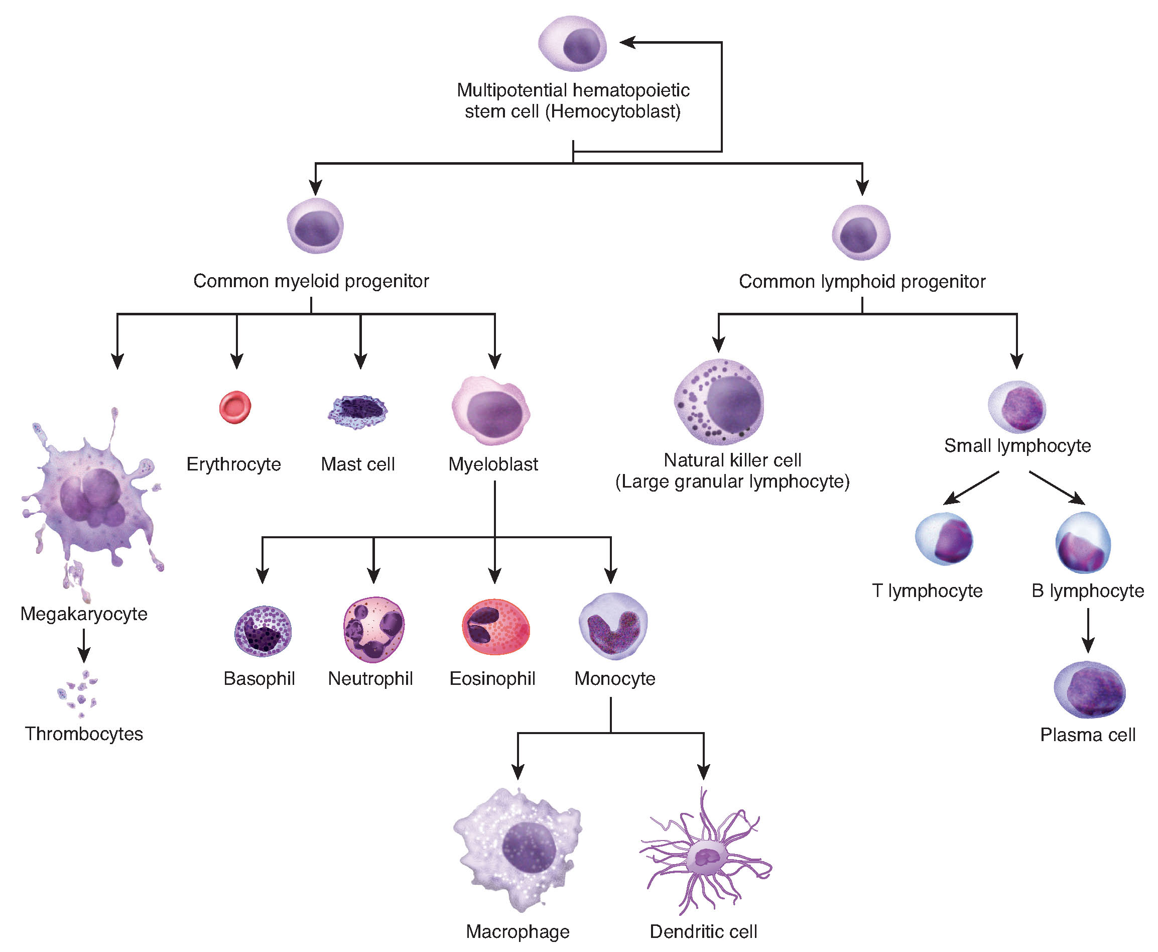
This flowchart shows the steps in which a multipotential hematopoietic stem cell differentiates into the different cell types in blood.
Figure 21.5 — 21.1 Anatomy of the Lymphatic and Immune Systems

Figure 21.5 Hematopoietic System of the Bone Marrow All the cells of the immune response as well as of the blood arise by differentiation from hematopoietic stem cells. Platelets are cell fragments involved in the clotting of blood.

J. Gordon Betts and colleagues, Anatomy And Physiology 2E, OpenStax. · CC BY-NC-SA 4.0 · Original image ↗ · Access for free ↗
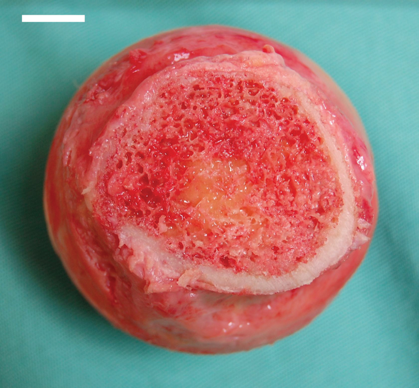
This photograph shows the bone marrow.
Figure 21.6 — 21.1 Anatomy of the Lymphatic and Immune Systems

Figure 21.6 Bone Marrow Red bone marrow fills the head of the femur, and a spot of yellow bone marrow is visible in the center. The white reference bar is 1 cm.

J. Gordon Betts and colleagues, Anatomy And Physiology 2E, OpenStax. · CC BY-NC-SA 4.0 · Original image ↗ · Access for free ↗
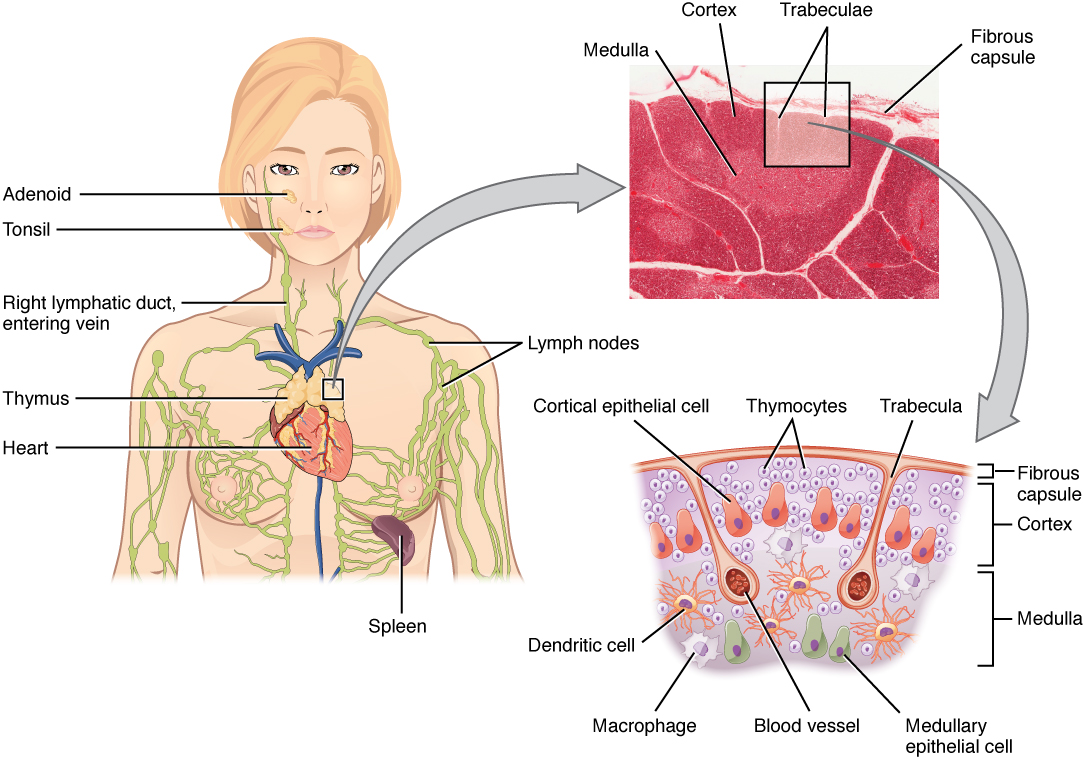
The left panel of this figure shows the head and chest of a woman and the location of the thymus is marked. The top right panel shows a micrograph of the thymus and the bottom right panel shows a magnified view of the structure of the thymus.
Figure 21.7 — 21.1 Anatomy of the Lymphatic and Immune Systems

Figure 21.7 Location, Structure, and Histology of the Thymus The thymus lies above the heart. The trabeculae and lobules, including the darkly staining cortex and the lighter staining medulla of each lobule, are clearly visible in the light micrograph of the thymus of a newborn. LM × 100. (Micrograph provided by the Regents of the University of Michigan Medical School © 2012)

J. Gordon Betts and colleagues, Anatomy And Physiology 2E, OpenStax. · CC BY-NC-SA 4.0 · Original image ↗ · Access for free ↗
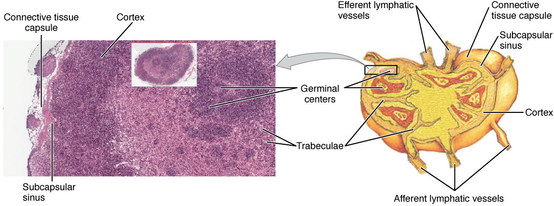
The left panel of this figure shows a micrograph of the cross section of a lymph node. The right panel shows the structure of a lymph node.
Figure 21.8 — 21.1 Anatomy of the Lymphatic and Immune Systems

Figure 21.8 Structure and Histology of a Lymph Node Lymph nodes are masses of lymphatic tissue located along the larger lymph vessels. The micrograph of the lymph nodes shows a germinal center, which consists of rapidly dividing B cells surrounded by a layer of T cells and other accessory cells. LM × 128. (Micrograph provided by the Regents of the University of Michigan Medical School © 2012)

J. Gordon Betts and colleagues, Anatomy And Physiology 2E, OpenStax. · CC BY-NC-SA 4.0 · Original image ↗ · Access for free ↗
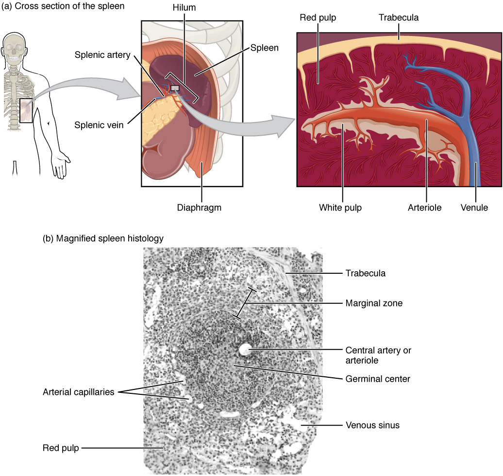
The top left panel shows the location of the spleen in the human body. The top center panel shows a close up view of the location of the spleen. The top right panel shows the blood vessels and spleen tissue. The bottom panel shows a histological micrograph.
Figure 21.9 — 21.1 Anatomy of the Lymphatic and Immune Systems

Figure 21.9 Spleen (a) The spleen is attached to the stomach. (b) A micrograph of spleen tissue shows the germinal center. The marginal zone is the region between the red pulp and white pulp, which sequesters particulate antigens from the circulation and presents these antigens to lymphocytes in the white pulp. EM × 660. (Micrograph provided by the Regents of the University of Michigan Medical School © 2012)

J. Gordon Betts and colleagues, Anatomy And Physiology 2E, OpenStax. · CC BY-NC-SA 4.0 · Original image ↗ · Access for free ↗
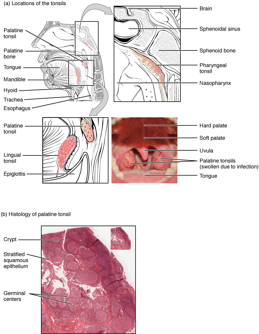
The top panel of this image shows the location of the tonsils. All the major parts are labeled. The bottom panel shows the histological micrograph of the tonsils.
Figure 21.10 — 21.1 Anatomy of the Lymphatic and Immune Systems

Figure 21.10 Locations and Histology of the Tonsils (a) The pharyngeal tonsil is located on the roof of the posterior superior wall of the nasopharynx. The palatine tonsils lay on each side of the pharynx. (b) A micrograph shows the palatine tonsil tissue. LM × 40. (Micrograph provided by the Regents of the University of Michigan Medical School © 2012)

J. Gordon Betts and colleagues, Anatomy And Physiology 2E, OpenStax. · CC BY-NC-SA 4.0 · Original image ↗ · Access for free ↗
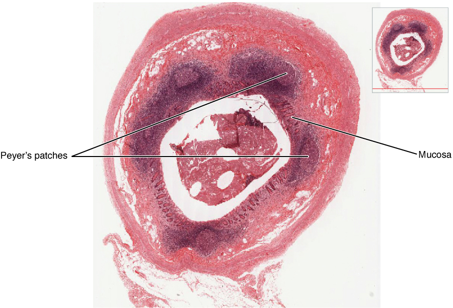
This figure shows a micrograph of a mucosa associated lymphoid tissue nodule.
Figure 21.11 — 21.1 Anatomy of the Lymphatic and Immune Systems

Figure 21.11 Mucosa-associated Lymphoid Tissue (MALT) Nodule LM × 40. (Micrograph provided by the Regents of the University of Michigan Medical School © 2012)

J. Gordon Betts and colleagues, Anatomy And Physiology 2E, OpenStax. · CC BY-NC-SA 4.0 · Original image ↗ · Access for free ↗
Type of lymphocyte Primary function
Generates diverse
T lymphocyte Secretes chemical messengers
Secretes
Destroys virally infected cells

Table 21.1. J. Gordon Betts and colleagues, Anatomy And Physiology 2E, OpenStax.

Source: 21.1 Anatomy of the Lymphatic and Immune Systems

21.2 Barrier Defenses and the Innate Immune Response — figures and tables
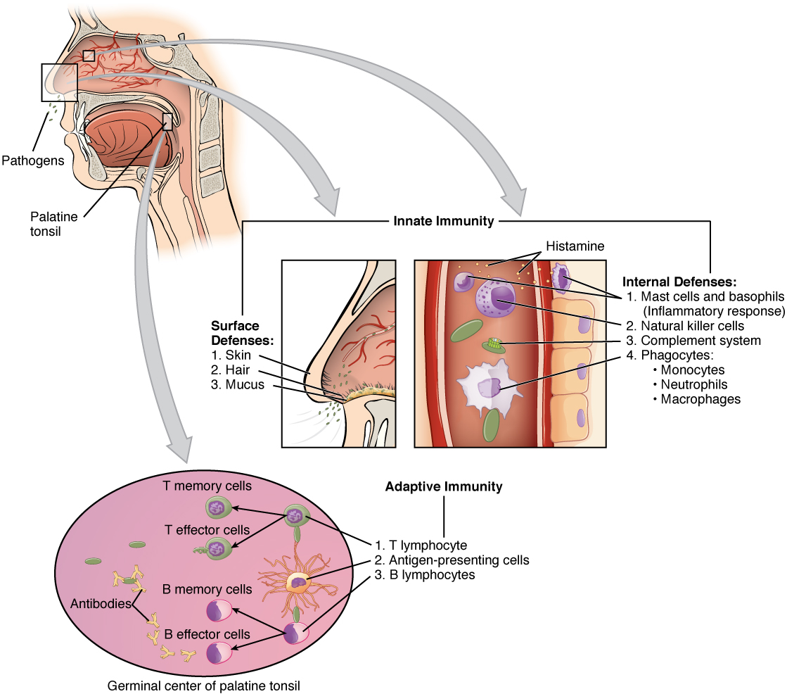
This figure shows a lateral view of a human face in the top left. A magnified callout shows the germinal center of the palatine tonsil. Another magnified view shows how the innate immune system works.
Figure 21.12 — 21.2 Barrier Defenses and the Innate Immune Response

Figure 21.12 Cooperation between Innate and Adaptive Immune Responses The innate immune system enhances adaptive immune responses so they can be more effective.

J. Gordon Betts and colleagues, Anatomy And Physiology 2E, OpenStax. · CC BY-NC-SA 4.0 · Original image ↗ · Access for free ↗
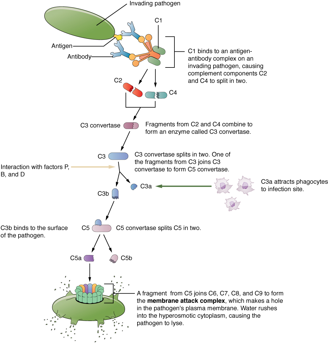
This flow chart shows an invading pathogen and the series of events that results in the complement cascade and function.
Figure 21.13 — 21.2 Barrier Defenses and the Innate Immune Response

Figure 21.13 Complement Cascade and Function The classical pathway, used during adaptive immune responses, occurs when C1 reacts with antibodies that have bound an antigen.

J. Gordon Betts and colleagues, Anatomy And Physiology 2E, OpenStax. · CC BY-NC-SA 4.0 · Original image ↗ · Access for free ↗
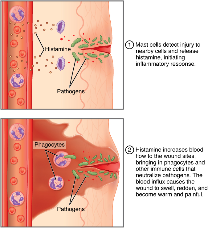
The top panel of this figure shows the mast cells detecting an injury and initiating an inflammatory response. The bottom panel shows the increase in blood flow in response to histamine.
Figure 21.14 — 21.2 Barrier Defenses and the Innate Immune Response

Figure 21.14 The inflammatory response Events resulting in warmth, redness, pain, and swelling, as well as the recruitment of phagocytes.

J. Gordon Betts and colleagues, Anatomy And Physiology 2E, OpenStax. · CC BY-NC-SA 4.0 · Original image ↗ · Access for free ↗
Site Specific defense Protective aspect
Skin Epidermal surface Keratinized cells of surface, Langerhans cells
Skin (sweat/secretions) Sweat glands, Low , washing action
Oral cavity Salivary glands Lysozyme
Stomach Gastrointestinal tract Low
Mucosal surfaces Mucosal Nonkeratinized epithelial cells
Normal flora (nonpathogenic ) Mucosal tissues Prevent from growing on mucosal surfaces

Table 21.2. J. Gordon Betts and colleagues, Anatomy And Physiology 2E, OpenStax.

Cell Cell type Primary location Function in the innate immune response
Agranulocyte Body cavities/organs
Granulocyte Blood
Monocyte Agranulocyte Blood Precursor of /

Table 21.3. J. Gordon Betts and colleagues, Anatomy And Physiology 2E, OpenStax.

Source: 21.2 Barrier Defenses and the Innate Immune Response

21.3 The Adaptive Immune Response: T lymphocytes and Their Functional Types — figures and tables
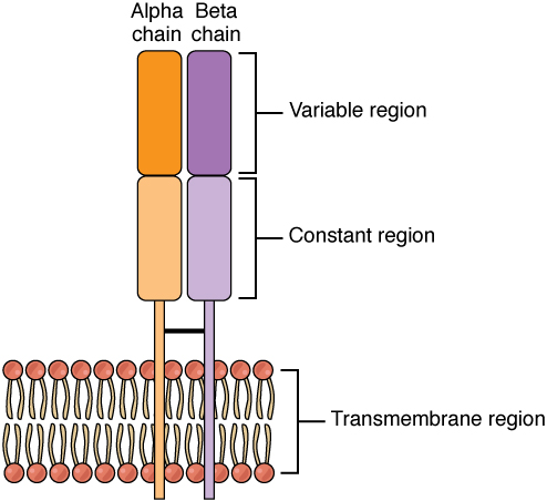
This figure shows the alpha beta T cell receptor in the plasma membrane.
Figure 21.15 — 21.3 The Adaptive Immune Response: T lymphocytes and Their Functional Types

Figure 21.15 Alpha-beta T Cell Receptor Notice the constant and variable regions of each chain, anchored by the transmembrane region.

J. Gordon Betts and colleagues, Anatomy And Physiology 2E, OpenStax. · CC BY-NC-SA 4.0 · Original image ↗ · Access for free ↗
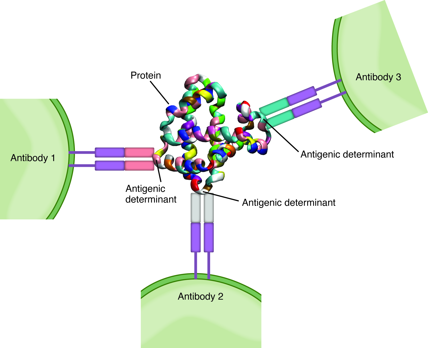
This figure shows three T cells and the antigenic determinants in the center.
Figure 21.16 — 21.3 The Adaptive Immune Response: T lymphocytes and Their Functional Types

Figure 21.16 Antigenic Determinants A typical protein antigen has multiple antigenic determinants, shown by the ability of T cells with three different specificities to bind to different parts of the same antigen.

J. Gordon Betts and colleagues, Anatomy And Physiology 2E, OpenStax. · CC BY-NC-SA 4.0 · Original image ↗ · Access for free ↗
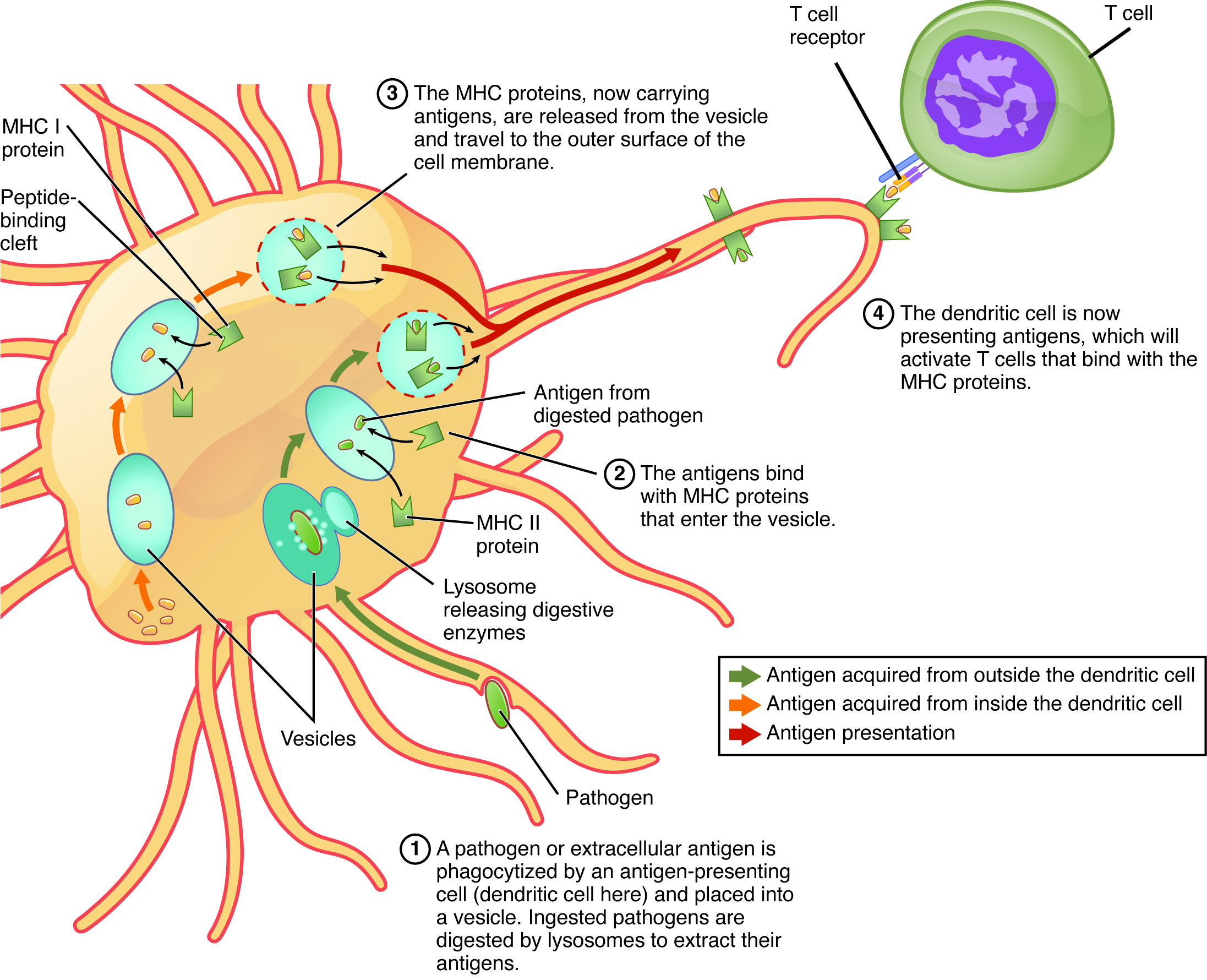
This figure shows how an antigen-presenting cell deals with a pathogen or extracellular antigen. The different steps are shown with text callouts.
Figure 21.17 — 21.3 The Adaptive Immune Response: T lymphocytes and Their Functional Types

Figure 21.17 Antigen Processing and Presentation

J. Gordon Betts and colleagues, Anatomy And Physiology 2E, OpenStax. · CC BY-NC-SA 4.0 · Original image ↗ · Access for free ↗
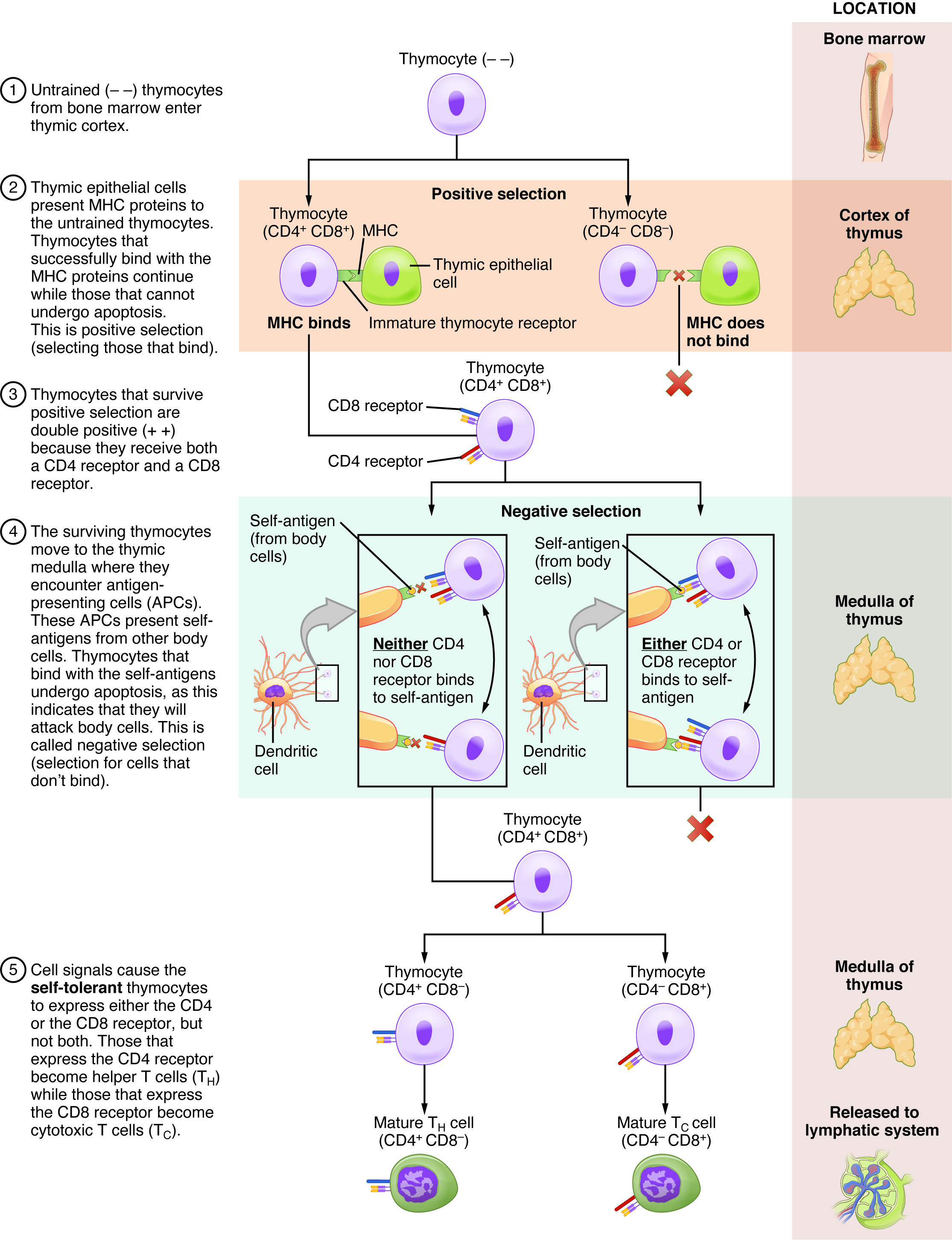
This multipart figure shows the different steps in the differentiation of a thymocyte into T cells. For each step of the process, accompanying text details the steps in the process. The right panel of this image shows the location of the different steps in the process.
Figure 21.18 — 21.3 The Adaptive Immune Response: T lymphocytes and Their Functional Types

Figure 21.18 Differentiation of T Cells within the Thymus Immature T-cells, called thymocytes, enter the thymus and go through a series of developmental stages that ensures both function and tolerance before they leave and become functional components of the adaptive immune response.

J. Gordon Betts and colleagues, Anatomy And Physiology 2E, OpenStax. · CC BY-NC-SA 4.0 · Original image ↗ · Access for free ↗
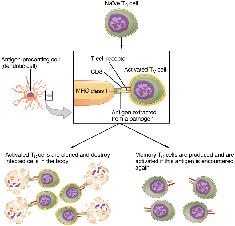
This flowchart shows the process in which a naïve T cell become activated T cells in the left part of the pathway and memory cells in the right part of the pathway.
Figure 21.19 — 21.3 The Adaptive Immune Response: T lymphocytes and Their Functional Types

Figure 21.19 Clonal Selection and Expansion of T Lymphocytes Stem cells differentiate into T cells with specific receptors, called clones. The clones with receptors specific for antigens on the pathogen are selected for and expanded.

J. Gordon Betts and colleagues, Anatomy And Physiology 2E, OpenStax. · CC BY-NC-SA 4.0 · Original image ↗ · Access for free ↗
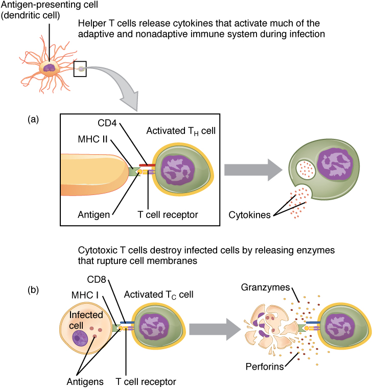
This figure shows the different steps in processing an extracellular pathogen.
Figure 21.20 — 21.3 The Adaptive Immune Response: T lymphocytes and Their Functional Types

Figure 21.20 Pathogen Presentation (a) CD4 is associated with helper and regulatory T cells. An extracellular pathogen is processed and presented in the binding cleft of a class II MHC molecule, and this interaction is strengthened by the CD4 molecule. (b) CD8 is associated with cytotoxic T cells. An intracellular pathogen is presented by a class I MHC molecule, and CD8 interacts with it.

J. Gordon Betts and colleagues, Anatomy And Physiology 2E, OpenStax. · CC BY-NC-SA 4.0 · Original image ↗ · Access for free ↗
Cell type Phagocytic? Function
Class I Many No Stimulates cytotoxic T cell immune response
Class II Yes Stimulates and presentation at primary infection site
Class II Dendritic Yes, in tissues Brings to regional
Class II Yes, internalizes surface Ig and Stimulates by

Table 21.4. J. Gordon Betts and colleagues, Anatomy And Physiology 2E, OpenStax.

T cell Main target Function Surface marker Cytokines or mediators
Tc Infected cells Cytotoxicity Intracellular Class I Perforins, granzymes, and fas
Th1 Helper Extracellular Class II Interferon-γ and TGF-β
Th2 Helper Extracellular Class II IL-4, IL-6, IL-10, and others
Treg Th cell Suppressor None , CD25 ? TGF-β and IL-10

Table 21.5. J. Gordon Betts and colleagues, Anatomy And Physiology 2E, OpenStax.

Source: 21.3 The Adaptive Immune Response: T lymphocytes and Their Functional Types

21.4 The Adaptive Immune Response: B-lymphocytes and — figures and tables

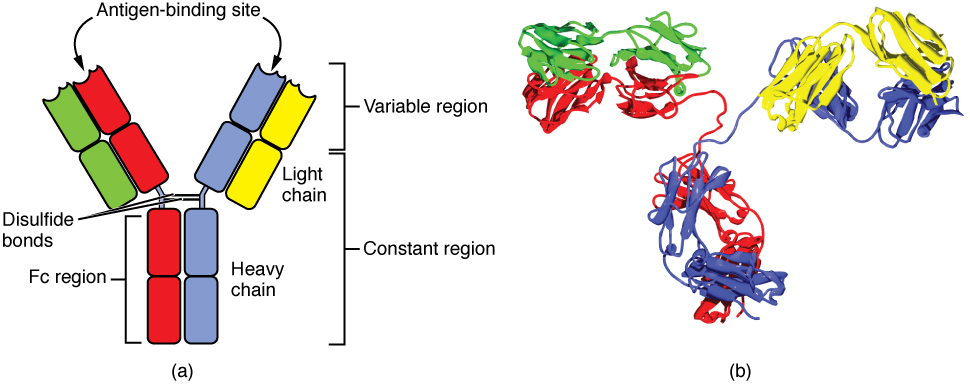
This diagram shows the four chain structure of a generic antibody.
Figure 21.21 — 21.4 The Adaptive Immune Response: B-lymphocytes and

Figure 21.21 Antibody and IgG2 Structures The typical four chain structure of a generic antibody (a) and the corresponding three-dimensional structure of the antibody IgG2 (b). (credit b: modification of work by Tim Vickers)

J. Gordon Betts and colleagues, Anatomy And Physiology 2E, OpenStax. (credit b: modification of work by Tim Vickers) · CC BY-NC-SA 4.0 · Original image ↗ · Access for free ↗
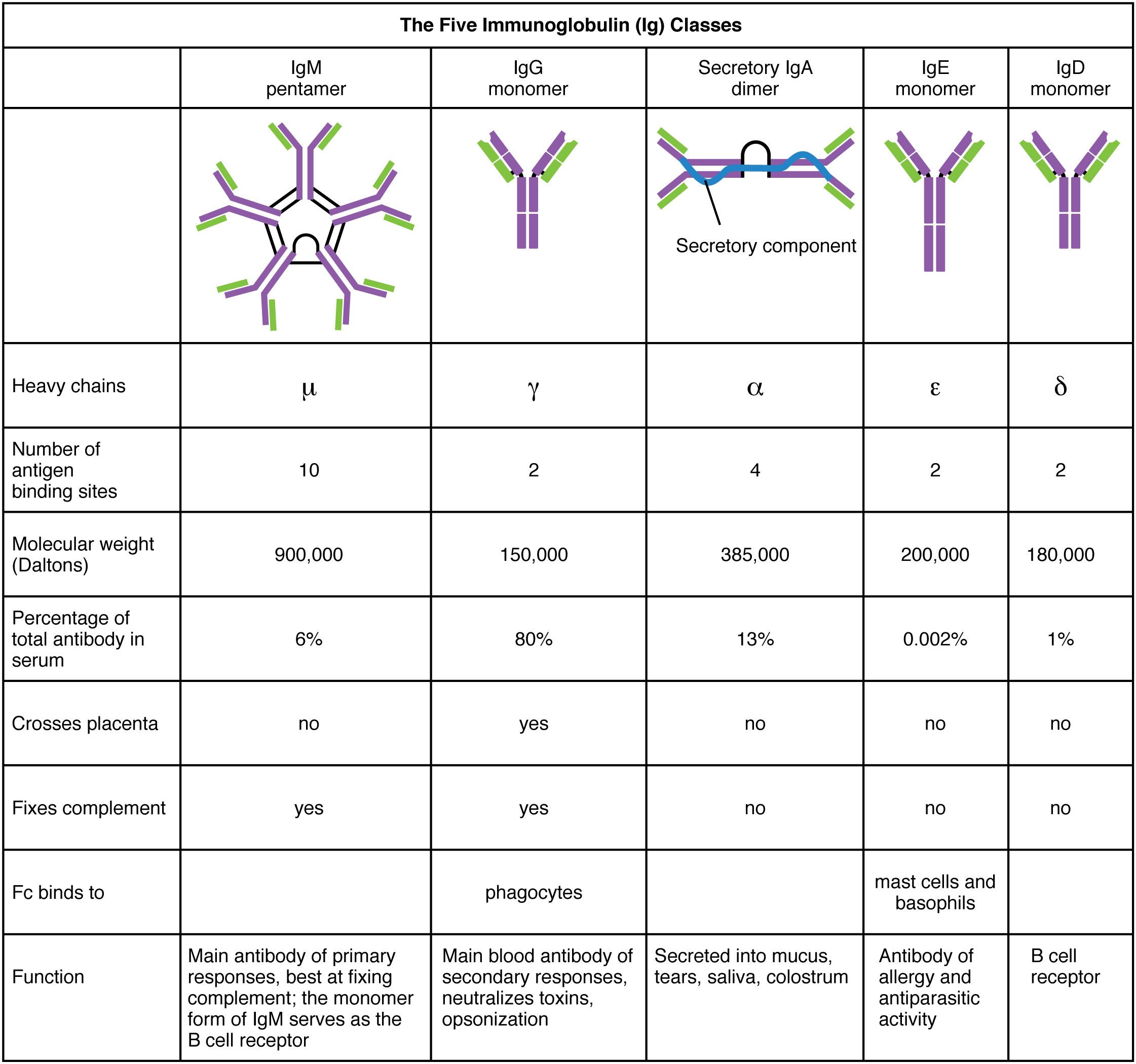
This table shows the five classes of the immunoglobulins. The table shows the molecular weight, number of antigen binding sites, and their function.
Figure 21.22 — 21.4 The Adaptive Immune Response: B-lymphocytes and

Figure 21.22 Five Classes of Antibodies

J. Gordon Betts and colleagues, Anatomy And Physiology 2E, OpenStax. · CC BY-NC-SA 4.0 · Original image ↗ · Access for free ↗
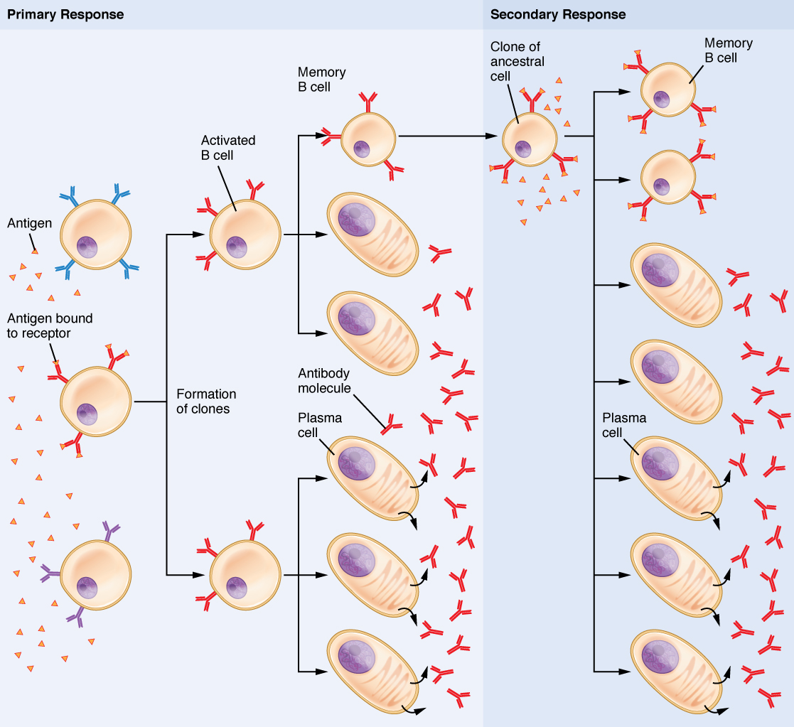
This flow chart shows how the clonal selection of B cells takes place. The left panel shows the primary response and the right panel shows the secondary response.
Figure 21.23 — 21.4 The Adaptive Immune Response: B-lymphocytes and

Figure 21.23 Clonal Selection of B Cells During a primary B cell immune response, both antibody-secreting plasma cells and memory B cells are produced. These memory cells lead to the differentiation of more plasma cells and memory B cells during secondary responses.

J. Gordon Betts and colleagues, Anatomy And Physiology 2E, OpenStax. · CC BY-NC-SA 4.0 · Original image ↗ · Access for free ↗
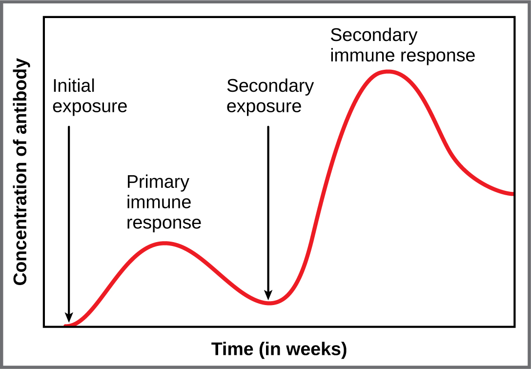
This graph shows the antibody concentration as a function of time in primary and secondary response.
Figure 21.24 — 21.4 The Adaptive Immune Response: B-lymphocytes and

Figure 21.24 Primary and Secondary Antibody Responses Antigen A is given once to generate a primary response and later to generate a secondary response. When a different antigen is given for the first time, a new primary response is made.

J. Gordon Betts and colleagues, Anatomy And Physiology 2E, OpenStax. · CC BY-NC-SA 4.0 · Original image ↗ · Access for free ↗
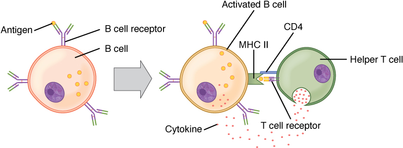
This diagram shows the binding of a B cell and a T cell.
Figure 21.25 — 21.4 The Adaptive Immune Response: B-lymphocytes and

Figure 21.25 T and B Cell Binding To elicit a response to a T cell-dependent antigen, the B and T cells must come close together. To become fully activated, the B cell must receive two signals from the native antigen and the T cell’s cytokines.

J. Gordon Betts and colleagues, Anatomy And Physiology 2E, OpenStax. · CC BY-NC-SA 4.0 · Original image ↗ · Access for free ↗
Natural Artificial
Active Adaptive immune response Vaccine response
Passive Trans-placental /breastfeeding Immune globulin injections

Table 21.6. J. Gordon Betts and colleagues, Anatomy And Physiology 2E, OpenStax.

Source: 21.4 The Adaptive Immune Response: B-lymphocytes and Antibodies

289 terms, alphabetically ordered

3′ end · ·

The end of a nucleic- strand associated with the sugar’s 3′ position. extends a strand at a free .

Read explanation · OpenStax ↗
3′ hydroxyl group · · ·

The at the sugar’s 3′ carbon that attacks the incoming ’s during strand extension.

Read explanation · OpenStax ↗
A site · ·

The site that accepts most incoming during elongation and at termination.

Read explanation · OpenStax ↗
acceleration

Rate of change of , measured in m/s².

Read explanation · OpenStax ↗
Acid · ·

In the Brønsted–Lowry model, a substance that can donate a . Acidic have a higher activity than water.

Read explanation · OpenStax ↗
Activation energy ·

The energy barrier that must overcome to reach a and undergo .

Read explanation · OpenStax ↗
Adenosine triphosphate

An energy-carrying ; its powers many cellular processes, including cycles.

Read explanation · OpenStax ↗
affinity maturation

Selection of variants with improved binding during an adaptive response.

Read explanation · OpenStax ↗
Allolactose

A isomer produced by that binds and relieves lac repression.

Read explanation · OpenStax ↗
Alpha carbon · ·

The central carbon next to the carboxyl carbon. In a standard it binds an , hydrogen, and .

Read explanation · OpenStax ↗
Alpha helix · · · · · ·

A helical stabilized by between a oxygen and an hydrogen four later.

Read explanation · OpenStax ↗
alveolar macrophage

A phagocytic immune cell removing particles and responding to threats in alveolar spaces.

Read explanation · OpenStax ↗
Amide · · ·

A group containing a carbon bonded to nitrogen. are amide linkages.

Read explanation · OpenStax ↗
Amino acid

A building block containing amino and and a characteristic .

Read explanation · OpenStax ↗
Amino acid residue · ·

The part of an incorporated into a chain. Residue can also describe another building block within a larger .

Read explanation · OpenStax ↗
Amino group · · ·

A nitrogen-containing . A free ’s amino group is typically (NH₃⁺) near physiological ; ’s ring nitrogen is an exception to this formula.

Read explanation · OpenStax ↗
Aminoacyl-tRNA · ·

A covalently linked to an at its , ready to deliver that during .

Read explanation · OpenStax ↗
Amphipathic

Having both a region and a region, as in a or .

Read explanation · OpenStax ↗
Amylose

The largely unbranched component of , made mainly of linked by alpha-1,4 .

Read explanation · OpenStax ↗
Analyte

A substance being measured or separated within a sample; the source table uses the term for the mixture under analysis.

Read explanation · Waters ↗
anergy

A state of functional lymphocyte unresponsiveness despite recognition.

Read explanation · OpenStax ↗
anomeric carbon

The former carbon of a sugar, which becomes the or hemiketal center upon cyclization.

Read explanation · OpenStax ↗
Antibody

An that recognizes a specific region () on an . Antibodies help immune defenses and are used as detection tools in .

Read explanation · OpenStax ↗
Anticodon

A triplet that pairs to an . The anticodon for 5′-AUG-3′ is aligned 3′-UAC-5′, or written 5′-CAU-3′.

Read explanation · NCBI Bookshelf / Garland Science ↗
Antigen

A or molecular structure specifically recognized by an or other adaptive immune .

Read explanation · OpenStax ↗
Antiparallel

Running in opposite directions, as with two strands or neighboring strands in some .

Read explanation · OpenStax ↗
Apoptosis

A regulated process of cell death that removes cells in development, tissue maintenance, and responses to damage.

Read explanation · OpenStax ↗
Aqueous solution ·

A in which water is the .

Read explanation · OpenStax ↗
Archaea ·

A of distinct from , with characteristic chemistry and -expression machinery.

Read explanation · OpenStax ↗
arteriole

A small artery whose smooth-muscle tone provides major control of local vascular resistance.

Read explanation · OpenStax ↗
Assay

A procedure used to detect a substance or measure its amount, activity, or another property.

Read explanation · Thermo Fisher ↗
Atom

The smallest unit of an element that retains its chemical identity; it has a surrounded by .

Read explanation · OpenStax ↗
attachment

Enduring emotional bond and expectations about another’s availability and responsiveness.

Read explanation · OpenStax ↗
autoimmunity

An immune response against self components that causes or contributes to tissue injury.

Read explanation · OpenStax ↗
B lymphocyte · ·

An immune-system lymphocyte lineage that can develop into -secreting cells.

Read explanation · OpenStax ↗
Bacteria ·

A of single-celled . Bacterial cells lack a -bound and are distinct from .

Read explanation · OpenStax ↗
Beta sheet · · · · · ·

A made of aligned segments connected by ; strands may be parallel or .

Read explanation · OpenStax ↗
Binding site

A region of a where another interacts selectively.

Read explanation · OpenStax ↗
Biomolecule

A made by or involved in living systems, including , , , and .

Read explanation · OpenStax ↗
Blotting ·

Transfer of separated or to a so a specific target can be detected.

Read explanation · Thermo Fisher ↗
Brønsted–Lowry base · · · · · · · · · · ·

In the Brønsted–Lowry model, a substance that can accept a . Basic solutions have lower activity than water.

Read explanation · OpenStax ↗
Buffer ·

A that resists changes when small amounts of or are added, commonly through a weak and its .

Read explanation · OpenStax ↗
Cancer

A group of diseases involving abnormal cells that can proliferate without normal restraints and invade tissues.

Read explanation · OpenStax ↗
Carbohydrate

A class of including sugars and their , used for energy storage, structure, and recognition.

Read explanation · OpenStax ↗
Carbonyl group · ·

A carbon double-bonded to oxygen, C=O; present in and many other .

Read explanation · OpenStax ↗
Carboxyl group · ·

An , COOH. In free it is usually to COO⁻ near physiological .

Read explanation · OpenStax ↗
Catalyst · ·

A substance that speeds a by providing a pathway with lower and is regenerated rather than consumed overall.

Read explanation · OpenStax ↗
CD4

A cell-surface immune coreceptor used with additional coreceptors for and entry.

Read explanation · OpenStax ↗
CD8

A T-cell coreceptor associated with recognition of – and many cytotoxic T cells.

Read explanation · OpenStax ↗
Cell proliferation

Increase in cell number through cell division; it should be distinguished experimentally from changes in survival.

Read explanation · OpenStax ↗
Cellulose

A structural in plant cell walls, with beta-1,4 linkages; also used as a support.

Read explanation · OpenStax ↗
Centimeter

A unit of length equal to one hundredth of a meter.

Read explanation · NIST ↗
central nervous system

Brain and spinal cord.

Read explanation · OpenStax ↗
central tolerance

Developmental processes that reduce strongly self-reactive lymphocytes in primary lymphoid organs.

Read explanation · OpenStax ↗
Centrifugation ·

Spinning a sample to accelerate separation of particles according to their behavior.

Read explanation · Beckman Coulter ↗
Chemical compound ·

A substance containing of two or more different chemically combined in a definite composition.

Read explanation · OpenStax ↗
Chemical element

A substance defined by having the same number of , such as carbon or oxygen.

Read explanation · OpenStax ↗
Chemical reaction ·

A process that rearranges and chemical bonds, converting into .

Read explanation · OpenStax ↗
chemotaxis

Biased movement in response to a chemical .

Read explanation · pmc.ncbi.nlm.nih.gov ↗
Chromatin

The complex of and associated , including , in .

Read explanation · OpenStax ↗
Chromatography

Separation of mixture components by their different interactions with a and a moving .

Read explanation · Waters ↗
Chromosome ·

An organized with associated that carries . A replicated chromosome has two .

Read explanation · OpenStax ↗
chylomicron

An intestinal lipoprotein particle carrying dietary and other into and blood.

Read explanation · Biology LibreTexts / Oregon State University ↗
Cisternae

Flattened -enclosed sacs, such as those forming the .

Read explanation · OpenStax ↗
class switching

A process changing heavy-chain constant region while retaining the original -binding variable region.

Read explanation · OpenStax ↗
clonal selection

Expansion of lymphocytes whose pre-existing recognize an with appropriate activation signals.

Read explanation · OpenStax ↗
Codon

A sequence of three that specifies an or a stop signal.

Read explanation · OpenStax ↗
Collagen

A major structural in , with a characteristic triple-helical arrangement.

Read explanation · OpenStax ↗
columnar

Describing epithelial cells taller than they are wide.

Read explanation · OpenStax ↗
complement

A system that can promote , , and attack against targets.

Read explanation · OpenStax ↗
Concentration

The amount of a substance in a specified volume or mass of mixture.

Read explanation · OpenStax ↗
Conformation ·

A spatial arrangement accessible by rotations and other motions that do not require breaking .

Read explanation · NCBI Bookshelf ↗
conjugate base

Species formed when an loses one .

Read explanation · OpenStax ↗
connective tissue

A tissue category generally characterized by cells distributed within substantial .

Read explanation · OpenStax ↗
cornea

Transparent anterior surface of the eye providing substantial refraction of incoming light.

Read explanation · OpenStax ↗
costimulation

Additional signaling needed alongside recognition for effective activation of many lymphocytes.

Read explanation · OpenStax ↗
Covalent bond · ·

A bond in which share pairs. and are covalent.

Read explanation · OpenStax ↗
culture

Learned and shared meanings, practices, values, and artifacts.

Read explanation · OpenStax ↗
Cyclic AMP ·

A signal. In the simplified E. coli lac model, low raises cAMP, which activates CAP.

Read explanation · OpenStax ↗
cyclic nucleotide

whose forms a cyclic within its sugar, such as .

Read explanation · OpenStax ↗
Cysteine ·

An with a . Two cysteines can form a .

Read explanation · OpenStax ↗
Cytoplasm

Cell contents inside the plasma and outside the , where one is present.

Read explanation · OpenStax ↗
Cytosol

The aqueous part of the outside -enclosed .

Read explanation · OpenStax ↗
Deletion

Loss of one or more from a sequence. The consequence depends on the affected region and length.

Read explanation · OpenStax ↗
dendritic cell

An immune cell specialized in capture and presentation to activate T cells.

Read explanation · OpenStax ↗
Density gradient

A fluid whose density varies with position, used to improve separation during .

Read explanation · Beckman Coulter ↗
Deoxyribonucleic acid

The that stores genetic information used to specify and sequences.

Read explanation · OpenStax ↗
Deprotonation

Removal of a from a or group; this changes its by minus one.

Read explanation · OpenStax ↗
Detergent

An substance that can associate with both water and material; detergents can disrupt or interactions.

Read explanation · Thermo Fisher ↗
differentiation

Acquisition of specialized cellular structures and functions through regulated and other changes.

Read explanation · OpenStax ↗
Disulfide bond · · ·

A covalent sulfur–sulfur bond formed by of two , which can stabilize structure.

Read explanation · NCBI Bookshelf ↗
DNA polymerase ·

An that synthesizes 5′ to 3′ using a template and a with a free .

Read explanation · OpenStax ↗
DNA replication

Copying so genetic information can be passed to daughter cells. Each guides a complementary new strand.

Read explanation · OpenStax ↗
edema

Excess fluid accumulation in interstitial tissue spaces.

Read explanation · OpenStax ↗
Effector

A that changes a ’s activity or binding behavior, often by stabilizing a particular .

Read explanation · Biology LibreTexts ↗
eicosanoid

Local signaling derived from a twenty-carbon polyunsaturated .

Read explanation · OpenStax ↗
Electric charge · · ·

An electrical property that can be positive or negative. Opposite charges attract and like charges repel; net charge is the sum of all charges on a particle.

Read explanation · OpenStax ↗
Electron

A negatively subatomic particle. Electron sharing and transfer determine much of chemical bonding.

Read explanation · OpenStax ↗
Electronegativity ·

An ’s tendency to attract shared in a chemical bond. Unequal attraction can make a bond .

Read explanation · OpenStax ↗
Endocrine signaling ·

Communication in which enter the circulation and act on distant target cells.

Read explanation · OpenStax ↗
endocytosis

Uptake of extracellular material or surface through inward budding of .

Read explanation · OpenStax ↗
Endomembrane system

The interacting and compartments involved in processes such as modification, transport, and .

Read explanation · OpenStax ↗
Endoplasmic reticulum

An interconnected network continuous with the ; it includes regions involved in processing and synthesis.

Read explanation · OpenStax ↗
Enzyme

A biological that speeds a without being consumed. Most enzymes are .

Read explanation · OpenStax ↗
epithelium ·

A polarized sheet of closely connected cells that covers surfaces, lines cavities, or forms glands.

Read explanation · OpenStax ↗
Epitope

The particular part of an recognized by an or immune .

Read explanation · OpenStax ↗
Eukaryote ·

An organism whose cells have a -enclosed ; animals, plants, fungi, and protists are eukaryotes.

Read explanation · OpenStax ↗
exocrine

Describing through ducts onto an epithelial surface or into a .

Read explanation · OpenStax ↗
Exon

A segment retained in a mature spliced . In an exon may contain coding sequence, untranslated sequence, or both.

Read explanation · OpenStax ↗
extracellular matrix

The network of and surrounding cells and providing structural and signaling functions.

Read explanation · OpenStax ↗
FACT complex ·

A -associated complex that helps machinery traverse and supports reorganization.

Read explanation · OpenStax ↗
Fatty acid

A attached to a chain; fatty acids are components of many .

Read explanation · OpenStax ↗
Fc

The constant-region fragment mediating many and interactions.

Read explanation · OpenStax ↗
fibroblast

A connective-tissue cell producing extracellular fibers and ground substance.

Read explanation · OpenStax ↗
fibrosis

Excess deposition of connective-tissue , often forming scar after injury.

Read explanation · OpenStax ↗
Functional group

A group of whose chemical behavior gives a characteristic and properties.

Read explanation · OpenStax ↗
Galactose

A six-carbon monosaccharide released along with when is hydrolyzed.

Read explanation · OpenStax ↗
Gene

A sequence that contributes to a functional or , together with the information needed to express it.

Read explanation · OpenStax ↗
Gene expression

Production of a functional from a , including and, for -coding , .

Read explanation · NCBI Bookshelf ↗
Genome

The complete genetic material of an organism or cell.

Read explanation · OpenStax ↗
Glucose

A six-carbon sugar that is a major cellular energy source and a building block of , , and .

Read explanation · OpenStax ↗
Glycerol

A three-carbon alcohol with three ; used in sample to increase density and in some .

Read explanation · OpenStax ↗
glycogen

A highly branched used for storage in animals.

Read explanation · Biology LibreTexts / Oregon State University ↗
Glycosidic bond

A involving a sugar’s . In a , an N-glycosidic bond links the sugar to the .

Read explanation · Biology LibreTexts / Oregon State University ↗
Golgi apparatus ·

A stack of compartments that modifies and sorts and traveling through the .

Read explanation · OpenStax ↗
Gradient

A change in a quantity, such as , , or density, across a distance or over time.

Read explanation · Thermo Fisher ↗
Heavy chain

The larger chain type in an ; its constant region helps determine the class.

Read explanation · OpenStax ↗
hemiacetal

Carbon bonded to both and alkoxy groups, usually formed by alcohol addition to a .

Read explanation · OpenStax ↗
Hemoglobin

The oxygen-carrying in , composed of four with heme groups.

Read explanation · OpenStax ↗
Histone

A -associated rich in . Core histones form the around which nucleosomal wraps.

Read explanation · OpenStax ↗
HIV

Human virus, an enveloped that uses and coreceptors for entry.

Read explanation · OpenStax ↗
Hormone

A chemical messenger released by cells that changes the activity of target cells with matching .

Read explanation · OpenStax ↗
Hydrocarbon · ·

A made only of carbon and hydrogen. Hydrocarbon regions are generally .

Read explanation · OpenStax ↗
Hydrogen bond ·

An attraction between a hydrogen bonded to an and another . hydrogen bonds stabilize .

Read explanation · OpenStax ↗
Hydrogen ion · ·

A positively hydrogen species. H⁺ denotes a , which is hydrated in water; its activity determines .

Read explanation · OpenStax ↗
Hydrolysis

Breaking a chemical bond by adding water. hydrolysis separates .

Read explanation · OpenStax ↗
Hydrophilic

Interacting favorably with water, typically because a group is or .

Read explanation · OpenStax ↗
Hydrophobic

Interacting poorly with water. often gather in a ’s interior.

Read explanation · OpenStax ↗
Hydroxide ion ·

The negatively OH⁻. Basic contain a higher hydroxide ion activity than water at the same temperature.

Read explanation · OpenStax ↗
Hydroxyl group ·

An oxygen bonded to hydrogen, –OH. Hydroxyl groups can and often increase .

Read explanation · OpenStax ↗
immunodeficiency

Inadequate immune function that compromises defense or regulation.

Read explanation · OpenStax ↗
Immunoglobulin

An or the -bound form of the same type of -binding .

Read explanation · OpenStax ↗
Immunoglobulin G

A class of abundant in blood. Each IgG has two and two , forming two -.

Read explanation · OpenStax ↗
Inducer

A signal that increases expression of a regulated ; does so by disabling the .

Read explanation · OpenStax ↗
inflammation

A coordinated tissue response recruiting defense and repair mechanisms, often involving increased blood flow and permeability.

Read explanation · OpenStax ↗
interstitial fluid

Fluid occupying extracellular spaces between tissue cells.

Read explanation · OpenStax ↗
Intron ·

A segment removed from a precursor during . Intronic signals or regulatory sequences can affect .

Read explanation · OpenStax ↗
Ion · ·

An or with a net because its numbers of and do not balance.

Read explanation · OpenStax ↗
isotype

An class defined by its constant-region structure and associated functions.

Read explanation · OpenStax ↗
Lac operon

The E. coli containing , , and , controlled by repression and CAP– activation.

Read explanation · OpenStax ↗
Lac repressor

The encoded by lacI; it binds lac and represses until an reduces its binding.

Read explanation · OpenStax ↗
lacA

The encoding a galactoside .

Read explanation · OpenStax ↗
lactation

Production and delivery of milk by mammary tissue.

Read explanation · OpenStax ↗
lacteal

An intestinal lymphatic capillary that receives absorbed -rich .

Read explanation · OpenStax ↗
Lactose

A disaccharide containing and ; its use by E. coli is supported by .

Read explanation · OpenStax ↗
Lactose permease

The transport that couples uptake to movement.

Read explanation · OpenStax ↗
lacY

The encoding , which transports into the cell.

Read explanation · OpenStax ↗
lacZ

The encoding , often used as an experimental .

Read explanation · OpenStax ↗
leukocyte

A white blood cell involved in defense and immune functions.

Read explanation · OpenStax ↗
ligament

A connective-tissue structure commonly linking bones and stabilizing joints.

Read explanation · OpenStax ↗
Ligand

A or that binds to a particular site on another , such as a or metal .

Read explanation · Cytiva ↗
Light chain

The smaller chain type in an ; it contributes to the -binding region.

Read explanation · OpenStax ↗
linkage

A tendency of nearby on the same to be inherited together.

Read explanation · OpenStax ↗
Lipid

A broad class of largely or , including fats, , and steroids.

Read explanation · OpenStax ↗
locus

The specific position of a or other feature.

Read explanation · OpenStax ↗
Logarithm · · ·

The exponent needed to obtain a number from a chosen fixed number. For decimal logarithms, log₁₀(100) = 2 because 10² = 100; uses the negative decimal logarithm of activity.

Read explanation · OpenStax ↗
Loss of function

or elimination of a ’s normal activity.

Read explanation · NCBI Bookshelf / Sinauer Associates ↗
Lumen · ·

The interior space of an or tube; the ER lumen is enclosed by the .

Read explanation · OpenStax ↗
lymph

Fluid collected from tissue spaces and transported through lymphatic vessels back toward the bloodstream.

Read explanation · OpenStax ↗
lymph node

A lymphoid structure that filters and supports presentation and adaptive responses.

Read explanation · OpenStax ↗
Lysosome

An -enclosed compartment that uses hydrolytic to digest and recycle cellular material.

Read explanation · OpenStax ↗
macrophage

A tissue phagocyte involved in clearance, signaling, and presentation.

Read explanation · OpenStax ↗
Matrix

A supporting material, such as a gel or the solid support in a column. Its properties influence movement and binding of sample .

Read explanation · Thermo Fisher ↗
Mediator ·

A multiprotein complex communicating regulatory inputs from to machinery.

Read explanation · OpenStax ↗
Membrane · · · ·

A thin boundary or sheet. Cell membranes are bilayers containing ; membranes are synthetic supports that hold or for detection.

Read explanation · OpenStax ↗
Messenger RNA ·

An that carries coding information used by to synthesize a .

Read explanation · OpenStax ↗
Metabolism

The chemical that build , break them down, and transform energy in a living system.

Read explanation · OpenStax ↗
MHC

Major histocompatibility complex displaying for T-cell recognition.

Read explanation · OpenStax ↗
MHC I

Class I presenting mainly intracellular to T cells.

Read explanation · OpenStax ↗
MHC II

Class II presenting mainly endocytosed to T cells.

Read explanation · OpenStax ↗
Mitochondrion ·

A with its own , central to aerobic production and several other processes.

Read explanation · OpenStax ↗
mitosis

division that separates duplicated into daughter , ordinarily preserving .

Read explanation · OpenStax ↗
Mobile phase

The moving fluid that carries a sample through a system.

Read explanation · Waters ↗
Molecular chaperone · ·

A that assists other in folding or prevents , without specifying their sequence.

Read explanation · NCBI Bookshelf ↗
Molecule

Two or more joined by . A molecule may be electrically or carry a .

Read explanation · OpenStax ↗
Monomer

A small molecular building block that can join other monomers to form a .

Read explanation · OpenStax ↗
natural killer cell · ·

An innate lymphocyte responding to a balance of activating and inhibitory signals on potential target cells.

Read explanation · OpenStax ↗
Neutral

Having no net . For a , neutral instead means equal and activities; its is about 7 at 25 °C.

Read explanation · OpenStax ↗
neutrophil

An abundant innate specialized in rapid antimicrobial responses and .

Read explanation · OpenStax ↗
Nitrogenous base · · ·

A nitrogen-containing ring component of ; its pairing interactions help specify sequences.

Read explanation · OpenStax ↗
Noncoding RNA · · · ·

An whose functional is rather than a , including , , and regulatory .

Read explanation · NCBI Bookshelf / Landes Bioscience ↗
Nonpolar ·

Lacking a substantial separation of . Nonpolar groups generally interact poorly with water.

Read explanation · OpenStax ↗
Nonspecific binding · ·

Unwanted binding outside the intended target interaction, often contributing background signal in an .

Read explanation · Thermo Fisher ↗
Nuclear envelope

The double enclosing the and continuous with the .

Read explanation · OpenStax ↗
Nucleic acid

A of , such as or , used in information storage, expression, and other cellular functions.

Read explanation · OpenStax ↗
Nucleosome

The basic unit of : wrapped around a .

Read explanation · OpenStax ↗
Nucleotide

A joined to a sugar and one or more .

Read explanation · OpenStax ↗
Nucleus ·

A -enclosed compartment containing most of a cell’s .

Read explanation · OpenStax ↗
Octamer

A complex made of eight . A core contains eight .

Read explanation · OpenStax ↗
Operator · ·

A regulatory sequence bound by a , often overlapping or near a .

Read explanation · OpenStax ↗
Operon

A group of from a shared into one , with regulatory that coordinates their expression.

Read explanation · OpenStax ↗
opsonization

Coating a target with that enhance its recognition and uptake by phagocytes.

Read explanation · OpenStax ↗
Organelle ·

A specialized structure within a cell that carries out particular functions, such as a or .

Read explanation · OpenStax ↗
Oxidation ·

Loss of or an increase in oxidation state. Oxidizing two can form a .

Read explanation · OpenStax ↗
passive immunity

Protection supplied by transferred or other immune rather than the recipient generating a new adaptive response.

Read explanation · OpenStax ↗
Pathogen

An infectious agent capable of causing disease in a host.

Read explanation · OpenStax ↗
Peptide

A chain of joined by ; the term commonly describes chains shorter than a typical .

Read explanation · OpenStax ↗
Peptide bond ·

A covalent between the carboxyl carbon of one and the amino nitrogen of the next.

Read explanation · OpenStax ↗
peripheral tolerance

Mechanisms restraining self-reactive lymphocytes after they leave primary lymphoid organs.

Read explanation · OpenStax ↗
pH

A measure of , defined as the negative of activity. Lower pH means greater ; pH influences .

Read explanation · OpenStax ↗
phagocytosis

Engulfment of large particles or cells into -bound compartments.

Read explanation · OpenStax ↗
Phosphate group ·

A phosphorus-containing group bonded to oxygens; phosphate groups occur in , , and modified .

Read explanation · OpenStax ↗
Phospholipid

An with a -containing region and tails; a major component of cellular .

Read explanation · OpenStax ↗
placenta

An organ formed from fetal and maternal contributions that supports exchange and functions during pregnancy.

Read explanation · OpenStax ↗
plasma cell

A differentiated B-lineage cell specialized in secreting .

Read explanation · OpenStax ↗
ploidy

The number of complete sets in a cell.

Read explanation · OpenStax ↗
Polar

Having an uneven distribution of . Polar groups often interact favorably with water.

Read explanation · OpenStax ↗
Polymer

A large made from many linked molecular building blocks.

Read explanation · OpenStax ↗
Polypeptide

A chain of connected by . A functional can contain one or several such chains.

Read explanation · OpenStax ↗
Porous material · · · ·

A material containing small spaces that can admit of suitable size.

Read explanation · Cytiva ↗
Pre-mRNA · · ·

A precursor to mature , commonly containing and requiring end processing and .

Read explanation · OpenStax ↗
Primary antibody

The that binds directly to the target .

Read explanation · Thermo Fisher ↗
Primer · ·

A short nucleic- strand paired with a template that provides a free for polymerase extension.

Read explanation · OpenStax ↗
Prokaryote ·

An organism whose cells lack a -bound ; and are prokaryotes.

Read explanation · OpenStax ↗
Proline ·

An whose loops back to the nitrogen, restricting rotation and often creating bends.

Read explanation · OpenStax ↗
Promoter

A region that recruits machinery and helps specify where and in which direction begins.

Read explanation · OpenStax ↗
prostaglandin

signaling formed from arachidonic through cyclooxygenase pathways.

Read explanation · OpenStax ↗
Proteasome

A cellular -degrading complex that breaks down many marked by particular chains.

Read explanation · NCBI Bookshelf ↗
Protein

One or more folded chains that perform biological roles such as , transport, signaling, or structural support.

Read explanation · OpenStax ↗
Protein aggregation · ·

Association of into larger clusters, often when exposed surfaces of unfolded stick together.

Read explanation · NCBI Bookshelf ↗
Protein backbone ·

The repeating nitrogen–alpha-carbon– chain of a , excluding its variable .

Read explanation · OpenStax ↗
Protein domain · ·

A region of a that forms a recognizable structural or functional unit.

Read explanation · OpenStax ↗
Proton

A positively subatomic particle. In – chemistry, donating a proton means transferring a .

Read explanation · OpenStax ↗
Protonation

Addition of a to an , , or ; this changes its by plus one.

Read explanation · OpenStax ↗
R group · ·

The variable side chain attached to an ’s . It determines identity, , , and chemical behavior.

Read explanation · OpenStax ↗
Reactant

A starting substance transformed during a .

Read explanation · OpenStax ↗
Reaction intermediate · ·

A species formed in one step and consumed in another. It occupies a local energy minimum between .

Read explanation · OpenStax ↗
Reaction product ·

A substance formed by a .

Read explanation · OpenStax ↗
Receptor

A that recognizes a particular signal and helps produce a cellular response.

Read explanation · OpenStax ↗
Red blood cell · ·

A blood cell that transports oxygen using ; mature human red blood cells lack a .

Read explanation · OpenStax ↗
Reduction

Gain of or a decrease in state. Reducing a restores two .

Read explanation · OpenStax ↗
refractive index

Ratio of vacuum light speed to phase speed in a medium.

Read explanation · OpenStax ↗
Refractive index detector

A detector that measures changes in when pass through it.

Read explanation · Waters ↗
Release factor ·

A that recognizes a stop signal and promotes of the – to release the .

Read explanation · OpenStax ↗
Reporter gene · ·

A with a readily measured used to study regulation; reporter output can depend on synthesis, decay, , and behavior.

Read explanation · NCBI Bookshelf / Sinauer Associates ↗
retrovirus

An virus replicating through a intermediate produced by .

Read explanation · OpenStax ↗
Reverse transcriptase

An that makes using an template. is a specialized example.

Read explanation · OpenStax ↗
Reverse transcription

Synthesis of from an template by ; it transfers information between .

Read explanation · OpenStax ↗
Ribonucleic acid ·

A with roles in , regulation, and ; carries -coding information.

Read explanation · OpenStax ↗
Ribosomal RNA · ·

An component of that contributes to their structure, decoding, and ; it is not into .

Read explanation · OpenStax ↗
Ribosome ·

A complex of and that translates into a .

Read explanation · OpenStax ↗
RNA polymerase II

The polymerase that transcribes -coding and many .

Read explanation · OpenStax ↗
RNA splicing · ·

Removal of and joining of in a precursor .

Read explanation · NCBI Bookshelf / Garland Science ↗
Rough endoplasmic reticulum ·

with attached to its surface during synthesis of destined for or the .

Read explanation · OpenStax ↗
sebaceous gland

A skin gland releasing -rich sebum into follicles or onto skin.

Read explanation · OpenStax ↗
Secondary antibody

An that recognizes the ; often carries a detectable label or .

Read explanation · Thermo Fisher ↗
Secondary structure

Local folding, such as and , stabilized mainly by .

Read explanation · OpenStax ↗
Secretory pathway ·

The route through the , , and used to deliver many to the cell surface, outside the cell, or compartments.

Read explanation · OpenStax ↗
Sedimentation ·

Movement of particles through a fluid under gravity or centrifugal .

Read explanation · Beckman Coulter ↗
Sister chromatid

One of the two copies of a produced by . An intact sister chromatid can supply a template for accurate repair.

Read explanation · Biology LibreTexts / Oregon State University ↗
Solubility ·

The amount of a substance that can dissolve in a under specified conditions. Soluble means able to dissolve; insoluble means only very slightly soluble in that .

Read explanation · OpenStax ↗
Solute

A dissolved component of a .

Read explanation · OpenStax ↗
Solution

A homogeneous mixture in which are dissolved in a .

Read explanation · OpenStax ↗
Solvent

The component of a that dissolves other substances, usually the component present in the greatest amount.

Read explanation · OpenStax ↗
spleen

A lymphoid organ filtering blood, supporting immune responses, and removing aged blood cells.

Read explanation · OpenStax ↗
Starch

A plant storage made of chains; its two main forms are and amylopectin.

Read explanation · OpenStax ↗
Stationary phase

The material that remains fixed in a system.

Read explanation · Waters ↗
stem cell

A cell capable of self-renewal and production of differentiating descendants.

Read explanation · OpenStax ↗
Subunit ·

An individual chain within a containing multiple chains.

Read explanation · OpenStax ↗
Telomerase

A ribonucleoprotein that uses its own template to extend a ’s 3′ end.

Read explanation · OpenStax ↗
Template strand · · ·

A preexisting strand whose sequence guides complementary nucleic- synthesis.

Read explanation · OpenStax ↗
Thiol group · · · ·

A sulfur bonded to hydrogen, –SH. thiols can form through .

Read explanation · OpenStax ↗
thymus

A lymphoid organ supporting T-cell development and selection.

Read explanation · OpenStax ↗
tolerance

Reduced effect of a given drug dose after repeated exposure.

Read explanation · OpenStax ↗
trabecula

A supporting strut or plate of spongy bone.

Read explanation · OpenStax ↗
Transacetylase

An transferring an acetyl group; the acetylates certain galactosides.

Read explanation · OpenStax ↗
Transcription

Synthesis of using a template.

Read explanation · OpenStax ↗
Transcription factor

A that helps recruit or regulate machinery, often by binding or other .

Read explanation · OpenStax ↗
Transcriptional repressor · ·

A regulatory that reduces , for example by obstructing polymerase access or recruiting other repressing factors.

Read explanation · OpenStax ↗
Transfer RNA ·

An adaptor that carries an and pairs its with a on .

Read explanation · OpenStax ↗
Transition state

A fleeting, high-energy arrangement at a barrier. It is an energy maximum along the path, rather than a stable .

Read explanation · OpenStax ↗
Translation ·

Synthesis of a on a using the sequence of in .

Read explanation · OpenStax ↗
triacylglycerol · ·

A esterified to three ; a major energy-storage .

Read explanation · Biology LibreTexts / Oregon State University ↗
Ubiquitin

A small attached to other to regulate their degradation, localization, or interactions.

Read explanation · Thermo Fisher ↗
vasodilation

Widening of a blood vessel through reduced smooth-muscle tone or other relaxing influences.

Read explanation · OpenStax ↗
velocity

Rate of change of position, including direction.

Read explanation · OpenStax ↗
venule

A small vessel collecting blood from capillary beds.

Read explanation · OpenStax ↗
Vesicle · · ·

A small -enclosed compartment that can transport material between cellular locations.

Read explanation · OpenStax ↗
β-Galactosidase ·

The -encoded that hydrolyzes and can also convert to .

Read explanation · OpenStax ↗

21.1 Anatomy of the Lymphatic and Immune Systems

OpenStax · 21.1 Anatomy of the Lymphatic and Immune Systems ↗
Instructional page including learning objectives, examples, figures, source tables and questions. Site navigation and embedded media players are omitted; source media links remain available.

Learning Objectives

By the end of this section, you will be able to:

  • Describe the structure and function of the lymphatic tissue ( fluid, vessels, ducts, and organs)
  • Describe the structure and function of the primary and secondary lymphatic organs
  • Discuss the cells of the immune system, how they function, and their relationship with the lymphatic system

The immune system is the complex collection of cells and organs that destroys or neutralizes that would otherwise cause disease or death. The lymphatic system, for most people, is associated with the immune system to such a degree that the two systems are virtually indistinguishable. The lymphatic system is the system of vessels, cells, and organs that carries excess fluids to the bloodstream and filters from the blood. The swelling of during an infection and the transport of lymphocytes via the lymphatic vessels are but two examples of the many connections between these critical organ systems.

Functions of the Lymphatic System

A major function of the lymphatic system is to drain body fluids and return them to the bloodstream. Blood pressure causes leakage of fluid from the capillaries, resulting in the accumulation of fluid in the interstitial space—that is, spaces between individual cells in the tissues. In humans, 20 liters of plasma is released into the interstitial space of the tissues each day due to capillary filtration. Once this filtrate is out of the bloodstream and in the tissue spaces, it is referred to as . Of this, 17 liters is reabsorbed directly by the blood vessels. But what happens to the remaining three liters? This is where the lymphatic system comes into play. It drains the excess fluid and empties it back into the bloodstream via a series of vessels, trunks, and ducts. is the term used to describe once it has entered the lymphatic system. When the lymphatic system is damaged in some way, such as by being blocked by cells or destroyed by injury, -rich accumulates (sometimes “backs up” from the vessels) in the tissue spaces. This inappropriate accumulation of fluid referred to as lymphedema may lead to serious medical consequences.

As the vertebrate immune system evolved, the network of lymphatic vessels became convenient avenues for transporting the cells of the immune system. Additionally, the transport of dietary and fat- vitamins absorbed in the gut uses this system.

Cells of the immune system not only use lymphatic vessels to make their way from interstitial spaces back into the circulation, but they also use as major staging areas for the development of critical immune responses. A is one of the small, bean-shaped organs located throughout the lymphatic system.

Interactive Link

Visit this website for an overview of the lymphatic system. What are the three main components of the lymphatic system?

Structure of the Lymphatic System

The lymphatic vessels begin as a blind ending, or closed at one end, capillaries, which feed into larger and larger lymphatic vessels, and eventually empty into the bloodstream by a series of ducts. Along the way, the travels through the , which are commonly found near the groin, armpits, neck, chest, and abdomen. Humans have about 500–600 throughout the body (Figure 21.2).

The left panel shows a female human body, and the entire lymphatic system is shown. The right panel shows magnified images of the thymus and the lymph node. All the major parts in the lymphatic system are labeled.
Figure 21.2 — 21.1 Anatomy of the Lymphatic and Immune Systems

Figure 21.2 Anatomy of the Lymphatic System Lymphatic vessels in the arms and legs convey lymph to the larger lymphatic vessels in the torso.

J. Gordon Betts and colleagues, Anatomy And Physiology 2E, OpenStax. · CC BY-NC-SA 4.0 · Original image ↗ · Access for free ↗

A major distinction between the lymphatic and cardiovascular systems in humans is that is not actively pumped by the heart, but is forced through the vessels by the movements of the body, the contraction of skeletal muscles during body movements, and breathing. One-way valves (semi-lunar valves) in lymphatic vessels keep the moving toward the heart. flows from the lymphatic capillaries, through lymphatic vessels, and then is dumped into the circulatory system via the lymphatic ducts located at the junction of the jugular and subclavian veins in the neck.

Lymphatic Capillaries

Lymphatic capillaries, also called the terminal lymphatics, are vessels where enters the lymphatic system to become fluid. Located in almost every tissue in the body, these vessels are interlaced among the and of the circulatory system in the soft connective tissues of the body (Figure 21.3). Exceptions are the , bone marrow, bones, teeth, and the of the eye, which do not contain vessels.

This image shows the lymph capillaries in the tissue spaces, and a magnified image shows the interstitial fluid and the lymph vessels. The major parts are labeled.
Figure 21.3 — 21.1 Anatomy of the Lymphatic and Immune Systems

Figure 21.3 Lymphatic Capillaries Lymphatic capillaries are interlaced with the arterioles and venules of the cardiovascular system. Collagen fibers anchor a lymphatic capillary in the tissue (inset). Interstitial fluid slips through spaces between the overlapping endothelial cells that compose the lymphatic capillary.

J. Gordon Betts and colleagues, Anatomy And Physiology 2E, OpenStax. · CC BY-NC-SA 4.0 · Original image ↗ · Access for free ↗

Lymphatic capillaries are formed by a one cell-thick layer of endothelial cells and represent the open end of the system, allowing to flow into them via overlapping cells (see Figure 21.3). When interstitial pressure is low, the endothelial flaps close to prevent “backflow.” As interstitial pressure increases, the spaces between the cells open up, allowing the fluid to enter. Entry of fluid into lymphatic capillaries is also enabled by the filaments that anchor the capillaries to surrounding structures. As interstitial pressure increases, the filaments pull on the endothelial cell flaps, opening up them even further to allow easy entry of fluid.

In the small intestine, lymphatic capillaries called are critical for the transport of dietary and - vitamins to the bloodstream. In the small intestine, dietary combine with other and , and enter the to form a milky fluid called chyle. The chyle then travels through the lymphatic system, eventually entering the bloodstream.

Larger Lymphatic Vessels, Trunks, and Ducts

The lymphatic capillaries empty into larger lymphatic vessels, which are similar to veins in terms of their three-tunic structure and the presence of valves. These one-way valves are located fairly close to one another, and each one causes a bulge in the lymphatic vessel, giving the vessels a beaded appearance (see Figure 21.3).

The superficial and deep lymphatics eventually merge to form larger lymphatic vessels known as lymphatic trunks. On the right side of the body, the right sides of the head, thorax, and right upper limb drain fluid into the right subclavian vein via the right lymphatic duct (Figure 21.4). On the left side of the body, the remaining portions of the body drain into the larger thoracic duct, which drains into the left subclavian vein. The thoracic duct itself begins just beneath the diaphragm in the chyli, a sac-like chamber that receives from the lower abdomen, pelvis, and lower limbs by way of the left and right lumbar trunks and the intestinal trunk.

This figure shows the lymphatic trunks and the duct system in the human body. Callouts to the left and right show the magnified views of the left and right jugular vein respectively.
Figure 21.4 — 21.1 Anatomy of the Lymphatic and Immune Systems

Figure 21.4 Major Trunks and Ducts of the Lymphatic System The thoracic duct drains a much larger portion of the body than does the right lymphatic duct.

J. Gordon Betts and colleagues, Anatomy And Physiology 2E, OpenStax. · CC BY-NC-SA 4.0 · Original image ↗ · Access for free ↗

The overall drainage system of the body is asymmetrical (see Figure 21.4). The right lymphatic duct receives from only the upper right side of the body. The from the rest of the body enters the bloodstream through the thoracic duct via all the remaining lymphatic trunks. In general, lymphatic vessels of the subcutaneous tissues of the skin, that is, the superficial lymphatics, follow the same routes as veins, whereas the deep lymphatic vessels of the viscera generally follow the paths of arteries.

The Organization of Immune Function

The immune system is a collection of barriers, cells, and that interact and communicate with each other in extraordinarily complex ways. The modern model of immune function is organized into three phases based on the timing of their effects. The three temporal phases consist of the following:

  • Barrier defenses such as the skin and mucous , which act instantaneously to prevent pathogenic invasion into the body tissues
  • The rapid but innate immune response, which consists of a variety of specialized cells and factors
  • The slower but more specific and effective adaptive immune response, which involves many cell types and factors, but is primarily controlled by white blood cells () known as lymphocytes, which help control immune responses

The cells of the blood, including all those involved in the immune response, arise in the bone marrow via various pathways from hematopoietic (Figure 21.5). In contrast with embryonic , hematopoietic are present throughout adulthood and allow for the continuous of blood cells to replace those lost to age or function. These cells can be divided into three classes based on function:

  • Phagocytic cells, which ingest to destroy them
  • Lymphocytes, which specifically coordinate the activities of adaptive immunity
  • Cells containing granules, which help mediate immune responses against parasites and intracellular such as viruses
This flowchart shows the steps in which a multipotential hematopoietic stem cell differentiates into the different cell types in blood.
Figure 21.5 — 21.1 Anatomy of the Lymphatic and Immune Systems

Figure 21.5 Hematopoietic System of the Bone Marrow All the cells of the immune response as well as of the blood arise by differentiation from hematopoietic stem cells. Platelets are cell fragments involved in the clotting of blood.

J. Gordon Betts and colleagues, Anatomy And Physiology 2E, OpenStax. · CC BY-NC-SA 4.0 · Original image ↗ · Access for free ↗

Lymphocytes: , T Cells, , and

As stated above, lymphocytes are the primary cells of adaptive immune responses (Table 21.1). The two basic types of lymphocytes, and T cells, are identical morphologically with a large central surrounded by a thin layer of . They are distinguished from each other by their surface markers as well as by the they secrete. While mature in red bone marrow and T cells mature in the , they both initially develop from bone marrow. T cells migrate from bone marrow to the gland where they further mature. and T cells are found in many parts of the body, circulating in the bloodstream and , and residing in secondary lymphoid organs, including the and , which will be described later in this section. The human body contains approximately 1012 lymphocytes.

are immune cells that function primarily by producing . An is any of the group of that binds specifically to -associated known as . An is a chemical structure on the surface of a that binds to T or . Once activated by binding to , differentiate into cells that secrete a form of their surface . These activated are known as .

T Cells

The T cell, on the other hand, does not secrete but performs a variety of functions in the adaptive immune response. Different T cell types have the ability to either secrete factors that communicate with other cells of the adaptive immune response or destroy cells infected with intracellular . The roles of T and B lymphocytes in the adaptive immune response will be discussed further in this chapter.

Another type of lymphocyte of importance is the . A is a that has differentiated in response to binding, and has thereby gained the ability to secrete . These cells differ in morphology from standard B and T cells in that they contain a large amount of packed with the -synthesizing machinery known as .

A fourth important lymphocyte is the , a participant in the innate immune response. A (NK) is a circulating blood cell that contains cytotoxic (cell-killing) granules in its extensive . It shares this mechanism with the cytotoxic T cells of the adaptive immune response. are among the body’s first lines of defense against viruses and certain types of .

Lymphocytes
Type of lymphocyte Primary function
Generates diverse
T lymphocyte Secretes chemical messengers
Secretes
Destroys virally infected cells
Table 21.1

Interactive Link

Visit this website to learn about the many different cell types in the immune system and their very specialized jobs. What is the role of the in an infection?

Primary Lymphoid Organs and Lymphocyte Development

Understanding the and development of B and T cells is critical to the understanding of the adaptive immune response. It is through this process that the body (ideally) learns to destroy only and leaves the body’s own cells relatively intact. The primary lymphoid organs are the bone marrow and gland. The lymphoid organs are where lymphocytes mature, proliferate, and are selected, which enables them to attack without harming the cells of the body.

Bone Marrow

In the embryo, blood cells are made in the yolk sac. As development proceeds, this function is taken over by the and liver. Later, the bone marrow takes over most hematopoietic functions, although the final stages of the of some cells may take place in other organs. The red bone marrow is a loose collection of cells where hematopoiesis occurs, and the yellow bone marrow is of energy storage, which consists largely of fat cells (Figure 21.6). The undergoes nearly all of its development in the red bone marrow, whereas the immature T cell, called a thymocyte, leaves the bone marrow and matures largely in the gland.

This photograph shows the bone marrow.
Figure 21.6 — 21.1 Anatomy of the Lymphatic and Immune Systems

Figure 21.6 Bone Marrow Red bone marrow fills the head of the femur, and a spot of yellow bone marrow is visible in the center. The white reference bar is 1 cm.

J. Gordon Betts and colleagues, Anatomy And Physiology 2E, OpenStax. · CC BY-NC-SA 4.0 · Original image ↗ · Access for free ↗

The gland is a bilobed organ found in the space between the sternum and the aorta of the heart (Figure 21.7). holds the lobes closely together but also separates them and forms a capsule.

The left panel of this figure shows the head and chest of a woman and the location of the thymus is marked. The top right panel shows a micrograph of the thymus and the bottom right panel shows a magnified view of the structure of the thymus.
Figure 21.7 — 21.1 Anatomy of the Lymphatic and Immune Systems

Figure 21.7 Location, Structure, and Histology of the Thymus The thymus lies above the heart. The trabeculae and lobules, including the darkly staining cortex and the lighter staining medulla of each lobule, are clearly visible in the light micrograph of the thymus of a newborn. LM × 100. (Micrograph provided by the Regents of the University of Michigan Medical School © 2012)

J. Gordon Betts and colleagues, Anatomy And Physiology 2E, OpenStax. · CC BY-NC-SA 4.0 · Original image ↗ · Access for free ↗

Interactive Link

View the University of Michigan WebScope to explore the tissue sample in greater detail.

The capsule further divides the into lobules via extensions called . The outer region of the organ is known as the cortex and contains large numbers of thymocytes with some epithelial cells, , and (two types of phagocytic cells that are derived from monocytes). The cortex is densely packed so it stains more intensely than the rest of the (see Figure 21.7). The medulla, where thymocytes migrate before leaving the , contains a less dense collection of thymocytes, epithelial cells, and .

Aging and the...

Immune System

By the year 2050, 25 percent of the population of the United States will be 60 years of age or older. The CDC estimates that 80 percent of those 60 years and older have one or more chronic disease associated with deficiencies of the immune systems. This loss of immune function with age is called immunosenescence. To treat this growing population, medical professionals must better understand the aging process. One major cause of age-related immune deficiencies is thymic involution, the shrinking of the gland that begins after puberty, at a rate of about three percent tissue loss per year, and continues until 35–45 years of age, when the rate declines to about one percent loss per year for the rest of one’s life. At that pace, the total loss of thymic and thymocytes would occur at about 120 years of age. Thus, this age is a theoretical limit to a healthy human lifespan.

Thymic involution has been observed in all vertebrate species that have a gland. Animal studies have shown that transplanted thymic grafts between inbred strains of mice involuted according to the age of the donor and not of the recipient, implying the process is genetically programmed. There is evidence that the thymic microenvironment, so vital to the development of naïve T cells, loses thymic epithelial cells according to the decreasing expression of the FOXN1 with age.

It is also known that thymic involution can be altered by levels. Sex such as estrogen and testosterone enhance involution, and the hormonal changes in pregnant people cause a temporary thymic involution that reverses itself, when the size of the and its levels return to normal, usually after ceases. What does all this tell us? Can we reverse immunosenescence, or at least slow it down? The potential is there for using thymic transplants from younger donors to keep thymic output of naïve T cells high. therapies that target are also seen as future possibilities. The more we learn through immunosenescence research, the more opportunities there will be to develop therapies, even though these therapies will likely take decades to develop. The ultimate goal is for everyone to live and be healthy longer, but there may be limits to immortality imposed by our and .

Secondary Lymphoid Organs and their Roles in Active Immune Responses

Lymphocytes develop and mature in the primary lymphoid organs, but they mount immune responses from the secondary lymphoid organs. A naïve lymphocyte is one that has left the primary organ and entered a secondary lymphoid organ. Naïve lymphocytes are fully functional immunologically, but have yet to encounter an to respond to. In addition to circulating in the blood and , lymphocytes concentrate in secondary lymphoid organs, which include the , , and lymphoid nodules. All of these tissues have many features in common, including the following:

  • The presence of lymphoid follicles, the sites of the formation of lymphocytes, with specific -rich and T cell-rich areas
  • An internal structure of reticular fibers with associated fixed
  • Germinal centers, which are the sites of rapidly dividing and differentiating B lymphocytes
  • Specialized post-capillary vessels known as high endothelial ; the cells lining these are thicker and more than normal endothelial cells, which allow cells from the blood to directly enter these tissues

function to remove debris and from the , and are thus sometimes referred to as the “filters of the ” (Figure 21.8). Any that infect the are taken up by the lymphatic capillaries and transported to a regional . and within this organ internalize and kill many of the that pass through, thereby removing them from the body. The is also the site of adaptive immune responses mediated by T cells, , and accessory cells of the adaptive immune system. Like the , the bean-shaped are surrounded by a tough capsule of and are separated into compartments by , the extensions of the capsule. In addition to the structure provided by the capsule and , the structural support of the is provided by a series of reticular fibers laid down by .

The left panel of this figure shows a micrograph of the cross section of a lymph node. The right panel shows the structure of a lymph node.
Figure 21.8 — 21.1 Anatomy of the Lymphatic and Immune Systems

Figure 21.8 Structure and Histology of a Lymph Node Lymph nodes are masses of lymphatic tissue located along the larger lymph vessels. The micrograph of the lymph nodes shows a germinal center, which consists of rapidly dividing B cells surrounded by a layer of T cells and other accessory cells. LM × 128. (Micrograph provided by the Regents of the University of Michigan Medical School © 2012)

J. Gordon Betts and colleagues, Anatomy And Physiology 2E, OpenStax. · CC BY-NC-SA 4.0 · Original image ↗ · Access for free ↗

Interactive Link

View the University of Michigan WebScope to explore the tissue sample in greater detail.

The major routes into the are via afferent lymphatic vessels (see Figure 21.8). Cells and fluid that leave the may do so by another set of vessels known as the efferent lymphatic vessels. enters the via the subcapsular sinus, which is occupied by , , and reticular fibers. Within the cortex of the are lymphoid follicles, which consist of germinal centers of rapidly dividing surrounded by a layer of T cells and other accessory cells. As the continues to flow through the node, it enters the medulla, which consists of medullary cords of and , and the medullary sinuses where the collects before leaving the node via the efferent lymphatic vessels.

In addition to the , the is a major secondary lymphoid organ (Figure 21.9). It is about 12 (5 in) long and is attached to the lateral border of the stomach via the gastrosplenic . The is a fragile organ without a strong capsule, and is dark red due to its extensive vascularization. The is sometimes called the “filter of the blood” because of its extensive vascularization and the presence of and that remove microbes and other materials from the blood, including dying . The also functions as the location of immune responses to blood-borne .

The top left panel shows the location of the spleen in the human body. The top center panel shows a close up view of the location of the spleen. The top right panel shows the blood vessels and spleen tissue. The bottom panel shows a histological micrograph.
Figure 21.9 — 21.1 Anatomy of the Lymphatic and Immune Systems

Figure 21.9 Spleen (a) The spleen is attached to the stomach. (b) A micrograph of spleen tissue shows the germinal center. The marginal zone is the region between the red pulp and white pulp, which sequesters particulate antigens from the circulation and presents these antigens to lymphocytes in the white pulp. EM × 660. (Micrograph provided by the Regents of the University of Michigan Medical School © 2012)

J. Gordon Betts and colleagues, Anatomy And Physiology 2E, OpenStax. · CC BY-NC-SA 4.0 · Original image ↗ · Access for free ↗

The is also divided by of , and within each splenic nodule is an area of red pulp, consisting of mostly , and white pulp, which resembles the lymphoid follicles of the . Upon entering the , the splenic artery splits into several (surrounded by white pulp) and eventually into sinusoids. Blood from the capillaries subsequently collects in the venous sinuses and leaves via the splenic vein. The red pulp consists of reticular fibers with fixed attached, free , and all of the other cells typical of the blood, including some lymphocytes. The white pulp surrounds a central and consists of germinal centers of dividing surrounded by T cells and accessory cells, including and . Thus, the red pulp primarily functions as a filtration system of the blood, using cells of the relatively immune response, and white pulp is where adaptive T and responses are mounted.

Lymphoid Nodules

The other lymphoid tissues, the lymphoid nodules, have a simpler architecture than the and in that they consist of a dense cluster of lymphocytes without a surrounding fibrous capsule. These nodules are located in the respiratory and digestive tracts, areas routinely exposed to environmental .

Tonsils are lymphoid nodules located along the inner surface of the pharynx and are important in developing immunity to oral (Figure 21.10). The tonsil located at the back of the throat, the pharyngeal tonsil, is sometimes referred to as the adenoid when swollen. Such swelling is an indication of an active immune response to infection. Histologically, tonsils do not contain a complete capsule, and the epithelial layer invaginates deeply into the interior of the tonsil to form tonsillar crypts. These structures, which accumulate all sorts of materials taken into the body through eating and breathing, actually “encourage” to penetrate deep into the tonsillar tissues where they are acted upon by numerous lymphoid follicles and eliminated. This seems to be the major function of tonsils—to help children’s bodies recognize, destroy, and develop immunity to common environmental so that they will be protected in their later lives. Tonsils are often removed in those children who have recurring throat infections, especially those involving the palatine tonsils on either side of the throat, whose swelling may interfere with their breathing and/or swallowing.

The top panel of this image shows the location of the tonsils. All the major parts are labeled. The bottom panel shows the histological micrograph of the tonsils.
Figure 21.10 — 21.1 Anatomy of the Lymphatic and Immune Systems

Figure 21.10 Locations and Histology of the Tonsils (a) The pharyngeal tonsil is located on the roof of the posterior superior wall of the nasopharynx. The palatine tonsils lay on each side of the pharynx. (b) A micrograph shows the palatine tonsil tissue. LM × 40. (Micrograph provided by the Regents of the University of Michigan Medical School © 2012)

J. Gordon Betts and colleagues, Anatomy And Physiology 2E, OpenStax. · CC BY-NC-SA 4.0 · Original image ↗ · Access for free ↗

Interactive Link

View the University of Michigan WebScope to explore the tissue sample in greater detail.

Mucosa-associated lymphoid tissue (MALT) consists of an of lymphoid follicles directly associated with the mucous . MALT makes up dome-shaped structures found underlying the mucosa of the gastrointestinal tract, breast tissue, lungs, and eyes. Peyer’s patches, a type of MALT in the small intestine, are especially important for immune responses against ingested substances (Figure 21.11). Peyer’s patches contain specialized endothelial cells called M (or microfold) cells that sample material from the intestinal and transport it to nearby follicles so that adaptive immune responses to potential can be mounted. A similar process occurs involving MALT in the mucosa and submucosa of the appendix. A blockage of the triggers these cells to elicit an inflammatory response that can lead to appendicitis.

This figure shows a micrograph of a mucosa associated lymphoid tissue nodule.
Figure 21.11 — 21.1 Anatomy of the Lymphatic and Immune Systems

Figure 21.11 Mucosa-associated Lymphoid Tissue (MALT) Nodule LM × 40. (Micrograph provided by the Regents of the University of Michigan Medical School © 2012)

J. Gordon Betts and colleagues, Anatomy And Physiology 2E, OpenStax. · CC BY-NC-SA 4.0 · Original image ↗ · Access for free ↗

Bronchus-associated lymphoid tissue (BALT) consists of lymphoid follicular structures with an overlying epithelial layer found along the bifurcations of the bronchi, and between bronchi and arteries. They also have the typically less-organized structure of other lymphoid nodules. These tissues, in addition to the tonsils, are effective against inhaled .

21.2 Barrier Defenses and the Innate Immune Response

OpenStax · 21.2 Barrier Defenses and the Innate Immune Response ↗
Instructional page including learning objectives, examples, figures, source tables and questions. Site navigation and embedded media players are omitted; source media links remain available.

Learning Objectives

By the end of this section, you will be able to:

  • Describe the barrier defenses of the body
  • Show how the innate immune response is important and how it helps guide and prepare the body for adaptive immune responses
  • Describe various factors that are part of the innate immune response
  • Explain the steps of and how they lead to destruction of a
  • Discuss early induced immune responses and their level of effectiveness

The immune system can be divided into two overlapping mechanisms to destroy : the innate immune response, which is relatively rapid but and thus not always effective, and the adaptive immune response, which is slower in its development during an initial infection with a , but is highly specific and effective at attacking a wide variety of (Figure 21.12).

This figure shows a lateral view of a human face in the top left. A magnified callout shows the germinal center of the palatine tonsil. Another magnified view shows how the innate immune system works.
Figure 21.12 — 21.2 Barrier Defenses and the Innate Immune Response

Figure 21.12 Cooperation between Innate and Adaptive Immune Responses The innate immune system enhances adaptive immune responses so they can be more effective.

J. Gordon Betts and colleagues, Anatomy And Physiology 2E, OpenStax. · CC BY-NC-SA 4.0 · Original image ↗ · Access for free ↗

Any discussion of the innate immune response usually begins with the physical barriers that prevent from entering the body, destroy them after they enter, or flush them out before they can establish themselves in the hospitable environment of the body’s soft tissues. Barrier defenses are part of the body’s most basic defense mechanisms. The barrier defenses are not a response to infections, but they are continuously working to protect against a broad range of .

The different modes of barrier defenses are associated with the external surfaces of the body, where may try to enter (Table 21.2). The primary barrier to the entrance of microorganisms into the body is the skin. Not only is the skin covered with a layer of dead, keratinized that is too dry for in which to grow, but as these cells are continuously sloughed off from the skin, they carry and other with them. Additionally, sweat and other skin secretions may lower , contain toxic , and physically wash microbes away.

Barrier Defenses
Site Specific defense Protective aspect
Skin Epidermal surface Keratinized cells of surface, Langerhans cells
Skin (sweat/secretions) Sweat glands, Low , washing action
Oral cavity Salivary glands Lysozyme
Stomach Gastrointestinal tract Low
Mucosal surfaces Mucosal Nonkeratinized epithelial cells
Normal flora (nonpathogenic ) Mucosal tissues Prevent from growing on mucosal surfaces
Table 21.2

Another barrier is the saliva in the mouth, which is rich in lysozyme—an that destroys by digesting their cell walls. The environment of the stomach, which is fatal to many , is also a barrier. Additionally, the mucus layer of the gastrointestinal tract, respiratory tract, reproductive tract, eyes, ears, and nose traps both microbes and debris, and facilitates their removal. In the case of the upper respiratory tract, ciliated epithelial cells move potentially contaminated mucus upwards to the mouth, where it is then swallowed into the digestive tract, ending up in the harsh environment of the stomach. Considering how often you breathe compared to how often you eat or perform other activities that expose you to , it is not surprising that multiple barrier mechanisms have evolved to work in concert to protect this vital area.

Cells of the Innate Immune Response

A phagocyte is a cell that is able to surround and engulf a particle or cell, a process called . The phagocytes of the immune system engulf other particles or cells, either to clean an area of debris, old cells, or to kill pathogenic organisms such as . The phagocytes are the body’s fast acting, first line of immunological defense against organisms that have breached barrier defenses and have entered the vulnerable tissues of the body.

Phagocytes: and

Many of the cells of the immune system have a phagocytic ability, at least at some point during their life cycles. is an important and effective mechanism of destroying during innate immune responses. The phagocyte takes the organism inside itself as a phagosome, which subsequently fuses with a and its digestive , effectively killing many . On the other hand, some including Mycobacteria tuberculosis, the cause of tuberculosis, may be resistant to these and are therefore much more difficult to clear from the body. , , and are the major phagocytes of the immune system.

A is an irregularly shaped phagocyte that is amoeboid in nature and is the most versatile of the phagocytes in the body. move through tissues and squeeze through capillary walls using pseudopodia. They not only participate in innate immune responses but have also evolved to cooperate with lymphocytes as part of the adaptive immune response. exist in many tissues of the body, either freely roaming through connective tissues or fixed to reticular fibers within specific tissues such as . When breach the body’s barrier defenses, are the first line of defense (Table 21.3). They are called different names, depending on the tissue: Kupffer cells in the liver, histiocytes in , and in the lungs.

A is a phagocytic cell that is attracted via from the bloodstream to infected tissues. These spherical cells are granulocytes. A granulocyte contains granules, which in turn contain a variety of vasoactive mediators such as histamine. In contrast, are agranulocytes. An agranulocyte has few or no granules. Whereas act like sentries, always on guard against infection, can be thought of as military reinforcements that are called into a battle to hasten the destruction of the enemy. Although, usually thought of as the primary -killing cell of the inflammatory process of the innate immune response, new research has suggested that play a role in the adaptive immune response as well, just as do.

A monocyte is a circulating precursor cell that differentiates into either a or , which can be rapidly attracted to areas of infection by signal of .

Phagocytic Cells of the Innate Immune System
Cell Cell type Primary location Function in the innate immune response
Agranulocyte Body cavities/organs
Granulocyte Blood
Monocyte Agranulocyte Blood Precursor of /
Table 21.3

are a type of lymphocyte that have the ability to induce , that is, , in cells infected with intracellular such as obligate intracellular and viruses. recognize these cells by mechanisms that are still not well understood, but that presumably involve their surface . can induce , in which a cascade of events inside the cell causes its own death by either of two mechanisms:

1) are able to respond to chemical signals and express the fas . The fas is a surface that binds to the fas on the surface of the infected cell, sending it apoptotic signals, thus killing the cell and the within it; or

2) The granules of the release perforins and granzymes. A perforin is a that forms in the of infected cells. A granzyme is a -digesting that enters the cell via the perforin and triggers intracellularly.

Both mechanisms are especially effective against virally infected cells. If is induced before the virus has the ability to synthesize and assemble all its components, no infectious virus will be released from the cell, thus preventing further infection.

Recognition of

Cells of the innate immune response, the phagocytic cells, and the cytotoxic recognize patterns of -specific , such as cell wall components or flagellar , using pattern recognition . A pattern recognition (PRR) is a -bound that recognizes characteristic features of a and released by stressed or damaged cells.

These , which are thought to have evolved prior to the adaptive immune response, are present on the cell surface whether they are needed or not. Their variety, however, is limited by two factors. First, the that each type must be encoded by a specific requires the cell to allocate most or all of its to make able to recognize all . Secondly, the variety of is limited by the finite surface area of the . Thus, the innate immune system must “get by” using only a limited number of that are active against as wide a variety of as possible. This strategy is in stark contrast to the approach used by the adaptive immune system, which uses large numbers of different , each highly specific to a particular .

Should the cells of the innate immune system come into contact with a species of they recognize, the cell will bind to the and initiate (or cellular in the case of an intracellular ) in an effort to destroy the offending microbe. vary somewhat according to cell type, but they usually include for components and for , discussed below.

Mediators of the Innate Immune Response

The previous discussions have alluded to chemical signals that can induce cells to change various physiological characteristics, such as the expression of a particular . These factors are during innate or early induced responses, and later during adaptive immune responses.

Cytokines and Chemokines

A cytokine is signaling that allows cells to communicate with each other over short distances. Cytokines are into the intercellular space, and the action of the cytokine induces the receiving cell to change its physiology. A chemokine is a chemical similar to cytokines except that its function is to attract cells () from longer distances.

Interactive Link

Visit this website to learn about phagocyte . Phagocyte is the movement of phagocytes according to the of chemical messengers in the form of interleukins and other chemokines. By what means does a phagocyte destroy a that it has ingested?

Early induced

Early induced are those that are not constitutively present in the body, but are made as they are needed early during the innate immune response. Interferons are an example of early induced . Cells infected with viruses secrete interferons that travel to adjacent cells and induce them to make antiviral . Thus, even though the initial cell is sacrificed, the surrounding cells are protected. Other early induced specific for cell wall components are mannose-binding and C-reactive , made in the liver, which bind specifically to polysaccharide components of the cell wall. Phagocytes such as have for these , and they are thus able to recognize them as they are bound to the . This brings the phagocyte and into close proximity and enhances the of the by the process known as . is the tagging of a for by the binding of an or an antimicrobial .

System

The system is a series of constitutively found in the blood plasma. As such, these are not considered part of the early induced immune response, even though they share features with some of the antibacterial of this class. Made in the liver, they have a variety of functions in the innate immune response, using what is known as the “alternate pathway” of activation. Additionally, functions in the adaptive immune response as well, in what is called the classical pathway. The system consists of several that enzymatically alter and fragment later in a series, which is why it is termed cascade. Once activated, the series of is irreversible, and releases fragments that have the following actions:

Figure 21.13 shows the classical pathway, which requires of the adaptive immune response. The alternate pathway does not require an to become activated.

This flow chart shows an invading pathogen and the series of events that results in the complement cascade and function.
Figure 21.13 — 21.2 Barrier Defenses and the Innate Immune Response

Figure 21.13 Complement Cascade and Function The classical pathway, used during adaptive immune responses, occurs when C1 reacts with antibodies that have bound an antigen.

J. Gordon Betts and colleagues, Anatomy And Physiology 2E, OpenStax. · CC BY-NC-SA 4.0 · Original image ↗ · Access for free ↗

The splitting of the C3 is the common step to both pathways. In the alternate pathway, C3 is activated spontaneously and, after reacting with the factor P, factor B, and factor D, splits apart. The larger fragment, C3b, binds to the surface of the and C3a, the smaller fragment, diffuses outward from the site of activation and attracts phagocytes to the site of infection. Surface-bound C3b then activates the rest of the cascade, with the last five , C5–C9, forming the -attack complex (MAC). The MAC can kill certain by disrupting their osmotic balance. The MAC is especially effective against a broad range of . The classical pathway is similar, except the early stages of activation require the presence of bound to , and thus is dependent on the adaptive immune response. The earlier fragments of the cascade also have important functions. Phagocytic cells such as and are attracted to an infection site by chemotactic attraction to smaller fragments. Additionally, once they arrive, their for surface-bound C3b opsonize the for and destruction.

Inflammatory Response

The hallmark of the innate immune response is . is something everyone has experienced. Stub a toe, cut a finger, or do any activity that causes tissue damage and will result, with its four characteristics: heat, redness, pain, and swelling (“” is sometimes mentioned as a fifth characteristic). It is important to note that does not have to be initiated by an infection, but can also be caused by tissue injuries. The release of damaged cellular contents into the site of injury is enough to stimulate the response, even in the absence of breaks in physical barriers that would allow to enter (by hitting your thumb with a hammer, for example). The inflammatory brings in phagocytic cells to the damaged area to clear cellular debris and to set the stage for wound repair (Figure 21.14).

The top panel of this figure shows the mast cells detecting an injury and initiating an inflammatory response. The bottom panel shows the increase in blood flow in response to histamine.
Figure 21.14 — 21.2 Barrier Defenses and the Innate Immune Response

Figure 21.14 The inflammatory response Events resulting in warmth, redness, pain, and swelling, as well as the recruitment of phagocytes.

J. Gordon Betts and colleagues, Anatomy And Physiology 2E, OpenStax. · CC BY-NC-SA 4.0 · Original image ↗ · Access for free ↗

This also brings in the cells of the innate immune system, allowing them to get of the sources of a possible infection. is part of a very basic form of immune response. The process not only brings fluid and cells into the site to destroy the and remove it and debris from the site, but also helps to isolate the site, limiting the spread of the . Acute is a short-term inflammatory response to an insult to the body. If the cause of the is not resolved, however, it can lead to chronic , which is associated with major tissue destruction and . Chronic is ongoing . It can be caused by foreign bodies, persistent , and autoimmune diseases such as rheumatoid arthritis.

There are four important parts to the inflammatory response:

  • Tissue Injury. The released contents of injured cells stimulate the release of mast cell granules and their potent inflammatory mediators such as histamine, leukotrienes, and prostaglandins. Histamine increases the diameter of local blood vessels (), causing an increase in blood flow. Histamine also increases the permeability of local capillaries, causing plasma to leak out and form . This causes the swelling associated with .
    Additionally, injured cells, phagocytes, and basophils are sources of inflammatory mediators, including prostaglandins and leukotrienes. Leukotrienes attract from the blood by and increase vascular permeability. Prostaglandins cause by relaxing vascular smooth muscle and are a major cause of the pain associated with . Nonsteroidal anti-inflammatory drugs such as aspirin and ibuprofen relieve pain by inhibiting production.
  • . Many inflammatory mediators such as histamine are vasodilators that increase the diameters of local capillaries. This causes increased blood flow and is responsible for the heat and redness of inflamed tissue. It allows greater access of the blood to the site of .
  • Increased Vascular Permeability. At the same time, inflammatory mediators increase the permeability of the local vasculature, causing leakage of fluid into the interstitial space, resulting in the swelling, or , associated with .
  • Recruitment of Phagocytes. Leukotrienes are particularly good at attracting from the blood to the site of infection by . Following an early infiltrate stimulated by cytokines, more are recruited to clean up the debris left over at the site. When local infections are severe, are attracted to the sites of infections in large numbers, and as they phagocytose the and subsequently die, their accumulated cellular remains are visible as pus at the infection site.

Overall, is valuable for many reasons. Not only are the killed and debris removed, but the increase in vascular permeability encourages the entry of clotting factors, the first step towards wound repair. also facilitates the transport of to by for the development of the adaptive immune response.

21.3 The Adaptive Immune Response: T lymphocytes and Their Functional Types

OpenStax · 21.3 The Adaptive Immune Response: T lymphocytes and Their Functional Types ↗
Instructional page including learning objectives, examples, figures, source tables and questions. Site navigation and embedded media players are omitted; source media links remain available.

Learning Objectives

By the end of this section, you will be able to:

  • Explain the advantages of the adaptive immune response over the innate immune response
  • List the various characteristics of an
  • Describe the types of T cell
  • Outline the steps of T cell development
  • Describe the major T cell types and their functions

Innate immune responses (and early induced responses) are in many cases ineffective at completely controlling growth. However, they slow growth and allow time for the adaptive immune response to strengthen and either control or eliminate the . The innate immune system also sends signals to the cells of the adaptive immune system, guiding them in how to attack the . Thus, these are the two important arms of the immune response.

The Benefits of the Adaptive Immune Response

The specificity of the adaptive immune response—its ability to specifically recognize and make a response against a wide variety of —is its great strength. , the small chemical groups often associated with , are recognized by on the surface of B and T lymphocytes. The adaptive immune response to these is so versatile that it can respond to nearly any . This increase in specificity comes because the adaptive immune response has a unique way to develop as many as 1014, or 100 trillion, different to recognize nearly every conceivable . How could so many different types of be encoded? And what about the many specificities of T cells? There is not nearly enough in a cell to have a separate for each specificity. The mechanism was finally worked out in the 1970s and 1980s using the new tools of molecular genetics.

Primary Disease and Immunological Memory

The immune system’s first exposure to a is called a primary adaptive response. Symptoms of a first infection, called primary disease, are always relatively severe because it takes time for an initial adaptive immune response to a to become effective.

Upon re-exposure to the same , a secondary adaptive immune response is generated, which is stronger and faster than the primary response. The secondary adaptive response often eliminates a before it can cause significant tissue damage or any symptoms. Without symptoms, there is no disease, and the individual is not even aware of the infection. This secondary response is the basis of immunological memory, which protects us from getting diseases repeatedly from the same . By this mechanism, an individual’s exposure to early in life spares the person from these diseases later in life.

Self Recognition

A third important feature of the adaptive immune response is its ability to distinguish between self-, those that are normally present in the body, and foreign , those that might be on a potential . As T and mature, there are mechanisms in place that prevent them from recognizing self-, preventing a damaging immune response against the body. These mechanisms are not 100 percent effective, however, and their breakdown leads to autoimmune diseases, which will be discussed later in this chapter.

T Cell-Mediated Immune Responses

The primary cells that control the adaptive immune response are the lymphocytes, the T and . T cells are particularly important, as they not only control a multitude of immune responses directly, but also control immune responses in many cases as well. Thus, many of the decisions about how to attack a are made at the T cell level, and knowledge of their functional types is crucial to understanding the functioning and regulation of adaptive immune responses as a whole.

T lymphocytes recognize based on a two-chain . The most common and important of these are the alpha-beta T cell (Figure 21.15).

This figure shows the alpha beta T cell receptor in the plasma membrane.
Figure 21.15 — 21.3 The Adaptive Immune Response: T lymphocytes and Their Functional Types

Figure 21.15 Alpha-beta T Cell Receptor Notice the constant and variable regions of each chain, anchored by the transmembrane region.

J. Gordon Betts and colleagues, Anatomy And Physiology 2E, OpenStax. · CC BY-NC-SA 4.0 · Original image ↗ · Access for free ↗

There are two chains in the T cell , and each chain consists of two . The variable region is furthest away from the T and is so named because its sequence varies between . In contrast, the constant region has less variation. The differences in the sequences of the variable are the molecular basis of the diversity of the can recognize. Thus, the - of the consists of the terminal ends of both chains, and the sequences of those two areas combine to determine its antigenic specificity. Each T cell produces only one type of and thus is specific for a single particular .

on are usually large and complex, and consist of many antigenic determinants. An antigenic determinant () is one of the small regions within an to which a can bind, and antigenic determinants are limited by the size of the itself. They usually consist of six or fewer in a , or one or two sugar moieties in a . Antigenic determinants on a are usually less diverse than on a . are found on cell walls and on (the ABO blood group ). are complex because of the variety of three-dimensional shapes that can assume, and are especially important for the immune responses to viruses and worm parasites. It is the interaction of the shape of the and the complementary shape of the of the - that accounts for the chemical basis of specificity (Figure 21.16).

This figure shows three T cells and the antigenic determinants in the center.
Figure 21.16 — 21.3 The Adaptive Immune Response: T lymphocytes and Their Functional Types

Figure 21.16 Antigenic Determinants A typical protein antigen has multiple antigenic determinants, shown by the ability of T cells with three different specificities to bind to different parts of the same antigen.

J. Gordon Betts and colleagues, Anatomy And Physiology 2E, OpenStax. · CC BY-NC-SA 4.0 · Original image ↗ · Access for free ↗

Processing and Presentation

Although Figure 21.16 shows T cell interacting with antigenic determinants directly, the mechanism that T cells use to recognize is, in reality, much more complex. T cells do not recognize free-floating or cell-bound as they appear on the surface of the . They only recognize on the surface of specialized cells called -presenting cells. are internalized by these cells. processing is a mechanism that enzymatically cleaves the into smaller pieces. The fragments are then brought to the cell’s surface and associated with a specialized type of -presenting known as a () . The is the cluster of that encode these -presenting . The association of the fragments with an on the surface of a cell is known as presentation and results in the recognition of by a T cell. This association of and occurs inside the cell, and it is the complex of the two that is brought to the surface. The -binding cleft is a small indentation at the end of the that is furthest away from the ; it is here that the processed fragment of sits. are capable of presenting a variety of , depending on the sequence, in their -binding clefts. It is the combination of the and the fragment of the original or that is actually physically recognized by the T cell (Figure 21.17).

This figure shows how an antigen-presenting cell deals with a pathogen or extracellular antigen. The different steps are shown with text callouts.
Figure 21.17 — 21.3 The Adaptive Immune Response: T lymphocytes and Their Functional Types

Figure 21.17 Antigen Processing and Presentation

J. Gordon Betts and colleagues, Anatomy And Physiology 2E, OpenStax. · CC BY-NC-SA 4.0 · Original image ↗ · Access for free ↗

Two distinct types of , class I and class II, play roles in presentation. Although produced from different , they both have similar functions. They bring processed to the surface of the cell via a and present the to the T cell and its . from different classes of , however, use different classes and take different routes through the cell to get to the surface for presentation. The basic mechanism, though, is the same. are processed by digestion, are brought into the of the cell, and then are expressed on the surface of the -presenting cell for recognition by a T cell.

Intracellular are typical of viruses, which replicate inside the cell, and certain other intracellular parasites and . These are processed in the by an complex known as the and are then brought into the by the transporter associated with processing (TAP) system, where they interact with class I and are eventually transported to the cell surface by a .

Extracellular , characteristic of many , parasites, and fungi that do not replicate inside the cell’s , are brought into the of the cell by -mediated . The resulting fuses with from the complex, which contain pre-formed class II . After fusion of these two and the association of and , the new makes its way to the cell surface.

Professional -presenting Cells

Many cell types express class I for the presentation of intracellular . These may then stimulate a cytotoxic T cell immune response, eventually destroying the cell and the within. This is especially important when it comes to the most common class of intracellular , the virus. Viruses infect nearly every tissue of the body, so all these tissues must necessarily be able to express class I or no T cell response can be made.

On the other hand, class II are expressed only on the cells of the immune system, specifically cells that affect other arms of the immune response. Thus, these cells are called “professional” -presenting cells to distinguish them from those that bear class I . The three types of professional presenters are , , and (Table 21.4).

stimulate T cells to release cytokines that enhance . also kill by (see Figure 21.17), but their major function is to bring to regional draining . The are the locations in which most T cell responses against of the interstitial tissues are mounted. are found in the skin and in the lining of mucosal surfaces, such as the nasopharynx, stomach, lungs, and intestines. may also present to T cells, which are necessary for certain types of responses, to be covered later in this chapter.

Classes of -presenting Cells
Cell type Phagocytic? Function
Class I Many No Stimulates cytotoxic T cell immune response
Class II Yes Stimulates and presentation at primary infection site
Class II Dendritic Yes, in tissues Brings to regional
Class II Yes, internalizes surface Ig and Stimulates by
Table 21.4

T Cell Development and

The process of eliminating T cells that might attack the cells of one’s own body is referred to as T cell . While thymocytes are in the cortex of the , they are referred to as “double negatives,” meaning that they do not bear the or that you can use to follow their pathways of (Figure 21.18). In the cortex of the , they are exposed to cortical epithelial cells. In a process known as positive selection, double-negative thymocytes bind to the they observe on the thymic , and the of “self” are selected. This mechanism kills many thymocytes during T cell . In , only two percent of the thymocytes that enter the leave it as mature, functional T cells.

This multipart figure shows the different steps in the differentiation of a thymocyte into T cells. For each step of the process, accompanying text details the steps in the process. The right panel of this image shows the location of the different steps in the process.
Figure 21.18 — 21.3 The Adaptive Immune Response: T lymphocytes and Their Functional Types

Figure 21.18 Differentiation of T Cells within the Thymus Immature T-cells, called thymocytes, enter the thymus and go through a series of developmental stages that ensures both function and tolerance before they leave and become functional components of the adaptive immune response.

J. Gordon Betts and colleagues, Anatomy And Physiology 2E, OpenStax. · CC BY-NC-SA 4.0 · Original image ↗ · Access for free ↗

Later, the cells become double positives that express both and markers and move from the cortex to the junction between the cortex and medulla. It is here that negative selection takes place. In negative selection, self- are brought into the from other parts of the body by professional -presenting cells. The T cells that bind to these self- are selected for negatively and are killed by . In summary, the only T cells left are those that can bind to of the body with foreign presented on their binding clefts, preventing an attack on one’s own body tissues, at least under normal circumstances. can be broken, however, by the development of an autoimmune response, to be discussed later in this chapter.

The cells that leave the become single positives, expressing either or , but not both (see Figure 21.18). The + T cells will bind to class II and the + cells will bind to class I . The discussion that follows explains the functions of these and how they can be used to differentiate between the different T cell functional types.

Mechanisms of T Cell-mediated Immune Responses

Mature T cells become activated by recognizing processed foreign in association with a self- and begin dividing rapidly by . This of T cells is called clonal expansion and is necessary to make the immune response strong enough to effectively control a . How does the body select only those T cells that are needed against a specific ? Again, the specificity of a T cell is based on the sequence and the three-dimensional shape of the - formed by the variable regions of the two chains of the T cell (Figure 21.19). is the process of binding only to those T cells that have specific to that . Each T cell that is activated has a specific “hard-wired” into its , and all of its progeny will have identical and T cell , forming clones of the original T cell.

This flowchart shows the process in which a naïve T cell become activated T cells in the left part of the pathway and memory cells in the right part of the pathway.
Figure 21.19 — 21.3 The Adaptive Immune Response: T lymphocytes and Their Functional Types

Figure 21.19 Clonal Selection and Expansion of T Lymphocytes Stem cells differentiate into T cells with specific receptors, called clones. The clones with receptors specific for antigens on the pathogen are selected for and expanded.

J. Gordon Betts and colleagues, Anatomy And Physiology 2E, OpenStax. · CC BY-NC-SA 4.0 · Original image ↗ · Access for free ↗

and Expansion

The theory was proposed by Frank Burnet in the 1950s. However, the term is not a complete description of the theory, as clonal expansion goes hand in glove with the selection process. The main tenet of the theory is that a typical individual has a multitude (1011) of different types of T cell clones based on their . In this use, a clone is a group of lymphocytes that share the same . Each clone is necessarily present in the body in low numbers. Otherwise, the body would not have room for lymphocytes with so many specificities.

Only those clones of lymphocytes whose are activated by the are stimulated to proliferate. Keep in mind that most have multiple antigenic determinants, so a T cell response to a typical involves a polyclonal response. A polyclonal response is the stimulation of multiple T cell clones. Once activated, the selected clones increase in number and make many copies of each cell type, each clone with its unique . By the time this process is complete, the body will have large numbers of specific lymphocytes available to fight the infection (see Figure 21.19).

The Cellular Basis of Immunological Memory

As already discussed, one of the major features of an adaptive immune response is the development of immunological memory.

During a primary adaptive immune response, both memory T cells and T cells are generated. Memory T cells are long-lived and can even persist for a lifetime. Memory cells are primed to act rapidly. Thus, any subsequent exposure to the will elicit a very rapid T cell response. This rapid, secondary adaptive response generates large numbers of T cells so fast that the is often overwhelmed before it can cause any symptoms of disease. This is what is meant by immunity to a disease. The same pattern of primary and secondary immune responses occurs in and the response, as will be discussed later in the chapter.

T Cell Types and their Functions

In the discussion of T cell development, you saw that mature T cells express either the marker or the marker, but not both. These markers are cell adhesion that keep the T cell in close contact with the -presenting cell by directly binding to the (to a different part of the than does the ). Thus, T cells and -presenting cells are held together in two ways: by or attaching to and by the T cell binding to (Figure 21.20).

This figure shows the different steps in processing an extracellular pathogen.
Figure 21.20 — 21.3 The Adaptive Immune Response: T lymphocytes and Their Functional Types

Figure 21.20 Pathogen Presentation (a) CD4 is associated with helper and regulatory T cells. An extracellular pathogen is processed and presented in the binding cleft of a class II MHC molecule, and this interaction is strengthened by the CD4 molecule. (b) CD8 is associated with cytotoxic T cells. An intracellular pathogen is presented by a class I MHC molecule, and CD8 interacts with it.

J. Gordon Betts and colleagues, Anatomy And Physiology 2E, OpenStax. · CC BY-NC-SA 4.0 · Original image ↗ · Access for free ↗

Although the correlation is not 100 percent, -bearing T cells are associated with helper functions and -bearing T cells are associated with cytotoxicity. These functional distinctions based on and markers are useful in defining the function of each type.

Helper T Cells and their Cytokines

Helper T cells (Th), bearing the , function by secreting cytokines that act to enhance other immune responses. There are two classes of Th cells, and they act on different components of the immune response. These cells are not distinguished by their surface but by the characteristic set of cytokines they secrete (Table 21.5).

Th1 cells are a type of helper T cell that secretes cytokines that regulate the immunological activity and development of a variety of cells, including and other types of T cells.

Th2 cells, on the other hand, are cytokine-secreting cells that act on to drive their into that make . In , T cell help is required for responses to most , and these are called T cell-dependent .

Cytotoxic T cells

Cytotoxic T cells (Tc) are T cells that kill target cells by inducing using the same mechanism as . They either express Fas , which binds to the fas on the target cell, or act by using perforins and granzymes contained in their granules. As was discussed earlier with , killing a virally infected cell before the virus can complete its cycle results in the production of no infectious particles. As more Tc cells are developed during an immune response, they overwhelm the ability of the virus to cause disease. In addition, each Tc cell can kill more than one target cell, making them especially effective. Tc cells are so important in the antiviral immune response that some speculate that this was the main reason the adaptive immune response evolved in the first place.

Regulatory T Cells

Regulatory T cells (Treg), or suppressor T cells, are the most recently discovered of the types listed here, so less is understood about them. In addition to , they bear the CD25 and FOXP3. Exactly how they function is still under investigation, but it is known that they suppress other T cell immune responses. This is an important feature of the immune response, because if clonal expansion during immune responses were allowed to continue uncontrolled, these responses could lead to autoimmune diseases and other medical issues.

Not only do T cells directly destroy , but they regulate nearly all other types of the adaptive immune response as well, as evidenced by the functions of the T cell types, their surface markers, the cells they work on, and the types of they work against (see Table 21.5).

Functions of T Cell Types and Their Cytokines
T cell Main target Function Surface marker Cytokines or mediators
Tc Infected cells Cytotoxicity Intracellular Class I Perforins, granzymes, and fas
Th1 Helper Extracellular Class II Interferon-γ and TGF-β
Th2 Helper Extracellular Class II IL-4, IL-6, IL-10, and others
Treg Th cell Suppressor None , CD25 ? TGF-β and IL-10
Table 21.5

21.4 The Adaptive Immune Response: B-lymphocytes and Antibodies
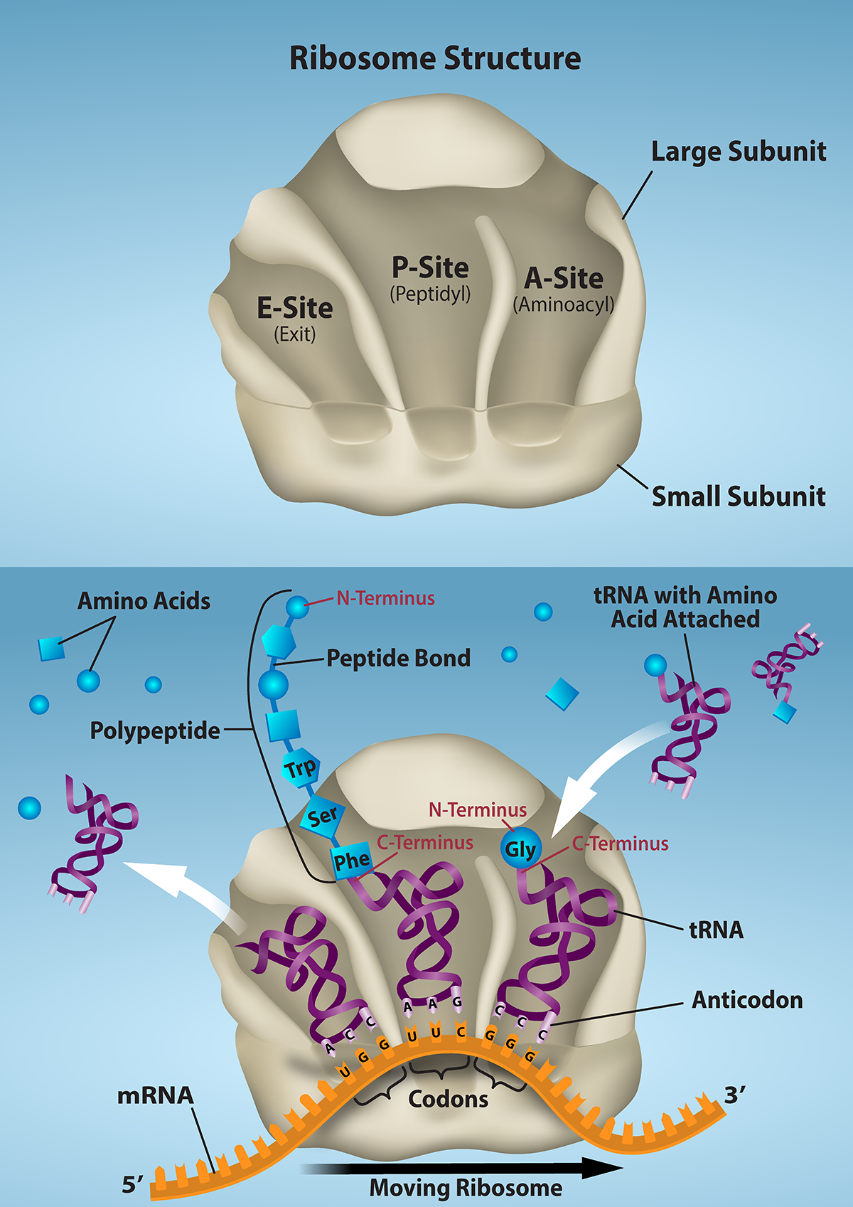
OpenStax · 21.4 The Adaptive Immune Response: B-lymphocytes and Antibodies ↗

Instructional page including learning objectives, examples, figures, source tables and questions. Site navigation and embedded media players are omitted; source media links remain available.

Learning Objectives

By the end of this section, you will be able to:

were the first component of the adaptive immune response to be characterized by scientists working on the immune system. It was already known that individuals who survived a infection were immune to re-infection with the same . Early microbiologists took serum from an immune patient and mixed it with a fresh of the same type of , then observed the under a microscope. The became clumped in a process called agglutination. When a different species was used, the agglutination did not happen. Thus, there was something in the serum of immune individuals that could specifically bind to and agglutinate .

Scientists now know the cause of the agglutination is an , also called an . What is an ? An is essentially a form of a . (In , surface is another name for the .) Not surprisingly, the same encode both the and the surface . One minor difference in the way these are synthesized distinguishes a naïve with on its surface from an -secreting with no on its surface. The of the have the exact same - and specificity as their precursors.

There are five different classes of found in humans: IgM, IgD, , IgA, and IgE. Each of these has specific functions in the immune response, so by learning about them, researchers can learn about the great variety of functions critical to many adaptive immune responses.

do not recognize in the complex fashion of T cells. can recognize native, unprocessed and do not require the participation of and -presenting cells.

and Activation

differentiate in the bone marrow. During the process of maturation, up to 100 trillion different clones of are generated, which is similar to the diversity of seen in T cells.

and the development of are not quite as well understood as it is in T cells. is the destruction or inactivation of that recognize self- in the bone marrow, and its role is critical and well established. In the process of clonal , immature that bind strongly to self- expressed on tissues are signaled to induce their own destruction by , removing them from the population. In the process of clonal , however, exposed to in the bone marrow are not physically deleted, but become unable to function.

Another mechanism called is a direct result of T cell . In , functional, mature leave the bone marrow but have yet to be exposed to self-. Most require signals from helper T cells (Th2) to proceed to make . When a binds to a self- but receives no signals from a nearby Th2 cell to produce , the cell is signaled to undergo and is destroyed. This is yet another example of the control that T cells have over the adaptive immune response.

After are activated by their binding to , they differentiate into . often leave the secondary lymphoid organs, where the response is generated, and migrate back to the bone marrow, where the whole process started. After secreting for a specific period, they die, as most of their energy is devoted to making and not to maintaining themselves. Thus, are said to be terminally differentiated.

The final of interest is the memory , which results from the clonal expansion of an activated . Memory function in a way similar to memory T cells. They lead to a stronger and faster secondary response when compared to the primary response, as illustrated below.

Structure

are glycoproteins consisting of two types of chains with attached . The and the are the two that form the . The main differences between the classes of are in the differences between their , but as you shall see, the have an important role, forming part of the - on the .

Four-chain Models of Structures

All have two identical and two identical . (Some contain multiple units of this four-chain structure.) The region of the is formed by the two coming together, usually linked by (Figure 21.21). The portion of the is important in that many cells of the immune system have . Cells having these can then bind to -coated , greatly increasing the specificity of the cells. At the other end of the are two identical -.

This diagram shows the four chain structure of a generic antibody.
Figure 21.21 — 21.4 The Adaptive Immune Response: B-lymphocytes and

Figure 21.21 Antibody and IgG2 Structures The typical four chain structure of a generic antibody (a) and the corresponding three-dimensional structure of the antibody IgG2 (b). (credit b: modification of work by Tim Vickers)

J. Gordon Betts and colleagues, Anatomy And Physiology 2E, OpenStax. (credit b: modification of work by Tim Vickers) · CC BY-NC-SA 4.0 · Original image ↗ · Access for free ↗

Five Classes of and their Functions

In general, have two basic functions. They can act as the or they can be , circulate, and bind to a , often labeling it for identification by other forms of the immune response. Of the five classes, notice that only two can function as the for naïve : IgM and IgD (Figure 21.22). Mature that leave the bone marrow express both IgM and IgD, but both have the same specificity. Only IgM is , however, and no other nonreceptor function for IgD has been discovered.

This table shows the five classes of the immunoglobulins. The table shows the molecular weight, number of antigen binding sites, and their function.
Figure 21.22 — 21.4 The Adaptive Immune Response: B-lymphocytes and

Figure 21.22 Five Classes of Antibodies

J. Gordon Betts and colleagues, Anatomy And Physiology 2E, OpenStax. · CC BY-NC-SA 4.0 · Original image ↗ · Access for free ↗

IgM consists of five four-chain structures (20 total chains with 10 identical -) and is thus the largest of the . IgM is usually the first made during a primary response. Its 10 - and large shape allow it to bind well to many surfaces. It is excellent at binding and activating the cascade, consistent with its role in promoting , , and cell lysis. Thus, it is a very effective against at early stages of a response. As the primary response proceeds, the produced in a can change to , IgA, or IgE by the process known as . is the change of one class to another. While the class of changes, the specificity and the - do not. Thus, the made are still specific to the that stimulated the initial IgM response.

is a major of late primary responses and the main of secondary responses in the blood. This is because occurs during primary responses. is a monomeric that clears from the blood and can activate (although not as well as IgM), taking advantage of its antibacterial activities. Furthermore, this class of is the one that crosses the to protect the developing fetus from disease exits the blood to the to fight extracellular .

IgA exists in two forms, a four-chain in the blood and an eight-chain structure, or dimer, in gland secretions of the mucous , including mucus, saliva, and tears. Thus, dimeric IgA is the only to leave the interior of the body to protect body surfaces. IgA is also of importance to newborns, because this is present in mother’s breast milk (colostrum), which serves to protect the infant from disease.

IgE is usually associated with allergies and anaphylaxis. It is present in the lowest in the blood, because its region binds strongly to an IgE-specific on the surfaces of mast cells. IgE makes mast cell degranulation very specific, such that if a person is allergic to peanuts, there will be peanut-specific IgE bound to their mast cells. In this person, eating peanuts will cause the mast cells to degranulate, sometimes causing severe allergic , including anaphylaxis, a severe, systemic allergic response that can cause death.



of

and expansion work much the same way in as in T cells. Only with appropriate specificity are selected for and expanded (Figure 21.23). Eventually, the secrete with antigenic specificity identical to those that were on the surfaces of the selected . Notice in the figure that both and memory are generated simultaneously.

This flow chart shows how the clonal selection of B cells takes place. The left panel shows the primary response and the right panel shows the secondary response.
Figure 21.23 — 21.4 The Adaptive Immune Response: B-lymphocytes and

Figure 21.23 Clonal Selection of B Cells During a primary B cell immune response, both antibody-secreting plasma cells and memory B cells are produced. These memory cells lead to the differentiation of more plasma cells and memory B cells during secondary responses.

J. Gordon Betts and colleagues, Anatomy And Physiology 2E, OpenStax. · CC BY-NC-SA 4.0 · Original image ↗ · Access for free ↗

Primary versus Secondary Responses

Primary and secondary responses as they relate to T cells were discussed earlier. This section will look at these responses with and production. Because are easily obtained from blood samples, they are easy to follow and graph (Figure 21.24). As you will see from the figure, the primary response to an (representing a ) is delayed by several days. This is the time it takes for the clones to expand and differentiate into . The level of produced is low, but it is sufficient for immune protection. The second time a person encounters the same , there is no time delay, and the amount of made is much higher. Thus, the response overwhelms the quickly and, in most situations, no symptoms are felt. When a different is used, another primary response is made with its low levels and time delay.

This graph shows the antibody concentration as a function of time in primary and secondary response.
Figure 21.24 — 21.4 The Adaptive Immune Response: B-lymphocytes and

Figure 21.24 Primary and Secondary Antibody Responses Antigen A is given once to generate a primary response and later to generate a secondary response. When a different antigen is given for the first time, a new primary response is made.

J. Gordon Betts and colleagues, Anatomy And Physiology 2E, OpenStax. · CC BY-NC-SA 4.0 · Original image ↗ · Access for free ↗

Active versus

Immunity to , and the ability to control growth so that damage to the tissues of the body is limited, can be acquired by (1) the active development of an immune response in the infected individual or (2) the passive transfer of immune components from an immune individual to a nonimmune one. Both active and have examples in the natural world and as part of medicine.

Active immunity is the resistance to acquired during an adaptive immune response within an individual (Table 21.6). Naturally acquired active immunity, the response to a , is the focus of this chapter. Artificially acquired active immunity involves the use of vaccines. A vaccine is a killed or weakened or its components that, when administered to a healthy individual, leads to the development of immunological memory (a weakened primary immune response) without causing much in the way of symptoms. Thus, with the use of vaccines, one can avoid the damage from disease that results from the first exposure to the , yet reap the benefits of protection from immunological memory. The advent of vaccines was one of the major medical advances of the twentieth century and led to the eradication of smallpox and the control of many infectious diseases, including polio, measles, and whooping cough.

Active versus
Natural Artificial
Active Adaptive immune response Vaccine response
Passive Trans-placental /breastfeeding Immune globulin injections
Table 21.6

arises from the transfer of to an individual without requiring them to mount their own active immune response. Naturally acquired is seen during fetal development. is transferred from the maternal circulation to the fetus via the , protecting the fetus from infection and protecting the newborn for the first few months of its life. As already stated, a newborn benefits from the IgA it obtains from milk during breastfeeding. The fetus and newborn thus benefit from the immunological memory based on the to which the pregnant person has been exposed. In medicine, artificially acquired usually involves injections of , taken from animals previously exposed to a specific . This treatment is a fast-acting method of temporarily protecting an individual who was possibly exposed to a . The downside to both types of is the lack of the development of immunological memory. Once the are transferred, they are effective for only a limited time before they degrade.

Interactive Link

Immunity can be acquired in an active or passive way, and it can be natural or artificial. Watch this video to see an animated discussion of passive and active immunity. What is an example of natural immunity acquired passively?

T cell-dependent versus T cell-independent

As discussed previously, Th2 cells secrete cytokines that drive the production of in a , responding to complex such as those made by . On the other hand, some are T cell independent. A T cell-independent usually is in the form of repeated moieties found on the cell walls of . Each on the surface has two , and the repeated nature of T cell-independent leads to crosslinking of the surface on the . The crosslinking is enough to activate it in the absence of T cell cytokines.

A T cell-dependent , on the other hand, usually is not repeated to the same degree on the and thus does not crosslink surface with the same efficiency. To elicit a response to such , the B and T cells must come close together (Figure 21.25). The must receive two signals to become activated. Its surface must recognize native . Some of this is internalized, processed, and presented to the Th2 cells on a class II . The T cell then binds using its and is activated to secrete cytokines that diffuse to the , finally activating it completely. Thus, the receives signals from both its surface and the T cell via its cytokines, and acts as a professional -presenting cell in the process.

This diagram shows the binding of a B cell and a T cell.
Figure 21.25 — 21.4 The Adaptive Immune Response: B-lymphocytes and

Figure 21.25 T and B Cell Binding To elicit a response to a T cell-dependent antigen, the B and T cells must come close together. To become fully activated, the B cell must receive two signals from the native antigen and the T cell’s cytokines.

J. Gordon Betts and colleagues, Anatomy And Physiology 2E, OpenStax. · CC BY-NC-SA 4.0 · Original image ↗ · Access for free ↗