Separate filtration, reabsorption, and secretion before predicting urine composition or clearance.
Map the nephron and outflow
Renal cortex contains glomeruli and much of the convoluted tubules; medulla contains loops of Henle and collecting ducts. Blood enters glomerular capillaries from an afferent arteriole and leaves through an efferent arteriole to peritubular capillaries or vasa recta. Filtrate enters Bowman’s space, proximal tubule, loop, distal tubule, and collecting duct. Urine drains through papillary ducts, calyces, renal pelvis, ureter, bladder, and urethra. Juxtamedullary nephrons with long loops support a strong medullary gradient.
Source: 25.4 Microscopic Anatomy of the Kidney · 25.5 Physiology of Urine Formation · 25.6 Tubular Reabsorption · 25.8 Endocrine Regulation of Kidney Function · 25.2 Gross Anatomy of Urine Transport · 26.4 Acid-Base Balance
Account for what enters and leaves tubules
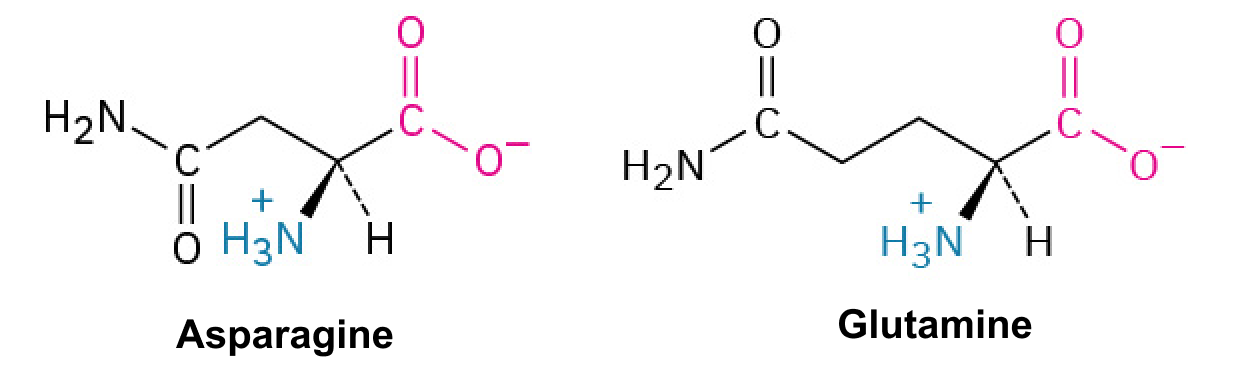
Glomerular filtration moves water and small solutes into Bowman’s space while retaining most cells and large proteins. Reabsorption returns tubular material to blood; secretion moves material from blood into tubules. Excretion rate = filtration − reabsorption + secretion. Filtered load equals GFR × plasma concentration for a freely filtered solute. Proximal transport normally reclaims most filtered glucose and amino acids, but transport maxima can be exceeded. Nitrogenous wastes include urea and creatinine. Clearance C = urine concentration × urine flow / plasma concentration; it is a volume per time, not a mass.
Source: 25.4 Microscopic Anatomy of the Kidney · 25.5 Physiology of Urine Formation · 25.6 Tubular Reabsorption · 25.8 Endocrine Regulation of Kidney Function · 25.2 Gross Anatomy of Urine Transport · 26.4 Acid-Base Balance
Build and use a medullary gradient
The descending loop is relatively water-permeable; the ascending limb reabsorbs salt while remaining relatively water-impermeable. Opposing flow and differing permeability multiply the corticomedullary gradient. Vasa recta countercurrent exchange limits gradient washout, and urea recycling contributes to deep-medullary osmolarity. ADH increases collecting-duct water permeability through aquaporin regulation, allowing water to leave along the gradient. Without ADH action, dilute urine can be excreted despite an existing gradient. Aldosterone promotes distal Na⁺ reabsorption and K⁺ secretion rather than directly replacing ADH’s water-channel action.
Source: 25.4 Microscopic Anatomy of the Kidney · 25.5 Physiology of Urine Formation · 25.6 Tubular Reabsorption · 25.8 Endocrine Regulation of Kidney Function · 25.2 Gross Anatomy of Urine Transport · 26.4 Acid-Base Balance
Regulate volume, osmolarity, pH, and voiding
Low effective circulating volume activates renin release, angiotensin II, aldosterone, thirst, and often ADH, supporting pressure and volume. Osmoreceptors regulate ADH and thirst independently of the volume sensors. Kidneys reclaim filtered bicarbonate and excrete net acid through H⁺ secretion buffered by phosphate and ammonium; lungs regulate CO₂ more rapidly. Natriuretic peptides favor Na⁺ loss under volume expansion. Bladder stretch triggers a spinal/brainstem micturition circuit: detrusor contracts and internal outflow resistance decreases, while the external skeletal-muscle sphincter remains under voluntary control until coordinated release.
Source: 25.4 Microscopic Anatomy of the Kidney · 25.5 Physiology of Urine Formation · 25.6 Tubular Reabsorption · 25.8 Endocrine Regulation of Kidney Function · 25.2 Gross Anatomy of Urine Transport · 26.4 Acid-Base Balance
Practice and reasoning
Question. A freely filtered marker has urine concentration 60 mg/mL, urine flow 2 mL/min, and plasma concentration 1 mg/mL. Find clearance. What would reduced ADH action do to urine concentration if the medullary gradient is intact?
Show reasoning
Clearance = 60×2/1 = 120 mL/min. This equals GFR only if the marker is neither reabsorbed nor secreted and the assumptions hold. Reduced ADH action lowers collecting-duct water permeability, increasing water excretion and producing more dilute urine; it does not imply the ascending limb suddenly becomes water-permeable.
Source: 25.4 Microscopic Anatomy of the Kidney · 25.5 Physiology of Urine Formation · 25.6 Tubular Reabsorption · 25.8 Endocrine Regulation of Kidney Function · 25.2 Gross Anatomy of Urine Transport · 26.4 Acid-Base Balance
25.4 Microscopic Anatomy of the Kidney — figures and tables
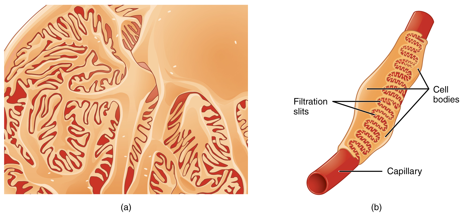
Figure 25.11 Podocytes Podocytes interdigitate with structures called pedicels and filter substances in a way similar to fenestrations. In (a), the large cell body can be seen at the top right corner, with branches extending from the cell body. The smallest finger-like extensions are the pedicels. Pedicels on one podocyte always interdigitate with the pedicels of another podocyte. (b) This capillary has three podocytes wrapped around it.
J. Gordon Betts and colleagues, Anatomy And Physiology 2E, OpenStax. · CC BY-NC-SA 4.0 · Original image ↗ · Access for free ↗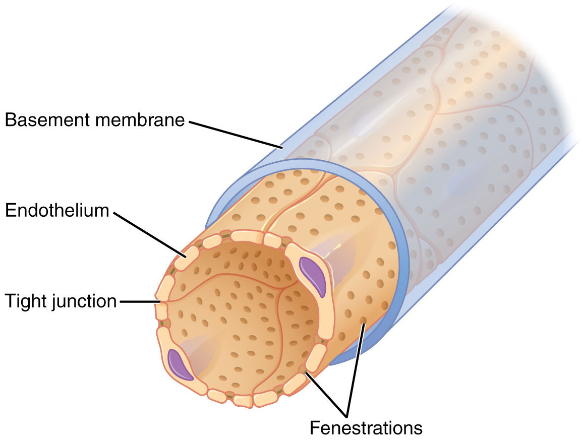
Figure 25.12 Fenestrated Capillary Fenestrations allow many substances to diffuse from the blood based primarily on size.
J. Gordon Betts and colleagues, Anatomy And Physiology 2E, OpenStax. · CC BY-NC-SA 4.0 · Original image ↗ · Access for free ↗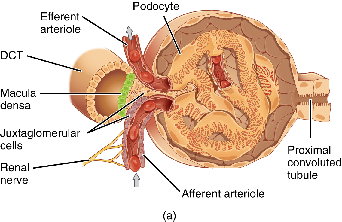
Figure 25.13 Juxtaglomerular Apparatus and Glomerulus (a) The JGA allows specialized cells to monitor the composition of the fluid in the DCT and adjust the glomerular filtration rate. (b) This micrograph shows the glomerulus and surrounding structures. LM × 1540. (Micrograph provided by the Regents of University of Michigan Medical School © 2012)
J. Gordon Betts and colleagues, Anatomy And Physiology 2E, OpenStax. · CC BY-NC-SA 4.0 · Original image ↗ · Access for free ↗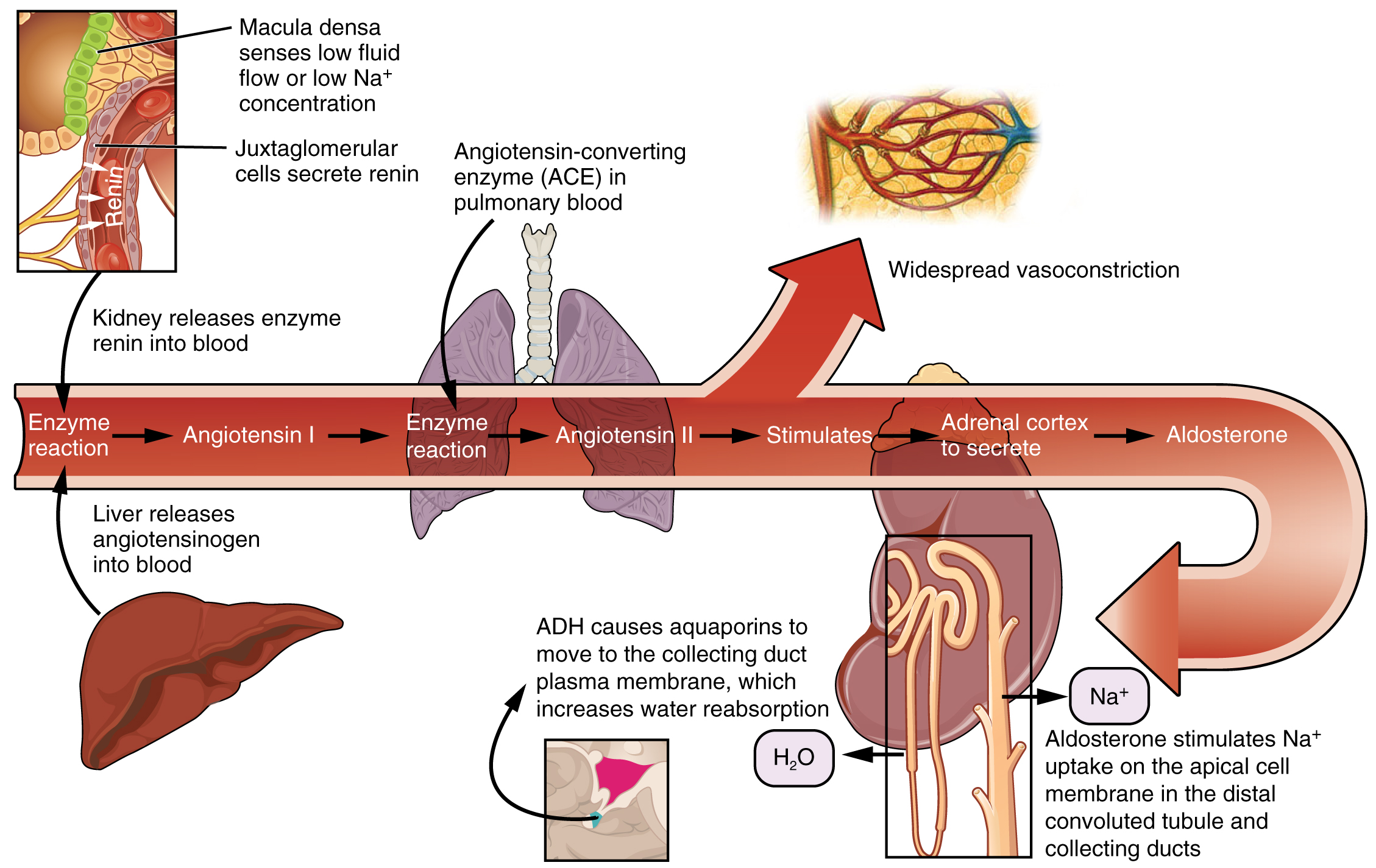
Figure 25.14 Conversion of Angiotensin I to Angiotensin II The enzyme renin converts the pro-enzyme angiotensin I; the lung-derived enzyme ACE converts angiotensin I into active angiotensin II.
J. Gordon Betts and colleagues, Anatomy And Physiology 2E, OpenStax. · CC BY-NC-SA 4.0 · Original image ↗ · Access for free ↗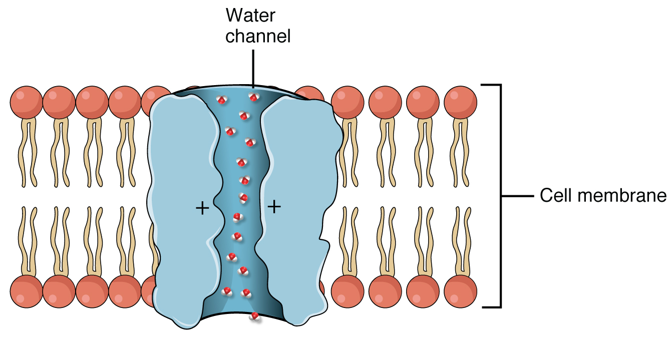
Figure 25.15 Aquaporin Water Channel Positive charges inside the channel prevent the leakage of electrolytes across the cell membrane, while allowing water to move due to osmosis.
J. Gordon Betts and colleagues, Anatomy And Physiology 2E, OpenStax. · CC BY-NC-SA 4.0 · Original image ↗ · Access for free ↗25.5 Physiology of Urine Formation — figures and tables
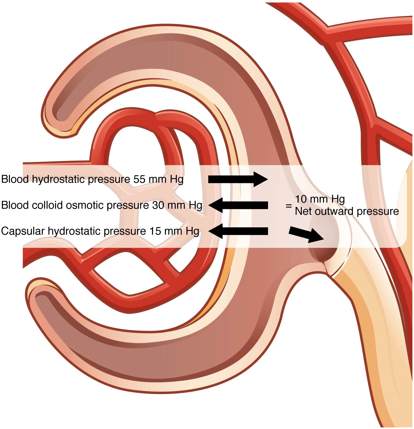
Figure 25.16 Net Filtration Pressure The NFP is the sum of osmotic and hydrostatic pressures.
J. Gordon Betts and colleagues, Anatomy And Physiology 2E, OpenStax. · CC BY-NC-SA 4.0 · Original image ↗ · Access for free ↗| Weakness |
| Lethargy |
| Shortness of breath |
| Widespread edema |
| Anemia |
| Metabolic acidosis |
| Metabolic alkalosis |
| Heart arrhythmias |
| Uremia (high urea level in the blood) |
| Loss of appetite |
| Fatigue |
| Excessive urination |
| Oliguria (too little urine output) |
Table 25.3. J. Gordon Betts and colleagues, Anatomy And Physiology 2E, OpenStax.
| Flow per minute (mL) | Calculation | |
|---|---|---|
| Renal blood flow | 1050 | Cardiac output is about 5000 mL/minute, of which 21 percent flows through the kidney.
5000*0.21 = 1050 mL blood/min |
| Renal plasma flow | 578 | Renal plasma flow equals the blood flow per minute times (1 minus the hematocrit). If a person has a hematocrit of 45, then the renal plasma flow is 55 percent.
1050*0.55 = 578 mL plasma/min |
| Glomerular filtration rate | 110 | The GFR is the amount of plasma entering Bowman’s capsule per minute. It is the renal plasma flow times the fraction that enters the renal capsule (19 percent).
578*0.19 = 110 mL filtrate/min |
| Urine | 1296 ml/day | The filtrate not recovered by the kidney is the urine that will be eliminated. It is the GFR times the fraction of the filtrate that is not reabsorbed (0.8 percent).
110*.008 = 0.9 mL urine /min Multiply urine/min times 60 minutes times 24 hours to get daily urine production. 0.9*60*24 = 1296 mL/day urine |
Table 25.4. J. Gordon Betts and colleagues, Anatomy And Physiology 2E, OpenStax.
25.6 Tubular Reabsorption — figures and tables
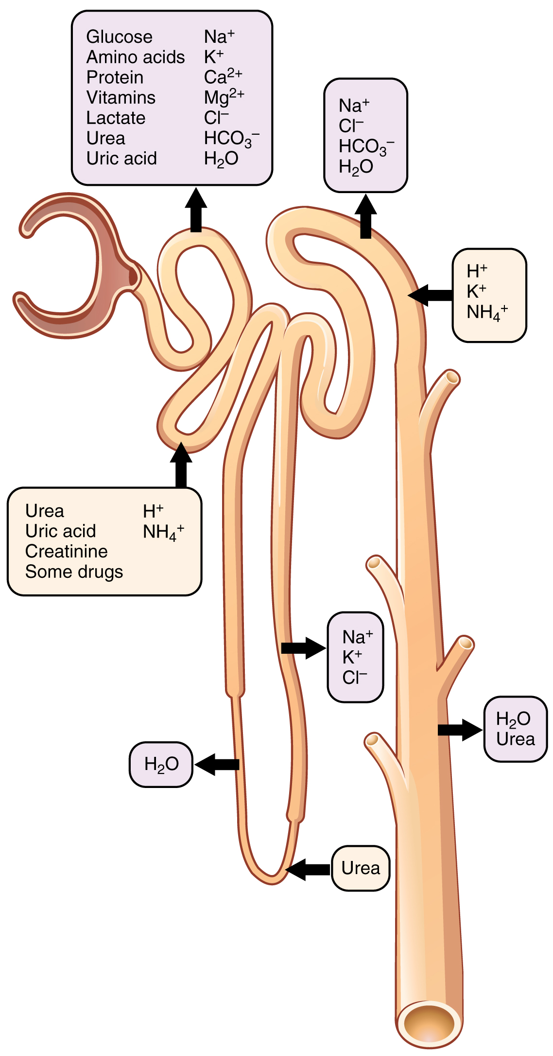
Figure 25.17 Locations of Secretion and Reabsorption in the Nephron
J. Gordon Betts and colleagues, Anatomy And Physiology 2E, OpenStax. · CC BY-NC-SA 4.0 · Original image ↗ · Access for free ↗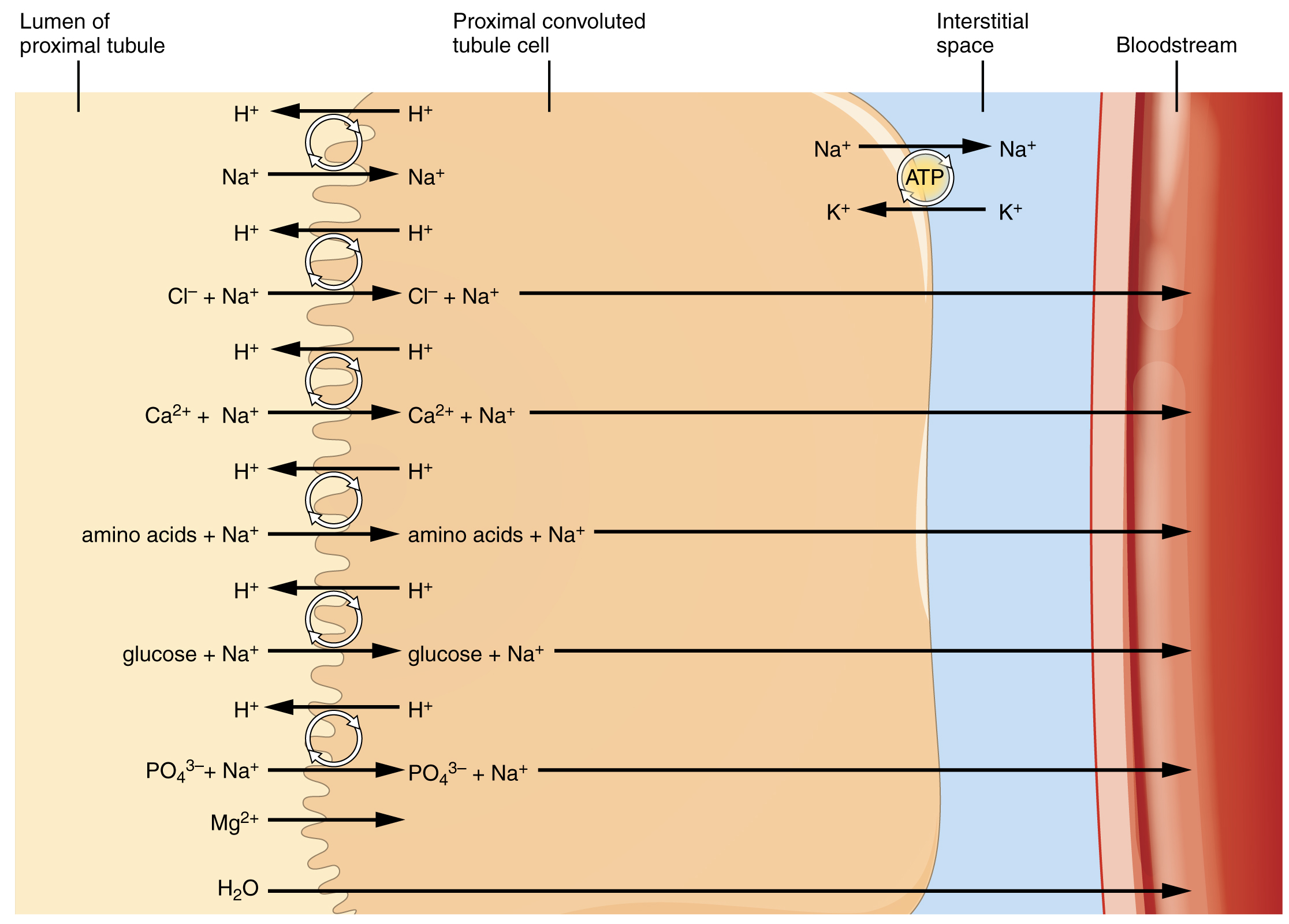
Figure 25.18 Substances Reabsorbed and Secreted by the PCT
J. Gordon Betts and colleagues, Anatomy And Physiology 2E, OpenStax. · CC BY-NC-SA 4.0 · Original image ↗ · Access for free ↗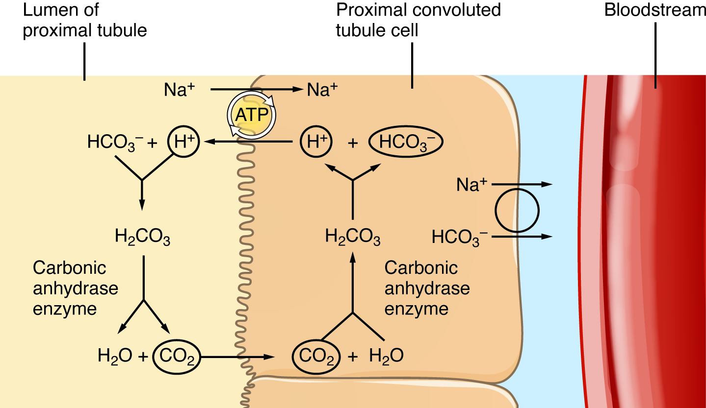
Figure 25.19 Reabsorption of Bicarbonate from the PCT
J. Gordon Betts and colleagues, Anatomy And Physiology 2E, OpenStax. · CC BY-NC-SA 4.0 · Original image ↗ · Access for free ↗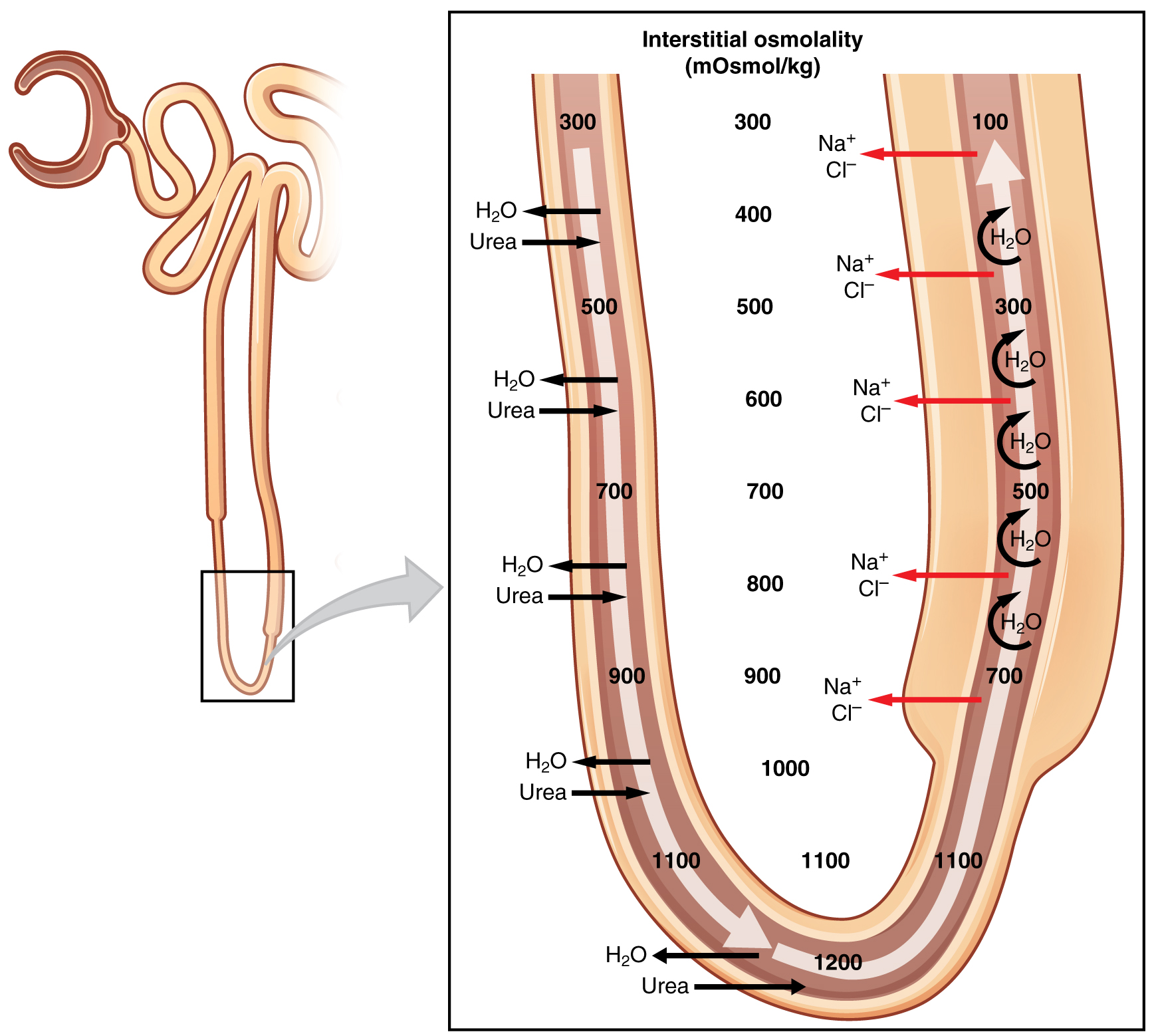
Figure 25.20 Countercurrent Multiplier System
J. Gordon Betts and colleagues, Anatomy And Physiology 2E, OpenStax. · CC BY-NC-SA 4.0 · Original image ↗ · Access for free ↗| Substance | PCT | Loop of Henle | DCT | Collecting ducts |
|---|---|---|---|---|
| Glucose | Almost 100 percent reabsorbed; secondary active transport with Na+ | |||
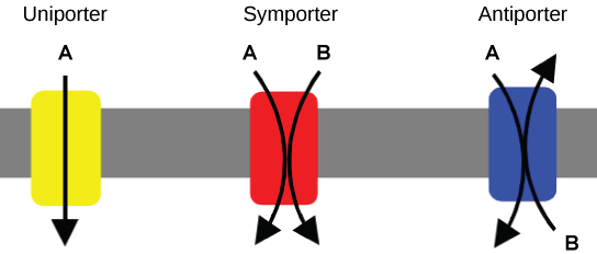
| Oligopeptides, proteins, amino acids | Almost 100 percent reabsorbed; symport with Na+ | |||
| Vitamins | Reabsorbed | |||
| Lactate | Reabsorbed | |||
| Creatinine | Secreted | |||
| Urea | 50 percent reabsorbed by diffusion; also secreted | Secretion, diffusion in descending limb | Reabsorption in medullary collecting ducts; diffusion | |
| Sodium | 65 percent actively reabsorbed | 25 percent reabsorbed in thick ascending limb; active transport | 5 percent reabsorbed; active | 5 percent reabsorbed, stimulated by aldosterone; active |
| Chloride | Reabsorbed, symport with Na+, diffusion | Reabsorbed in thin and thick ascending limb; diffusion in ascending limb | Reabsorbed; diffusion | Reabsorbed; symport |
| Water | 67 percent reabsorbed osmotically with solutes | 15 percent reabsorbed in descending limb; osmosis | 8 percent reabsorbed if ADH; osmosis | Variable amounts reabsorbed, controlled by ADH, osmosis |
| HCO3- | 80–90 percent symport reabsorption with Na+ | Reabsorbed, symport with Na+ and antiport with Cl–; in ascending limb | Reabsorbed antiport with Cl– | |
| H+ | Secreted; diffusion | Secreted; active | Secreted; active | |
| NH4+ | Secreted; diffusion | Secreted; diffusion | Secreted; diffusion | |
| Some drugs | Secreted | Secreted; active | Secreted; active | |
| Potassium | 65 percent reabsorbed; diffusion | 20 percent reabsorbed in thick ascending limb; symport | Secreted; active | Secretion controlled by aldosterone; active |
| Calcium | Reabsorbed; diffusion | Reabsorbed in thick ascending limb; diffusion | Reabsorbed if parathyroid hormone present; active | |
| Magnesium | Reabsorbed; diffusion | Reabsorbed in thick ascending limb; diffusion | Reabsorbed | |
| Phosphate | 85 percent reabsorbed, inhibited by parathyroid hormone, diffusion | Reabsorbed; diffusion |
Table 25.5. J. Gordon Betts and colleagues, Anatomy And Physiology 2E, OpenStax.
| Substance | Amount filtered (grams) | Amount reabsorbed (grams) | Amount in urine (grams) |
|---|---|---|---|
| Water | 180 L | 179 L | 1 L |
| Proteins | 10–20 | 10–20 | 0 |
| Chlorine | 630 | 625 | 5 |
| Sodium | 540 | 537 | 3 |
| Bicarbonate | 300 | 299.7 | 0.3 |
| Glucose | 180 | 180 | 0 |
| Urea | 53 | 28 | 25 |
| Potassium | 28 | 24 | 4 |
| Uric acid | 8.5 | 7.7 | 0.8 |
| Creatinine | 1.4 | 0 | 1.4 |
Table 25.6. J. Gordon Betts and colleagues, Anatomy And Physiology 2E, OpenStax.
Source: 25.6 Tubular Reabsorption
25.8 Endocrine Regulation of Kidney Function — figures and tables
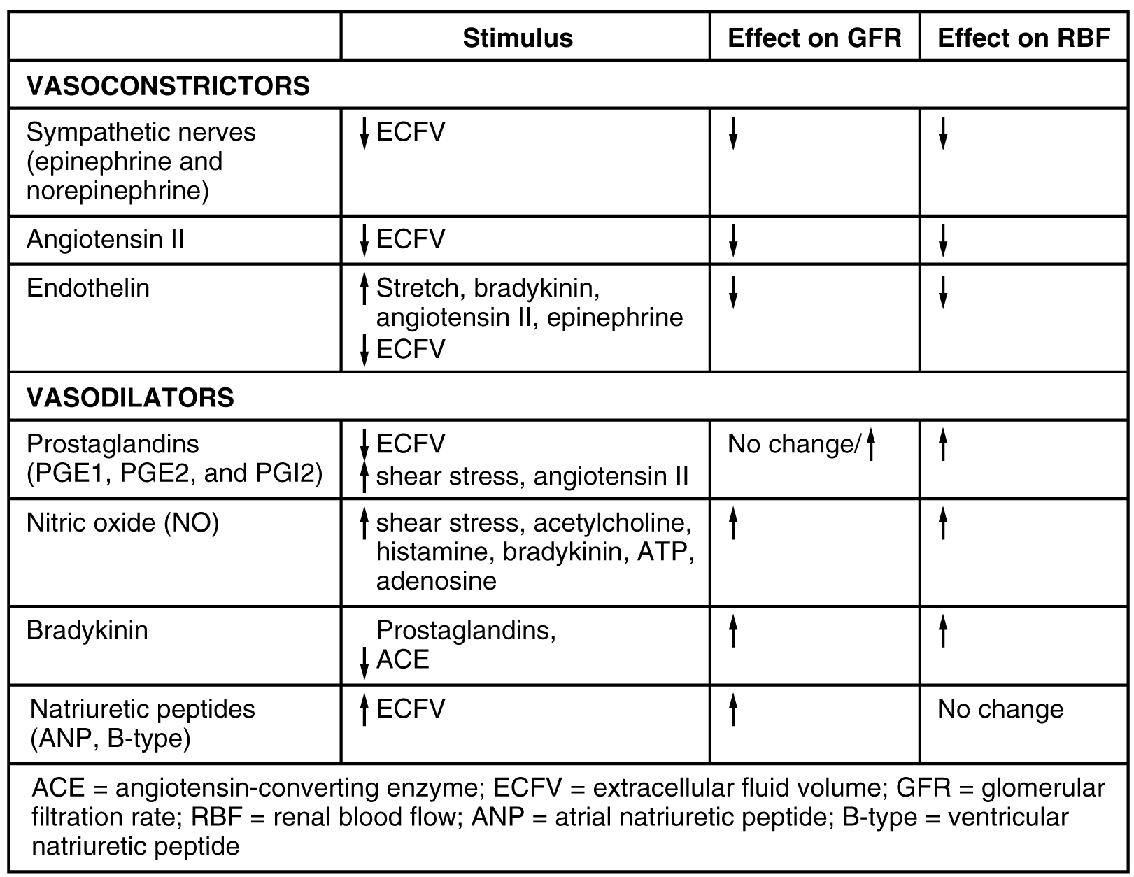
Figure 25.21 Major Hormones That Influence GFR and RFB
J. Gordon Betts and colleagues, Anatomy And Physiology 2E, OpenStax. · CC BY-NC-SA 4.0 · Original image ↗ · Access for free ↗25.2 Gross Anatomy of Urine Transport — figures and tables
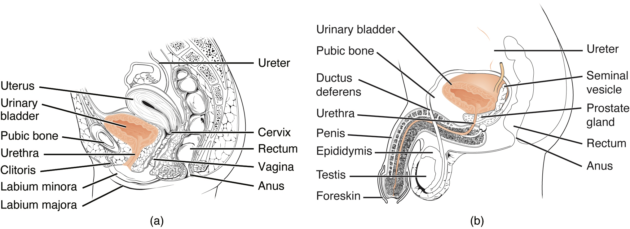
Figure 25.3 Female and Male Urethras The urethra transports urine from the bladder to the outside of the body. This image shows (a) a female urethra and (b) a male urethra.
J. Gordon Betts and colleagues, Anatomy And Physiology 2E, OpenStax. · CC BY-NC-SA 4.0 · Original image ↗ · Access for free ↗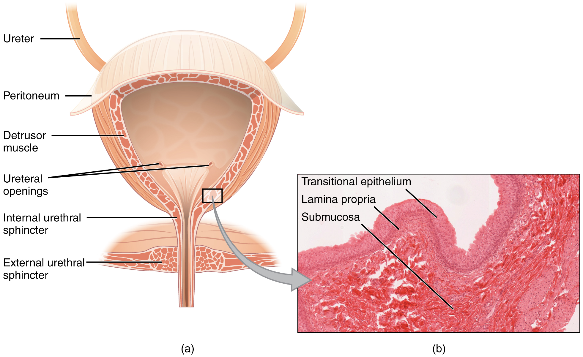
Figure 25.4 Bladder (a) Anterior cross section of the bladder. (b) The detrusor muscle of the bladder (source: monkey tissue) LM × 448. (Micrograph provided by the Regents of the University of Michigan Medical School © 2012)
J. Gordon Betts and colleagues, Anatomy And Physiology 2E, OpenStax. · CC BY-NC-SA 4.0 · Original image ↗ · Access for free ↗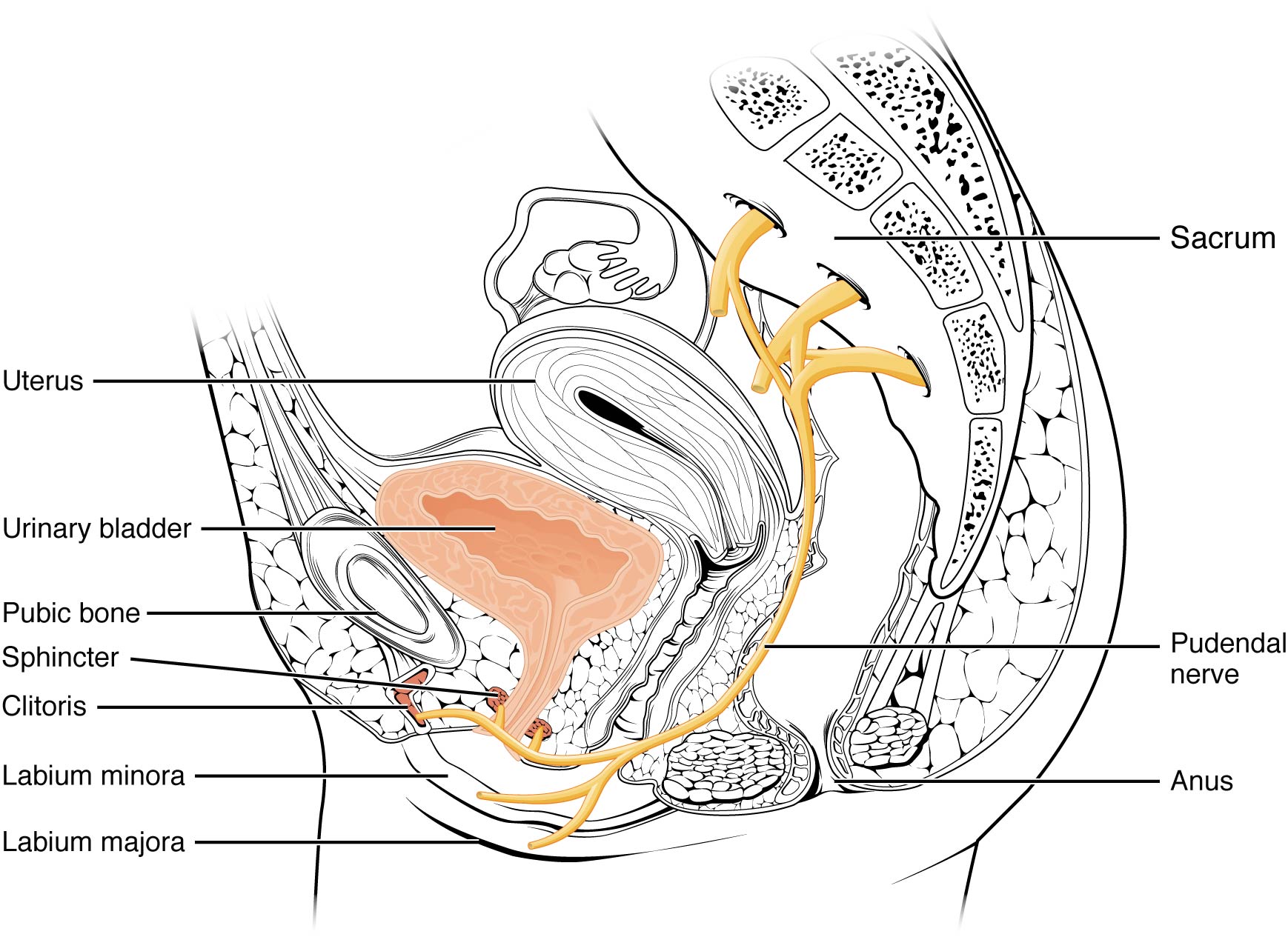
Figure 25.5 Nerves Innervating the Urinary System
J. Gordon Betts and colleagues, Anatomy And Physiology 2E, OpenStax. · CC BY-NC-SA 4.0 · Original image ↗ · Access for free ↗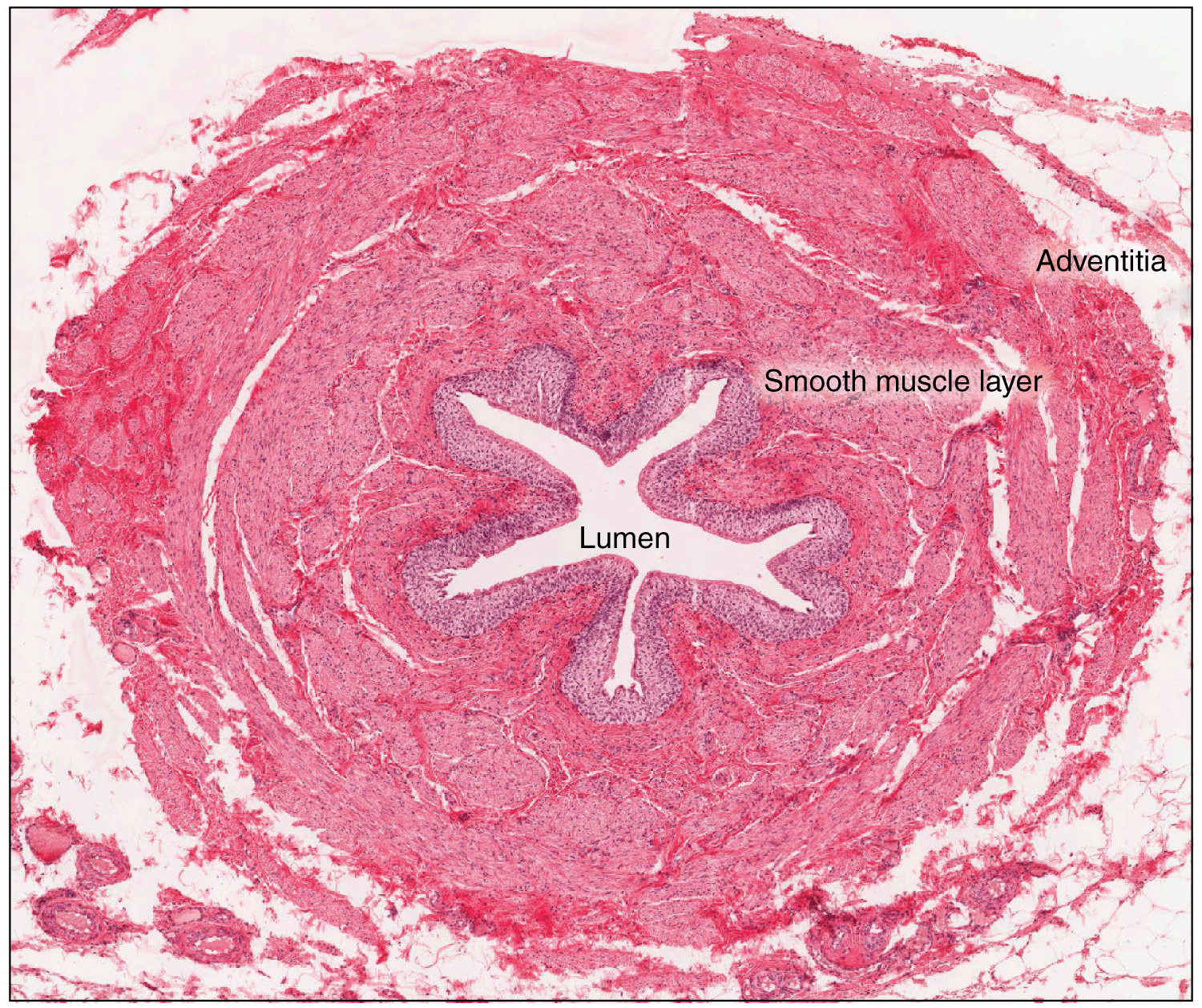
Figure 25.6 Ureter Peristaltic contractions help to move urine through the lumen with contributions from fluid pressure and gravity. LM × 128. (Micrograph provided by the Regents of the University of Michigan Medical School © 2012)
J. Gordon Betts and colleagues, Anatomy And Physiology 2E, OpenStax. · CC BY-NC-SA 4.0 · Original image ↗ · Access for free ↗26.4 Acid-Base Balance — figures and tables
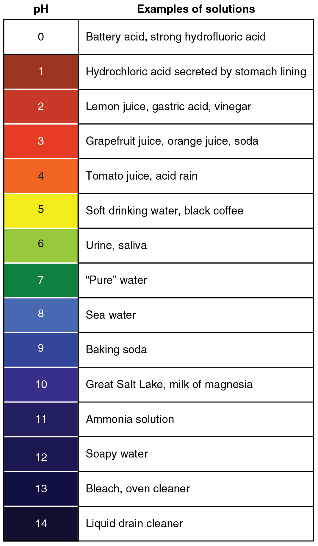
Figure 26.15 The pH Scale This chart shows where many common substances fall on the pH scale.
J. Gordon Betts and colleagues, Anatomy And Physiology 2E, OpenStax. · CC BY-NC-SA 4.0 · Original image ↗ · Access for free ↗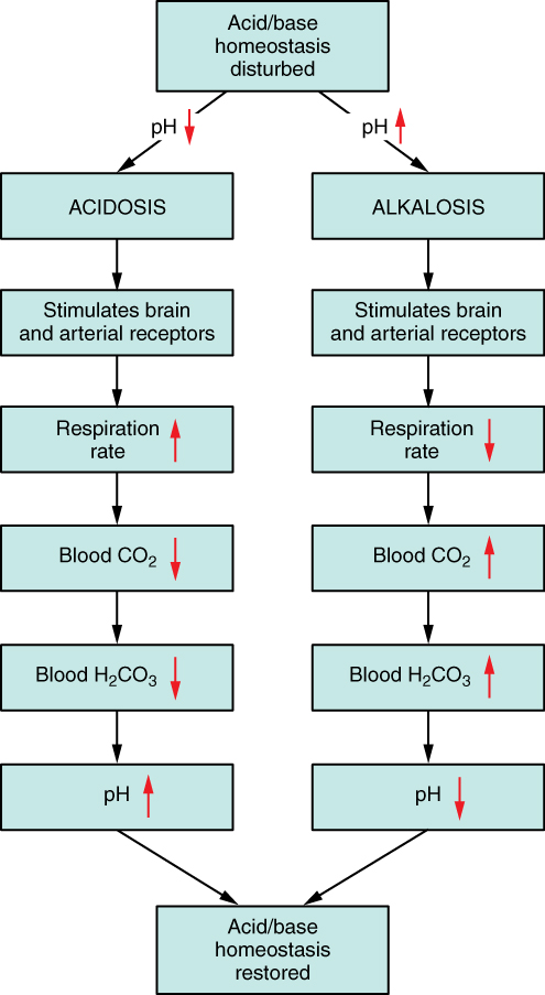
Figure 26.16 Respiratory Regulation of Blood pH The respiratory system can reduce blood pH by removing CO 2 from the blood.
J. Gordon Betts and colleagues, Anatomy And Physiology 2E, OpenStax. · CC BY-NC-SA 4.0 · Original image ↗ · Access for free ↗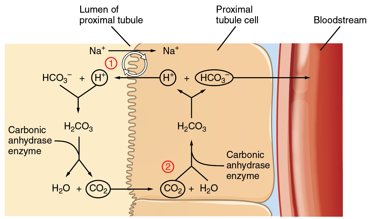
Figure 26.17 Conservation of Bicarbonate in the Kidney Tubular cells are not permeable to bicarbonate; thus, bicarbonate is conserved rather than reabsorbed. Steps 1 and 2 of bicarbonate conservation are indicated.
J. Gordon Betts and colleagues, Anatomy And Physiology 2E, OpenStax. · CC BY-NC-SA 4.0 · Original image ↗ · Access for free ↗Source: 26.4 Acid-Base Balance