Foundation 3 · 3B · Integrated organ systems

Digestion and absorption

Sources ↗

Mechanical processing and chemical digestion make nutrients available for selective intestinal transport.

Follow the alimentary canal

Food enters the mouth for mastication and saliva exposure, passes through pharynx and esophagus, and reaches the stomach. The small intestine comprises duodenum, jejunum, and ileum; most digestion and absorption occur there. The large intestine includes cecum and colon, followed by rectum and anal canal. regulate passage and reduce backflow. propels contents; mixes them. Accessory organs deliver secretions without food passing through their tissue.

Source: 23.2 Digestive System Processes and Regulation · 23.4 The Stomach · 23.6 Accessory Organs in Digestion: The Liver, Pancreas, and Gallbladder · 23.7 Chemical Digestion and Absorption: A Closer Look

Gastric chemistry and protection

Stomach muscle mixes food into . secrete HCl and ; release , which becomes in . begins substantial digestion. unfolds and limits many microbes, but it does not replace specificity. A mucus– barrier, epithelial junctions, blood flow, and repair protect the stomach. later supports vitamin B12 absorption in the ileum. , histamine, vagal activity, and inhibitory feedback coordinate and emptying.

Source: 23.2 Digestive System Processes and Regulation · 23.4 The Stomach · 23.6 Accessory Organs in Digestion: The Liver, Pancreas, and Gallbladder · 23.7 Chemical Digestion and Absorption: A Closer Look

Liver, gallbladder, and

The liver makes bile, processes portal nutrients, stores and mobilizes fuels, and modifies or clears many substances. The gallbladder stores and concentrates bile, releasing it into the duodenum. emulsify fat and support micelles but are not digestive . pancreatic ducts deliver and ; are commonly as zymogens to limit self-digestion. converts to , which activates additional pancreatic precursors. islets independently regulate blood fuels through and .

Source: 23.2 Digestive System Processes and Regulation · 23.4 The Stomach · 23.6 Accessory Organs in Digestion: The Liver, Pancreas, and Gallbladder · 23.7 Chemical Digestion and Absorption: A Closer Look

Surface area and transport routes

Folds, , and expand small-intestinal surface area. Pancreatic neutralizes gastric , enabling intestinal . Brush-border complete digestion of selected and . Monosaccharides and amino- enter ; and uptake commonly use Na⁺-coupled transport. travel in micelles to , are reassembled, and leave largely as through . Water follows osmotic ; colon recovers additional water and electrolytes. Transporter means absorption does not increase without limit.

Source: 23.2 Digestive System Processes and Regulation · 23.4 The Stomach · 23.6 Accessory Organs in Digestion: The Liver, Pancreas, and Gallbladder · 23.7 Chemical Digestion and Absorption: A Closer Look

Neural, hormonal, and microbial coordination

The coordinates local motility and , with autonomic modulation. supports gastric functions; responds to duodenal and promotes ; responds to fats and , promoting pancreatic and gallbladder contraction while slowing gastric emptying. Colonic microbes ferment material not absorbed earlier, producing short-chain and contributing some vitamins. Microbial effects depend on community and context. Defecation combines involuntary smooth-muscle reflexes with voluntary external- control.

Source: 23.2 Digestive System Processes and Regulation · 23.4 The Stomach · 23.6 Accessory Organs in Digestion: The Liver, Pancreas, and Gallbladder · 23.7 Chemical Digestion and Absorption: A Closer Look

Practice and reasoning

Question. A blocked bile duct reduces fat absorption. Does adding more pancreatic solve the main problem? Predict a useful difference between and dietary long-chain fat leaving the intestine.

Show reasoning

digests and cannot replace bile- emulsification and micelle formation. normally reaches hepatic directly after absorption; much long-chain dietary fat first leaves as in before entering systemic blood. The defect and must target the relevant physical and chemical step.

Source: 23.2 Digestive System Processes and Regulation · 23.4 The Stomach · 23.6 Accessory Organs in Digestion: The Liver, Pancreas, and Gallbladder · 23.7 Chemical Digestion and Absorption: A Closer Look

23.2 Digestive System Processes and Regulation — figures and tables
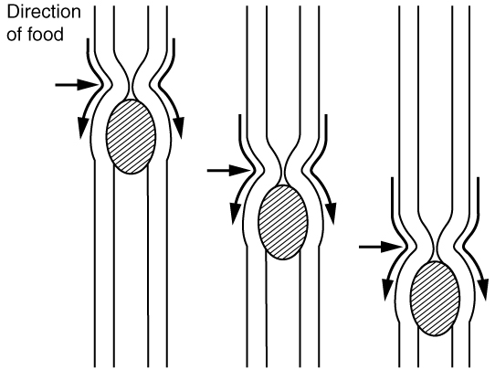
This image shows the peristaltic movement of food. In the left image, the food bolus is towards the top of the esophagus and arrows pointing downward show the direction of movement of the peristaltic wave. In the center image, the food bolus and the wave movement are closer to the center of the esophagus and in the right image, the bolus and the wave are close to the bottom end of the esophagus.
Figure 23.5 — 23.2 Digestive System Processes and Regulation

Figure 23.5 Peristalsis Peristalsis moves food through the digestive tract with alternating waves of muscle contraction and relaxation.

J. Gordon Betts and colleagues, Anatomy And Physiology 2E, OpenStax. · CC BY-NC-SA 4.0 · Original image ↗ · Access for free ↗
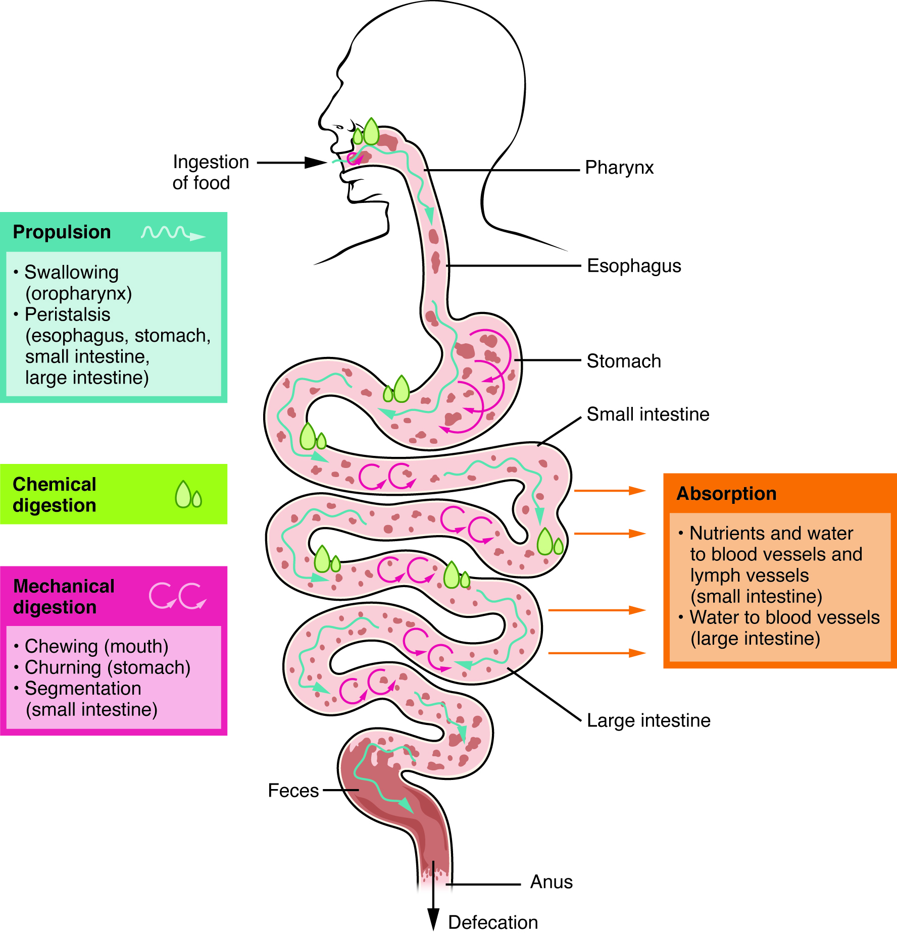
This image shows the different processes involved in digestion. The image shows how food travels from the mouth through the major organs. Associated textboxes list the different processes such as propulsion, chemical and mechanical digestion and absorption near the organs where they take place.
Figure 23.6 — 23.2 Digestive System Processes and Regulation

Figure 23.6 Digestive Processes The digestive processes are ingestion, propulsion, mechanical digestion, chemical digestion, absorption, and defecation.

J. Gordon Betts and colleagues, Anatomy And Physiology 2E, OpenStax. · CC BY-NC-SA 4.0 · Original image ↗ · Access for free ↗
Organ Major functions Other functions
Mouth
  • Ingests food
  • Chews and mixes food
  • Begins chemical breakdown of
  • Moves food into the pharynx
  • Begins breakdown of via lingual
  • Moistens and dissolves food, allowing you to taste it
  • Cleans and lubricates the teeth and oral cavity
  • Has some antimicrobial activity
Pharynx
  • Propels food from the oral cavity to the esophagus
  • Lubricates food and passageways
Esophagus
  • Propels food to the stomach
  • Lubricates food and passageways
Stomach
  • Mixes and churns food with gastric juices to form
  • Begins chemical breakdown of
  • Releases food into the duodenum as
  • Absorbs some fat- substances (for example, alcohol, aspirin)
  • Possesses antimicrobial functions
Small intestine
  • Provides optimal medium for enzymatic activity
Accessory organs
  • -rich pancreatic juices help neutralize and provide optimal environment for enzymatic activity
Large intestine
  • Further breaks down food
  • Absorbs most residual water, electrolytes, and vitamins produced by enteric
  • Propels feces toward rectum
  • Eliminates feces
  • Food is concentrated and temporarily stored prior to defecation
  • Mucus eases passage of feces through colon

Table 23.3. J. Gordon Betts and colleagues, Anatomy And Physiology 2E, OpenStax.

Source: 23.2 Digestive System Processes and Regulation

23.4 The Stomach — figures and tables

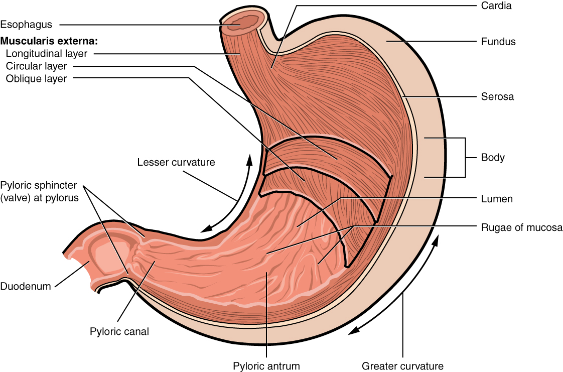
This image shows a cross-section of the stomach, and the major parts: the cardia, fundus, body and pylorus are labeled.
Figure 23.15 — 23.4 The Stomach

Figure 23.15 Stomach The stomach has four major regions: the cardia, fundus, body, and pylorus. The addition of an inner oblique smooth muscle layer gives the muscularis the ability to vigorously churn and mix food.

J. Gordon Betts and colleagues, Anatomy And Physiology 2E, OpenStax. · CC BY-NC-SA 4.0 · Original image ↗ · Access for free ↗
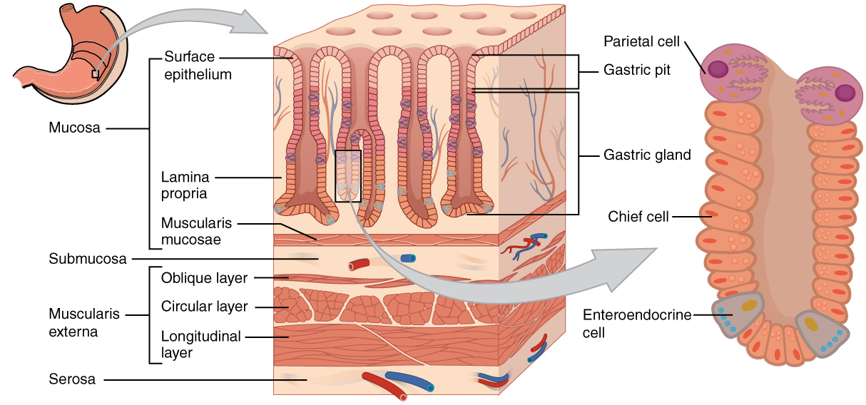
This diagram shows the histological cross-section of the stomach. The left panel shows the stomach and the center panel shows a magnified view of a small region including the epithelium and the gastric glands. The right panel shows a further magnification of the mucosa and the different cell types are labeled.
Figure 23.16 — 23.4 The Stomach

Figure 23.16 Histology of the Stomach The stomach wall is adapted for the functions of the stomach. In the epithelium, gastric pits lead to gastric glands that secrete gastric juice. The gastric glands (one gland is shown enlarged on the right) contain different types of cells that secrete a variety of enzymes, including hydrochloride acid, which activates the protein-digesting enzyme pepsin.

J. Gordon Betts and colleagues, Anatomy And Physiology 2E, OpenStax. · CC BY-NC-SA 4.0 · Original image ↗ · Access for free ↗
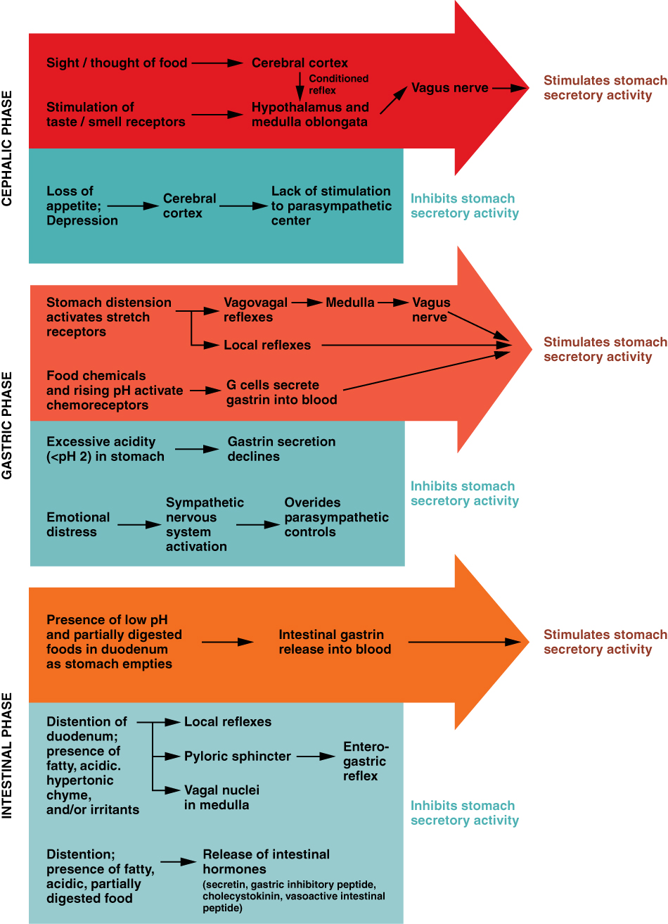
This flowchart shows the three different phases of gastric secretion. The top panel shows the cephalic phase, the middle panel shows the gastric phase and the bottom panel shows the intestinal phase.
Figure 23.17 — 23.4 The Stomach

Figure 23.17 The Three Phases of Gastric Secretion Gastric secretion occurs in three phases: cephalic, gastric, and intestinal. During each phase, the secretion of gastric juice can be stimulated or inhibited.

J. Gordon Betts and colleagues, Anatomy And Physiology 2E, OpenStax. · CC BY-NC-SA 4.0 · Original image ↗ · Access for free ↗
Production site Production stimulus Target organ Action
Stomach mucosa, mainly G cells of the pyloric antrum Presence of and in stomach Stomach Increases by gastric glands; promotes gastric emptying
Stomach mucosa, mainly G cells of the pyloric antrum Presence of and in stomach Small intestine Promotes intestinal muscle contraction
Stomach mucosa, mainly G cells of the pyloric antrum Presence of and in stomach Ileocecal valve Relaxes valve
Stomach mucosa, mainly G cells of the pyloric antrum Presence of and in stomach Large intestine Triggers mass movements
Stomach mucosa, mainly fundus Fasting state (levels increase just prior to meals) Regulates food intake, primarily by stimulating hunger and
Histamine Stomach mucosa Presence of food in the stomach Stomach Stimulates to release HCl
Serotonin Stomach mucosa Presence of food in the stomach Stomach Contracts stomach muscle
Somatostatin Mucosa of stomach, especially pyloric antrum; also duodenum Presence of food in the stomach; sympathetic stimulation Stomach Restricts all gastric secretions, gastric motility, and emptying
Somatostatin Mucosa of stomach, especially pyloric antrum; also duodenum Presence of food in the stomach; sympathetic stimulation Restricts pancreatic secretions
Somatostatin Mucosa of stomach, especially pyloric antrum; also duodenum Presence of food in the stomach; sympathetic stimulation Small intestine Reduces intestinal absorption by reducing blood flow

Table 23.6. J. Gordon Betts and colleagues, Anatomy And Physiology 2E, OpenStax.

Source: 23.4 The Stomach

23.6 Accessory Organs in Digestion: The Liver, , and Gallbladder — figures and tables

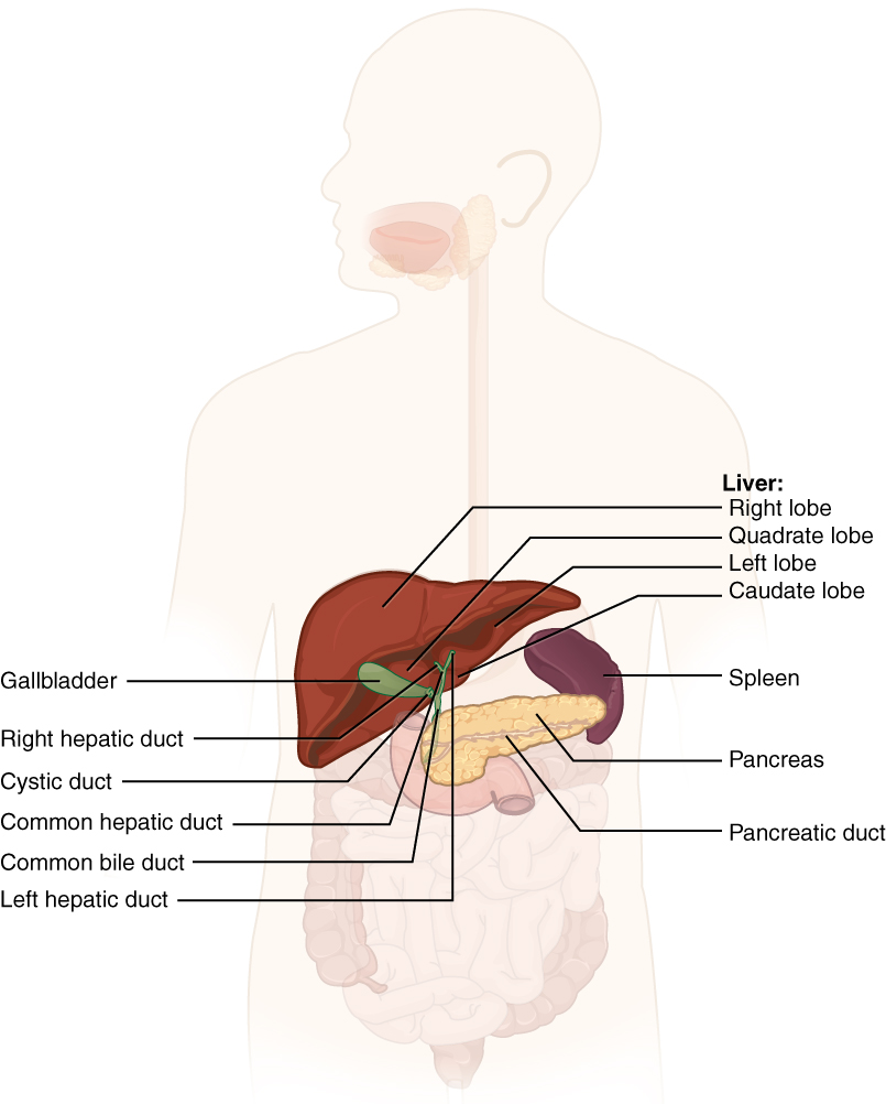
This diagram shows the accessory organs of the digestive system. The liver, spleen, pancreas, gallbladder and their major parts are shown.
Figure 23.24 — 23.6 Accessory Organs in Digestion: The Liver, , and Gallbladder

Figure 23.24 Accessory Organs The liver, pancreas, and gallbladder are considered accessory digestive organs, but their roles in the digestive system are vital.

J. Gordon Betts and colleagues, Anatomy And Physiology 2E, OpenStax. · CC BY-NC-SA 4.0 · Original image ↗ · Access for free ↗
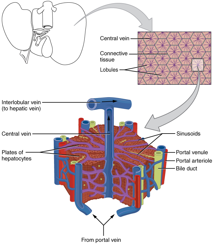
This image shows the microscopic anatomy of the liver. The top panel shows the liver; the center panel shows a magnified view of the connective tissue and the lobules. The bottom panel shows a further magnified view of a lobule, identifying the veins, bile duct and the sinusoids.
Figure 23.25 — 23.6 Accessory Organs in Digestion: The Liver, , and Gallbladder

Figure 23.25 Microscopic Anatomy of the Liver The liver receives oxygenated blood from the hepatic artery and nutrient-rich deoxygenated blood from the hepatic portal vein.

J. Gordon Betts and colleagues, Anatomy And Physiology 2E, OpenStax. · CC BY-NC-SA 4.0 · Original image ↗ · Access for free ↗
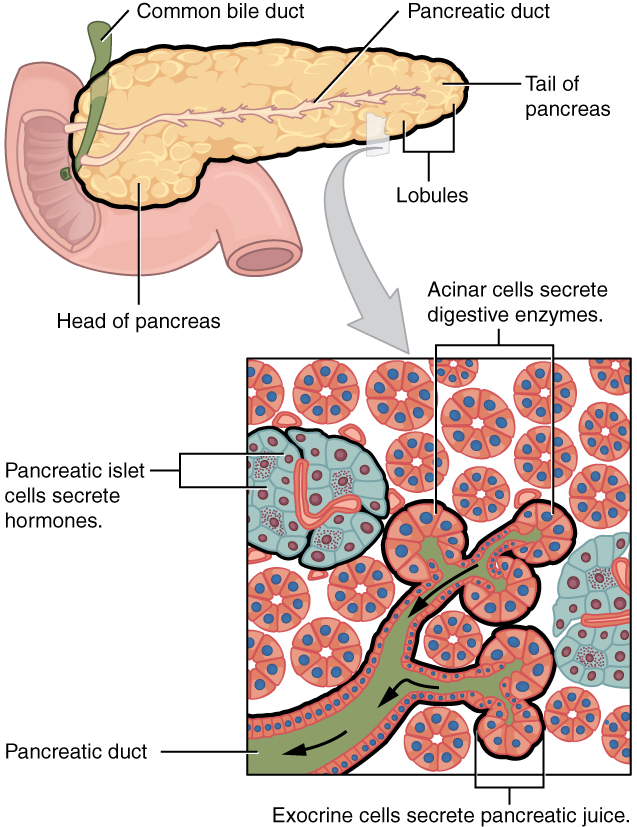
This figure shows the pancreas and its major parts. A magnified view of a small region of the pancreas shows the pancreatic islet cells, the acinar cells and the pancreatic duct.
Figure 23.26 — 23.6 Accessory Organs in Digestion: The Liver, , and Gallbladder

Figure 23.26 Exocrine and Endocrine Pancreas The pancreas has a head, a body, and a tail. It delivers pancreatic juice to the duodenum through the pancreatic duct.

J. Gordon Betts and colleagues, Anatomy And Physiology 2E, OpenStax. · CC BY-NC-SA 4.0 · Original image ↗ · Access for free ↗
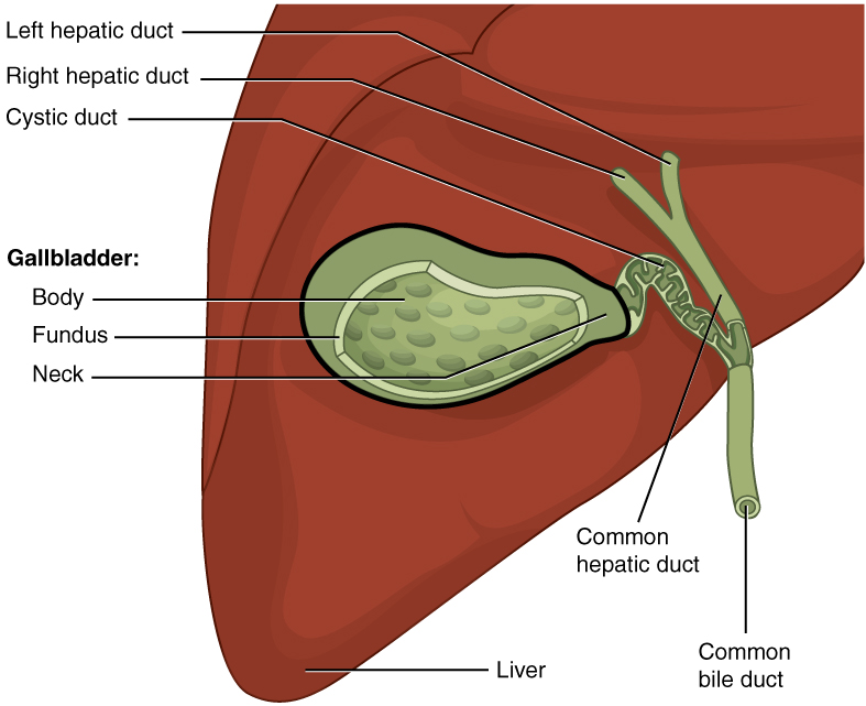
This figure shows the gallbladder and its major parts are labeled.
Figure 23.27 — 23.6 Accessory Organs in Digestion: The Liver, , and Gallbladder

Figure 23.27 Gallbladder The gallbladder stores and concentrates bile, and releases it into the two-way cystic duct when it is needed by the small intestine.

J. Gordon Betts and colleagues, Anatomy And Physiology 2E, OpenStax. · CC BY-NC-SA 4.0 · Original image ↗ · Access for free ↗

Source: 23.6 Accessory Organs in Digestion: The Liver, Pancreas, and Gallbladder

23.7 Chemical Digestion and Absorption: A Closer Look — figures and tables
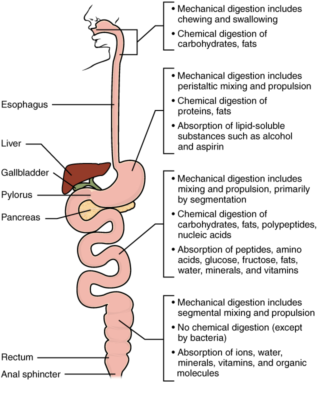
This diagram identifies the functions of mechanical and chemical digestion and absorption at each organ. Next to each organ, a callout identifies which steps of digestion take place in that particular organ.
Figure 23.28 — 23.7 Chemical Digestion and Absorption: A Closer Look

Figure 23.28 Digestion and Absorption Digestion begins in the mouth and continues as food travels through the small intestine. Most absorption occurs in the small intestine.

J. Gordon Betts and colleagues, Anatomy And Physiology 2E, OpenStax. · CC BY-NC-SA 4.0 · Original image ↗ · Access for free ↗
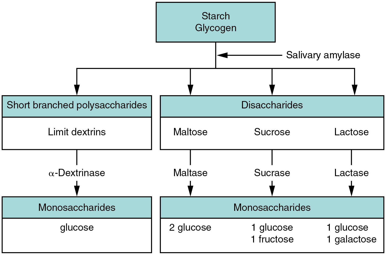
This flow chart shows the steps in digestion of carbohydrates. The different levels shown are starch and glycogen, disaccharides and monosaccharides. Under each type of sugar, examples and the enzymes responsible for digestion are listed.
Figure 23.29 — 23.7 Chemical Digestion and Absorption: A Closer Look

Figure 23.29 Carbohydrate Digestion Flow Chart Carbohydrates are broken down into their monomers in a series of steps.

J. Gordon Betts and colleagues, Anatomy And Physiology 2E, OpenStax. · CC BY-NC-SA 4.0 · Original image ↗ · Access for free ↗
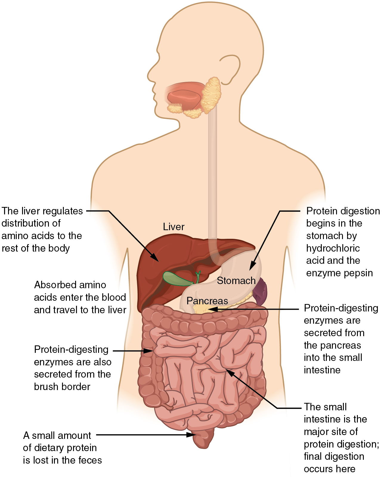
This diagrams shows the human digestive system and identifies the role of each organ in protein digestion. A text call-out next to each organ details the specific function.
Figure 23.30 — 23.7 Chemical Digestion and Absorption: A Closer Look

Figure 23.30 Digestion of Protein The digestion of protein begins in the stomach and is completed in the small intestine.

J. Gordon Betts and colleagues, Anatomy And Physiology 2E, OpenStax. · CC BY-NC-SA 4.0 · Original image ↗ · Access for free ↗
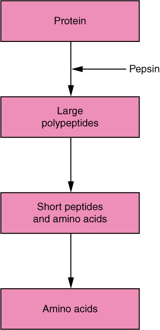
This flow chart shows the different steps in the digestion of protein. The four steps shown are protein, large polypeptides, short peptides and amino acids and amino acids.
Figure 23.31 — 23.7 Chemical Digestion and Absorption: A Closer Look

Figure 23.31 Digestion of Protein Flow Chart Proteins are successively broken down into their amino acid components.

J. Gordon Betts and colleagues, Anatomy And Physiology 2E, OpenStax. · CC BY-NC-SA 4.0 · Original image ↗ · Access for free ↗
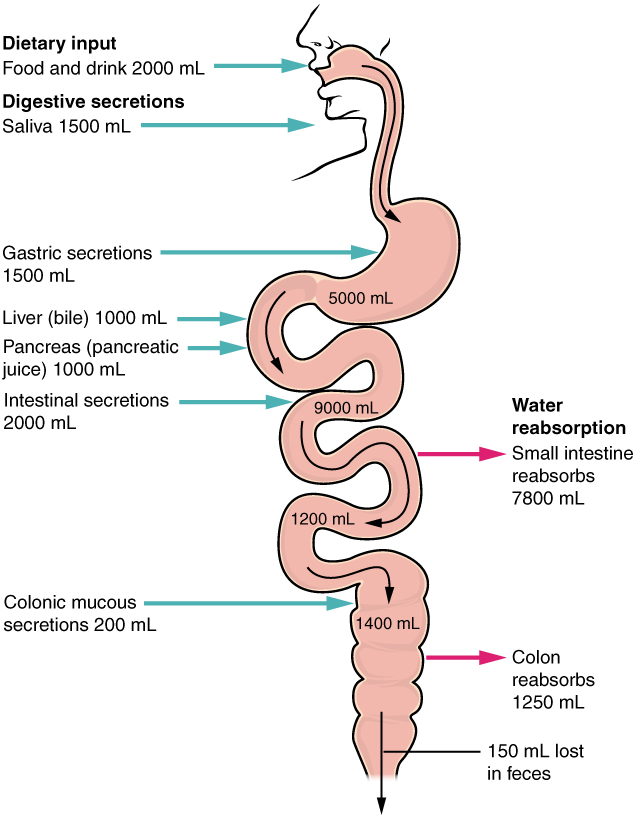
This image shows the human digestive system. Next to each organ, a text callout identifies how water and digestive secretions such as saliva and bile are processed.
Figure 23.32 — 23.7 Chemical Digestion and Absorption: A Closer Look

Figure 23.32 Digestive Secretions and Absorption of Water Absorption is a complex process, in which nutrients from digested food are harvested.

J. Gordon Betts and colleagues, Anatomy And Physiology 2E, OpenStax. · CC BY-NC-SA 4.0 · Original image ↗ · Access for free ↗
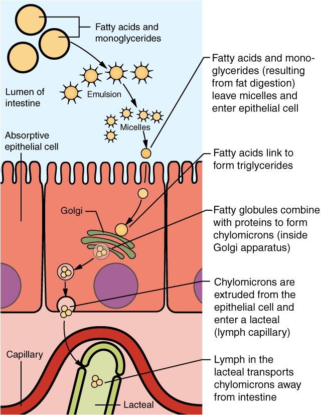
This diagram shows how lipids are absorbed from the lumen of the intestine into the lacteals. The fatty acid micelles are shown to enter the epithelial cell and form chylomicrons inside the Golgi apparatus. Then, the chylomicrons are extruded from the epithelial cell and are taken up by the lacteals.
Figure 23.33 — 23.7 Chemical Digestion and Absorption: A Closer Look

Figure 23.33 Lipid Absorption Unlike amino acids and simple sugars, lipids are transformed as they are absorbed through epithelial cells.

J. Gordon Betts and colleagues, Anatomy And Physiology 2E, OpenStax. · CC BY-NC-SA 4.0 · Original image ↗ · Access for free ↗
Category Name Source
Salivary Lingual Lingual glands Free , and mono- and diglycerides
Salivary Salivary glands Polysaccharides Disaccharides and trisaccharides
Gastric Gastric and monoacylglycerides
Gastric *
α-Dextrinase Small intestine α-Dextrins
Small intestine
Lactase Small intestine and
Maltase Small intestine Maltose
Nucleosidases and Small intestine Phosphates, , and
Peptidases Small intestine
Sucrase Small intestine Sucrose and fructose
Pancreatic Carboxy-peptidase* Pancreatic acinar cells at the carboxyl end of and
Pancreatic Chymotrypsin* Pancreatic acinar cells
Pancreatic Elastase* Pancreatic acinar cells
Pancreatic Pancreatic acinar cells
  • Ribonuclease: ribonucleic
  • Deoxyribonuclease: deoxyribonucleic
Pancreatic Pancreatic Pancreatic acinar cells Polysaccharides (starches) α-Dextrins, disaccharides (maltose), trisaccharides (maltotriose)
Pancreatic Pancreatic Pancreatic acinar cells that have been emulsified by and monoacylglycerides
Pancreatic * Pancreatic acinar cells

Table 23.8 *These enzymes have been activated by other substances.. J. Gordon Betts and colleagues, Anatomy And Physiology 2E, OpenStax.

Source Substance
Monosaccharides: , , and fructose
Single , dipeptides, and tripeptides
Monoacylglycerides, , and free
sugars, phosphates, and

Table 23.9. J. Gordon Betts and colleagues, Anatomy And Physiology 2E, OpenStax.

Food Breakdown Absorption mechanism Entry to bloodstream Destination
Co-transport with sodium Capillary blood in Liver via hepatic portal vein
Fructose Capillary blood in Liver via hepatic portal vein
Co-transport with sodium Capillary blood in Liver via hepatic portal vein
Long-chain into intestinal cells, where they are combined with to create of Systemic circulation via entering thoracic duct
Monoacylglycerides into intestinal cells, where they are combined with to create of Systemic circulation via entering thoracic duct
Short-chain Simple Capillary blood in Liver via hepatic portal vein
Simple Capillary blood in Liver via hepatic portal vein
digestion Active transport via carriers Capillary blood in Liver via hepatic portal vein

Table 23.10. J. Gordon Betts and colleagues, Anatomy And Physiology 2E, OpenStax.

Source: 23.7 Chemical Digestion and Absorption: A Closer Look

283 terms, alphabetically ordered

acceleration

Rate of change of , measured in m/s².

Read explanation · OpenStax ↗
Acid · ·

In the Brønsted–Lowry model, a substance that can donate a . Acidic have a higher activity than water.

Read explanation · OpenStax ↗
action potential

A regenerative -voltage change produced by voltage-dependent -channel activity.

Read explanation · OpenStax ↗
Activation energy ·

The energy barrier that must overcome to reach a and undergo .

Read explanation · OpenStax ↗
Active catalytic-site concentration ·

The total of active sites used to calculate from . It equals active when each has one site.

Read explanation · Biology LibreTexts ↗
Active site

The region of an that binds its and carries out .

Read explanation · OpenStax ↗
adaptation

A heritable feature shaped by selection because it improves reproductive success in a relevant environment.

Read explanation · OpenStax ↗
Adenosine triphosphate

An energy-carrying ; its powers many cellular processes, including cycles.

Read explanation · OpenStax ↗
adenylyl cyclase

An converting to in cell signaling.

Read explanation · OpenStax ↗
ADH ·

Antidiuretic hormone, or vasopressin, synthesized in hypothalamic and released from the posterior pituitary to promote renal water conservation.

Read explanation · OpenStax ↗
Allele

One version or copy of a at a ; a typical cell carries two copies of an autosomal .

Read explanation · NCBI Bookshelf / Sinauer Associates ↗
Allolactose

A isomer produced by that binds and relieves lac repression.

Read explanation · OpenStax ↗
Alpha carbon · ·

The central carbon next to the carboxyl carbon. In a standard it binds an , hydrogen, and .

Read explanation · OpenStax ↗
Alpha helix · · · · · ·

A helical stabilized by between a oxygen and an hydrogen four later.

Read explanation · OpenStax ↗
Amide · · ·

A group containing a carbon bonded to nitrogen. are amide linkages.

Read explanation · OpenStax ↗
Amino acid

A building block containing amino and and a characteristic .

Read explanation · OpenStax ↗
Amino acid residue · ·

The part of an incorporated into a chain. Residue can also describe another building block within a larger .

Read explanation · OpenStax ↗
Amino group · · ·

A nitrogen-containing . A free ’s amino group is typically (NH₃⁺) near physiological ; ’s ring nitrogen is an exception to this formula.

Read explanation · OpenStax ↗
Amphipathic

Having both a region and a region, as in a or .

Read explanation · OpenStax ↗
Amylase

An that hydrolyzes certain in to produce smaller .

Read explanation · OpenStax ↗
Amylose

The largely unbranched component of , made mainly of linked by alpha-1,4 .

Read explanation · OpenStax ↗
Anion ·

An with a negative net .

Read explanation · OpenStax ↗
anomeric carbon

The former carbon of a sugar, which becomes the or hemiketal center upon cyclization.

Read explanation · OpenStax ↗
Antiparallel

Running in opposite directions, as with two strands or neighboring strands in some .

Read explanation · OpenStax ↗
apical

Referring to the epithelial surface facing a or external space.

Read explanation · OpenStax ↗
Aqueous solution ·

A in which water is the .

Read explanation · OpenStax ↗
Archaea ·

A of distinct from , with characteristic chemistry and -expression machinery.

Read explanation · OpenStax ↗
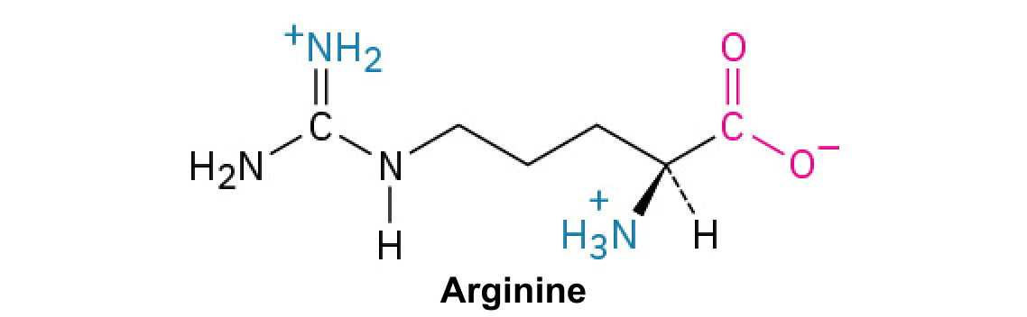
Arginine ·

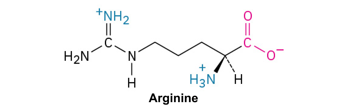
A with a , usually positively near physiological .

Read explanation · OpenStax ↗
Atom

The smallest unit of an element that retains its chemical identity; it has a surrounded by .

Read explanation · OpenStax ↗
axon

Neuronal process that usually conducts toward terminals.

Read explanation · OpenStax ↗
Bacteria ·

A of single-celled . Bacterial cells lack a -bound and are distinct from .

Read explanation · OpenStax ↗
Beta sheet · · · · · ·

A made of aligned segments connected by ; strands may be parallel or .

Read explanation · OpenStax ↗
bicarbonate ·

The HCO3− , a major extracellular component and form of carbon dioxide transport.

Read explanation · OpenStax ↗
bile salt

An -derived component of bile that supports fat emulsification and micelle formation.

Read explanation · OpenStax ↗
bilirubin

A heme-breakdown pigment processed by the liver and excreted through bile.

Read explanation · OpenStax ↗
Biomolecule

A made by or involved in living systems, including , , , and .

Read explanation · OpenStax ↗
Blotting ·

Transfer of separated or to a so a specific target can be detected.

Read explanation · Thermo Fisher ↗
brush border

The dense microvillar surface of absorptive epithelial cells, containing transporters and digestive .

Read explanation · OpenStax ↗
Brønsted–Lowry base · · · · · · · · · · ·

In the Brønsted–Lowry model, a substance that can accept a . Basic solutions have lower activity than water.

Read explanation · OpenStax ↗
Buffer ·

A that resists changes when small amounts of or are added, commonly through a weak and its .

Read explanation · OpenStax ↗
Cancer

A group of diseases involving abnormal cells that can proliferate without normal restraints and invade tissues.

Read explanation · OpenStax ↗
Carbohydrate

A class of including sugars and their , used for energy storage, structure, and recognition.

Read explanation · OpenStax ↗
Carbonyl group · ·

A carbon double-bonded to oxygen, C=O; present in and many other .

Read explanation · OpenStax ↗
Carboxyl group · ·

An , COOH. In free it is usually to COO⁻ near physiological .

Read explanation · OpenStax ↗
Catalyst · ·

A substance that speeds a by providing a pathway with lower and is regenerated rather than consumed overall.

Read explanation · OpenStax ↗
Cation ·

An with a positive net .

Read explanation · OpenStax ↗
Cellulose

A structural in plant cell walls, with beta-1,4 linkages; also used as a support.

Read explanation · OpenStax ↗
Centimeter

A unit of length equal to one hundredth of a meter.

Read explanation · NIST ↗
central nervous system

Brain and spinal cord.

Read explanation · OpenStax ↗
Centrifugation ·

Spinning a sample to accelerate separation of particles according to their behavior.

Read explanation · Beckman Coulter ↗
Chemical compound ·

A substance containing of two or more different chemically combined in a definite composition.

Read explanation · OpenStax ↗
Chemical element

A substance defined by having the same number of , such as carbon or oxygen.

Read explanation · OpenStax ↗
Chemical reaction ·

A process that rearranges and chemical bonds, converting into .

Read explanation · OpenStax ↗
chemoreceptor

A sensory responding to chemical conditions, including oxygen, carbon dioxide, or .

Read explanation · OpenStax ↗
chief cell

A gastric cell secreting and selected digestive .

Read explanation · OpenStax ↗
cholecystokinin

An intestinal promoting pancreatic and gallbladder contraction while influencing gastric emptying.

Read explanation · OpenStax ↗
cholesterol

An animal that modulates packing and serves as a precursor for and bile .

Read explanation · OpenStax ↗
Chromatin

The complex of and associated , including , in .

Read explanation · OpenStax ↗
Chromatography

Separation of mixture components by their different interactions with a and a moving .

Read explanation · Waters ↗
Chromatography resin ·

The solid or beads in a column, often carrying chemical groups that determine separation behavior.

Read explanation · Cytiva ↗
Chromosome ·

An organized with associated that carries . A replicated chromosome has two .

Read explanation · OpenStax ↗
chylomicron

An intestinal lipoprotein particle carrying dietary and other into and blood.

Read explanation · Biology LibreTexts / Oregon State University ↗
chyme

The partly digested mixture of food and gastric secretions entering the small intestine.

Read explanation · OpenStax ↗
Codon

A sequence of three that specifies an or a stop signal.

Read explanation · OpenStax ↗
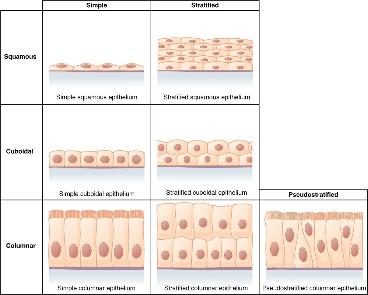
columnar

Describing epithelial cells taller than they are wide.

Read explanation · OpenStax ↗
Concentration

The amount of a substance in a specified volume or mass of mixture.

Read explanation · OpenStax ↗
conjugate base

Species formed when an loses one .

Read explanation · OpenStax ↗
Copy number ·

The number of copies of a segment in a defined sample or ; it is distinct from or abundance.

Read explanation · NHGRI / NIH ↗
Covalent bond · ·

A bond in which share pairs. and are covalent.

Read explanation · OpenStax ↗
culture

Learned and shared meanings, practices, values, and artifacts.

Read explanation · OpenStax ↗
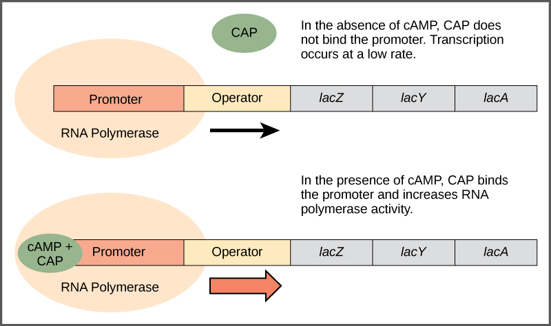
Cyclic AMP ·

A signal. In the simplified E. coli lac model, low raises cAMP, which activates CAP.

Read explanation · OpenStax ↗
cyclic nucleotide

whose forms a cyclic within its sugar, such as .

Read explanation · OpenStax ↗
Cysteine ·

An with a . Two cysteines can form a .

Read explanation · OpenStax ↗
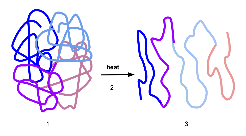
Denaturation · ·

Loss of a ’s , often with . This usually preserves the chain’s .

Read explanation · NCBI Bookshelf ↗
Density gradient

A fluid whose density varies with position, used to improve separation during .

Read explanation · Beckman Coulter ↗
Deoxyribonucleic acid

The that stores genetic information used to specify and sequences.

Read explanation · OpenStax ↗
Deoxyribose

The five-carbon sugar in ; its 2′ carbon bears H instead of the found in .

Read explanation · OpenStax ↗
Deprotonation

Removal of a from a or group; this changes its by minus one.

Read explanation · OpenStax ↗
Detergent

An substance that can associate with both water and material; detergents can disrupt or interactions.

Read explanation · Thermo Fisher ↗
Diffusion

Net spreading of particles down a due to random molecular motion.

Read explanation · OpenStax ↗
Diploid

Having two sets; comparisons of must account for and sample composition.

Read explanation · NHGRI / NIH ↗
Dissociation · ·

Separation of a chemical species or molecular complex into smaller components; dissociation releases a .

Read explanation · OpenStax ↗
Disulfide bond · · ·

A covalent sulfur–sulfur bond formed by of two , which can stabilize structure.

Read explanation · NCBI Bookshelf ↗
DNA replication

Copying so genetic information can be passed to daughter cells. Each guides a complementary new strand.

Read explanation · OpenStax ↗
dominant

Describing an whose specified is expressed in a ; dominance does not imply greater frequency or .

Read explanation · OpenStax ↗
Electric charge · · ·

An electrical property that can be positive or negative. Opposite charges attract and like charges repel; net charge is the sum of all charges on a particle.

Read explanation · OpenStax ↗
electrochemical gradient

The combined and electrical driving forces acting on an across space or a .

Read explanation · OpenStax ↗
Electron

A negatively subatomic particle. Electron sharing and transfer determine much of chemical bonding.

Read explanation · OpenStax ↗
Electronegativity ·

An ’s tendency to attract shared in a chemical bond. Unequal attraction can make a bond .

Read explanation · OpenStax ↗
Endocrine signaling ·

Communication in which enter the circulation and act on distant target cells.

Read explanation · OpenStax ↗
endocytosis

Uptake of extracellular material or surface through inward budding of .

Read explanation · OpenStax ↗
Endomembrane system

The interacting and compartments involved in processes such as modification, transport, and .

Read explanation · OpenStax ↗
Endoplasmic reticulum

An interconnected network continuous with the ; it includes regions involved in processing and synthesis.

Read explanation · OpenStax ↗
endothelium

The cellular lining of blood and lymphatic vessels, involved in barrier and signaling functions.

Read explanation · OpenStax ↗
enteric nervous system

Neural networks in the gastrointestinal wall coordinating motility, , and local responses.

Read explanation · OpenStax ↗
enterocyte

An absorptive intestinal epithelial cell.

Read explanation · OpenStax ↗
enteropeptidase

A small-intestinal brush-border activating to .

Read explanation · OpenStax ↗
Enzyme

A biological that speeds a without being consumed. Most enzymes are .

Read explanation · OpenStax ↗
Enzyme saturation · ·

The high- limit in which sites spend almost all their time processing and the rate approaches .

Read explanation · Biology LibreTexts ↗
epithelium ·

A polarized sheet of closely connected cells that covers surfaces, lines cavities, or forms glands.

Read explanation · OpenStax ↗
Eukaryote ·

An organism whose cells have a -enclosed ; animals, plants, fungi, and protists are eukaryotes.

Read explanation · OpenStax ↗
exocrine

Describing through ducts onto an epithelial surface or into a .

Read explanation · OpenStax ↗
Experimental control ·

A comparison sample or condition used to distinguish the effect being tested from other influences.

Read explanation · Thermo Fisher ↗
facilitated diffusion

Passive movement down an through a selective channel or .

Read explanation · OpenStax ↗
FACT complex ·

A -associated complex that helps machinery traverse and supports reorganization.

Read explanation · OpenStax ↗
Fatty acid

A attached to a chain; fatty acids are components of many .

Read explanation · OpenStax ↗
fitness

Relative reproductive contribution of a or organism in a particular environment.

Read explanation · OpenStax ↗
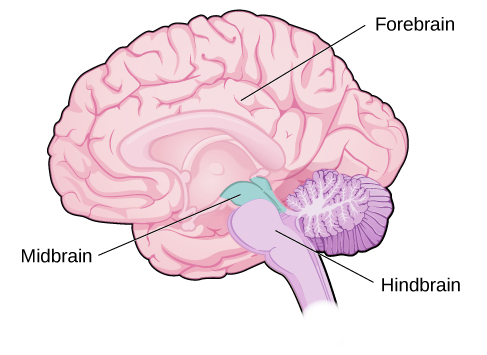
forebrain

Major developmental brain division including cerebral hemispheres, , and .

Read explanation · OpenStax ↗
Fraction · ·

A portion of a sample collected during a separation, such as the fluid leaving a column during a time interval.

Read explanation · Waters ↗
Functional group

A group of whose chemical behavior gives a characteristic and properties.

Read explanation · OpenStax ↗
Galactose

A six-carbon monosaccharide released along with when is hydrolyzed.

Read explanation · OpenStax ↗
gastrin

A released mainly from gastric G cells that promotes gastric and other gastric functions.

Read explanation · OpenStax ↗
Gene

A sequence that contributes to a functional or , together with the information needed to express it.

Read explanation · OpenStax ↗
Gene expression

Production of a functional from a , including and, for -coding , .

Read explanation · NCBI Bookshelf ↗
Genome

The complete genetic material of an organism or cell.

Read explanation · OpenStax ↗
genotype

The allelic composition of an organism at one or more specified .

Read explanation · OpenStax ↗
ghrelin

A associated with hunger signaling, produced mainly by the stomach.

Read explanation · OpenStax ↗
Gi

A heterotrimeric G- family whose activated α commonly inhibits .

Read explanation · OpenStax ↗
Glucagon

A pancreatic that helps raise , especially by promoting release from the liver.

Read explanation · OpenStax ↗
Glucose

A six-carbon sugar that is a major cellular energy source and a building block of , , and .

Read explanation · OpenStax ↗
Glycerol

A three-carbon alcohol with three ; used in sample to increase density and in some .

Read explanation · OpenStax ↗
glycogen

A highly branched used for storage in animals.

Read explanation · Biology LibreTexts / Oregon State University ↗
Glycosidic bond

A involving a sugar’s . In a , an N-glycosidic bond links the sugar to the .

Read explanation · Biology LibreTexts / Oregon State University ↗
Golgi apparatus ·

A stack of compartments that modifies and sorts and traveling through the .

Read explanation · OpenStax ↗
Gradient

A change in a quantity, such as , , or density, across a distance or over time.

Read explanation · Thermo Fisher ↗
Guanidinium group · ·

The positively form of a guanidine group, with distributed over three nitrogens; found in ’s .

Read explanation · OpenStax ↗
hemiacetal

Carbon bonded to both and alkoxy groups, usually formed by alcohol addition to a .

Read explanation · OpenStax ↗
Hemoglobin

The oxygen-carrying in , composed of four with heme groups.

Read explanation · OpenStax ↗
hepatic portal circulation

The vascular route carrying blood from much of the digestive tract to liver before return to systemic circulation.

Read explanation · OpenStax ↗
heterozygote ·

An individual with two different at a specified .

Read explanation · OpenStax ↗
Histone

A -associated rich in . Core histones form the around which nucleosomal wraps.

Read explanation · OpenStax ↗
Hormone

A chemical messenger released by cells that changes the activity of target cells with matching .

Read explanation · OpenStax ↗
Hydrocarbon · ·

A made only of carbon and hydrogen. Hydrocarbon regions are generally .

Read explanation · OpenStax ↗
Hydrogen bond ·

An attraction between a hydrogen bonded to an and another . hydrogen bonds stabilize .

Read explanation · OpenStax ↗
Hydrogen ion · ·

A positively hydrogen species. H⁺ denotes a , which is hydrated in water; its activity determines .

Read explanation · OpenStax ↗
Hydrolysis

Breaking a chemical bond by adding water. hydrolysis separates .

Read explanation · OpenStax ↗
Hydrophilic

Interacting favorably with water, typically because a group is or .

Read explanation · OpenStax ↗
Hydrophobic

Interacting poorly with water. often gather in a ’s interior.

Read explanation · OpenStax ↗
Hydroxide ion ·

The negatively OH⁻. Basic contain a higher hydroxide ion activity than water at the same temperature.

Read explanation · OpenStax ↗
Hydroxyl group ·

An oxygen bonded to hydrogen, –OH. Hydroxyl groups can and often increase .

Read explanation · OpenStax ↗
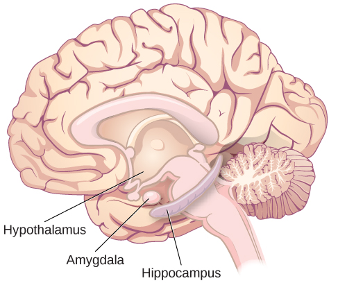
hypothalamus

region coordinating homeostatic, , and autonomic functions.

Read explanation · OpenStax ↗
Inducer

A signal that increases expression of a regulated ; does so by disabling the .

Read explanation · OpenStax ↗
inflammation

A coordinated tissue response recruiting defense and repair mechanisms, often involving increased blood flow and permeability.

Read explanation · OpenStax ↗
Insulin

A from that lowers by promoting uptake and storage and reducing production.

Read explanation · OpenStax ↗
interstitial fluid

Fluid occupying extracellular spaces between tissue cells.

Read explanation · OpenStax ↗
intrinsic factor

A gastric glycoprotein required for efficient vitamin B12 absorption in the terminal ileum.

Read explanation · OpenStax ↗
Ion · ·

An or with a net because its numbers of and do not balance.

Read explanation · OpenStax ↗
Lac operon

The E. coli containing , , and , controlled by repression and CAP– activation.

Read explanation · OpenStax ↗
Lac repressor

The encoded by lacI; it binds lac and represses until an reduces its binding.

Read explanation · OpenStax ↗
lacA

The encoding a galactoside .

Read explanation · OpenStax ↗
lacteal

An intestinal lymphatic capillary that receives absorbed -rich .

Read explanation · OpenStax ↗
Lactose

A disaccharide containing and ; its use by E. coli is supported by .

Read explanation · OpenStax ↗
Lactose permease

The transport that couples uptake to movement.

Read explanation · OpenStax ↗
lacY

The encoding , which transports into the cell.

Read explanation · OpenStax ↗
lacZ

The encoding , often used as an experimental .

Read explanation · OpenStax ↗
ligament

A connective-tissue structure commonly linking bones and stabilizing joints.

Read explanation · OpenStax ↗
linkage

A tendency of nearby on the same to be inherited together.

Read explanation · OpenStax ↗
Lipase

An that hydrolyzes ester bonds in , such as the bonds joining to .

Read explanation · OpenStax ↗
Lipid

A broad class of largely or , including fats, , and steroids.

Read explanation · OpenStax ↗
lipoprotein lipase

A capillary-associated releasing from circulating lipoprotein .

Read explanation · Biology LibreTexts / Oregon State University ↗
locus

The specific position of a or other feature.

Read explanation · OpenStax ↗
Logarithm · · ·

The exponent needed to obtain a number from a chosen fixed number. For decimal logarithms, log₁₀(100) = 2 because 10² = 100; uses the negative decimal logarithm of activity.

Read explanation · OpenStax ↗
Loss of function

or elimination of a ’s normal activity.

Read explanation · NCBI Bookshelf / Sinauer Associates ↗
Lumen · ·

The interior space of an or tube; the ER lumen is enclosed by the .

Read explanation · OpenStax ↗
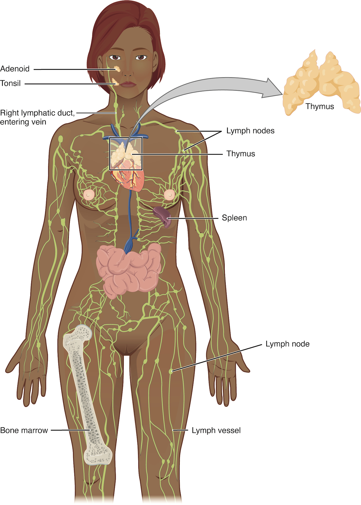
lymph

Fluid collected from tissue spaces and transported through lymphatic vessels back toward the bloodstream.

Read explanation · OpenStax ↗
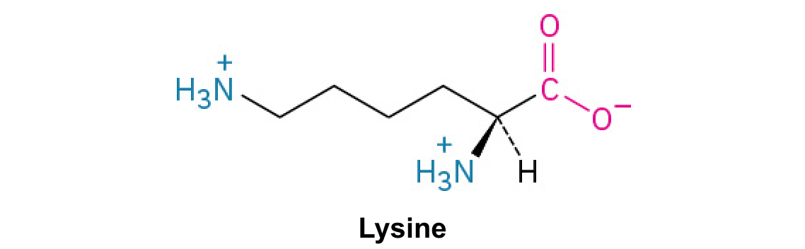
Lysine ·

A with a terminal , usually positively near physiological .

Read explanation · OpenStax ↗
Lysosome

An -enclosed compartment that uses hydrolytic to digest and recycle cellular material.

Read explanation · OpenStax ↗
Matrix

A supporting material, such as a gel or the solid support in a column. Its properties influence movement and binding of sample .

Read explanation · Thermo Fisher ↗
Maximum reaction rate · ·

The limiting as becomes saturating. For a fixed under given conditions, Vmax = times .

Read explanation · Biology LibreTexts ↗
Membrane · · · ·

A thin boundary or sheet. Cell membranes are bilayers containing ; membranes are synthetic supports that hold or for detection.

Read explanation · OpenStax ↗
Messenger RNA ·

An that carries coding information used by to synthesize a .

Read explanation · OpenStax ↗
Metabolism

The chemical that build , break them down, and transform energy in a living system.

Read explanation · OpenStax ↗
microvillus

A microscopic cell-surface projection increasing surface area, often for absorption.

Read explanation · OpenStax ↗
Mitochondrion ·

A with its own , central to aerobic production and several other processes.

Read explanation · OpenStax ↗
Mobile phase

The moving fluid that carries a sample through a system.

Read explanation · Waters ↗
Molecular chaperone · ·

A that assists other in folding or prevents , without specifying their sequence.

Read explanation · NCBI Bookshelf ↗
Molecule

Two or more joined by . A molecule may be electrically or carry a .

Read explanation · OpenStax ↗
Monomer

A small molecular building block that can join other monomers to form a .

Read explanation · OpenStax ↗
Native structure · · ·

A ’s functional folded arrangement under its normal conditions.

Read explanation · NCBI Bookshelf ↗
nephron

A kidney functional unit consisting of a filtration apparatus and associated tubular segments.

Read explanation · OpenStax ↗
neuron

An electrically excitable cell specialized for receiving and transmitting information.

Read explanation · OpenStax ↗
Neutral

Having no net . For a , neutral instead means equal and activities; its is about 7 at 25 °C.

Read explanation · OpenStax ↗
Nitrogenous base · · ·

A nitrogen-containing ring component of ; its pairing interactions help specify sequences.

Read explanation · OpenStax ↗
Nonpolar ·

Lacking a substantial separation of . Nonpolar groups generally interact poorly with water.

Read explanation · OpenStax ↗
Nuclear envelope

The double enclosing the and continuous with the .

Read explanation · OpenStax ↗
Nuclease

An that cleaves in or .

Read explanation · Biology LibreTexts / Oregon State University ↗
Nucleic acid

A of , such as or , used in information storage, expression, and other cellular functions.

Read explanation · OpenStax ↗
Nucleosome

The basic unit of : wrapped around a .

Read explanation · OpenStax ↗
Nucleotide

A joined to a sugar and one or more .

Read explanation · OpenStax ↗
Nucleus ·

A -enclosed compartment containing most of a cell’s .

Read explanation · OpenStax ↗
Octamer

A complex made of eight . A core contains eight .

Read explanation · OpenStax ↗
Operator · ·

A regulatory sequence bound by a , often overlapping or near a .

Read explanation · OpenStax ↗
Operon

A group of from a shared into one , with regulatory that coordinates their expression.

Read explanation · OpenStax ↗
Organelle ·

A specialized structure within a cell that carries out particular functions, such as a or .

Read explanation · OpenStax ↗
osmoreceptor

A sensory cell responding to osmotic conditions and helping regulate thirst and .

Read explanation · OpenStax ↗
Oxidation ·

Loss of or an increase in oxidation state. Oxidizing two can form a .

Read explanation · OpenStax ↗
Pancreas

An organ with digestive and functions; its islets contain cells that secrete and .

Read explanation · OpenStax ↗
Pancreatic beta cell ·

An cell in a pancreatic islet that secretes .

Read explanation · OpenStax ↗
parietal cell

A gastric cell secreting hydrochloric and .

Read explanation · OpenStax ↗
Pentose

A sugar with five carbon , such as or .

Read explanation · OpenStax ↗
Pepsin

A stomach that functions in conditions and breaks into smaller .

Read explanation · OpenStax ↗
pepsinogen

The inactive precursor of gastric .

Read explanation · OpenStax ↗
Peptide

A chain of joined by ; the term commonly describes chains shorter than a typical .

Read explanation · OpenStax ↗
Peptide bond ·

A covalent between the carboxyl carbon of one and the amino nitrogen of the next.

Read explanation · OpenStax ↗
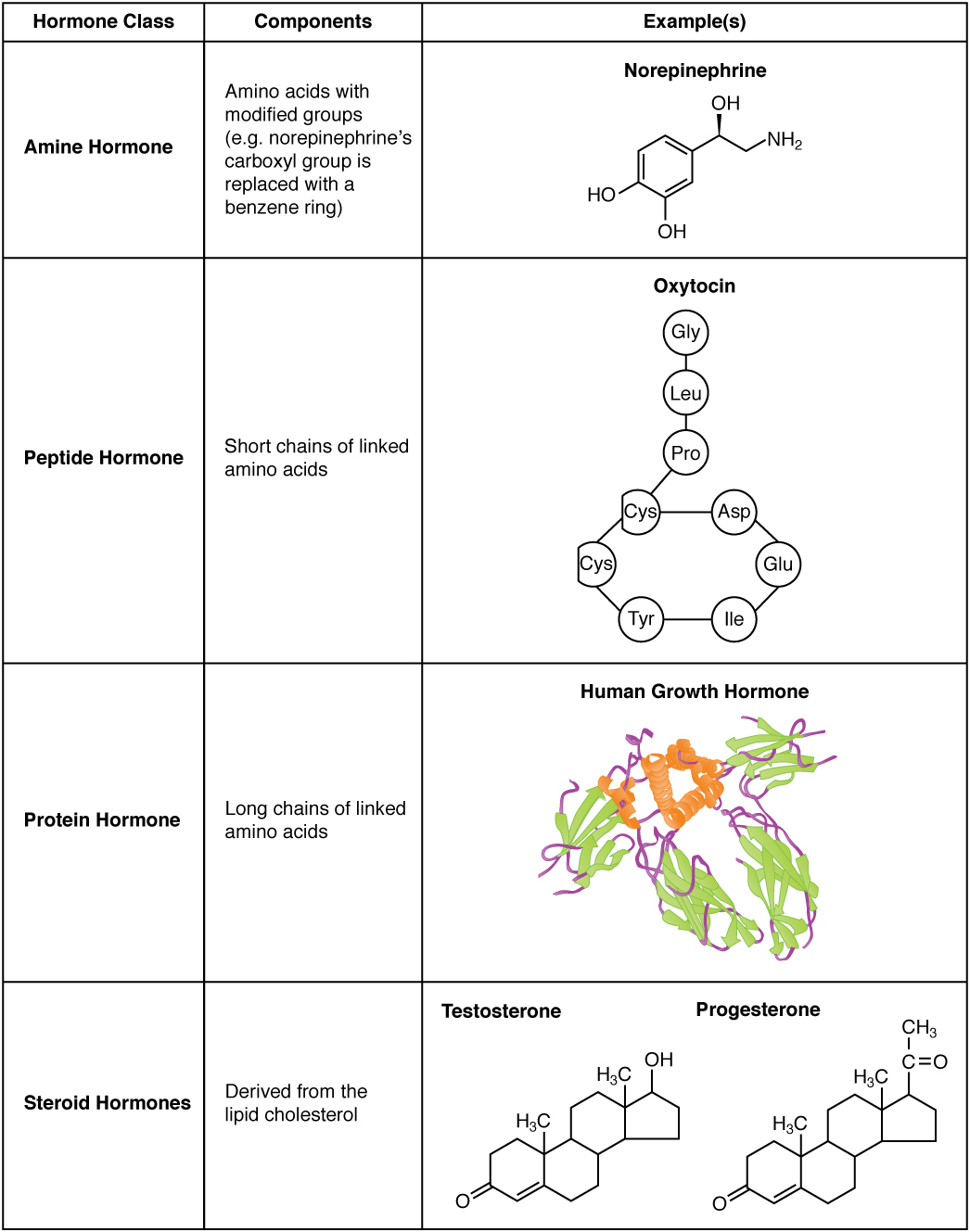
Peptide hormone

An amino--based chemical messenger released by ; and are examples.

Read explanation · OpenStax ↗
peristalsis

Coordinated waves of muscular contraction that propel material through a tubular organ.

Read explanation · OpenStax ↗
pH

A measure of , defined as the negative of activity. Lower pH means greater ; pH influences .

Read explanation · OpenStax ↗
phenotype

A measurable trait produced by interacting with development and environment.

Read explanation · OpenStax ↗
phosphatase

An removing a by .

Read explanation · OpenStax ↗
Phosphate group ·

A phosphorus-containing group bonded to oxygens; phosphate groups occur in , , and modified .

Read explanation · OpenStax ↗
Phosphodiester bond · · ·

A in which connects two sugars. In and it links the 3′ oxygen of one sugar to the 5′ oxygen of the next.

Read explanation · OpenStax ↗
Phospholipid

An with a -containing region and tails; a major component of cellular .

Read explanation · OpenStax ↗
ploidy

The number of complete sets in a cell.

Read explanation · OpenStax ↗
Polar

Having an uneven distribution of . Polar groups often interact favorably with water.

Read explanation · OpenStax ↗
Polymer

A large made from many linked molecular building blocks.

Read explanation · OpenStax ↗
Polypeptide

A chain of connected by . A functional can contain one or several such chains.

Read explanation · OpenStax ↗
Porous material · · · ·

A material containing small spaces that can admit of suitable size.

Read explanation · Cytiva ↗
Prokaryote ·

An organism whose cells lack a -bound ; and are prokaryotes.

Read explanation · OpenStax ↗
Proline ·

An whose loops back to the nitrogen, restricting rotation and often creating bends.

Read explanation · OpenStax ↗
Promoter

A region that recruits machinery and helps specify where and in which direction begins.

Read explanation · OpenStax ↗
Protease · ·

An that hydrolyzes in or .

Read explanation · OpenStax ↗
Protein

One or more folded chains that perform biological roles such as , transport, signaling, or structural support.

Read explanation · OpenStax ↗
Protein aggregation · ·

Association of into larger clusters, often when exposed surfaces of unfolded stick together.

Read explanation · NCBI Bookshelf ↗
Protein backbone ·

The repeating nitrogen–alpha-carbon– chain of a , excluding its variable .

Read explanation · OpenStax ↗
Protein domain · ·

A region of a that forms a recognizable structural or functional unit.

Read explanation · OpenStax ↗
Proton

A positively subatomic particle. In – chemistry, donating a proton means transferring a .

Read explanation · OpenStax ↗
Protonation

Addition of a to an , , or ; this changes its by plus one.

Read explanation · OpenStax ↗
PTH

Parathyroid hormone, a signal that raises blood calcium through effects on bone, kidney, and vitamin D activation.

Read explanation · OpenStax ↗
R group · ·

The variable side chain attached to an ’s . It determines identity, , , and chemical behavior.

Read explanation · OpenStax ↗
Reactant

A starting substance transformed during a .

Read explanation · OpenStax ↗
Reaction intermediate · ·

A species formed in one step and consumed in another. It occupies a local energy minimum between .

Read explanation · OpenStax ↗
Reaction product ·

A substance formed by a .

Read explanation · OpenStax ↗
Reaction rate · ·

How quickly are consumed or form, usually expressed as a change per unit time.

Read explanation · OpenStax ↗
Receptor

A that recognizes a particular signal and helps produce a cellular response.

Read explanation · OpenStax ↗
Red blood cell · ·

A blood cell that transports oxygen using ; mature human red blood cells lack a .

Read explanation · OpenStax ↗
Reduction

Gain of or a decrease in state. Reducing a restores two .

Read explanation · OpenStax ↗
Reporter gene · ·

A with a readily measured used to study regulation; reporter output can depend on synthesis, decay, , and behavior.

Read explanation · NCBI Bookshelf / Sinauer Associates ↗
Rescue experiment

An experiment restoring the altered component or function to test whether it accounts for an observed .

Read explanation · NCBI Bookshelf / Sinauer Associates ↗
Ribonucleic acid ·

A with roles in , regulation, and ; carries -coding information.

Read explanation · OpenStax ↗
Ribose

The five-carbon sugar in , with a at the 2′ carbon.

Read explanation · OpenStax ↗
Ribosome ·

A complex of and that translates into a .

Read explanation · OpenStax ↗
Salt

An electrically made from and . Dissolved salts can screen or compete with – interactions.

Read explanation · Cytiva ↗
satiety

The state of reduced motivation to eat following food intake and related physiological signals.

Read explanation · OpenStax ↗
secondary active transport

Uphill movement of one substance coupled to downhill movement of another across a .

Read explanation · OpenStax ↗
Secondary structure

Local folding, such as and , stabilized mainly by .

Read explanation · OpenStax ↗
secretin

An intestinal released in response to that promotes -rich .

Read explanation · OpenStax ↗
Secretory pathway ·

The route through the , , and used to deliver many to the cell surface, outside the cell, or compartments.

Read explanation · OpenStax ↗
Sedimentation ·

Movement of particles through a fluid under gravity or centrifugal .

Read explanation · Beckman Coulter ↗
segmentation

Mixing contractions of the intestine that distribute contents and promote contact with mucosa.

Read explanation · OpenStax ↗
Sister chromatid

One of the two copies of a produced by . An intact sister chromatid can supply a template for accurate repair.

Read explanation · Biology LibreTexts / Oregon State University ↗
sodium–potassium ATPase ·

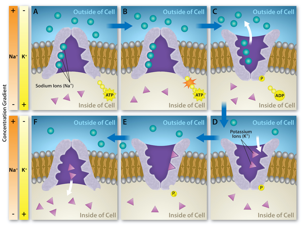
The -driven pump exporting three Na+ and importing two K+ per cycle.

Read explanation · OpenStax ↗
Solubility ·

The amount of a substance that can dissolve in a under specified conditions. Soluble means able to dissolve; insoluble means only very slightly soluble in that .

Read explanation · OpenStax ↗
Solute

A dissolved component of a .

Read explanation · OpenStax ↗
Solution

A homogeneous mixture in which are dissolved in a .

Read explanation · OpenStax ↗
Solvent

The component of a that dissolves other substances, usually the component present in the greatest amount.

Read explanation · OpenStax ↗
sphincter

A ring or zone of muscle regulating passage through an opening.

Read explanation · OpenStax ↗
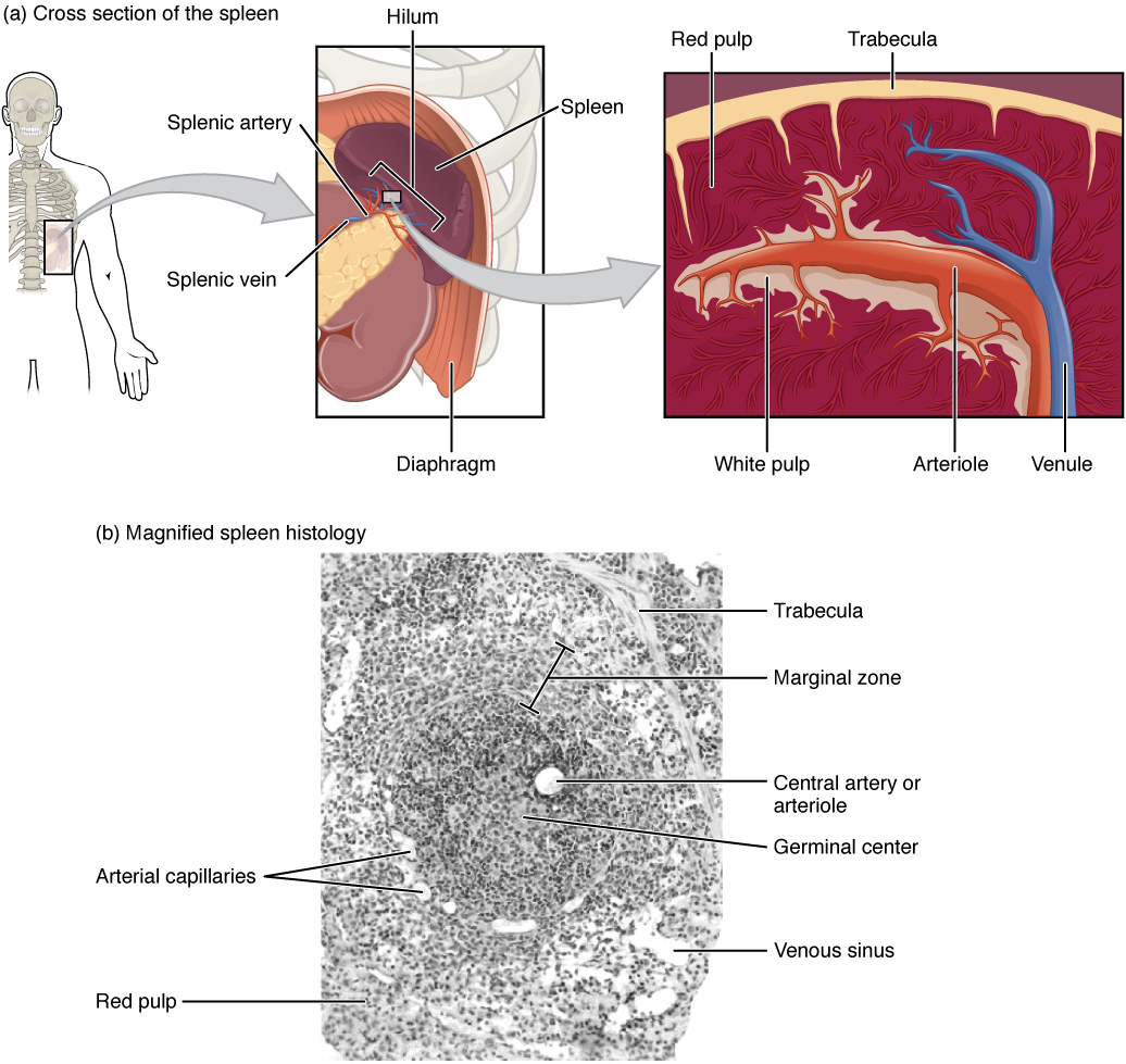
spleen

A lymphoid organ filtering blood, supporting immune responses, and removing aged blood cells.

Read explanation · OpenStax ↗
Starch

A plant storage made of chains; its two main forms are and amylopectin.

Read explanation · OpenStax ↗
Stationary phase

The material that remains fixed in a system.

Read explanation · Waters ↗
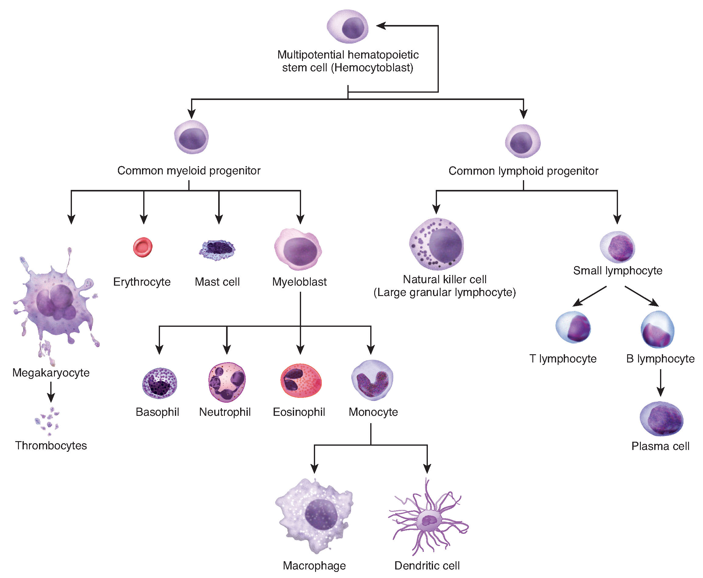
stem cell

A cell capable of self-renewal and production of differentiating descendants.

Read explanation · OpenStax ↗
steroid hormone

A -derived with fused-ring structure, commonly acting through intracellular .

Read explanation · OpenStax ↗
sterol

A containing fused rings and a ; is a major animal example.

Read explanation · OpenStax ↗
Substrate

A that an binds and chemically transforms.

Read explanation · OpenStax ↗
Subunit ·

An individual chain within a containing multiple chains.

Read explanation · OpenStax ↗
Template strand · · ·

A preexisting strand whose sequence guides complementary nucleic- synthesis.

Read explanation · OpenStax ↗
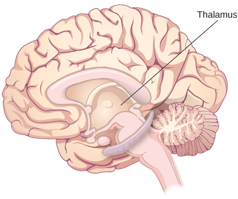
thalamus

structure containing relays and circuits connecting sensory, motor, and cortical systems.

Read explanation · OpenStax ↗
Thiol group · · · ·

A sulfur bonded to hydrogen, –SH. thiols can form through .

Read explanation · OpenStax ↗
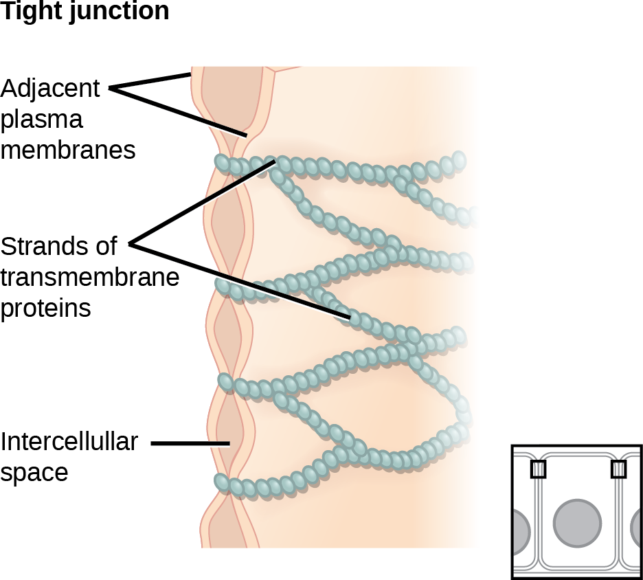
tight junction

A cell junction that restricts paracellular passage and helps preserve epithelial .

Read explanation · OpenStax ↗
Transacetylase

An transferring an acetyl group; the acetylates certain galactosides.

Read explanation · OpenStax ↗
Transcription

Synthesis of using a template.

Read explanation · OpenStax ↗
Transcriptional repressor · ·

A regulatory that reduces , for example by obstructing polymerase access or recruiting other repressing factors.

Read explanation · OpenStax ↗
Transition state

A fleeting, high-energy arrangement at a barrier. It is an energy maximum along the path, rather than a stable .

Read explanation · OpenStax ↗
Translation ·

Synthesis of a on a using the sequence of in .

Read explanation · OpenStax ↗
triacylglycerol · ·

A esterified to three ; a major energy-storage .

Read explanation · Biology LibreTexts / Oregon State University ↗
triad

Social group of three members.

Read explanation · OpenStax ↗
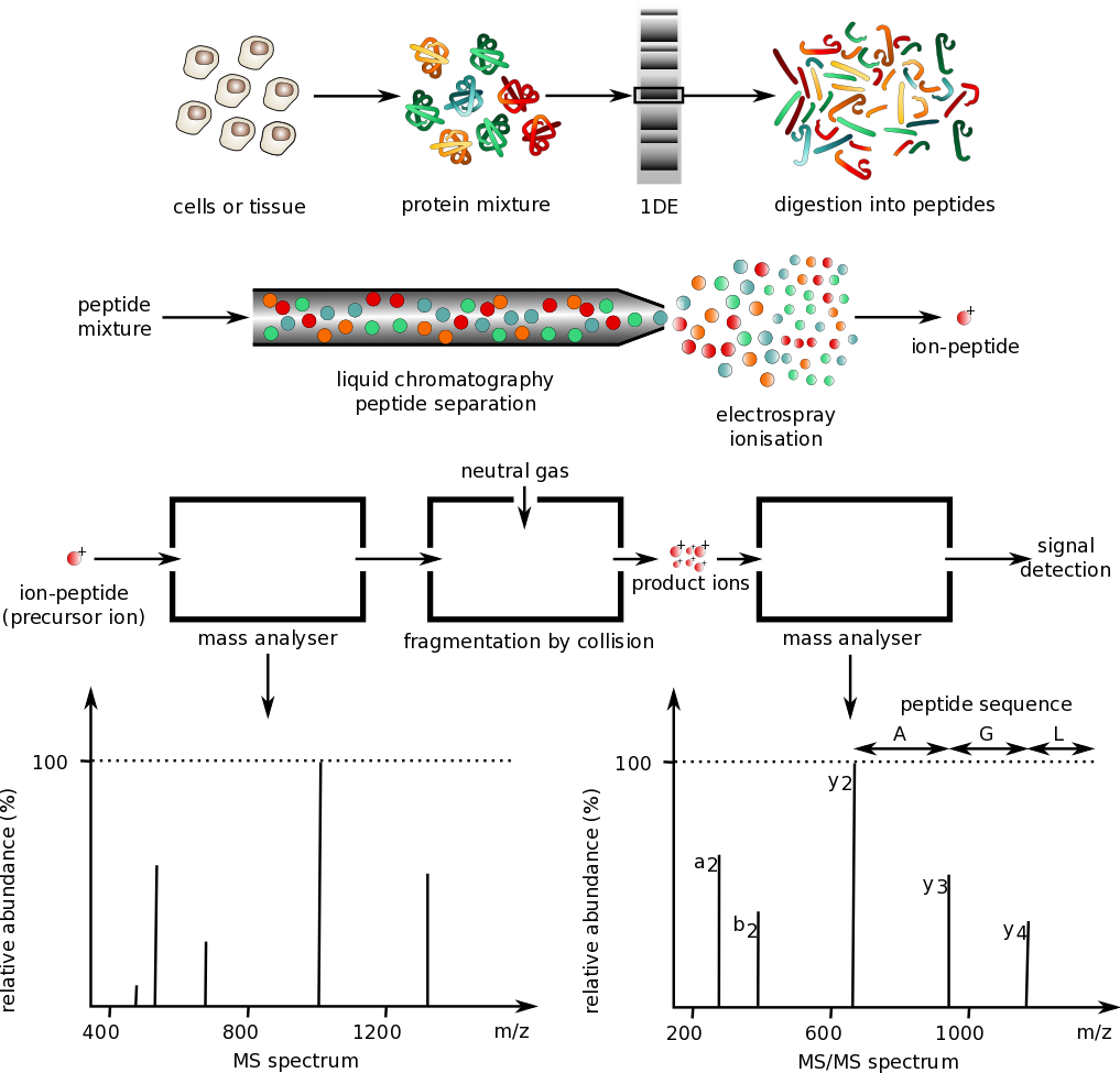
Trypsin

A commonly used to digest into , usually cleaving after or unless followed by .

Read explanation · Thermo Fisher ↗
trypsinogen

An inactive pancreatic precursor converted to in the intestine.

Read explanation · OpenStax ↗
tubular reabsorption

Movement of material from fluid back toward the blood.

Read explanation · OpenStax ↗
Turnover number · · ·

The maximum number of cycles per per unit time, calculated as /. Units are inverse time, commonly s⁻¹.

Read explanation · IUBMB ↗
velocity

Rate of change of position, including direction.

Read explanation · OpenStax ↗
Vesicle · · ·

A small -enclosed compartment that can transport material between cellular locations.

Read explanation · OpenStax ↗
villus

A small-intestinal mucosal projection increasing absorptive surface area.

Read explanation · OpenStax ↗
β-Galactosidase ·

The -encoded that hydrolyzes and can also convert to .

Read explanation · OpenStax ↗

23.2 Digestive System Processes and Regulation

OpenStax · 23.2 Digestive System Processes and Regulation ↗
Instructional page including learning objectives, examples, figures, source tables and questions. Site navigation and embedded media players are omitted; source media links remain available.

Learning Objectives

By the end of this section, you will be able to:

  • Discuss six fundamental activities of the digestive system, giving an example of each
  • Compare and contrast the neural and hormonal involved in digestion

The digestive system uses mechanical and chemical activities to break food down into absorbable substances during its journey through the digestive system. Table 23.3 provides an overview of the basic functions of the digestive organs.

Interactive Link

Visit this site for an overview of digestion of food in different regions of the digestive tract. Note the route of non-fat nutrients from the small intestine to their release as nutrients to the body.

Functions of the Digestive Organs
Organ Major functions Other functions
Mouth
  • Ingests food
  • Chews and mixes food
  • Begins chemical breakdown of
  • Moves food into the pharynx
  • Begins breakdown of via lingual
  • Moistens and dissolves food, allowing you to taste it
  • Cleans and lubricates the teeth and oral cavity
  • Has some antimicrobial activity
Pharynx
  • Propels food from the oral cavity to the esophagus
  • Lubricates food and passageways
Esophagus
  • Propels food to the stomach
  • Lubricates food and passageways
Stomach
  • Mixes and churns food with gastric juices to form
  • Begins chemical breakdown of
  • Releases food into the duodenum as
  • Absorbs some fat- substances (for example, alcohol, aspirin)
  • Possesses antimicrobial functions
Small intestine
  • Provides optimal medium for enzymatic activity
Accessory organs
  • -rich pancreatic juices help neutralize and provide optimal environment for enzymatic activity
Large intestine
  • Further breaks down food
  • Absorbs most residual water, electrolytes, and vitamins produced by enteric
  • Propels feces toward rectum
  • Eliminates feces
  • Food is concentrated and temporarily stored prior to defecation
  • Mucus eases passage of feces through colon
Table 23.3

Digestive Processes

The processes of digestion include six activities: ingestion, propulsion, mechanical or physical digestion, chemical digestion, absorption, and defecation.

The first of these processes, ingestion, refers to the entry of food into the alimentary canal through the mouth. There, the food is chewed and mixed with saliva, which contains that begin breaking down the in the food plus some digestion via lingual . Chewing increases the surface area of the food and allows an appropriately sized bolus to be produced.

Food leaves the mouth when the tongue and pharyngeal muscles propel it into the esophagus. This act of swallowing, the last voluntary act until defecation, is an example of propulsion, which refers to the movement of food through the digestive tract. It includes both the voluntary process of swallowing and the involuntary process of . consists of sequential, alternating waves of contraction and relaxation of alimentary wall smooth muscles, which act to propel food along (Figure 23.5). These waves also play a role in mixing food with digestive juices. is so powerful that foods and liquids you swallow enter your stomach even if you are standing on your head.

This image shows the peristaltic movement of food. In the left image, the food bolus is towards the top of the esophagus and arrows pointing downward show the direction of movement of the peristaltic wave. In the center image, the food bolus and the wave movement are closer to the center of the esophagus and in the right image, the bolus and the wave are close to the bottom end of the esophagus.
Figure 23.5 — 23.2 Digestive System Processes and Regulation

Figure 23.5 Peristalsis Peristalsis moves food through the digestive tract with alternating waves of muscle contraction and relaxation.

J. Gordon Betts and colleagues, Anatomy And Physiology 2E, OpenStax. · CC BY-NC-SA 4.0 · Original image ↗ · Access for free ↗

Digestion includes both mechanical and chemical processes. Mechanical digestion is a purely physical process that does not change the chemical nature of the food. Instead, it makes the food smaller to increase both surface area and mobility. It includes mastication, or chewing, as well as tongue movements that help break food into smaller bits and mix food with saliva. Although there may be a tendency to think that mechanical digestion is limited to the first steps of the digestive process, it occurs after the food leaves the mouth, as well. The mechanical churning of food in the stomach serves to further break it apart and expose more of its surface area to digestive juices, creating an “soup” called . , which occurs mainly in the small intestine, consists of localized contractions of circular muscle of the muscularis layer of the alimentary canal. These contractions isolate small sections of the intestine, moving their contents back and forth while continuously subdividing, breaking up, and mixing the contents. By moving food back and forth in the intestinal , mixes food with digestive juices and facilitates absorption.

In chemical digestion, starting in the mouth, digestive secretions break down complex food into their chemical building blocks (for example, into separate ). These secretions vary in composition, but typically contain water, various , , and . The process is completed in the small intestine.

Food that has been broken down is of no value to the body unless it enters the bloodstream and its nutrients are put to work. This occurs through the process of absorption, which takes place primarily within the small intestine. There, most nutrients are absorbed from the of the alimentary canal into the bloodstream through the epithelial cells that make up the mucosa. are absorbed into and are transported via the lymphatic vessels to the bloodstream (the subclavian veins near the heart). The details of these processes will be discussed later.

In defecation, the final step in digestion, undigested materials are removed from the body as feces.

Aging and the...

Digestive System: From Appetite Suppression to Constipation

Age-related changes in the digestive system begin in the mouth and can affect virtually every aspect of the digestive system. Taste buds become less sensitive, so food isn’t as appetizing as it once was. A slice of pizza is a challenge, not a treat, when you have lost teeth, your gums are diseased, and your salivary glands aren’t producing enough saliva. Swallowing can be difficult, and ingested food moves slowly through the alimentary canal because of reduced strength and tone of muscular tissue. Neurosensory feedback is also dampened, slowing the transmission of messages that stimulate the release of and .

Pathologies that affect the digestive organs—such as hiatal hernia, gastritis, and peptic ulcer disease—can occur at greater frequencies as you age. Problems in the small intestine may include duodenal ulcers, maldigestion, and malabsorption. Problems in the large intestine include hemorrhoids, diverticular disease, and constipation. Conditions that affect the function of accessory organs—and their abilities to deliver pancreatic and bile to the small intestine—include jaundice, acute pancreatitis, cirrhosis, and gallstones.

In some cases, a single organ is in of a digestive process. For example, ingestion occurs only in the mouth and defecation only in the anus. However, most digestive processes involve the interaction of several organs and occur gradually as food moves through the alimentary canal (Figure 23.6).

This image shows the different processes involved in digestion. The image shows how food travels from the mouth through the major organs. Associated textboxes list the different processes such as propulsion, chemical and mechanical digestion and absorption near the organs where they take place.
Figure 23.6 — 23.2 Digestive System Processes and Regulation

Figure 23.6 Digestive Processes The digestive processes are ingestion, propulsion, mechanical digestion, chemical digestion, absorption, and defecation.

J. Gordon Betts and colleagues, Anatomy And Physiology 2E, OpenStax. · CC BY-NC-SA 4.0 · Original image ↗ · Access for free ↗

Some chemical digestion occurs in the mouth. Some absorption can occur in the mouth and stomach, for example, alcohol and aspirin.

Regulatory Mechanisms

Neural and regulatory mechanisms work to maintain the optimal conditions in the needed for digestion and absorption. These regulatory mechanisms, which stimulate digestive activity through mechanical and chemical activity, are controlled both extrinsically and intrinsically.

Neural

The walls of the alimentary canal contain a variety of sensors that help regulate digestive functions. These include mechanoreceptors, , and , which are capable of detecting mechanical, chemical, and osmotic stimuli, respectively. For example, these can sense when the presence of food has caused the stomach to expand, whether food particles have been sufficiently broken down, how much liquid is present, and the type of nutrients in the food (, , and/or ). Stimulation of these provokes an appropriate reflex that furthers the process of digestion. This may entail sending a message that activates the glands that secrete digestive juices into the , or it may mean the stimulation of muscles within the alimentary canal, thereby activating and that move food along the intestinal tract.

The walls of the entire alimentary canal are embedded with nerve plexuses that interact with the and other nerve plexuses—either within the same digestive organ or in different ones. These interactions prompt several types of reflexes. Extrinsic nerve plexuses orchestrate long reflexes, which involve the central and autonomic nervous systems and work in response to stimuli from outside the digestive system. Short reflexes, on the other hand, are orchestrated by intrinsic nerve plexuses within the alimentary canal wall. These two plexuses and their connections were introduced earlier as the . Short reflexes regulate activities in one area of the digestive tract and may coordinate local peristaltic movements and stimulate digestive secretions. For example, the sight, smell, and taste of food initiate long reflexes that begin with a sensory delivering a signal to the medulla oblongata. The response to the signal is to stimulate cells in the stomach to begin secreting digestive juices in preparation for incoming food. In contrast, food that distends the stomach initiates short reflexes that cause cells in the stomach wall to increase their of digestive juices.

Hormonal

A variety of are involved in the digestive process. The main digestive of the stomach is , which is in response to the presence of food. stimulates the of gastric by the of the stomach mucosa. Other are produced and act upon the gut and its accessory organs. produced by the duodenum include , which stimulates a watery of by the ; (), which stimulates the of pancreatic and bile from the liver and release of bile from the gallbladder; and gastric inhibitory , which inhibits gastric and slows gastric emptying and motility. These are by specialized epithelial cells, called endocrinocytes, located in the mucosal of the stomach and small intestine. These then enter the bloodstream, through which they can reach their target organs.

23.4 The Stomach

OpenStax · 23.4 The Stomach ↗
Instructional page including learning objectives, examples, figures, source tables and questions. Site navigation and embedded media players are omitted; source media links remain available.

Learning Objectives

By the end of this section, you will be able to:

  • Label on a diagram the four main regions of the stomach, its curvatures, and its
  • Identify the four main types of secreting cells in gastric glands, and their important
  • Explain why the stomach does not digest itself
  • Describe the mechanical and chemical digestion of food entering the stomach

Although a minimal amount of digestion occurs in the mouth, chemical digestion really gets underway in the stomach. An expansion of the alimentary canal that lies immediately inferior to the esophagus, the stomach links the esophagus to the first part of the small intestine (the duodenum) and is relatively fixed in place at its esophageal and duodenal ends. In between, however, it can be a highly active structure, contracting and continually changing position and size. These contractions provide mechanical assistance to digestion. The empty stomach is only about the size of your fist, but can stretch to hold as much as 4 liters of food and fluid, or more than 75 times its empty volume, and then return to its resting size when empty. Although you might think that the size of a person’s stomach is related to how much food that individual consumes, body weight does not correlate with stomach size. Rather, when you eat greater quantities of food—such as at holiday dinner—you stretch the stomach more than when you eat less.

Popular tends to refer to the stomach as the location where all digestion takes place. Of course, this is not true. An important function of the stomach is to serve as a temporary holding chamber. You can ingest a meal far more quickly than it can be digested and absorbed by the small intestine. Thus, the stomach holds food and parses only small amounts into the small intestine at a time. Foods are not processed in the order they are eaten; rather, they are mixed together with digestive juices in the stomach until they are converted into , which is released into the small intestine.

As you will see in the sections that follow, the stomach plays several important roles in chemical digestion, including the continued digestion of and the initial digestion of and . Little if any nutrient absorption occurs in the stomach, with the exception of the negligible amount of nutrients in alcohol.

Structure

There are four main regions in the stomach: the cardia, fundus, body, and pylorus (Figure 23.15). The cardia (or cardiac region) is the point where the esophagus connects to the stomach and through which food passes into the stomach. Located inferior to the diaphragm, above and to the left of the cardia, is the dome-shaped fundus. Below the fundus is the body, the main part of the stomach. The funnel-shaped pylorus connects the stomach to the duodenum. The wider end of the funnel, the pyloric antrum, connects to the body of the stomach. The narrower end is called the pyloric canal, which connects to the duodenum. The smooth muscle pyloric is located at this latter point of connection and stomach emptying. In the absence of food, the stomach deflates inward, and its mucosa and submucosa fall into a large fold called a ruga.

This image shows a cross-section of the stomach, and the major parts: the cardia, fundus, body and pylorus are labeled.
Figure 23.15 — 23.4 The Stomach

Figure 23.15 Stomach The stomach has four major regions: the cardia, fundus, body, and pylorus. The addition of an inner oblique smooth muscle layer gives the muscularis the ability to vigorously churn and mix food.

J. Gordon Betts and colleagues, Anatomy And Physiology 2E, OpenStax. · CC BY-NC-SA 4.0 · Original image ↗ · Access for free ↗

The convex lateral surface of the stomach is called the greater curvature; the concave medial border is the lesser curvature. The stomach is held in place by the lesser omentum, which extends from the liver to the lesser curvature, and the greater omentum, which runs from the greater curvature to the posterior abdominal wall.

Histology

The wall of the stomach is made of the same four layers as most of the rest of the alimentary canal, but with to the mucosa and muscularis for the unique functions of this organ. In addition to the typical circular and longitudinal smooth muscle layers, the muscularis has an inner oblique smooth muscle layer (Figure 23.16). As a result, in addition to moving food through the canal, the stomach can vigorously churn food, mechanically breaking it down into smaller particles.

This diagram shows the histological cross-section of the stomach. The left panel shows the stomach and the center panel shows a magnified view of a small region including the epithelium and the gastric glands. The right panel shows a further magnification of the mucosa and the different cell types are labeled.
Figure 23.16 — 23.4 The Stomach

Figure 23.16 Histology of the Stomach The stomach wall is adapted for the functions of the stomach. In the epithelium, gastric pits lead to gastric glands that secrete gastric juice. The gastric glands (one gland is shown enlarged on the right) contain different types of cells that secrete a variety of enzymes, including hydrochloride acid, which activates the protein-digesting enzyme pepsin.

J. Gordon Betts and colleagues, Anatomy And Physiology 2E, OpenStax. · CC BY-NC-SA 4.0 · Original image ↗ · Access for free ↗

The stomach mucosa’s epithelial lining consists only of surface mucus cells, which secrete a protective coat of mucus. A vast number of gastric pits dot the surface of the , giving it the appearance of a well-used pincushion, and mark the entry to each gastric gland, which secretes a complex digestive fluid referred to as gastric juice.

Although the walls of the gastric pits are made up primarily of mucus cells, the gastric glands are made up of different types of cells. The glands of the cardia and pylorus are composed primarily of mucus-secreting cells. Cells that make up the pyloric antrum secrete mucus and a number of , including the majority of the stimulatory , . The much larger glands of the fundus and body of the stomach, the site of most chemical digestion, produce most of the gastric secretions. These glands are made up of a variety of secretory cells. These include , , mucous neck cells, and enteroendocrine cells.

—Located primarily in the middle region of the gastric glands are , which are among the most highly differentiated of the body’s epithelial cells. These relatively large cells produce both hydrochloric (HCl) and . HCl is responsible for the high ( 1.5 to 3.5) of the stomach contents and is needed to activate the -digesting , . The also kills much of the you ingest with food and helps to , making them more available for enzymatic digestion. is a glycoprotein necessary for the absorption of vitamin B12 in the small intestine.

—Located primarily in the basal regions of gastric glands are , which secrete , the inactive proenzyme form of . HCl is necessary for the conversion of to .

Mucous neck cells—Gastric glands in the upper part of the stomach contain mucous neck cells that secrete thin, mucus that is much different from the mucus by the goblet cells of the surface . The role of this mucus is not currently known.

Enteroendocrine cells—Finally, enteroendocrine cells found in the gastric glands secrete various into the of the lamina propria. These include , which is released mainly by enteroendocrine G cells.

Table 23.6 describes the digestive functions of important by the stomach.

Interactive Link

Watch this animation that depicts the structure of the stomach and how this structure functions in the initiation of digestion. This view of the stomach shows the characteristic rugae. What is the function of these rugae?

by the Stomach
Production site Production stimulus Target organ Action
Stomach mucosa, mainly G cells of the pyloric antrum Presence of and in stomach Stomach Increases by gastric glands; promotes gastric emptying
Stomach mucosa, mainly G cells of the pyloric antrum Presence of and in stomach Small intestine Promotes intestinal muscle contraction
Stomach mucosa, mainly G cells of the pyloric antrum Presence of and in stomach Ileocecal valve Relaxes valve
Stomach mucosa, mainly G cells of the pyloric antrum Presence of and in stomach Large intestine Triggers mass movements
Stomach mucosa, mainly fundus Fasting state (levels increase just prior to meals) Regulates food intake, primarily by stimulating hunger and
Histamine Stomach mucosa Presence of food in the stomach Stomach Stimulates to release HCl
Serotonin Stomach mucosa Presence of food in the stomach Stomach Contracts stomach muscle
Somatostatin Mucosa of stomach, especially pyloric antrum; also duodenum Presence of food in the stomach; sympathetic stimulation Stomach Restricts all gastric secretions, gastric motility, and emptying
Somatostatin Mucosa of stomach, especially pyloric antrum; also duodenum Presence of food in the stomach; sympathetic stimulation Restricts pancreatic secretions
Somatostatin Mucosa of stomach, especially pyloric antrum; also duodenum Presence of food in the stomach; sympathetic stimulation Small intestine Reduces intestinal absorption by reducing blood flow
Table 23.6

Gastric

The of gastric juice is controlled by both nerves and . Stimuli in the brain, stomach, and small intestine activate or inhibit gastric juice production. This is why the three phases of gastric are called the cephalic, gastric, and intestinal phases (Figure 23.17). However, once gastric begins, all three phases can occur simultaneously.

This flowchart shows the three different phases of gastric secretion. The top panel shows the cephalic phase, the middle panel shows the gastric phase and the bottom panel shows the intestinal phase.
Figure 23.17 — 23.4 The Stomach

Figure 23.17 The Three Phases of Gastric Secretion Gastric secretion occurs in three phases: cephalic, gastric, and intestinal. During each phase, the secretion of gastric juice can be stimulated or inhibited.

J. Gordon Betts and colleagues, Anatomy And Physiology 2E, OpenStax. · CC BY-NC-SA 4.0 · Original image ↗ · Access for free ↗

The cephalic phase (reflex phase) of gastric , which is relatively brief, takes place before food enters the stomach. The smell, taste, sight, or thought of food triggers this phase. For example, when you bring a piece of sushi to your lips, impulses from in your taste buds or the nose are relayed to your brain, which returns signals that increase gastric to prepare your stomach for digestion. This enhanced is a conditioned reflex, meaning it occurs only if you like or want a particular food. Depression and loss of appetite can suppress the cephalic reflex.

The gastric phase of lasts 3 to 4 hours, and is set in motion by local neural and hormonal mechanisms triggered by the entry of food into the stomach. For example, when your sushi reaches the stomach, it creates distention that activates the stretch . This stimulates parasympathetic to release acetylcholine, which then provokes increased of gastric juice. Partially digested , caffeine, and rising stimulate the release of from enteroendocrine G cells, which in turn induces to increase their production of HCl, which is needed to create an environment for the conversion of to , and digestion. Additionally, the release of activates vigorous smooth muscle contractions. However, it should be noted that the stomach does have a natural means of avoiding excessive and potential heartburn. Whenever levels drop too low, cells in the stomach react by suspending HCl and increasing mucous secretions.

The intestinal phase of gastric has both excitatory and inhibitory . The duodenum has a major role in regulating the stomach and its emptying. When partially digested food fills the duodenum, intestinal mucosal cells release a called intestinal (enteric) , which further excites gastric juice . This stimulatory activity is brief, however, because when the intestine distends with , the enterogastric reflex inhibits . One of the effects of this reflex is to close the pyloric , which blocks additional from entering the duodenum.

The Mucosal Barrier

The mucosa of the stomach is exposed to the highly corrosive of gastric juice. Gastric that can digest can also digest the stomach itself. The stomach is protected from self-digestion by the mucosal barrier. This barrier has several components. First, the stomach wall is covered by a thick coating of -rich mucus. This mucus forms a physical barrier, and its neutralize . Second, the epithelial cells of the stomach's mucosa meet at , which block gastric juice from penetrating the underlying tissue layers. Finally, located where gastric glands join the gastric pits quickly replace damaged epithelial mucosal cells, when the epithelial cells are shed. In , the surface of the stomach is completely replaced every 3 to 6 days.

Homeostatic Imbalances

Ulcers: When the Mucosal Barrier Breaks Down

As effective as the mucosal barrier is, it is not a “fail-safe” mechanism. Sometimes, gastric juice eats away at the superficial lining of the stomach mucosa, creating erosions, which mostly heal on their own. Deeper and larger erosions are called ulcers.

Why does the mucosal barrier break down? A number of factors can interfere with its ability to protect the stomach lining. The majority of all ulcers are caused by either excessive intake of non-steroidal anti-inflammatory drugs (NSAIDs), including aspirin, or Helicobacter pylori infection.

Antacids help relieve symptoms of ulcers such as “burning” pain and indigestion. When ulcers are caused by NSAID use, switching to other classes of pain relievers allows healing. When caused by H. pylori infection, antibiotics are effective.

A potential complication of ulcers is perforation: Perforated ulcers create a hole in the stomach wall, resulting in peritonitis ( of the peritoneum). These ulcers must be repaired surgically.

Digestive Functions of the Stomach

The stomach participates in virtually all the digestive activities with the exception of ingestion and defecation. Although almost all absorption takes place in the small intestine, the stomach does absorb some drugs, such as alcohol and aspirin.

Mechanical Digestion

Within a few moments after food enters your stomach, mixing waves begin to occur at intervals of approximately 20 seconds. A mixing wave is a unique type of that mixes and softens the food with gastric juices to create . The initial mixing waves are relatively gentle, but these are followed by more intense waves, starting at the body of the stomach and increasing in force as they reach the pylorus. It is fair to say that long before your sushi exits through the pyloric , it bears little resemblance to the sushi you ate.

The pylorus, which holds around 30 mL (1 fluid ounce) of , acts as a filter, permitting only liquids and small food particles to pass through the mostly, but not fully, closed pyloric . In a process called gastric emptying, rhythmic mixing waves force about 3 mL of at a time through the pyloric and into the duodenum. Release of a greater amount of at one time would overwhelm the capacity of the small intestine to handle it. The rest of the is pushed back into the body of the stomach, where it continues mixing. This process is repeated when the next mixing waves force more into the duodenum.

Gastric emptying is regulated by both the stomach and the duodenum. The presence of in the duodenum activates that inhibit gastric . This prevents additional from being released by the stomach before the duodenum is ready to process it.

Chemical Digestion

The fundus plays an important role, because it stores both undigested food and gases that are released during the process of chemical digestion. Food may sit in the fundus of the stomach for a while before being mixed with the . While the food is in the fundus, the digestive activities of continue until the food begins mixing with the . Ultimately, mixing waves incorporate this food with the , the of which inactivates and activates lingual . Lingual then begins breaking down into free , and mono- and diglycerides.

The breakdown of begins in the stomach through the actions of HCl and the .

Its numerous digestive functions notwithstanding, there is only one stomach function necessary to life: the production of . The intestinal absorption of vitamin B12, which is necessary for both the production of mature and normal neurological functioning, cannot occur without . People who undergo total gastrectomy (stomach removal)—for life-threatening stomach , for example—can survive with minimal digestive dysfunction if they receive vitamin B12 injections.

The contents of the stomach are completely emptied into the duodenum within 2 to 4 hours after you eat a meal. Different types of food take different amounts of time to process. Foods heavy in empty fastest, followed by high- foods. Meals with a high content remain in the stomach the longest. Since in the small intestine digest fats slowly, food can stay in the stomach for 6 hours or longer when the duodenum is processing fatty . However, note that this is still a of the 24 to 72 hours that full digestion typically takes from start to finish.

23.6 Accessory Organs in Digestion: The Liver, Pancreas, and Gallbladder

OpenStax · 23.6 Accessory Organs in Digestion: The Liver, Pancreas, and Gallbladder ↗
Instructional page including learning objectives, examples, figures, source tables and questions. Site navigation and embedded media players are omitted; source media links remain available.

Learning Objectives

By the end of this section, you will be able to:

  • State the main digestive roles of the liver, , and gallbladder
  • Identify three main features of liver histology that are critical to its function
  • Discuss the composition and function of bile
  • Identify the major types of and present in pancreatic juice

Chemical digestion in the small intestine relies on the activities of three accessory digestive organs: the liver, , and gallbladder (Figure 23.24). The digestive role of the liver is to produce bile and export it to the duodenum. The gallbladder primarily stores, concentrates, and releases bile. The produces pancreatic juice, which contains digestive and , and delivers it to the duodenum.

This diagram shows the accessory organs of the digestive system. The liver, spleen, pancreas, gallbladder and their major parts are shown.
Figure 23.24 — 23.6 Accessory Organs in Digestion: The Liver, , and Gallbladder

Figure 23.24 Accessory Organs The liver, pancreas, and gallbladder are considered accessory digestive organs, but their roles in the digestive system are vital.

J. Gordon Betts and colleagues, Anatomy And Physiology 2E, OpenStax. · CC BY-NC-SA 4.0 · Original image ↗ · Access for free ↗

The Liver

The liver is the largest gland in the body, weighing about three pounds in an adult. It is also one of the most important organs. In addition to being an accessory digestive organ, it plays a number of roles in and regulation. The liver lies inferior to the diaphragm in the right upper quadrant of the abdominal cavity and receives protection from the surrounding ribs.

The liver is divided into two primary lobes: a large right lobe and a much smaller left lobe. In the right lobe, some anatomists also identify an inferior quadrate lobe and a posterior caudate lobe, which are defined by internal features. The liver is connected to the abdominal wall and diaphragm by five peritoneal folds referred to as . These are the falciform , the coronary , two lateral , and the ligamentum teres hepatis. The falciform and ligamentum teres hepatis are actually remnants of the umbilical vein, and separate the right and left lobes anteriorly. The lesser omentum tethers the liver to the lesser curvature of the stomach.

The porta hepatis (“gate to the liver”) is where the hepatic artery and hepatic portal vein enter the liver. These two vessels, along with the common hepatic duct, run behind the lateral border of the lesser omentum on the way to their destinations. As shown in Figure 23.25, the hepatic artery delivers oxygenated blood from the heart to the liver. The hepatic portal vein delivers partially deoxygenated blood containing nutrients absorbed from the small intestine and actually supplies more oxygen to the liver than do the much smaller hepatic arteries. In addition to nutrients, drugs and toxins are also absorbed. After processing the bloodborne nutrients and toxins, the liver releases nutrients needed by other cells back into the blood, which drains into the central vein and then through the hepatic vein to the inferior vena cava. With this , all blood from the alimentary canal passes through the liver. This largely explains why the liver is the most common site for the metastasis of cancers that originate in the alimentary canal.

This image shows the microscopic anatomy of the liver. The top panel shows the liver; the center panel shows a magnified view of the connective tissue and the lobules. The bottom panel shows a further magnified view of a lobule, identifying the veins, bile duct and the sinusoids.
Figure 23.25 — 23.6 Accessory Organs in Digestion: The Liver, , and Gallbladder

Figure 23.25 Microscopic Anatomy of the Liver The liver receives oxygenated blood from the hepatic artery and nutrient-rich deoxygenated blood from the hepatic portal vein.

J. Gordon Betts and colleagues, Anatomy And Physiology 2E, OpenStax. · CC BY-NC-SA 4.0 · Original image ↗ · Access for free ↗

Histology

The liver has three main components: hepatocytes, bile canaliculi, and hepatic sinusoids. A hepatocyte is the liver’s main cell type, accounting for around 80 percent of the liver's volume. These cells play a role in a wide variety of secretory, , and functions. Plates of hepatocytes called hepatic laminae radiate outward from the central vein in each hepatic lobule.

Between adjacent hepatocytes, grooves in the provide room for each bile canaliculus (plural = canaliculi). These small ducts accumulate the bile produced by hepatocytes. From here, bile flows first into bile ductules and then into bile ducts. The bile ducts unite to form the larger right and left hepatic ducts, which themselves merge and exit the liver as the common hepatic duct. This duct then joins with the cystic duct from the gallbladder, forming the common bile duct through which bile flows into the small intestine.

A hepatic sinusoid is an open, blood space formed by fenestrated capillaries from nutrient-rich hepatic portal veins and oxygen-rich hepatic arteries. Hepatocytes are tightly packed around the fenestrated of these spaces, giving them easy access to the blood. From their central position, hepatocytes process the nutrients, toxins, and waste materials carried by the blood. Materials such as are processed and excreted into the bile canaliculi. Other materials including , , and are processed and into the sinusoids or just stored in the cells until called upon. The hepatic sinusoids combine and send blood to a central vein. Blood then flows through a hepatic vein into the inferior vena cava. This means that blood and bile flow in opposite directions. The hepatic sinusoids also contain star-shaped reticuloendothelial cells (Kupffer cells), phagocytes that remove dead red and white blood cells, , and other foreign material that enter the sinusoids. The portal is a distinctive arrangement around the perimeter of hepatic lobules, consisting of three basic structures: a bile duct, a hepatic artery branch, and a hepatic portal vein branch.

Bile

Recall that are , that is, they do not dissolve in water. Thus, before they can be digested in the watery environment of the small intestine, large globules must be broken down into smaller globules, a process called emulsification. Bile is a mixture by the liver to accomplish the emulsification of in the small intestine.

Hepatocytes secrete about one liter of bile each day. A yellow-brown or yellow-green ( 7.6 to 8.6), bile is a mixture of water, , bile pigments, (such as lecithin), electrolytes, , and . The components most critical to emulsification are and , which have a () region as well as a () region. The region interacts with the large , whereas the region interacts with the watery in the intestine. This results in the large globules being pulled apart into many tiny fragments of about 1 µm in diameter. This change dramatically increases the surface area available for -digesting activity. This is the same way dish soap works on fats mixed with water.

act as emulsifying agents, so they are also important for the absorption of digested . While most constituents of bile are eliminated in feces, are reclaimed by the enterohepatic circulation. Once reach the ileum, they are absorbed and returned to the liver in the hepatic . The hepatocytes then excrete the into newly formed bile. Thus, this precious resource is recycled.

, the main bile pigment, is a waste produced when the removes old or damaged from the circulation. These breakdown , including , iron, and toxic , are transported to the liver via the splenic vein of the hepatic portal system. In the liver, and iron are recycled, whereas is excreted in the bile. It accounts for the green color of bile. is eventually transformed by intestinal into stercobilin, a brown pigment that gives your stool its characteristic color! In some disease states, bile does not enter the intestine, resulting in white (‘acholic’) stool with a high fat content, since virtually no fats are broken down or absorbed.

Hepatocytes work non-stop, but bile production increases when fatty enters the duodenum and stimulates the of the gut . Between meals, bile is produced but conserved. The valve-like hepatopancreatic ampulla closes, allowing bile to divert to the gallbladder, where it is concentrated and stored until the next meal.

Interactive Link

Watch this video to see the structure of the liver and how this structure supports the functions of the liver, including the processing of nutrients, toxins, and wastes. At rest, about 1500 mL of blood per minute flow through the liver. What percentage of this blood flow comes from the hepatic portal system?

The

The soft, oblong, glandular lies transversely in the retroperitoneum behind the stomach. Its head is nestled into the “c-shaped” curvature of the duodenum with the body extending to the left about 15.2 (6 in) and ending as a tapering tail in the hilum of the . It is a curious mix of (secreting digestive ) and (releasing into the blood) functions (Figure 23.26).

This figure shows the pancreas and its major parts. A magnified view of a small region of the pancreas shows the pancreatic islet cells, the acinar cells and the pancreatic duct.
Figure 23.26 — 23.6 Accessory Organs in Digestion: The Liver, , and Gallbladder

Figure 23.26 Exocrine and Endocrine Pancreas The pancreas has a head, a body, and a tail. It delivers pancreatic juice to the duodenum through the pancreatic duct.

J. Gordon Betts and colleagues, Anatomy And Physiology 2E, OpenStax. · CC BY-NC-SA 4.0 · Original image ↗ · Access for free ↗

The part of the arises as little grape-like cell clusters, each called an acinus (plural = acini), located at the terminal ends of pancreatic ducts. These acinar cells secrete -rich pancreatic juice into tiny merging ducts that form two ducts. The larger duct fuses with the common bile duct (carrying bile from the liver and gallbladder) just before entering the duodenum via a common opening (the hepatopancreatic ampulla). The smooth muscle of the hepatopancreatic ampulla the release of pancreatic juice and bile into the small intestine. The second and smaller pancreatic duct, the accessory duct (duct of Santorini), runs from the directly into the duodenum, approximately 1 inch above the hepatopancreatic ampulla. When present, it is a persistent remnant of pancreatic development.

Scattered through the sea of acini are small islands of , the islets of Langerhans. These vital cells produce the pancreatic , , , and somatostatin.

Pancreatic Juice

The produces over a liter of pancreatic juice each day. Unlike bile, it is clear and composed mostly of water along with some , sodium , and several digestive . Sodium is responsible for the slight alkalinity of pancreatic juice ( 7.1 to 8.2), which serves to the gastric juice in , inactivate from the stomach, and create an optimal environment for the activity of -sensitive digestive in the small intestine. Pancreatic are active in the digestion of sugars, , and fats.

The produces -digesting in their inactive forms. These are activated in the duodenum. If produced in an active form, they would digest the (which is exactly what occurs in the disease, pancreatitis). The intestinal stimulates the activation of from of the , which in turn changes the pancreatic procarboxypeptidase and chymotrypsinogen into their active forms, carboxypeptidase and chymotrypsin.

The that digest (), fat (), and () are in their active forms, since they do not attack the as do the -digesting .

Pancreatic

Regulation of pancreatic is the job of and the parasympathetic nervous system. The entry of into the duodenum stimulates the release of , which in turn causes the duct cells to release -rich pancreatic juice. The presence of and fats in the duodenum stimulates the of , which then stimulates the acini to secrete -rich pancreatic juice and enhances the activity of . Parasympathetic regulation occurs mainly during the cephalic and gastric phases of gastric , when vagal stimulation prompts the of pancreatic juice.

Usually, the secretes just enough to counterbalance the amount of HCl produced in the stomach. enter the blood when is by the . Thus, the blood draining from the neutralizes the blood draining from the stomach, maintaining the of the venous blood that flows to the liver.

The Gallbladder

The gallbladder is 8–10 (~3–4 in) long and is nested in a shallow area on the posterior aspect of the right lobe of the liver. This muscular sac stores, concentrates, and, when stimulated, propels the bile into the duodenum via the common bile duct. It is divided into three regions. The fundus is the widest portion and tapers medially into the body, which in turn narrows to become the neck. The neck angles slightly superiorly as it approaches the hepatic duct. The cystic duct is 1–2 (less than 1 in) long and turns inferiorly as it bridges the neck and hepatic duct.

The simple of the gallbladder mucosa is organized in rugae, similar to those of the stomach. There is no submucosa in the gallbladder wall. The wall’s middle, muscular coat is made of smooth muscle fibers. When these fibers contract, the gallbladder’s contents are ejected through the cystic duct and into the bile duct (Figure 23.27). Visceral peritoneum reflected from the liver capsule holds the gallbladder against the liver and forms the outer coat of the gallbladder. The gallbladder's mucosa absorbs water and from bile, concentrating it by up to 10-fold.

This figure shows the gallbladder and its major parts are labeled.
Figure 23.27 — 23.6 Accessory Organs in Digestion: The Liver, , and Gallbladder

Figure 23.27 Gallbladder The gallbladder stores and concentrates bile, and releases it into the two-way cystic duct when it is needed by the small intestine.

J. Gordon Betts and colleagues, Anatomy And Physiology 2E, OpenStax. · CC BY-NC-SA 4.0 · Original image ↗ · Access for free ↗

23.7 Chemical Digestion and Absorption: A Closer Look
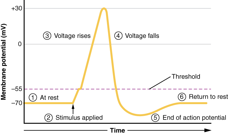
OpenStax · 23.7 Chemical Digestion and Absorption: A Closer Look ↗

Instructional page including learning objectives, examples, figures, source tables and questions. Site navigation and embedded media players are omitted; source media links remain available.

Learning Objectives

By the end of this section, you will be able to:

As you have learned, the process of mechanical digestion is relatively simple. It involves the physical breakdown of food but does not alter its chemical makeup. Chemical digestion, on the other hand, is a complex process that reduces food into its chemical building blocks, which are then absorbed to nourish the cells of the body (Figure 23.28). In this section, you will look more closely at the processes of chemical digestion and absorption.

This diagram identifies the functions of mechanical and chemical digestion and absorption at each organ. Next to each organ, a callout identifies which steps of digestion take place in that particular organ.
Figure 23.28 — 23.7 Chemical Digestion and Absorption: A Closer Look

Figure 23.28 Digestion and Absorption Digestion begins in the mouth and continues as food travels through the small intestine. Most absorption occurs in the small intestine.

J. Gordon Betts and colleagues, Anatomy And Physiology 2E, OpenStax. · CC BY-NC-SA 4.0 · Original image ↗ · Access for free ↗

Chemical Digestion

Large food (for example, , , , and starches) must be broken down into that are small enough to be absorbed by the lining of the alimentary canal. This is accomplished by through . The many involved in chemical digestion are summarized in Table 23.8.

The Digestive
Category Name Source
Salivary Lingual Lingual glands Free , and mono- and diglycerides
Salivary Salivary glands Polysaccharides Disaccharides and trisaccharides
Gastric Gastric and monoacylglycerides
Gastric *
α-Dextrinase Small intestine α-Dextrins
Small intestine
Lactase Small intestine and
Maltase Small intestine Maltose
Nucleosidases and Small intestine Phosphates, , and
Peptidases Small intestine
Sucrase Small intestine Sucrose and fructose
Pancreatic Carboxy-peptidase* Pancreatic acinar cells at the carboxyl end of and
Pancreatic Chymotrypsin* Pancreatic acinar cells
Pancreatic Elastase* Pancreatic acinar cells
Pancreatic Pancreatic acinar cells
  • Ribonuclease: ribonucleic
  • Deoxyribonuclease: deoxyribonucleic
Pancreatic Pancreatic Pancreatic acinar cells Polysaccharides (starches) α-Dextrins, disaccharides (maltose), trisaccharides (maltotriose)
Pancreatic Pancreatic Pancreatic acinar cells that have been emulsified by and monoacylglycerides
Pancreatic * Pancreatic acinar cells
Table 23.8 *These have been activated by other substances.

Digestion

The average American diet is about 50 percent , which may be classified according to the number of they contain of simple sugars (monosaccharides and disaccharides) and/or complex sugars (polysaccharides). , , and fructose are the three monosaccharides that are commonly consumed and are readily absorbed. Your digestive system is also able to break down the disaccharide sucrose (regular table sugar: + fructose), (milk sugar: + ), and maltose (grain sugar: + ), and the polysaccharides and (chains of monosaccharides). Your bodies do not produce that can break down most fibrous polysaccharides, such as . While indigestible polysaccharides do not provide any nutritional value, they do provide dietary fiber, which helps propel food through the alimentary canal.

The chemical digestion of starches begins in the mouth and has been reviewed above.

In the small intestine, pancreatic does the ‘heavy lifting’ for and digestion (Figure 23.29). After amylases break down into smaller fragments, the α-dextrinase starts working on α-dextrin, breaking off one unit at a time. Three hydrolyze sucrose, , and maltose into monosaccharides. Sucrase splits sucrose into one of fructose and one of ; maltase breaks down maltose and maltotriose into two and three , respectively; and lactase breaks down into one of and one of . Insufficient lactase can lead to intolerance.

This flow chart shows the steps in digestion of carbohydrates. The different levels shown are starch and glycogen, disaccharides and monosaccharides. Under each type of sugar, examples and the enzymes responsible for digestion are listed.
Figure 23.29 — 23.7 Chemical Digestion and Absorption: A Closer Look

Figure 23.29 Carbohydrate Digestion Flow Chart Carbohydrates are broken down into their monomers in a series of steps.

J. Gordon Betts and colleagues, Anatomy And Physiology 2E, OpenStax. · CC BY-NC-SA 4.0 · Original image ↗ · Access for free ↗

Digestion

are composed of linked by to form long chains. Digestion reduces them to their constituent . You usually consume about 15 to 20 percent of your total calorie intake as .

The digestion of starts in the stomach, where HCl and break into smaller , which then travel to the small intestine (Figure 23.30). Chemical digestion in the small intestine is continued by pancreatic , including chymotrypsin and , each of which act on specific bonds in sequences. At the same time, the cells of the secrete such as aminopeptidase and dipeptidase, which further break down chains. This results in small enough to enter the bloodstream (Figure 23.31).

This diagrams shows the human digestive system and identifies the role of each organ in protein digestion. A text call-out next to each organ details the specific function.
Figure 23.30 — 23.7 Chemical Digestion and Absorption: A Closer Look
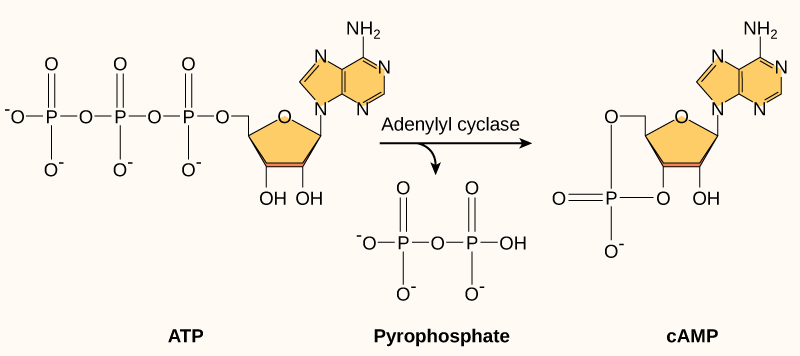
Figure 23.30 Digestion of Protein The digestion of protein begins in the stomach and is completed in the small intestine.

J. Gordon Betts and colleagues, Anatomy And Physiology 2E, OpenStax. · CC BY-NC-SA 4.0 · Original image ↗ · Access for free ↗
This flow chart shows the different steps in the digestion of protein. The four steps shown are protein, large polypeptides, short peptides and amino acids and amino acids.
Figure 23.31 — 23.7 Chemical Digestion and Absorption: A Closer Look

Figure 23.31 Digestion of Protein Flow Chart Proteins are successively broken down into their amino acid components.

J. Gordon Betts and colleagues, Anatomy And Physiology 2E, OpenStax. · CC BY-NC-SA 4.0 · Original image ↗ · Access for free ↗

Digestion

A healthy diet limits intake to 35 percent of total calorie intake. The most common dietary are , which are made up of a bound to three chains. Small amounts of dietary and are also consumed.

The three responsible for digestion are lingual , gastric , and pancreatic . However, because the is the only consequential source of , virtually all digestion occurs in the small intestine. Pancreatic breaks down each into two free and a monoglyceride. The include both short-chain (less than 10 to 12 carbons) and long-chain .

Digestion

The and are found in most of the foods you eat. Two types of pancreatic are responsible for their digestion: deoxyribonuclease, which digests , and ribonuclease, which digests . The produced by this digestion are further broken down by two intestinal (nucleosidase and ) into , phosphates, and , which can be absorbed through the alimentary canal wall. The large food that must be broken down into are summarized Table 23.9

Absorbable Food Substances
Source Substance
Monosaccharides: , , and fructose
Single , dipeptides, and tripeptides
Monoacylglycerides, , and free
sugars, phosphates, and
Table 23.9

Absorption

The mechanical and digestive processes have one goal: to convert food into small enough to be absorbed by the epithelial cells of the intestinal . The absorptive capacity of the alimentary canal is almost endless. Each day, the alimentary canal processes up to 10 liters of food, liquids, and secretions, yet less than one liter enters the large intestine. Almost all ingested food, 80 percent of electrolytes, and 90 percent of water are absorbed in the small intestine. Although the entire small intestine is involved in the absorption of water and , most absorption of and occurs in the jejunum. Notably, and vitamin B12 are absorbed in the terminal ileum. By the time passes from the ileum into the large intestine, it is essentially indigestible food (mainly plant fibers like ), some water, and millions of (Figure 23.32).

This image shows the human digestive system. Next to each organ, a text callout identifies how water and digestive secretions such as saliva and bile are processed.
Figure 23.32 — 23.7 Chemical Digestion and Absorption: A Closer Look
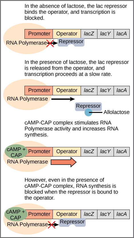
Figure 23.32 Digestive Secretions and Absorption of Water Absorption is a complex process, in which nutrients from digested food are harvested.

J. Gordon Betts and colleagues, Anatomy And Physiology 2E, OpenStax. · CC BY-NC-SA 4.0 · Original image ↗ · Access for free ↗

Absorption can occur through five mechanisms: (1) active transport, (2) passive , (3) , (4) co-transport (or ), and (5) . As you will recall from Chapter 3, active transport refers to the movement of a substance across a going from an area of lower to an area of higher (up the ). In this type of transport, within the act as “pumps,” using cellular energy () to move the substance. Passive refers to the movement of substances from an area of higher to an area of lower , while refers to the movement of substances from an area of higher to an area of lower using a in the . Co-transport uses the movement of one through the from higher to lower to power the movement of another from lower to higher. Finally, is a transportation process in which the engulfs material. It requires energy, generally in the form of .

Because the cell’s plasma is made up of , water- nutrients must use transport embedded in the to enter cells. Moreover, substances cannot pass between the epithelial cells of the intestinal mucosa because these cells are bound together by . Thus, substances can only enter blood capillaries by passing through the surfaces of epithelial cells and into the . Water- nutrients enter the capillary blood in the and travel to the liver via the hepatic portal vein.

In contrast to the water- nutrients, - nutrients can diffuse through the plasma . Once inside the cell, they are packaged for transport via the of the cell and then enter the of the to be transported by lymphatic vessels to the systemic circulation via the thoracic duct. The absorption of most nutrients through the mucosa of the intestinal requires active transport fueled by . The routes of absorption for each food category are summarized in Table 23.10.

Absorption in the Alimentary Canal
Food Breakdown Absorption mechanism Entry to bloodstream Destination
Co-transport with sodium Capillary blood in Liver via hepatic portal vein
Fructose Capillary blood in Liver via hepatic portal vein
Co-transport with sodium Capillary blood in Liver via hepatic portal vein
Long-chain into intestinal cells, where they are combined with to create of Systemic circulation via entering thoracic duct
Monoacylglycerides into intestinal cells, where they are combined with to create of Systemic circulation via entering thoracic duct
Short-chain Simple Capillary blood in Liver via hepatic portal vein
Simple Capillary blood in Liver via hepatic portal vein
digestion Active transport via carriers Capillary blood in Liver via hepatic portal vein
Table 23.10

Absorption

All are absorbed in the form of monosaccharides. The small intestine is highly efficient at this, absorbing monosaccharides at an estimated rate of 120 grams per hour. All normally digested dietary are absorbed; indigestible fibers are eliminated in the feces. The monosaccharides and are transported into the epithelial cells by common carriers via (that is, co-transport with sodium ). The monosaccharides leave these cells via and enter the capillaries through intercellular clefts. The monosaccharide fructose (which is in fruit) is absorbed and transported by alone. The monosaccharides combine with the transport immediately after the disaccharides are broken down.

Absorption

Active transport mechanisms, primarily in the duodenum and jejunum, absorb most as their breakdown , . Almost all (95 to 98 percent) is digested and absorbed in the small intestine. The type of that transports an varies. Most carriers are linked to the active transport of sodium. Short chains of two (dipeptides) or three (tripeptides) are also transported actively. However, after they enter the absorptive epithelial cells, they are broken down into their before leaving the cell and entering the capillary blood via .

Absorption

About 95 percent of are absorbed in the small intestine. not only speed up digestion, they are also essential to the absorption of the end of digestion. Short-chain are relatively water and can enter the absorptive cells () directly. The small size of short-chain enables them to be absorbed by via simple , and then take the same path as monosaccharides and into the blood capillary of a .

The large and long-chain and monoacylglycerides are not so easily suspended in the watery intestinal . However, and lecithin resolve this issue by enclosing them in a micelle, which is a tiny sphere with () ends facing the watery environment and tails turned to the interior, creating a receptive environment for the long-chain . The core also includes and fat- vitamins. Without micelles, would sit on the surface of and never come in contact with the absorptive surfaces of the epithelial cells. Micelles can easily squeeze between and get very near the cell surface. At this point, substances exit the micelle and are absorbed via simple .

The free and monoacylglycerides that enter the epithelial cells are reincorporated into . The are mixed with and , and surrounded with a coat. This new complex, called a , is a water- lipoprotein. After being processed by the , are released from the cell (Figure 23.33). Too big to pass through the basement of blood capillaries, instead enter the large of . The come together to form the lymphatic vessels. The are transported in the lymphatic vessels and empty through the thoracic duct into the subclavian vein of the circulatory system. Once in the bloodstream, the breaks down the of the into free and . These breakdown then pass through capillary walls to be used for energy by cells or stored in adipose tissue as fat. Liver cells combine the remaining remnants with , forming lipoproteins that transport in the blood.

This diagram shows how lipids are absorbed from the lumen of the intestine into the lacteals. The fatty acid micelles are shown to enter the epithelial cell and form chylomicrons inside the Golgi apparatus. Then, the chylomicrons are extruded from the epithelial cell and are taken up by the lacteals.
Figure 23.33 — 23.7 Chemical Digestion and Absorption: A Closer Look

Figure 23.33 Lipid Absorption Unlike amino acids and simple sugars, lipids are transformed as they are absorbed through epithelial cells.

J. Gordon Betts and colleagues, Anatomy And Physiology 2E, OpenStax. · CC BY-NC-SA 4.0 · Original image ↗ · Access for free ↗

Absorption

The of digestion— sugars, , and —are transported by carriers across the via active transport. These then enter the bloodstream.

Mineral Absorption

The electrolytes absorbed by the small intestine are from both secretions and ingested foods. Since electrolytes into in water, most are absorbed via active transport throughout the entire small intestine. During absorption, co-transport mechanisms result in the accumulation of sodium inside the cells, whereas anti-port mechanisms reduce the potassium inside the cells. To restore the sodium-potassium across the , a requiring pumps sodium out and potassium in.

In general, all minerals that enter the intestine are absorbed, whether you need them or not. Iron and calcium are exceptions; they are absorbed in the duodenum in amounts that meet the body’s current requirements, as follows:

Iron—The ionic iron needed for the production of is absorbed into mucosal cells via active transport. Once inside mucosal cells, ionic iron binds to the ferritin, creating iron-ferritin complexes that store iron until needed. When the body has enough iron, most of the stored iron is lost when worn-out epithelial cells slough off. When the body needs iron because, for example, it is lost during acute or chronic bleeding, there is increased uptake of iron from the intestine and accelerated release of iron into the bloodstream. Since females experience significant iron loss during menstruation, they have around four times as many iron transport in their intestinal epithelial cells as do males.

Calcium—Blood levels of ionic calcium determine the absorption of dietary calcium. When blood levels of ionic calcium drop, () by the parathyroid glands stimulates the release of calcium from bone and increases the of calcium by the kidneys. also upregulates the activation of vitamin D in the kidney, which then facilitates intestinal calcium absorption.

Vitamin Absorption

The small intestine absorbs the vitamins that occur naturally in food and supplements. Fat- vitamins (A, D, E, and K) are absorbed along with dietary in micelles via simple . This is why you are advised to eat some fatty foods when you take fat- vitamin supplements. Most water- vitamins (including most B vitamins and vitamin C) also are absorbed by simple . An exception is vitamin B12, which is a very large . in the stomach binds to vitamin B12, preventing its digestion and creating a complex that binds to mucosal in the terminal ileum, where it is taken up by .

Water Absorption

Each day, about nine liters of fluid enter the small intestine. About 2.3 liters are ingested in foods and beverages, and the rest is from secretions. About 90 percent of this water is absorbed in the small intestine. Water absorption is driven by the of the water: The of water is higher in than it is in epithelial cells. Thus, water moves down its from the into cells. As noted earlier, much of the remaining water is then absorbed in the colon.