PCR cycles through denaturation, primer annealing, and extension by a thermostableDNA polymerase. Each primer provides a free 3′ hydroxyl. Two primers bind opposite strands with their 3′ ends facing into the target region; extension proceeds 5′→3′. The interval between primer boundaries determines the eventual product. Ideal doubling gives N = N₀ × 2ⁿ, but efficiency and reagent depletion limit real amplification. PCR copies DNA in a tube; cloning propagates DNA in a vector/host. Reverse transcription converts RNA to cDNA before RNA-derived targets can be amplified.
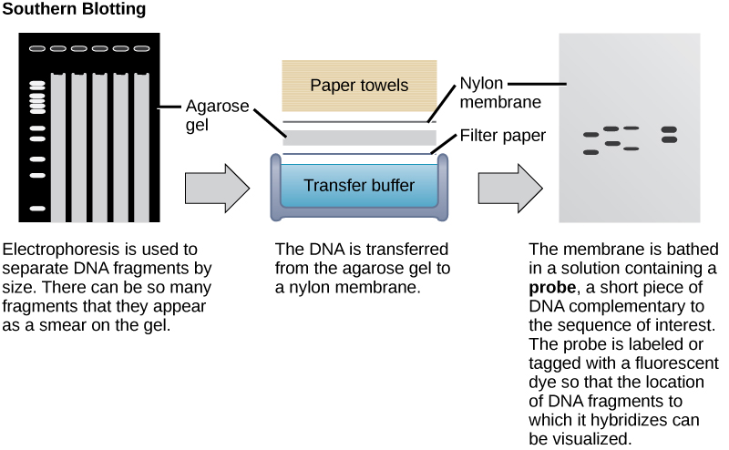
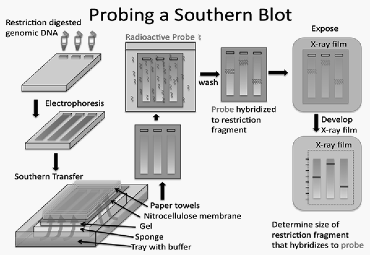
DNA is negatively charged and moves toward the positive electrode in agaroseelectrophoresis; smaller linear fragments usually migrate farther. A size ladder calibrates fragment length. A Southern blot transfers separated DNA to a membrane and uses a complementary probe after strand denaturation. Band position indicates fragment size and intensity can support abundance comparisons within assay limits. Northern blots detect RNA, and Western blots detect proteins using antibodies. A changed restriction site can alter a Southern band without changing the total number of gene copies.
Read sequence from terminated or parallel products
Sanger sequencing uses occasional chain-terminating dideoxynucleotides lacking a 3′ hydroxyl. Order labeled fragments from shortest to longest to read the newly synthesized strand 5′→3′; reverse-complement it when asked for the template. Massively parallel sequencing reads many fragments, then uses alignment or assembly; repeated regions and inadequate coverage complicate interpretation. Distinguish a sequencing read from a consensus, and a variant from a proven functional cause. Include quality checks and an independent method when a consequential result is uncertain.
Question.Primers bound to opposite strands face inward across a 300-base region. Starting from 5 ideal templates, how many copies after 8 cycles? A Sanger ladder read shortest to longest gives AGTC: what is the template written 5′→3′?
Show reasoning
Ideal copies = 5 × 2⁸ = 1280. This is an upper-model calculation; actual yield depends on efficiency. The synthesized strand is 5′-AGTC-3′ and the template is antiparallel 3′-TCAG-5′, or 5′-GACT-3′. Outward-facing primers do not exponentially amplify that intervening segment in the usual linear-template assay.
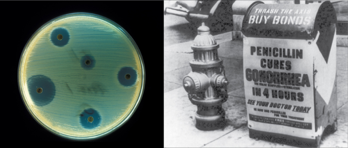
Figure 17.2 Fungi, bacteria, and other organisms that have antimicrobial properties produce antibiotics. The first antibiotic discovered was penicillin. Pharmaceutical companies now commercially produce and test antibiotics for their potential to inhibit bacterial growth. (credit "advertisement": modification of work by NIH; credit "test plate": modification of work by Don Stalons/CDC; scale-bar data from Matt Russell)
Mary Ann Clark, Matthew Douglas, and Jung Choi, Biology 2E, OpenStax. (credit "advertisement": modification of work by NIH; credit "test plate": modification of work by Don Stalons/CDC; scale-bar data from Matt Russell) · CC BY-NC-SA 4.0
· Original image ↗ · Access for free ↗Figure 17.3 — 17.1 Biotechnology
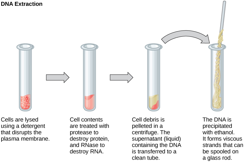
Figure 17.3 This diagram shows the basic method of DNA extraction.
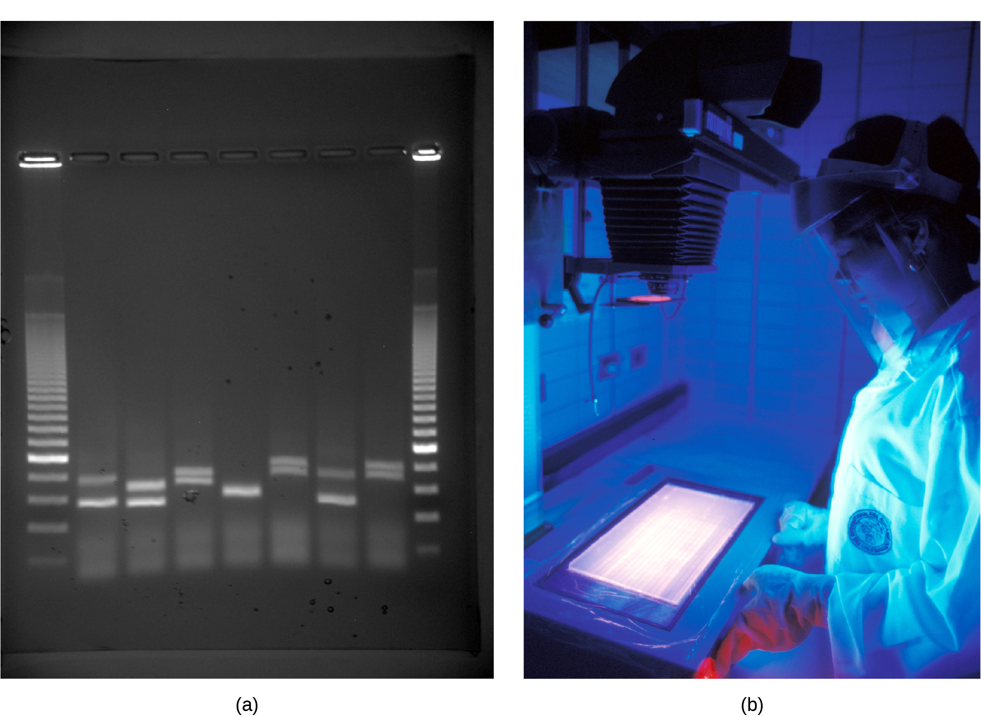
Figure 17.4 a) Shown are DNA fragments from seven samples run on a gel, stained with a fluorescent dye, and viewed under UV light; and b) a researcher from International Rice Research Institute, reviewing DNA profiles using UV light. (credit: a: James Jacob, Tompkins Cortland Community College b: International Rice Research Institute)
Mary Ann Clark, Matthew Douglas, and Jung Choi, Biology 2E, OpenStax. (credit: a: James Jacob, Tompkins Cortland Community College b: International Rice Research Institute) · CC BY-NC-SA 4.0
· Original image ↗ · Access for free ↗Figure 17.5 — 17.1 Biotechnology
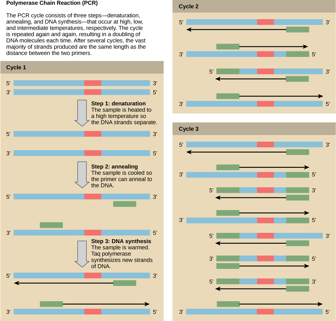
Figure 17.5 Scientists use polymerase chain reaction, or PCR, to amplify a specific DNA sequence. Primers—short pieces of DNA complementary to each end of the target sequence combine with genomic DNA, Taq polymerase, and deoxynucleotides. Taq polymerase is a DNA polymerase isolated from the thermostable bacterium Thermus aquaticus that is able to withstand the high temperatures that scientists use in PCR. Thermus aquaticus grows in the Lower Geyser Basin of Yellowstone National Park. Reverse transcriptase PCR (RT-PCR) is similar to PCR, but cDNA is made from an RNA template before PCR begins.
Figure 17.6 Scientists use Southern blotting to find a particular sequence in a DNA sample. Scientists separate DNA fragments on a gel, transfer them to a nylon membrane, and incubate them with a DNA probe complementary to the sequence of interest. Northern blotting is similar to Southern blotting, but scientists run RNA on the gel instead of DNA. In Western blotting, scientists run proteins on a gel and detect them using antibodies.
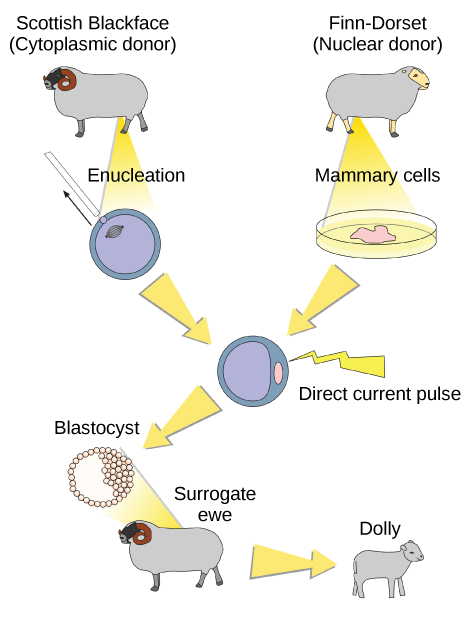
Figure 17.8 Dolly the sheep was the first mammal to be cloned. To create Dolly, they removed the nucleus from a donor egg cell. They then introduced the nucleus from a second sheep into the cell, which divided to the blastocyst stage before they implanted it in a surrogate mother. (credit: modification of work by "Squidonius"/Wikimedia Commons)
Mary Ann Clark, Matthew Douglas, and Jung Choi, Biology 2E, OpenStax. (credit: modification of work by "Squidonius"/Wikimedia Commons) · CC BY-NC-SA 4.0
· Original image ↗ · Access for free ↗Figure 17.9 — 17.1 Biotechnology
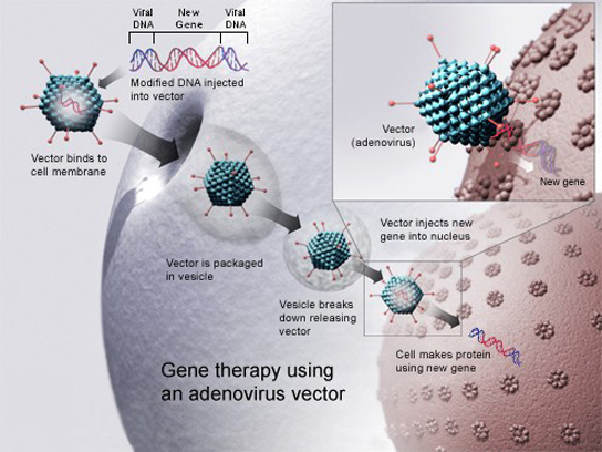
Figure 17.9 Gene therapy using an adenovirus vector can be used to cure certain genetic diseases in which a person has a defective gene. (credit: NIH)
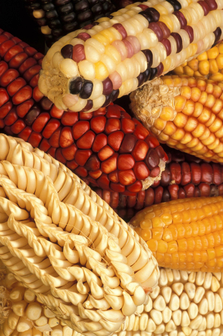
Figure 17.10 Corn, a major agricultural crop used to create products for a variety of industries, is often modified through plant biotechnology. (credit: Keith Weller, USDA)
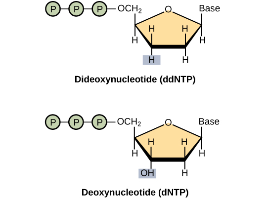
Figure 17.13 A dideoxynucleotide is similar in structure to a deoxynucleotide, but is missing the 3' hydroxyl group (indicated by the box). When a dideoxynucleotide is incorporated into a DNA strand, DNA synthesis stops.
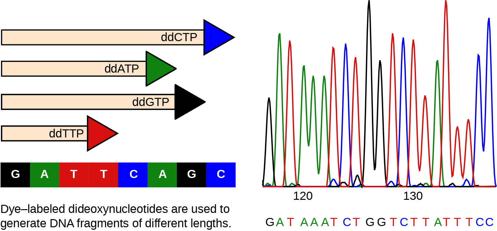
Figure 17.14 This figure illustrates Frederick Sanger's dideoxy chain termination method. Using dideoxynucleotides, the DNA fragment can terminate at different points. The DNA separates on the basis of size, and we can read these bands based on the fragments’ size.
Figure 14.7 DNA has (a) a double helix structure and (b) phosphodiester bonds; the dotted lines between Thymine and Adenine and Guanine and Cytosine represent hydrogen bonds. The (c) major and minor grooves are binding sites for DNA binding proteins during processes such as transcription (the copying of RNA from DNA) and replication.
Figure 14.7 DNA has (a) a double helix structure and (b) phosphodiester bonds; the dotted lines between Thymine and Adenine and Guanine and Cytosine represent hydrogen bonds. The (c) major and minor grooves are binding sites for DNA binding proteins during processes such as transcription (the copying of RNA from DNA) and replication.
Figure 10.16 The role of normal p53 is to monitor DNA and the supply of oxygen (hypoxia is a condition of reduced oxygen supply). If damage is detected, p53 triggers repair mechanisms. If repairs are unsuccessful, p53 signals apoptosis. A cell with an abnormal p53 protein cannot repair damaged DNA and thus cannot signal apoptosis. Cells with abnormal p53 can become cancerous. (credit: modification of work by Thierry Soussi)
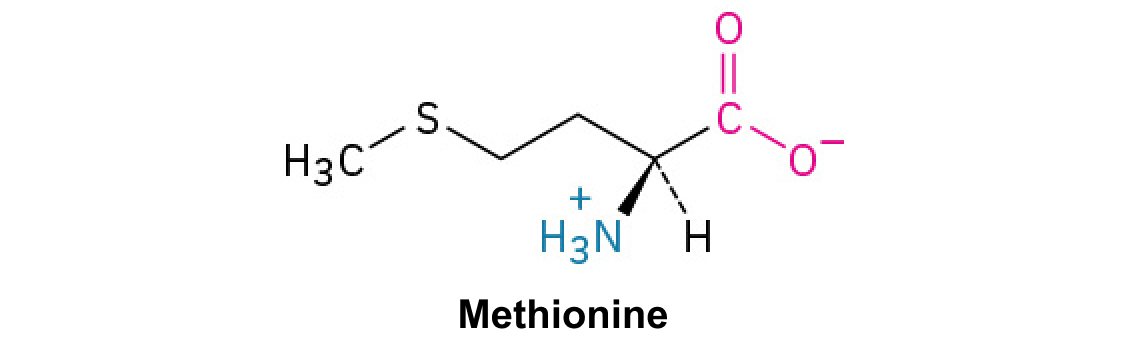
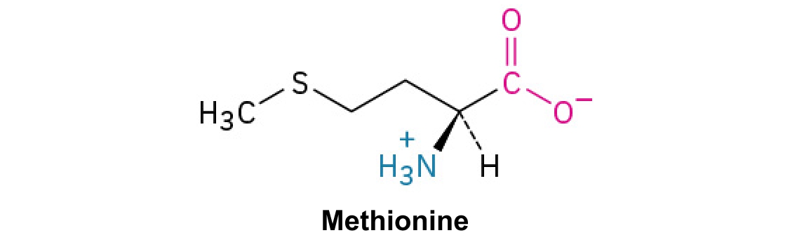
A nitrogen-containing functional group. A free amino acid’s amino group is typically protonated (NH₃⁺) near physiological pH; proline’s ring nitrogen is an exception to this formula.
Figure 10.15 Cell Division and Apoptosis. In an adult organism, normal cell division is balanced by apoptosis (programmed cell death) to maintain a constant cell number in homeostasis. Either an increase in cell division or a decrease in apoptosis leads an increase in the number of cells and tumor formation. Credit: Rao, A. and Ryan, K. Department of Biology, Texas A&M University.
Rao, A. and Ryan, K. Department of Biology, Texas A&M University. Mary Ann Clark, Matthew Douglas, and Jung Choi, Biology 2e, OpenStax. · CC BY-NC-SA 4.0
· Original image ↗ · Access for free ↗
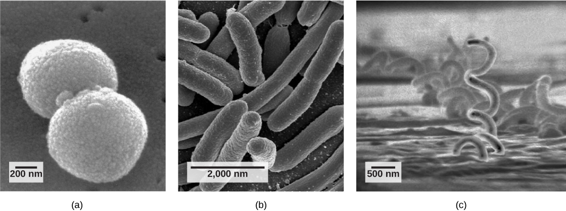
Figure 22.9 Common prokaryotic cell types. Prokaryotes fall into three basic categories based on their shape, visualized here using scanning electron microscopy: (a) cocci, or spherical (a pair is shown); (b) bacilli, or rod-shaped; and (c) spirilli, or spiral-shaped. (credit a: modification of work by Janice Haney Carr, Dr. Richard Facklam, CDC; credit c: modification of work by Dr. David Cox; scale-bar data from Matt Russell)
Mary Ann Clark, Matthew Douglas, and Jung Choi, Biology 2E, OpenStax. (credit a: modification of work by Janice Haney Carr, Dr. Richard Facklam, CDC; credit c: modification of work by Dr. David Cox; scale-bar data from Matt Russell) · CC BY-NC-SA 4.0
· Original image ↗ · Access for free ↗
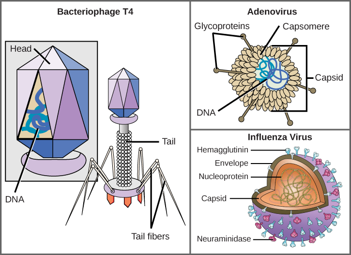
Figure 21.5 Complex Viruses. Viruses can be either complex or relatively simple in shape. This figure shows three relatively complex virions: the bacteriophage T4, with its DNA-containing head group and tail fibers that attach to host cells; adenovirus, which uses spikes from its capsid to bind to host cells; and the influenza virus, which uses glycoproteins embedded in its envelope to bind to host cells. The influenza virus also has matrix proteins, internal to the envelope, which help stabilize the virion’s shape. (credit “bacteriophage, adenovirus”: modification of work by NCBI, NIH; credit "influenza virus": modification of work by Dan Higgins, Centers for Disease Control and Prevention)
Mary Ann Clark, Matthew Douglas, and Jung Choi, Biology 2E, OpenStax. (credit “bacteriophage, adenovirus”: modification of work by NCBI, NIH; credit "influenza virus": modification of work by Dan Higgins, Centers for Disease Control and Prevention) · CC BY-NC-SA 4.0
· Original image ↗ · Access for free ↗
Figure 10.15 Cell Division and Apoptosis. In an adult organism, normal cell division is balanced by apoptosis (programmed cell death) to maintain a constant cell number in homeostasis. Either an increase in cell division or a decrease in apoptosis leads an increase in the number of cells and tumor formation. Credit: Rao, A. and Ryan, K. Department of Biology, Texas A&M University.
Rao, A. and Ryan, K. Department of Biology, Texas A&M University. Mary Ann Clark, Matthew Douglas, and Jung Choi, Biology 2e, OpenStax. · CC BY-NC-SA 4.0
· Original image ↗ · Access for free ↗
Figure 10.15 Cell Division and Apoptosis. In an adult organism, normal cell division is balanced by apoptosis (programmed cell death) to maintain a constant cell number in homeostasis. Either an increase in cell division or a decrease in apoptosis leads an increase in the number of cells and tumor formation. Credit: Rao, A. and Ryan, K. Department of Biology, Texas A&M University.
Rao, A. and Ryan, K. Department of Biology, Texas A&M University. Mary Ann Clark, Matthew Douglas, and Jung Choi, Biology 2e, OpenStax. · CC BY-NC-SA 4.0
· Original image ↗ · Access for free ↗
A DNAcarrier, such as a plasmid, used to introduce and maintain an inserted sequence in a host; an expression vector also provides signals for producing the encoded RNA or protein.
Figure 15.4 This figure shows the genetic code for translating each nucleotide triplet in mRNA into an amino acid or a termination signal in a protein. (credit: modification of work by NIH)
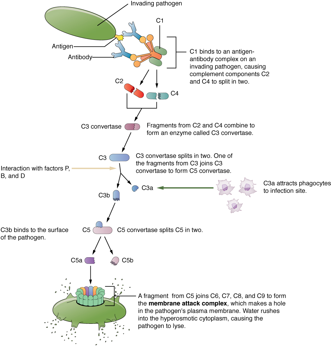
Figure 21.13 Complement Cascade and Function The classical pathway, used during adaptive immune responses, occurs when C1 reacts with antibodies that have bound an antigen.
Figure 14.14 First Components of DNA Replication. As DNA replication begins, DNA Helicase, a large enzyme, separates the two strands of DNA so that they can act as templates for replication. Single-strand binding proteins bind to each strand to stabilize and prevent them from reforming the double helix. Primase, an RNA polymerase, binds to the single stranded DNA and synthesizes a short RNA primer in the 5’ to 3’ direction that is antiparallel to the parental strand. This RNA primer allows for DNA polymerase to begin replicating the DNA. Topoisomerase binds to the double helix upstream of the replication fork to prevent additional coiling by making small cuts in one of the DNA strands.
Credit: Rao, A., Ryan, K. Fletcher, S. and Tag, A. Department of Biology, Texas A&M University.
Figure 14.5 The purines have a double ring structure with a six-membered ring fused to a five-membered ring. Pyrimidines are smaller in size; they have a single six-membered ring structure.
Figure 17.14 This figure illustrates Frederick Sanger's dideoxy chain termination method. Using dideoxynucleotides, the DNA fragment can terminate at different points. The DNA separates on the basis of size, and we can read these bands based on the fragments’ size.
Figure 14.14 First Components of DNA Replication. As DNA replication begins, DNA Helicase, a large enzyme, separates the two strands of DNA so that they can act as templates for replication. Single-strand binding proteins bind to each strand to stabilize and prevent them from reforming the double helix. Primase, an RNA polymerase, binds to the single stranded DNA and synthesizes a short RNA primer in the 5’ to 3’ direction that is antiparallel to the parental strand. This RNA primer allows for DNA polymerase to begin replicating the DNA. Topoisomerase binds to the double helix upstream of the replication fork to prevent additional coiling by making small cuts in one of the DNA strands.
Credit: Rao, A., Ryan, K. Fletcher, S. and Tag, A. Department of Biology, Texas A&M University.
Figure 14.14 First Components of DNA Replication. As DNA replication begins, DNA Helicase, a large enzyme, separates the two strands of DNA so that they can act as templates for replication. Single-strand binding proteins bind to each strand to stabilize and prevent them from reforming the double helix. Primase, an RNA polymerase, binds to the single stranded DNA and synthesizes a short RNA primer in the 5’ to 3’ direction that is antiparallel to the parental strand. This RNA primer allows for DNA polymerase to begin replicating the DNA. Topoisomerase binds to the double helix upstream of the replication fork to prevent additional coiling by making small cuts in one of the DNA strands.
Credit: Rao, A., Ryan, K. Fletcher, S. and Tag, A. Department of Biology, Texas A&M University.
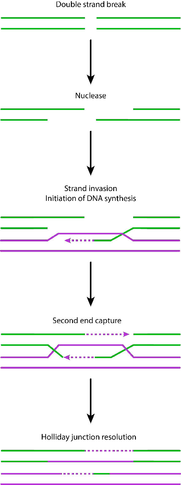
Figure 7.47 - Repair of DSBs by homologous recombination - Image by Pehr Jacobson
Pehr Jacobson, via Kevin Ahern, Indira Rajagopal, and Taralyn Tan, Biochemistry Free For All, Oregon State University, via Biology LibreTexts. · CC BY-NC-SA 4.0
· Original image ↗ · Access for free ↗
Figure 14.14 First Components of DNA Replication. As DNA replication begins, DNA Helicase, a large enzyme, separates the two strands of DNA so that they can act as templates for replication. Single-strand binding proteins bind to each strand to stabilize and prevent them from reforming the double helix. Primase, an RNA polymerase, binds to the single stranded DNA and synthesizes a short RNA primer in the 5’ to 3’ direction that is antiparallel to the parental strand. This RNA primer allows for DNA polymerase to begin replicating the DNA. Topoisomerase binds to the double helix upstream of the replication fork to prevent additional coiling by making small cuts in one of the DNA strands.
Credit: Rao, A., Ryan, K. Fletcher, S. and Tag, A. Department of Biology, Texas A&M University.
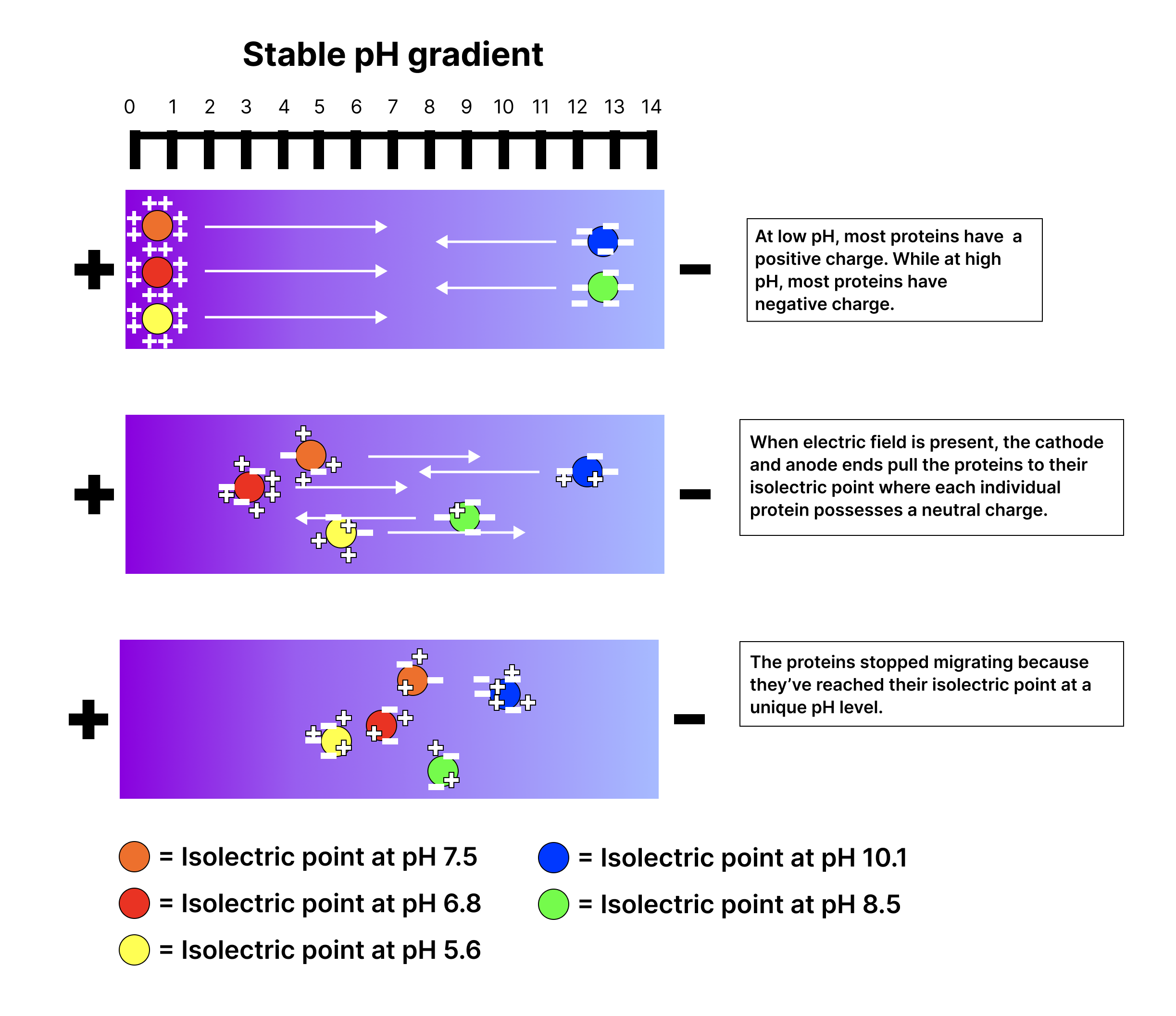
An electrical property that can be positive or negative. Opposite charges attract and like charges repel; net charge is the sum of all charges on a particle.
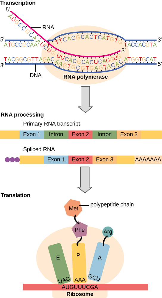
Figure 15.11 Eukaryotic pre-mRNA processing. In addition to 5’ Cap and 3’ Poly-A Tail addition, introns must be precisely removed and exons joined to generate a functional mRNA. Nucleotides upstream (towards the 5’cap) of the translation START codon are part of the 5’ untranslated region (5’ UTR). Nucleotides downstream (towards 3’end) of the STOP codon form the 3’ UTR. Both 5’ and 3’ UTRs are important for regulating translation initiation and mRNA stability. Credit: Rao, A., Ryan, K. Fletcher, S. and Tag, A. Department of Biology, Texas A&M University.
Rao, A., Ryan, K. Fletcher, S. and Tag, A. Department of Biology, Texas A&M University.; Mary Ann Clark, Matthew Douglas, and Jung Choi, Biology 2e, OpenStax. · CC BY-NC-SA 4.0
· Original image ↗ · Access for free ↗
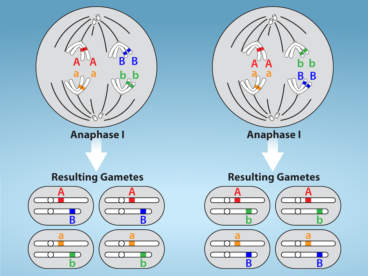
Figure 11.5 Random, independent assortment during metaphase I is demonstrated by considering a cell with a set of two chromosomes (n = 2).
There are two possible homologous chromosome arrangements at the equatorial plane in metaphase I, that are then separated during anaphase I.
The total possible number of different gametes is 2 n , where n equals the number of chromosomes in a set. In this example, there are four possible
genetic combinations for the gametes. With n = 23 in human cells, there are over eight million possible combinations of paternal and maternal
chromosomes. Credit: Rao, A.and Fletcher, S. Department of Biology, Texas A&M University.
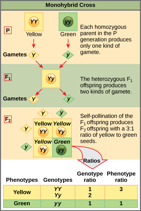
Figure 12.4 In the P generation, pea plants that are true-breeding for the dominant yellow phenotype are crossed with plants with the recessive green phenotype. This cross produces F 1 heterozygotes with a yellow phenotype. Punnett square analysis can be used to predict the genotypes of the F 2 generation.
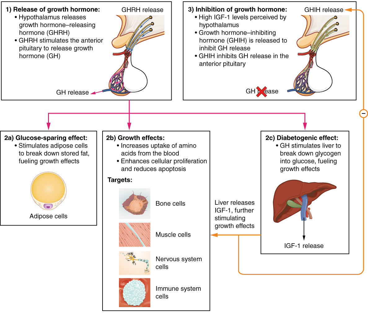
Figure 17.10 Hormonal Regulation of Growth Growth hormone (GH) directly accelerates the rate of protein synthesis in skeletal muscle and bones. Insulin-like growth factor 1 (IGF-1) is activated by growth hormone and indirectly supports the formation of new proteins in muscle cells and bone.
Figure 15.11 Eukaryotic pre-mRNA processing. In addition to 5’ Cap and 3’ Poly-A Tail addition, introns must be precisely removed and exons joined to generate a functional mRNA. Nucleotides upstream (towards the 5’cap) of the translation START codon are part of the 5’ untranslated region (5’ UTR). Nucleotides downstream (towards 3’end) of the STOP codon form the 3’ UTR. Both 5’ and 3’ UTRs are important for regulating translation initiation and mRNA stability. Credit: Rao, A., Ryan, K. Fletcher, S. and Tag, A. Department of Biology, Texas A&M University.
Rao, A., Ryan, K. Fletcher, S. and Tag, A. Department of Biology, Texas A&M University.; Mary Ann Clark, Matthew Douglas, and Jung Choi, Biology 2e, OpenStax. · CC BY-NC-SA 4.0
· Original image ↗ · Access for free ↗
The exponent needed to obtain a number from a chosen fixed number. For decimal logarithms, log₁₀(100) = 2 because 10² = 100; pH uses the negative decimal logarithm of hydrogen ion activity.
Figure 10.16 The role of normal p53 is to monitor DNA and the supply of oxygen (hypoxia is a condition of reduced oxygen supply). If damage is detected, p53 triggers repair mechanisms. If repairs are unsuccessful, p53 signals apoptosis. A cell with an abnormal p53 protein cannot repair damaged DNA and thus cannot signal apoptosis. Cells with abnormal p53 can become cancerous. (credit: modification of work by Thierry Soussi)
Figure 4.18 Membrane and secretory proteins are synthesized in the rough endoplasmic reticulum (RER). The RER also sometimes modifies proteins. In this illustration, a (green) integral membrane protein is modified by attachment of a (purple) carbohydrate in the ER. Vesicles with the integral protein bud from the ER and fuse with the Golgi apparatus' cis face. As the protein passes along the Golgi’s cisternae, the addition of more carbohydrates further modifies it. After its synthesis is complete, it exits as an integral membrane protein of the vesicle that buds from the Golgi’s trans face. When the vesicle fuses with the cell membrane, the protein becomes an integral portion of that cell membrane. (credit: modification of work by Magnus Manske)
A supporting material, such as a porous gel or the solid support in a chromatography column. Its properties influence movement and binding of sample molecules.
A thin boundary or sheet. Cell membranes are lipid bilayers containing proteins; blotting membranes are synthetic supports that hold proteins or nucleic acids for detection.
Figure 15.11 Eukaryotic pre-mRNA processing. In addition to 5’ Cap and 3’ Poly-A Tail addition, introns must be precisely removed and exons joined to generate a functional mRNA. Nucleotides upstream (towards the 5’cap) of the translation START codon are part of the 5’ untranslated region (5’ UTR). Nucleotides downstream (towards 3’end) of the STOP codon form the 3’ UTR. Both 5’ and 3’ UTRs are important for regulating translation initiation and mRNA stability. Credit: Rao, A., Ryan, K. Fletcher, S. and Tag, A. Department of Biology, Texas A&M University.
Rao, A., Ryan, K. Fletcher, S. and Tag, A. Department of Biology, Texas A&M University.; Mary Ann Clark, Matthew Douglas, and Jung Choi, Biology 2e, OpenStax. · CC BY-NC-SA 4.0
· Original image ↗ · Access for free ↗
Figure 4.18 Membrane and secretory proteins are synthesized in the rough endoplasmic reticulum (RER). The RER also sometimes modifies proteins. In this illustration, a (green) integral membrane protein is modified by attachment of a (purple) carbohydrate in the ER. Vesicles with the integral protein bud from the ER and fuse with the Golgi apparatus' cis face. As the protein passes along the Golgi’s cisternae, the addition of more carbohydrates further modifies it. After its synthesis is complete, it exits as an integral membrane protein of the vesicle that buds from the Golgi’s trans face. When the vesicle fuses with the cell membrane, the protein becomes an integral portion of that cell membrane. (credit: modification of work by Magnus Manske)
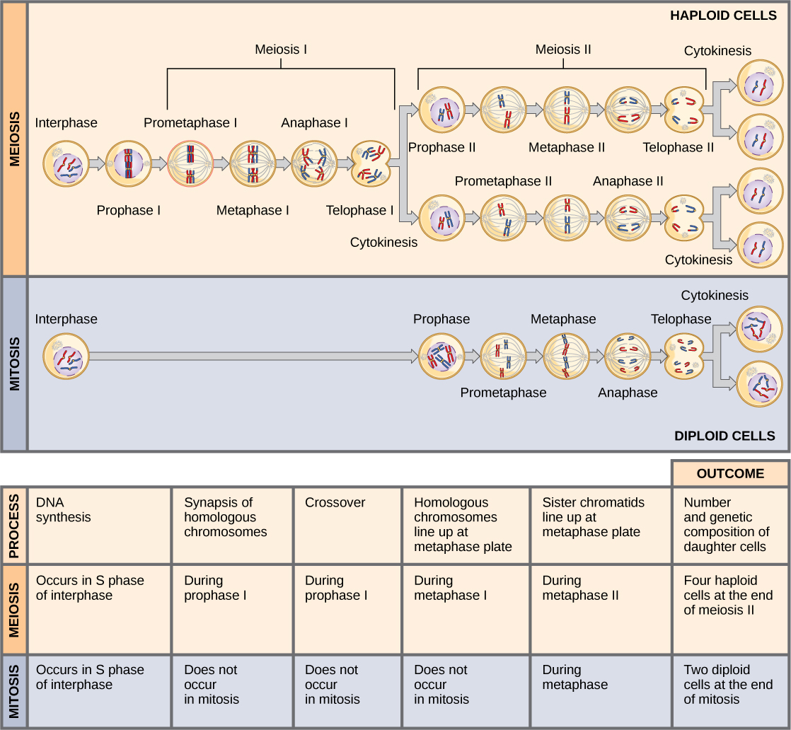
Figure 11.8 Meiosis and mitosis are both preceded by one cycle of DNA replication; however, meiosis includes two nuclear divisions. The four daughter cells resulting from meiosis are haploid and genetically distinct. The daughter cells resulting from mitosis are diploid and identical to the parent cell.
Figure 17.5 Scientists use polymerase chain reaction, or PCR, to amplify a specific DNA sequence. Primers—short pieces of DNA complementary to each end of the target sequence combine with genomic DNA, Taq polymerase, and deoxynucleotides. Taq polymerase is a DNA polymerase isolated from the thermostable bacterium Thermus aquaticus that is able to withstand the high temperatures that scientists use in PCR. Thermus aquaticus grows in the Lower Geyser Basin of Yellowstone National Park. Reverse transcriptase PCR (RT-PCR) is similar to PCR, but cDNA is made from an RNA template before PCR begins.
Figure 12.4 In the P generation, pea plants that are true-breeding for the dominant yellow phenotype are crossed with plants with the recessive green phenotype. This cross produces F 1 heterozygotes with a yellow phenotype. Punnett square analysis can be used to predict the genotypes of the F 2 generation.
Figure 14.7 DNA has (a) a double helix structure and (b) phosphodiester bonds; the dotted lines between Thymine and Adenine and Guanine and Cytosine represent hydrogen bonds. The (c) major and minor grooves are binding sites for DNA binding proteins during processes such as transcription (the copying of RNA from DNA) and replication.
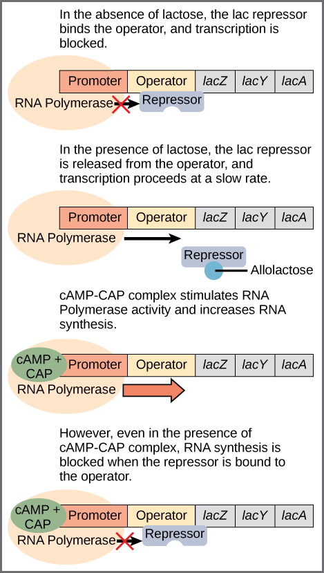
Figure 16.6 Regulation of the lac operon. Transcription of the lac operon is carefully regulated so that its expression only occurs when glucose is limited and lactose is present to serve as an alternative fuel source.
Figure 14.14 First Components of DNA Replication. As DNA replication begins, DNA Helicase, a large enzyme, separates the two strands of DNA so that they can act as templates for replication. Single-strand binding proteins bind to each strand to stabilize and prevent them from reforming the double helix. Primase, an RNA polymerase, binds to the single stranded DNA and synthesizes a short RNA primer in the 5’ to 3’ direction that is antiparallel to the parental strand. This RNA primer allows for DNA polymerase to begin replicating the DNA. Topoisomerase binds to the double helix upstream of the replication fork to prevent additional coiling by making small cuts in one of the DNA strands.
Credit: Rao, A., Ryan, K. Fletcher, S. and Tag, A. Department of Biology, Texas A&M University.
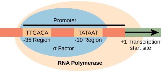
Figure 15.8 The σ subunit of prokaryotic RNA polymerase recognizes consensus sequences found in the promoter region upstream of the transcription start site. The σ subunit dissociates from the polymerase after transcription has been initiated.
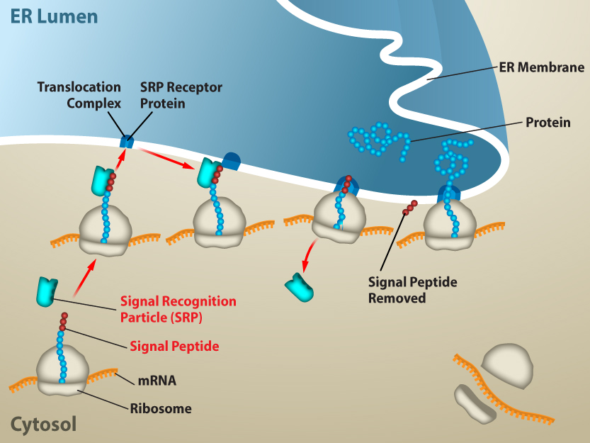
Figure 15.20 Proteins are co-translationally targeted into the ER for secretion. Proteins that will be secreted from the cell will contain a signal sequence at the N-terminus. The signal will be recognized by SRP as soon as the amino acids emerge from the ribosome, and the ribosome will be targeted to the translocation channel in the ER membrane. The rest of the protein will go directly from the ribosome, across the ER membrane and into the ER lumen. From the ER, proteins can be secreted from the cell via vesicle trafficking. Credit: Rao, A. and Ryan, K. Department of Biology, Texas A&M University.
Rao, A. and Ryan, K. Department of Biology, Texas A&M University.; Mary Ann Clark, Matthew Douglas, and Jung Choi, Biology 2e, OpenStax. · CC BY-NC-SA 4.0
· Original image ↗ · Access for free ↗
Figure 14.14 First Components of DNA Replication. As DNA replication begins, DNA Helicase, a large enzyme, separates the two strands of DNA so that they can act as templates for replication. Single-strand binding proteins bind to each strand to stabilize and prevent them from reforming the double helix. Primase, an RNA polymerase, binds to the single stranded DNA and synthesizes a short RNA primer in the 5’ to 3’ direction that is antiparallel to the parental strand. This RNA primer allows for DNA polymerase to begin replicating the DNA. Topoisomerase binds to the double helix upstream of the replication fork to prevent additional coiling by making small cuts in one of the DNA strands.
Credit: Rao, A., Ryan, K. Fletcher, S. and Tag, A. Department of Biology, Texas A&M University.
Figure 12.19 Reaction diagrams for an endothermic process in the absence (red curve) and presence (blue curve) of a catalyst. The catalyzed pathway involves a two-step mechanism (note the presence of two transition states) and an intermediate species (represented by the valley between the two transitions states).
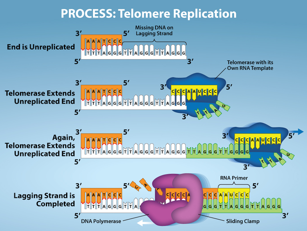
Figure 14.15 The ends of linear chromosomes are maintained by the action of the telomerase enzyme. Credit: Rao, A. and Fletcher, S. Department of Biology, Texas A&M University.
Figure 15.3 Instructions on DNA are transcribed onto messenger RNA. Ribosomes are able to read the genetic information inscribed on a strand of messenger RNA and use this information to string amino acids together into a protein.
Figure 14.5 The purines have a double ring structure with a six-membered ring fused to a five-membered ring. Pyrimidines are smaller in size; they have a single six-membered ring structure.
Figure 15.13 Pre-mRNA splicing involves the precise removal of introns from the primary RNA transcript. The splicing process is catalyzed by protein complexes called spliceosomes that are composed of proteins and RNA molecules called small nuclear RNAs (snRNAs). Spliceosomes recognize sequences at the 5' and 3' end of the intron. Rao, A. and Ryan, K. Department of Biology, Texas A&M University.
Rao, A. and Ryan, K. Department of Biology, Texas A&M University.; Mary Ann Clark, Matthew Douglas, and Jung Choi, Biology 2e, OpenStax. · CC BY-NC-SA 4.0
· Original image ↗ · Access for free ↗
Sanger sequencing
A DNA sequencing method identifying bases from differently terminated synthesis products.
Figure 7.45 - Non-homologous end joining (left) versus homologous recombination (right) - Wikipedia
Wikipedia, as credited by Kevin Ahern, Indira Rajagopal, and Taralyn Tan, Biochemistry Free For All, Oregon State University, via Biology LibreTexts. · Source attribution
· Original image ↗ · Access for free ↗
The amount of a substance that can dissolve in a solvent under specified conditions. Soluble means able to dissolve; insoluble means only very slightly soluble in that solvent.
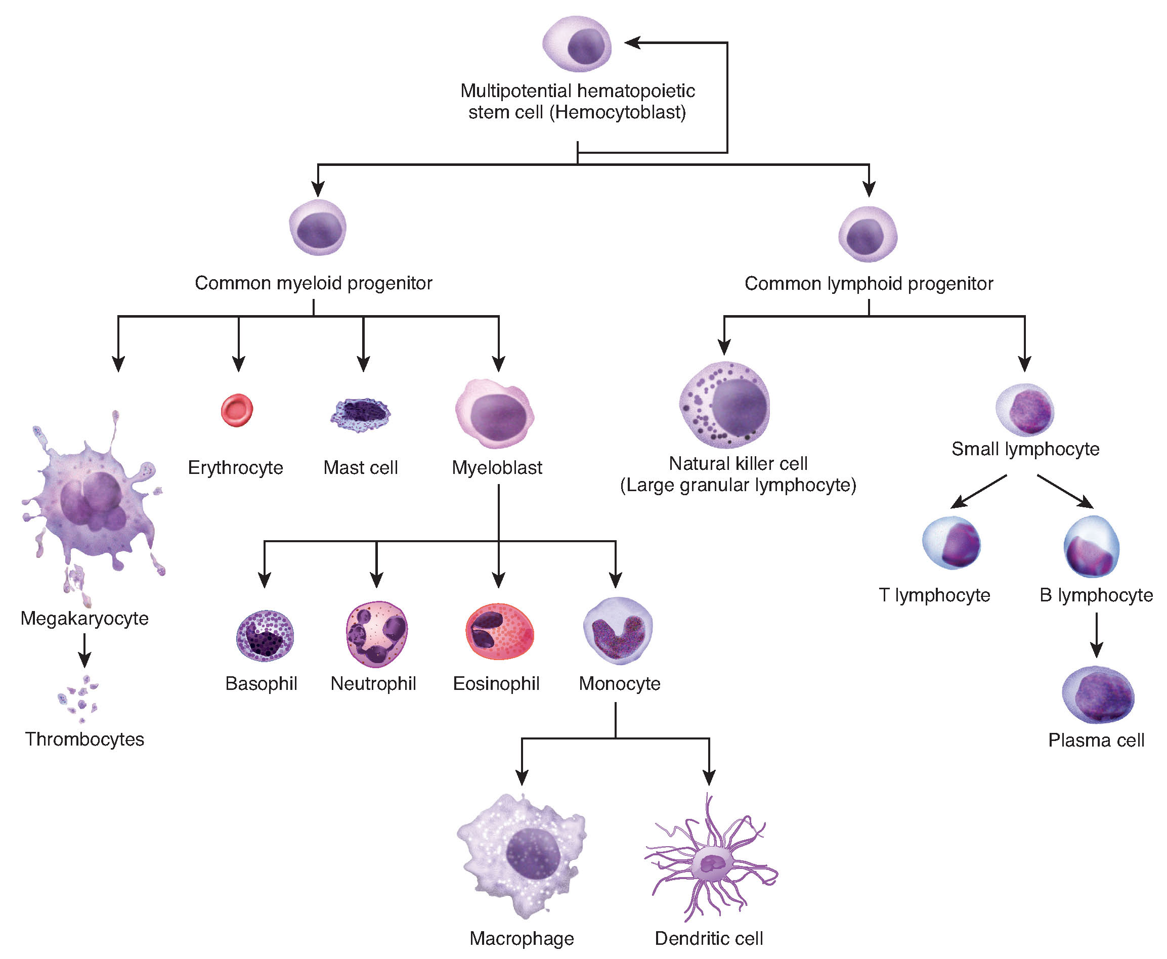
Figure 3.34 Hematopoiesis The process of hematopoiesis involves the differentiation of multipotent cells into blood and immune cells. The multipotent hematopoietic stem cells give rise to many different cell types, including the cells of the immune system and red blood cells.
Figure 14.15 The ends of linear chromosomes are maintained by the action of the telomerase enzyme. Credit: Rao, A. and Fletcher, S. Department of Biology, Texas A&M University.
Figure 15.7 Messenger RNA is a copy of protein-coding information in the coding strand of DNA, with the substitution of U in the RNA for T in the coding sequence. However, new RNA nucleotides base pair with the nucleotides of the template strand. RNA is synthesized in its 5'-3' direction, using the enzyme RNA polymerase. As the template is read, the DNA unwinds ahead of the polymerase and then rewinds behind it.
A fleeting, high-energy arrangement at a reaction barrier. It is an energy maximum along the reaction path, rather than a stable reaction intermediate.
Figure 12.19 Reaction diagrams for an endothermic process in the absence (red curve) and presence (blue curve) of a catalyst. The catalyzed pathway involves a two-step mechanism (note the presence of two transition states) and an intermediate species (represented by the valley between the two transitions states).
Figure 15.18 Translation begins when an initiator tRNA anticodon recognizes a start codon on mRNA bound to a small ribosomal subunit. The large ribosomal subunit joins the small subunit, and a second tRNA is recruited. As the mRNA moves relative to the ribosome, successive tRNAs move through the ribosome and the polypeptide chain is formed. Entry of a release factor into the A site terminates translation and the components dissociate.
Figure 10.15 Cell Division and Apoptosis. In an adult organism, normal cell division is balanced by apoptosis (programmed cell death) to maintain a constant cell number in homeostasis. Either an increase in cell division or a decrease in apoptosis leads an increase in the number of cells and tumor formation. Credit: Rao, A. and Ryan, K. Department of Biology, Texas A&M University.
Rao, A. and Ryan, K. Department of Biology, Texas A&M University. Mary Ann Clark, Matthew Douglas, and Jung Choi, Biology 2e, OpenStax. · CC BY-NC-SA 4.0
· Original image ↗ · Access for free ↗
No matching terms. Try a different name or abbreviation.
17.1 Biotechnology
OpenStax · 17.1 Biotechnology ↗ Instructional page including learning objectives, examples, figures, source tables and questions. Site navigation and embedded media players are omitted; source media links remain available.
Learning Objectives
By the end of this section, you will be able to do the following:
Describe biotechnology uses in medicine and agriculture
Biotechnology is the use of biological agents for technological advancement. Biotechnology was used for breeding livestock and crops long before people understood the scientific basis of these techniques. Since the discovery of the structure of DNA in 1953, the biotechnology field has grown rapidly through both academic research and private companies. The primary applications of this technology are in medicine (vaccine and antibiotic production) and agriculture (crop genetic modification in order to increase yields). Biotechnology also has many industrial applications, such as fermentation, treating oil spills, and producing biofuels (Figure 17.2).
Figure 17.2 — 17.1 Biotechnology
Figure 17.2 Fungi, bacteria, and other organisms that have antimicrobial properties produce antibiotics. The first antibiotic discovered was penicillin. Pharmaceutical companies now commercially produce and test antibiotics for their potential to inhibit bacterial growth. (credit "advertisement": modification of work by NIH; credit "test plate": modification of work by Don Stalons/CDC; scale-bar data from Matt Russell)
Mary Ann Clark, Matthew Douglas, and Jung Choi, Biology 2E, OpenStax. (credit "advertisement": modification of work by NIH; credit "test plate": modification of work by Don Stalons/CDC; scale-bar data from Matt Russell) · CC BY-NC-SA 4.0
· Original image ↗ · Access for free ↗
Basic Techniques to Manipulate Genetic Material (DNA and RNA)
To study or manipulate nucleic acids, one must first isolate or extract the DNA or RNA from the cells. Researchers use various techniques to extract different types of DNA (Figure 17.3). Most nucleic acid extraction techniques involve steps to break open the cell and use enzymatic reactions to destroy all macromolecules that are not desired (such as unwanted molecule degradation and separation from the DNA sample). A lysis buffer (a solution which is mostly a detergent) breaks cells. Note that lysis means "to split". These enzymes break apart lipidmolecules in the cell membranes and nuclearmembranes. Enzymes such as proteases and ribonucleases (RNases) break down proteins and RNA, respectively. Using alcohol precipitates the DNA. Human genomic DNA is usually visible as a gelatinous, white mass. One can store the DNA samples frozen at –80°C for several years.
Figure 17.3 — 17.1 Biotechnology
Figure 17.3 This diagram shows the basic method of DNA extraction.
Scientists perform RNA analysis to study gene expression patterns in cells. RNA is naturally very unstable because RNAses are commonly present in nature and very difficult to inactivate. Similar to DNA, RNA extraction involves using various buffers and enzymes to inactivate macromolecules and preserve the RNA.
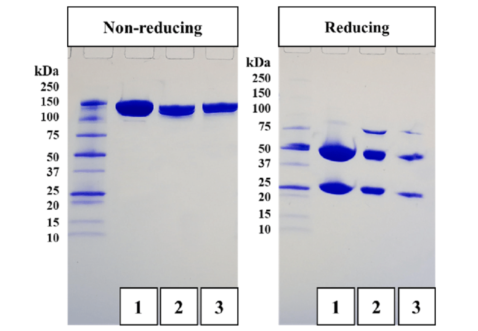
Because nucleic acids are negatively chargedions at neutral or basic pH in an aqueous environment, an electric field can mobilize them. Gel electrophoresis is a technique that scientists use to separate molecules on the basis of size, using this charge. One can separate the nucleic acids as whole chromosomes or fragments. The nucleic acids load into a slot near the semisolid, porous gel matrix's negative electrode, and pulled toward the positive electrode at the gel's opposite end. Smaller molecules move through the gel's pores faster than larger molecules. This difference in the migration rate separates the fragments on the basis of size. There are molecular weight standard samples that researchers can run alongside the molecules to provide a size comparison. We can observe nucleic acids in a gel matrix using various fluorescent or colored dyes. Distinct nucleic acid fragments appear as bands at specific distances from the gel's top (the negative electrode end) on the basis of their size (Figure 17.4). A mixture of genomic DNA fragments of varying sizes appear as a long smear; whereas, uncut genomic DNA is usually too large to run through the gel and forms a single large band at the gel's top.
Figure 17.4 — 17.1 Biotechnology
Figure 17.4 a) Shown are DNA fragments from seven samples run on a gel, stained with a fluorescent dye, and viewed under UV light; and b) a researcher from International Rice Research Institute, reviewing DNA profiles using UV light. (credit: a: James Jacob, Tompkins Cortland Community College b: International Rice Research Institute)
Mary Ann Clark, Matthew Douglas, and Jung Choi, Biology 2E, OpenStax. (credit: a: James Jacob, Tompkins Cortland Community College b: International Rice Research Institute) · CC BY-NC-SA 4.0
· Original image ↗ · Access for free ↗
Although genomic DNA is visible to the naked eye when it is extracted in bulk, DNA analysis often requires focusing on one or more specific genome regions. Polymerase chain reaction (PCR) is a technique that scientists use to amplify specific DNA regions for further analysis (Figure 17.5). Researchers use PCR for many purposes in laboratories, such as cloning gene fragments to analyze genetic diseases, identifying contaminant foreign DNA in a sample, and amplifying DNA for sequencing. More practical applications include determining paternity and detecting genetic diseases.
Figure 17.5 — 17.1 Biotechnology
Figure 17.5 Scientists use polymerase chain reaction, or PCR, to amplify a specific DNA sequence. Primers—short pieces of DNA complementary to each end of the target sequence combine with genomic DNA, Taq polymerase, and deoxynucleotides. Taq polymerase is a DNA polymerase isolated from the thermostable bacterium Thermus aquaticus that is able to withstand the high temperatures that scientists use in PCR. Thermus aquaticus grows in the Lower Geyser Basin of Yellowstone National Park. Reverse transcriptase PCR (RT-PCR) is similar to PCR, but cDNA is made from an RNA template before PCR begins.
Figure 17.6 Scientists use Southern blotting to find a particular sequence in a DNA sample. Scientists separate DNA fragments on a gel, transfer them to a nylon membrane, and incubate them with a DNA probe complementary to the sequence of interest. Northern blotting is similar to Southern blotting, but scientists run RNA on the gel instead of DNA. In Western blotting, scientists run proteins on a gel and detect them using antibodies.
In general, the word “cloning” means the creation of a perfect replica; however, in biology, the re-creation of a whole organism is referred to as “reproductive cloning.” Long before attempts were made to clone an entire organism, researchers learned how to reproduce desired regions or fragments of the genome, a process that is referred to as molecular cloning. The technique offered methods to create new medicines and to overcome difficulties with existing ones. When Lydia Villa-Komaroff, working in the Gilbert Lab at Harvard, published the first paper outlining the technique for producing synthetic insulin, diabetes researchers and patients received new hope in fighting the disease. Insulin at that time was only produced using pig and cow pancreases, and the life-saving substance was often in short supply. Synthetic insulin, once mass produced, would solve that problem for many patients. These early discoveries led to the "BioTech Boom," and spurred continued research and funding for newer and better ways to improve health.
Cloning small genome fragments allows researchers to manipulate and study specific genes (and their proteinproducts), or noncoding regions in isolation. A plasmid, or vector, is a small circular DNAmolecule that replicates independently of the chromosomalDNA. In cloning, scientists can use the plasmidmolecules to provide a "folder" in which to insert a desired DNA fragment. Plasmids are usually introduced into a bacterial host for proliferation. In the bacterial context, scientists call the DNA fragment from the human genome (or the genome of another studied organism) foreign DNA, or a transgene, to differentiate it from the bacterium's DNA, or the host DNA.
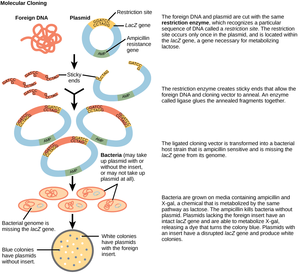
Plasmids occur naturally in bacterial populations (such as Escherichia coli) and have genes that can contribute favorable traits to the organism, such as antibiotic resistance (the ability to be unaffected by antibiotics). Scientists have repurposed and engineered plasmids as vectors for molecular cloning and the large-scale production of important reagents, such as insulin and human growth hormone. An important feature of plasmidvectors is the ease with which scientists can introduce a foreign DNA fragment via the multiple cloning site (MCS). The MCS is a short DNA sequence containing multiple sites that different commonly available restriction endonucleases can cut. Restriction endonucleases recognize specific DNA sequences and cut them in a predictable manner. They are naturally produced by bacteria as a defense mechanism against foreign DNA. Many restriction endonucleases make staggered cuts in the two DNA strands, such that the cut ends have a 2- or 4-base single-stranded overhang. Because these overhangs are capable of annealing with complementary overhangs, we call them “sticky ends.” Adding the enzymeDNA ligase permanently joins the DNA fragments via phosphodiester bonds. In this way, scientists can splice any DNA fragment generated by restriction endonuclease cleavage between the plasmidDNA's two ends that has been cut with the same restriction endonuclease (Figure 17.7).
Plasmids with foreign DNA inserted into them are called recombinant DNAmolecules because they are created artificially and do not occur in nature. They are also called chimeric molecules because the origin of different molecule parts of the molecules can be traced back to different species of biological organisms or even to chemical synthesis. We call proteins that are expressed from recombinant DNAmoleculesrecombinant proteins. Not all recombinant plasmids are capable of expressing genes. The recombinant DNA may need to move into a different vector (or host) that is better designed for gene expression. Scientists may also engineer plasmids to express proteins only when certain environmental factors stimulate them, so they can control the recombinant proteins' expression.
Visual Connection
Figure 17.7 — 17.1 Biotechnology
Figure 17.7 This diagram shows the steps involved in molecular cloning.
You are working in a molecular biology lab and, unbeknownst to you, your lab partner left the foreign genomic DNA that you are planning to clone on the lab bench overnight instead of storing it in the freezer. As a result, it was degraded by nucleases, but still used in the experiment. The plasmid, on the other hand, is fine. What results would you expect from your molecular cloning experiment?
Unicellular organisms, such as bacteria and yeast, naturally produce clones of themselves when they replicate asexually by binary fission; this is known as cellular cloning. The nuclearDNA duplicates by the process of mitosis, which creates an exact replica of the genetic material.
Reproductive Cloning
Reproductive cloning is a method scientists use to clone or identically copy an entire multicellular organism. Most multicellular organisms undergo reproduction by sexual means, which involves genetic hybridization of two individuals (parents), making it impossible to generate an identical copy or a clone of either parent. Recent advances in biotechnology have made it possible to artificially induce mammal asexual reproduction in the laboratory.
Parthenogenesis, or “virgin birth,” occurs when an embryo grows and develops without egg fertilization. This is a form of asexual reproduction. An example of parthenogenesis occurs in species in which the female lays an egg and if the egg is fertilized, it is a diploid egg and the individual develops into a female. If the egg is not fertilized, it remains a haploid egg and develops into a male. The unfertilized egg is a parthenogenic, or virgin egg. Some insects and reptiles lay parthenogenic eggs that can develop into adults.
Sexual reproduction requires two cells. When the haploid egg and sperm cells fuse, a diploidzygote results. The zygotenucleus contains the genetic information to produce a new individual. However, early embryonic development requires the cytoplasmic material contained in the egg cell. This idea forms the basis for reproductive cloning. Therefore, if we replace the egg cell's haploidnucleus with a diploidnucleus from the cell of any individual of the same species (a donor), it will become a zygote that is genetically identical to the donor. Somatic cellnuclear transfer is the technique of transferring a diploidnucleus into an enucleated egg. Scientists can use it for either therapeutic cloning or reproductive cloning.
The first cloned mammal was Dolly, a sheep born in 1996. The reproductive cloning success rate at the time was very low. Dolly lived for seven years and died of respiratory complications (Figure 17.8). There is speculation that because the cell DNA belongs to an older individual, DNA's age may affect a cloned individual's life expectancy. Since Dolly, scientists have cloned successfully several animals such as horses, bulls, and goats, although these animals often exhibit facial, limb, and cardiac abnormalities. There have been attempts at producing cloned human embryos as sources of embryonic stem cells for therapeutic purposes. Therapeutic cloning produces stem cells in the attempt to remedy detrimental diseases or defects (unlike reproductive cloning, which aims to reproduce an organism). While some have met therapeutic cloning efforts with resistance because of bioethical considerations, new discoveries have identified additional opportunities for stem cell therapies. For example, Freda Miller and Elaine Fuchs, working independently, discovered stem cells in different layers of the skin. These cells help the skin repair itself, and their discovery may have applications in treatments of skin disease and potentially other conditions, such as nerve damage.
Visual Connection
Figure 17.8 — 17.1 Biotechnology
Figure 17.8 Dolly the sheep was the first mammal to be cloned. To create Dolly, they removed the nucleus from a donor egg cell. They then introduced the nucleus from a second sheep into the cell, which divided to the blastocyst stage before they implanted it in a surrogate mother. (credit: modification of work by "Squidonius"/Wikimedia Commons)
Do you think Dolly was a Finn-Dorset or a Scottish Blackface sheep?
Genetic Engineering
Genetic engineering is the alteration of an organism’s genotype using recombinant DNA technology to modify an organism’s DNA to achieve desirable traits. The addition of foreign DNA in the form of recombinant DNAvectors generated by molecular cloning is the most common method of genetic engineering. The organism that receives the recombinant DNA is a genetically modified organism (GMO). If the foreign DNA comes from a different species, the host organism is transgenic. Scientists have genetically modified bacteria, plants, and animals since the early 1970s for academic, medical, agricultural, and industrial purposes. In the US, GMOs such as Roundup-ready soybeans and borer-resistant corn are part of many common processed foods.
Although classical methods of studying gene function began with a given phenotype and determined the genetic basis of that phenotype, modern techniques allow researchers to start at the DNA sequence level and ask: "What does this gene or DNA element do?" This technique, reverse genetics, has resulted in reversing the classic genetic methodology. This method would be similar to damaging a body part to determine its function. An insect that loses a wing cannot fly, which means that the wing's function is flight. The classical genetic method would compare insects that cannot fly with insects that can fly, and observe that the non-flying insects have lost wings. Similarly, mutating or deleting genes provides researchers with clues about gene function. We collectively call the methods they use to disable gene function genetargeting. Genetargeting is the use of recombinant DNAvectors to alter a particular gene's expression, either by introducing mutations in a gene, or by eliminating a certain gene's expression by deleting a part or all of the gene sequence from the organism's genome.
Biotechnology in Medicine and Agriculture
It is easy to see how biotechnology can be used for medicinal purposes. Knowledge of the genetic makeup of our species, the genetic basis of heritable diseases, and the invention of technology to manipulate and fix mutant genes provides methods to treat disease. Biotechnology in agriculture can enhance resistance to disease, pest, and environmental stress, and improve both crop yield and quality.
Scientists call the process of testing for suspected genetic defects before administering treatment genetic diagnosis by genetic testing. Depending on the inheritance patterns of a disease-causing gene, family members are advised to undergo genetic testing. For example, doctors usually advise people diagnosed with breast cancer to have a biopsy so that the medical team can determine the genetic basis of cancer development. Doctors base treatment plans on genetic test findings that determine the type of cancer. If inherited genemutations cause the cancer, doctors also advise other female relatives to undergo genetic testing and periodic screening for breast cancer. Doctors also offer genetic testing for fetuses (or embryos with in vitro fertilization) to determine the presence or absence of disease-causing genes in families with specific debilitating diseases.
Gene therapy is a genetic engineering technique used to cure disease. In its simplest form, it involves the introduction of a good gene at a random location in the genome to aid the cure of a disease that is caused by a mutated gene. The good gene is usually introduced into diseased cells as part of a vector transmitted by a virus that can infect the host cell and deliver the foreign DNA (Figure 17.9). More advanced forms of gene therapy try to correct the mutation at the original site in the genome, such as is the case with treatment of severe combined immunodeficiency (SCID).
Figure 17.9 — 17.1 Biotechnology
Figure 17.9 Gene therapy using an adenovirus vector can be used to cure certain genetic diseases in which a person has a defective gene. (credit: NIH)
Traditional vaccination strategies use weakened or inactive forms of microorganisms to mount the initial immune response. Modern techniques use the genes of microorganisms cloned into vectors to mass produce the desired antigen. Doctors then introduce the antigen into the body to stimulate the primary immune response and trigger immune memory. The medical field has used genes cloned from the influenza virus to combat the constantly changing strains of this virus.
Antibiotics are a biotechnological product. Microorganisms, such as fungi, naturally produce them to attain an advantage over bacterial populations. Cultivating and manipulating fungal cells produces antibiotics.
Scientists used recombinant DNA technology to produce large-scale quantities of human insulin in E. coli as early as 1978. Previously, it was only possible to treat diabetes with pig insulin, which caused allergic reactions in humans because of differences in the geneproduct. In addition, doctors use human growth hormone (HGH) to treat growth disorders in children. Researchers cloned the HGH gene from a cDNA library and inserted it into E. coli cells by cloning it into a bacterialvector.
Transgenic Animals
Although several recombinant proteins in medicine are successfully produced in bacteria, some proteins require a eukaryotic animal host for proper processing. For this reason, the desired genes are cloned and expressed in animals, such as sheep, goats, chickens, and mice. We call animals that have been modified to express recombinant DNA transgenic animals. Several human proteins are expressed in transgenic sheep and goat milk, and some are expressed in chicken eggs. Scientists have used mice extensively for expressing and studying recombinant gene and mutation effects.
Transgenic Plants
Manipulating the DNA of plants (i.e., creating GMOs) has helped to create desirable traits, such as disease resistance, herbicide and pesticide resistance, better nutritional value, and better shelf-life (Figure 17.10). Plants are the most important source of food for the human population. Farmers developed ways to select for plant varieties with desirable traits long before modern-day biotechnology practices were established.
Figure 17.10 — 17.1 Biotechnology
Figure 17.10 Corn, a major agricultural crop used to create products for a variety of industries, is often modified through plant biotechnology. (credit: Keith Weller, USDA)
We call plants that have received recombinant DNA from other species transgenic plants. Because they are not natural, government agencies closely monitor transgenic plants and other GMOs to ensure that they are fit for human consumption and do not endanger other plant and animal life. Because foreign genes can spread to other species in the environment, extensive testing is required to ensure ecological stability. Staples like corn, potatoes, and tomatoes were the first crop plants that scientists genetically engineered.
Transformation of Plants Using Agrobacterium tumefaciens
Gene transfer occurs naturally between species in microbial populations. Many viruses that cause human diseases, such as cancer, act by incorporating their DNA into the human genome. In plants, tumors caused by the bacteriumAgrobacterium tumefaciens occur by DNA transfer from the bacterium to the plant. Although the tumors do not kill the plants, they stunt the plants and they become more susceptible to harsh environmental conditions. A. tumefaciens affects many plants such as walnuts, grapes, nut trees, and beets. Artificially introducing DNA into plant cells is more challenging than in animal cells because of the thick plant cell wall.
Researchers used the natural transfer of DNA from Agrobacterium to a plant host to introduce DNA fragments of their choice into plant hosts. In nature, the disease-causing A. tumefaciens have a set of plasmids, Ti plasmids (tumor-inducing plasmids), that contain genes to produce tumors in plants. DNA from the Ti plasmid integrates into the infected plant cell’s genome. Researchers manipulate the Ti plasmids to remove the tumor-causing genes and insert the desired DNA fragment for transfer into the plant genome. The Ti plasmids carry antibiotic resistance genes to aid selection and researchers can propagate them in E. coli cells as well.
Bacillus thuringiensis (Bt) is a bacterium that produces protein crystals during sporulation that are toxic to many insect species that affect plants. Insects need to ingest Bt toxin in order to activate the toxin. Insects that have eaten Bt toxin stop feeding on the plants within a few hours. After the toxin activates in the insects' intestines, they die within a couple of days. Modern biotechnology has allowed plants to encode their own crystal Bt toxin that acts against insects. Scientists have cloned the crystal toxin genes from Bt and introduced them into plants. Bt toxin is safe for the environment, nontoxic to humans and other mammals, and organic farmers have approved it as a natural insecticide.
Flavr Savr Tomato
The first GM crop on the market was the Flavr Savr Tomato in 1994. Scientists used antisense RNA technology to slow the softening and rotting process caused by fungal infections, which led to increased shelf life of the GM tomatoes. Additional genetic modification improved the tomato's flavor. The Flavr Savr tomato did not successfully stay in the market because of problems maintaining and shipping the crop.
17.3 Whole-Genome Sequencing
OpenStax · 17.3 Whole-Genome Sequencing ↗ Instructional page including learning objectives, examples, figures, source tables and questions. Site navigation and embedded media players are omitted; source media links remain available.
Learning Objectives
By the end of this section, you will be able to do the following:
Although there have been significant advances in the medical sciences in recent years, doctors are still confounded by some diseases, and they are using whole-genome sequencing to discover the root of the problem. Whole-genome sequencing is a process that determines an entire genome’s DNA sequence. Whole-genome sequencing is a brute-force approach to problem solving when there is a genetic basis at the core of a disease. Several laboratories now provide services to sequence, analyze, and interpret entire genomes.
For example, whole-exome sequencing is a lower-cost alternative to whole genome sequencing. In exome sequencing, the doctor sequences only the DNA’s coding, exon-producing regions. In 2010, doctors used whole-exome sequencing to save a young boy whose intestines had multiple mysterious abscesses. The child had several colon operations with no relief. Finally, they performed whole-exome sequencing, which revealed a defect in a pathway that controlsapoptosis (programmed cell death). The doctors used a bone-marrow transplant to overcome this genetic disorder, leading to a cure for the boy. He was the first person to receive successful treatment based on a whole-exome sequencing diagnosis. Today, human genome sequencing is more readily available and results are available within two days for about $1000.
Strategies Used in Sequencing Projects
The basic sequencing technique used in all modern day sequencing projects is the chain termination method (also known as the dideoxy method), which Fred Sanger developed in the 1970s. The chain termination method involves DNA replication of a single-stranded template by using a primer and a regular deoxynucleotide (dNTP), which is a monomer, or a single DNA unit. The primer and dNTP mix with a small proportion of fluorescently labeled dideoxynucleotides (ddNTPs). The ddNTPs are monomers that are missing a hydroxyl group (–OH) at the site at which another nucleotide usually attaches to form a chain (Figure 17.13). Scientists label each ddNTP with a different color of fluorophore. Every time a ddNTP incorporates in the growing complementary strand, it terminates the DNA replication process, which results in multiple short strands of replicated DNA that each terminate at a different point during replication. When gel electrophoresis processes the reaction mixture after separating into single strands, the multiple newly replicated DNA strands form a ladder because of the differing sizes. Because the ddNTPs are fluorescently labeled, each band on the gel reflects the DNA strand’s size and the ddNTP that terminated the reaction. The different colors of the fluorophore-labeled ddNTPs help identify the ddNTP incorporated at that position. Reading the gel on the basis of each band’s color on the ladder produces the template strand’s sequence (Figure 17.14).
Figure 17.13 A dideoxynucleotide is similar in structure to a deoxynucleotide, but is missing the 3' hydroxyl group (indicated by the box). When a dideoxynucleotide is incorporated into a DNA strand, DNA synthesis stops.
Figure 17.14 This figure illustrates Frederick Sanger's dideoxy chain termination method. Using dideoxynucleotides, the DNA fragment can terminate at different points. The DNA separates on the basis of size, and we can read these bands based on the fragments’ size.
Early Strategies: Shotgun Sequencing and Pair-Wise End Sequencing
In shotgun sequencing method, several DNA fragment copies cut randomly into many smaller pieces (somewhat like what happens to a round shot cartridge when fired from a shotgun). All of the segments sequence using the chain-sequencing method. Then, with sequence computer assistance, scientists can analyze the fragments to see where their sequences overlap. By matching overlapping sequences at each fragment’s end, scientists can reform the entire DNA sequence. A larger sequence that is assembled from overlapping shorter sequences is called a contig. As an analogy, consider that someone has four copies of a landscape photograph that you have never seen before and know nothing about how it should appear. The person then rips up each photograph with their hands, so that different size pieces are present from each copy. The person then mixes all of the pieces together and asks you to reconstruct the photograph. In one of the smaller pieces you see a mountain. In a larger piece, you see that the same mountain is behind a lake. A third fragment shows only the lake, but it reveals that there is a cabin on the shore of the lake. Therefore, from looking at the overlapping information in these three fragments, you know that the picture contains a mountain behind a lake that has a cabin on its shore. This is the principle behind reconstructing entire DNA sequences using shotgun sequencing.
Originally, shotgun sequencing only analyzed one end of each fragment for overlaps. This was sufficient for sequencing small genomes. However, the desire to sequence larger genomes, such as that of a human, led to developing double-barrel shotgun sequencing, or pairwise-end sequencing. In pairwise-end sequencing, scientists analyze each fragment’s end for overlap. Pairwise-end sequencing is, therefore, more cumbersome than shotgun sequencing, but it is easier to reconstruct the sequence because there is more available information.
Next-generation Sequencing
Since 2005, automated sequencing techniques used by laboratories are under the umbrella of next-generation sequencing (deep sequencing or massively parallel sequencing), which is a group of automated techniques used for rapid DNA sequencing. These automated low-cost sequencers can generate sequences of hundreds of thousands or millions of short fragments (25 to 500 base pairs) in the span of one day. These sequencers use sophisticated software to get through the cumbersome process of putting all the fragments in order.
Evolution Connection
Comparing Sequences
A sequence alignment is an arrangement of proteins, DNA, or RNA. Scientists use it to identify similar regions between cell types or species, which may indicate function or structure conservation. We can use sequence alignments to construct phylogenetic trees. The following website uses a software program called BLAST (basic local alignment search tool).
Under Web Blast,” click “Nucleotide Blast.” Input the following sequence into the large "query sequence" box: ATTGCTTCGATTGCA. Below the box, locate the "Organism" field and type "human" or "Homo sapiens". Then click “BLAST” to
compare the inputted sequence against the human genome’s known sequences. The search will take some amount of time before the results are displayed. The result is that this sequence occurs in over a hundred places in the human genome. A list of descriptions of the matches is presented. Click on the Graphic Summary tab above the results and the results will be presented with the horizontal bars and you will see a short description of each of the matching hits. Pick one of the hits near the top of the list and click on alignment, and in the new window click on "Graphics". This will bring you to a page that shows the sequence’s location within the entire human genome. You can move the slider that looks like a colored flag back and forth to view the sequences immediately around the selected gene. You can then return to your selected sequence by clicking the "ATG" button.
British biochemist and Nobel Prize winner Fred Sanger used a bacterial virus, the bacteriophage fx174 (5368 base pairs), to completely sequence the first genome. Other scientists later sequenced several other organelle and viral genomes. American biotechnologist, biochemist, geneticist, and businessman Craig Venter sequenced the bacteriumHaemophilus influenzae in the 1980s. Approximately 74 different laboratories collaborated on sequencing the genome of the yeast Saccharomyces cerevisiae, which began in 1989 and was completed in 1996, because it was 60 times bigger than any other genome sequencing. By 1997, the genome sequences of two important model organisms were available: the bacteriumEscherichia coli K12 and the yeast Saccharomyces cerevisiae. We now know the genomes of other model organisms, such as the mouse Mus musculus, the fruit fly Drosophila melanogaster, the nematode Caenorhabditis. elegans, and humans Homo sapiens. Researchers perform extensive basic research in model organisms because they can apply the information to genetically similar organisms. A model organism is a species that researchers use as a model to understand the biological processes in other species that the model organism represents. Having entire genomes sequenced helps with the research efforts in these model organisms. The process of attaching biological information to gene sequences is genome annotation. Annotating gene sequences helps with basic experiments in molecular biology, such as designing PCRprimers and RNA targets.
Link to Learning
Click through each genome sequencing step at this site.
DNA microarrays are methods that scientists use to detect gene expression by analyzing different DNA fragments that are fixed to a glass slide or a silicon chip to identify active genes and sequences. We can discover almost one million genotypic abnormalities using microarrays; whereas, whole-genome sequencing can provide information about all six billion base pairs in the human genome. Although studying genome sequencing with medical applications is interesting, this discipline dwells on abnormal gene function. Knowing about the entire genome will allow researchers to discover future onset diseases and other genetic disorders early. This will allow for more informed decisions about lifestyle, medication, and having children. Genomics is still in its infancy, although someday it may become routine to use whole-genome sequencing to screen every newborn to detect genetic abnormalities.
In addition to disease and medicine, genomics can contribute to developing novel enzymes that convert biomass to biofuel, which results in higher crop and fuel production, and lower consumer cost. This knowledge should allow better methods of control over the microbes that industry uses to produce biofuels. Genomics could also improve monitoring methods that measure the impact of pollutants on ecosystems and help clean up environmental contaminants. Genomics has aided in developing agrochemicals and pharmaceuticals that could benefit medical science and agriculture.
It sounds great to have all the knowledge we can get from whole-genome sequencing; however, humans have a responsibility to use this knowledge wisely. Otherwise, it could be easy to misuse the power of such knowledge, leading to discrimination based on a person's genetics, human genetic engineering, and other ethical concerns. This information could also lead to legal issues regarding health and privacy.