DNA stores information in its base sequence. Complementary, antiparallel strands allow that information to be copied; the sugar–phosphate backbone gives each strand a direction.
Nucleotides and the backbone
- Nucleoside = nitrogenous base + sugar. Nucleotide = nucleoside + one or more phosphate groups.
- DNA uses deoxyribose, which has H at the 2′ carbon; RNA uses ribose, which has 2′-OH. The base attaches at 1′. The 3′-OH supports chain extension.
- Purines have two rings: adenine (A) and guanine (G). Pyrimidines have one: cytosine (C), thymine (T), and uracil (U). DNA normally uses T; RNA uses U.
- A phosphodiester bond links the 3′ oxygen of one sugar to the 5′ phosphate of the next nucleotide. The phosphate groups make the backbone negatively charged.

Figure 14.5 The purines have a double ring structure with a six-membered ring fused to a five-membered ring. Pyrimidines are smaller in size; they have a single six-membered ring structure.
Mary Ann Clark, Matthew Douglas, and Jung Choi, Biology 2e, OpenStax. · CC BY-NC-SA 4.0 · Original image ↗ · Access for free ↗Pairing and the double helix
In ordinary B-DNA, two antiparallel strands form a right-handed double helix. The sugar–phosphate backbones face outward; stacked bases face inward. A pairs with T through two hydrogen bonds; G pairs with C through three. Pairing a purine with a pyrimidine keeps the helix width uniform.
Hydrogen bonds give pairing specificity; base stacking also stabilizes the helix. Major and minor grooves expose chemical patterns that DNA-binding proteins can recognize. B-DNA is about 2 nm wide, with about 10 base pairs per turn and 0.34 nm between adjacent base pairs.

Figure 14.7 DNA has (a) a double helix structure and (b) phosphodiester bonds; the dotted lines between Thymine and Adenine and Guanine and Cytosine represent hydrogen bonds. The (c) major and minor grooves are binding sites for DNA binding proteins during processes such as transcription (the copying of RNA from DNA) and replication.
Mary Ann Clark, Matthew Douglas, and Jung Choi, Biology 2e, OpenStax. · CC BY-NC-SA 4.0 · Original image ↗ · Access for free ↗Rosalind Franklin’s X-ray diffraction work helped reveal the helical structure. The diffraction pattern below is retained from the source.
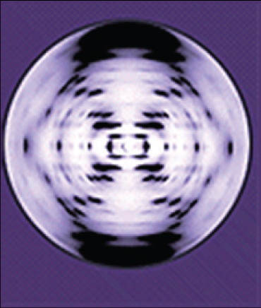
Figure 14.6 The X-ray diffraction pattern of DNA, which helped to elucidate its double-helix structure.
Mary Ann Clark, Matthew Douglas, and Jung Choi, Biology 2e, OpenStax. · CC BY-NC-SA 4.0 · Original image ↗ · Access for free ↗Chargaff’s rules. For double-stranded DNA, total A = T and G = C. These equalities need not hold within one isolated strand. If A is 30% of all bases in a double-stranded sample, T is 30% and G and C are each 20%.
Read and write sequences 5′ → 3′
The labels 5′ and 3′ refer to sugar carbon positions. Complementary strands run in opposite directions. For 5′-AGTC-3′, the aligned complement is 3′-TCAG-5′. Written in the usual 5′ → 3′ convention, that complement is 5′-GACT-3′: the reverse complement.
Check yourself. Write the complementary strand of 5′-AAGC-3′ in the 5′ → 3′ direction.
Show the answer
First pair the bases: 3′-TTCG-5′. Then reverse the written order: 5′-GCTT-3′. Replacing each letter without reversing gives the wrong direction.
Packing DNA into cells
- A typical bacterial chromosome is circular and occupies the nucleoid. Supercoiling compacts DNA; topoisomerases change this winding by cutting and resealing the backbone.
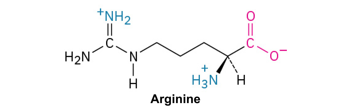
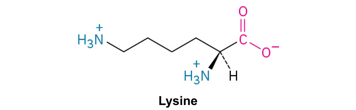
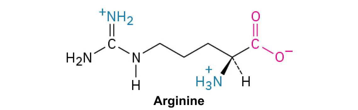
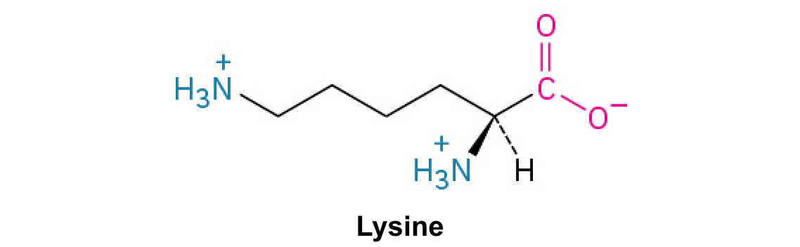
- Eukaryotic nuclear chromosomes are linear. DNA wraps around a histone octamer to form a nucleosome; linker DNA joins neighboring nucleosomes. Lysine- and arginine-rich histones interact with negatively charged DNA.
- Chromatin is DNA plus associated proteins. Euchromatin is generally more accessible for transcription; heterochromatin is more compact. The source diagram shows a classical hierarchy of chromosome compaction.

Figure 14.10 A eukaryote contains a well-defined nucleus, whereas in prokaryotes, the chromosome lies in the cytoplasm in an area called the nucleoid.
Mary Ann Clark, Matthew Douglas, and Jung Choi, Biology 2e, OpenStax. · CC BY-NC-SA 4.0 · Original image ↗ · Access for free ↗
Figure 14.11 These figures illustrate the compaction of the eukaryotic chromosome.
Mary Ann Clark, Matthew Douglas, and Jung Choi, Biology 2e, OpenStax. · CC BY-NC-SA 4.0 · Original image ↗ · Access for free ↗