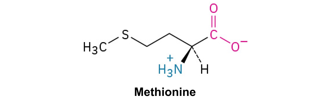
Ribosomes read mRNA 5′ → 3′ and build protein from N terminus to C terminus. Correct synthesis, folding, modification, and localization all contribute to a functional protein.
The machinery and its direction
- The small ribosomal subunit helps decode mRNA; the large subunit contains the peptidyl-transferase center. Ribosomal RNA contributes directly to catalysis, so the ribosome is a ribozyme.
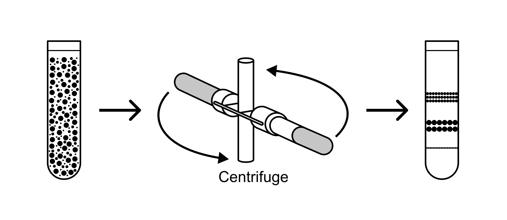
- Bacterial ribosomes are 70S, assembled from 30S and 50S subunits. Eukaryotic cytosolic ribosomes are 80S, from 40S and 60S. Svedberg values describe sedimentation and are not additive.
- The ribosome reads mRNA 5′ → 3′ and adds residues to the growing chain’s C terminus, producing the protein N terminus first. Multiple ribosomes can translate the same mRNA as a polysome.
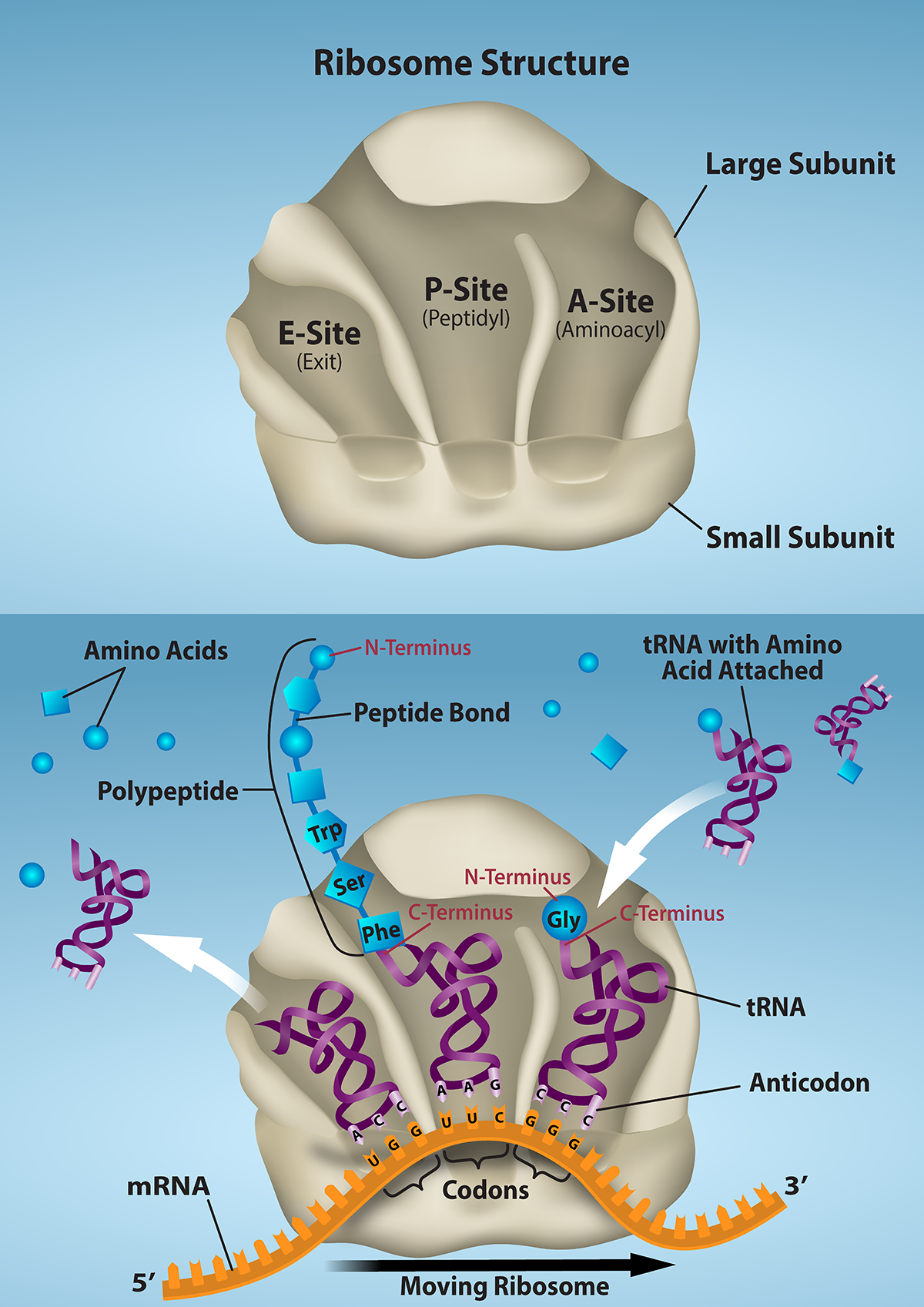
Figure 15.16 The ribosome and its function. The ribosome is responsible for translating the mRNA into protein. A. The ribosome consists of a large and small ribosomal subunit. Assembly of the subunits on the mRNA forms three tRNA binding sites. B. During translation, charged tRNAs enter the Acceptor site, and the anticodon on the tRNA base pairs with the codon in the mRNA. After the incoming amino acid forms a peptide bond with the growing polypeptide chain, the ribosome will move three nucleotides toward the 3’ end of the mRNA. This movement will transfer the tRNA with the growing polypeptide to the Peptidyl-tRNA binding site and allow the empty tRNA to exit at the Exit site. Credit: Rao, A., Ryan, K. and Fletcher, S. Department of Biology, Texas A&M University.
Rao, A., Ryan, K. and Fletcher, S. Department of Biology, Texas A&M University.; Mary Ann Clark, Matthew Douglas, and Jung Choi, Biology 2e, OpenStax. · CC BY-NC-SA 4.0 · Original image ↗ · Access for free ↗Source: Biology 2e §15.5 — Translation, folding, and targeting
Attach the right amino acid first
An aminoacyl-tRNA synthetase recognizes an amino acid and its appropriate tRNA, then attaches the amino acid to the tRNA’s terminal 3′ adenosine. Charging consumes ATP → AMP + PPi; pyrophosphate hydrolysis helps drive the reaction. Many synthetases edit incorrectly activated or attached amino acids.
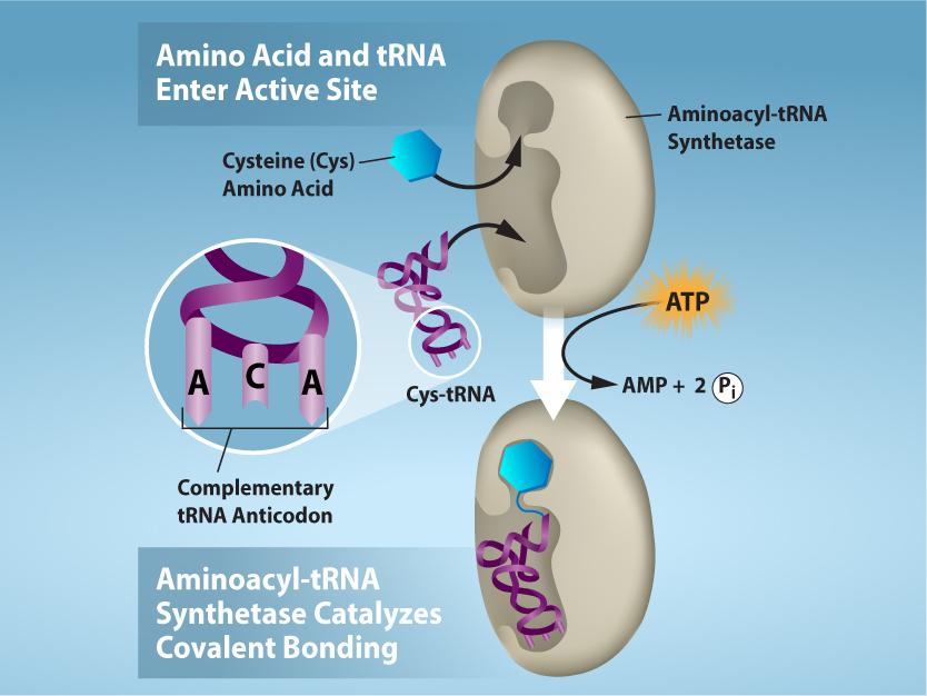
Figure 15.17 Charging of tRNAs with correct amino acids. Aminoacyl-tRNA synthetases catalyze covalent bond formation between the tRNA and the correct amino acid in preparation for translation. Because there are multiple amino acids, there are multiple different tRNA synthetases. All of the synthetases require energy, in the form of ATP, to make sure the correct amino acid is attached to the tRNA with the correct anticodon sequence. Credit: Rao, A., Ryan, K. and Tag, A. Department of Biology, Texas A&M University.
Rao, A., Ryan, K. and Tag, A. Department of Biology, Texas A&M University.; Mary Ann Clark, Matthew Douglas, and Jung Choi, Biology 2e, OpenStax. · CC BY-NC-SA 4.0 · Original image ↗ · Access for free ↗Charging versus decoding. The ribosome primarily checks codon–anticodon pairing. A tRNA with a correct anticodon but an incorrectly attached amino acid can deliver that wrong amino acid. Charging accuracy and decoding accuracy are separate requirements.
Source: Biology 2e §15.5 — Translation, folding, and targeting
Start in the correct frame
- In bacteria, the Shine–Dalgarno sequence on mRNA pairs with small-subunit rRNA to position the start codon. Initiation factors organize assembly; IF-2 helps deliver the initiator tRNA carrying formylmethionine (fMet).
- In typical eukaryotic cap-dependent initiation, initiation factors recruit the 40S subunit and an initiator Met-tRNA near the 5′ cap. Scanning favors an AUG in a suitable Kozak context; the first AUG is not an absolute rule.
- The initiator tRNA occupies the P site. After the large subunit joins and initiation factors leave, elongation can begin. Ordinary internal AUG codons use an elongator methionine tRNA rather than starting a new chain.

Figure 15.18 Translation begins when an initiator tRNA anticodon recognizes a start codon on mRNA bound to a small ribosomal subunit. The large ribosomal subunit joins the small subunit, and a second tRNA is recruited. As the mRNA moves relative to the ribosome, successive tRNAs move through the ribosome and the polypeptide chain is formed. Entry of a release factor into the A site terminates translation and the components dissociate.
Mary Ann Clark, Matthew Douglas, and Jung Choi, Biology 2e, OpenStax. · CC BY-NC-SA 4.0 · Original image ↗ · Access for free ↗Source: Biology 2e §15.5 — Translation, folding, and targeting
Follow the A → P → E cycle
- Delivery: an elongation factor and GTP help bring a charged tRNA to the A site; codon–anticodon matching selects the appropriate adaptor.
- Peptide transfer: the amino group on the A-site amino acid attacks the bond linking the growing peptide to the P-site tRNA. The peptide is transferred to the A-site tRNA; the P-site tRNA becomes uncharged.
- Translocation: another elongation factor uses GTP to move the ribosome by one codon toward the mRNA’s 3′ end. The peptidyl-tRNA moves to P, the empty tRNA moves toward E, and A becomes available.
- The energy stored in the aminoacyl/peptidyl-tRNA ester linkage supports peptide transfer. ATP is used during charging and GTP during factor-driven steps; ribosomal peptide transfer is not simply condensation of two free amino acids.
Factor names. In bacteria, EF-Tu delivers aminoacyl-tRNA and EF-G drives translocation. Eukaryotic eEF1A and eEF2 perform the corresponding roles.
Source: Biology 2e §15.5 — Translation, folding, and targeting
Release factors recognize stop codons
When UAA, UAG, or UGA enters the A site, a release factor recognizes the stop signal. The peptidyl-transferase center uses water to hydrolyze the bond between the completed peptide and its P-site tRNA. The peptide is released and the ribosome recycled. There is no standard stop-codon tRNA carrying a “stop amino acid.”

Figure 15.19 Translation Termination is Active. Translation is terminated when a STOP codon is in the A-site of the ribosome. Since there are no tRNAs corresponding to the STOP codons, the Release Factor protein enters and catalyzes the hydrolysis between the last amino acid and its tRNA. This hydrolysis releases the free carboxyl terminus (C-term) of the protein. Additional factors use the energy in GTP hydrolysis to disassemble the large and small ribosomal subunits and mRNA. Credit: Rao, A. and Ryan, K. Department of Biology, Texas A&M University.
Rao, A. and Ryan, K. Department of Biology, Texas A&M University.; Mary Ann Clark, Matthew Douglas, and Jung Choi, Biology 2e, OpenStax. · CC BY-NC-SA 4.0 · Original image ↗ · Access for free ↗Source: Biology 2e §15.5 — Translation, folding, and targeting
A new chain still needs to mature
- Folding brings residues into the structure needed for function. Chaperones assist folding and limit aggregation; they do not change the encoded amino-acid order.
- Proteolytic cleavage can remove a signal peptide or activate a precursor such as proinsulin. Cleavage changes the final chain even though the translated sequence came from the original mRNA.
- Phosphorylation can change activity or binding; glycosylation can influence folding, stability, recognition, and trafficking. Disulfide-bond formation can stabilize secreted proteins. Effects depend on the protein and the modified site.
Timing matters. Folding, targeting, and some modifications can begin while translation is still in progress. “Protein maturation” does not mean every step waits until the entire chain has been released.
Source: Biology 2e §15.5 — Translation, folding, and targeting · Biology 2e §4.4 — The protein secretory pathway
Sequence signals direct the destination
For many secreted and membrane proteins, a hydrophobic ER signal sequence emerges from the ribosome and binds the signal recognition particle (SRP). SRP directs the complex to the rough ER, where a translocon allows synthesis to continue into the lumen or membrane. A cleavable signal peptide is part of the encoded sequence, not a tag added afterward.
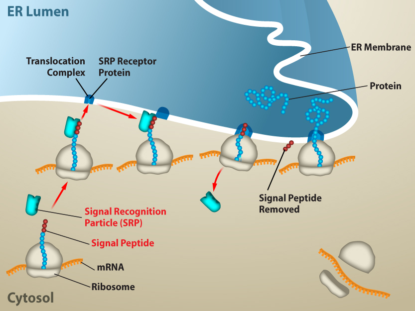
Figure 15.20 Proteins are co-translationally targeted into the ER for secretion. Proteins that will be secreted from the cell will contain a signal sequence at the N-terminus. The signal will be recognized by SRP as soon as the amino acids emerge from the ribosome, and the ribosome will be targeted to the translocation channel in the ER membrane. The rest of the protein will go directly from the ribosome, across the ER membrane and into the ER lumen. From the ER, proteins can be secreted from the cell via vesicle trafficking. Credit: Rao, A. and Ryan, K. Department of Biology, Texas A&M University.
Rao, A. and Ryan, K. Department of Biology, Texas A&M University.; Mary Ann Clark, Matthew Douglas, and Jung Choi, Biology 2e, OpenStax. · CC BY-NC-SA 4.0 · Original image ↗ · Access for free ↗Proteins without an ER targeting signal are generally translated on free cytosolic ribosomes. Some remain in the cytosol; other targeting signals direct completed proteins to destinations such as the nucleus or mitochondria. Free and ER-bound ribosomes are not permanently different kinds of ribosome, and not all targeting signals are cleaved.

Figure 4.18 Membrane and secretory proteins are synthesized in the rough endoplasmic reticulum (RER). The RER also sometimes modifies proteins. In this illustration, a (green) integral membrane protein is modified by attachment of a (purple) carbohydrate in the ER. Vesicles with the integral protein bud from the ER and fuse with the Golgi apparatus' cis face. As the protein passes along the Golgi’s cisternae, the addition of more carbohydrates further modifies it. After its synthesis is complete, it exits as an integral membrane protein of the vesicle that buds from the Golgi’s trans face. When the vesicle fuses with the cell membrane, the protein becomes an integral portion of that cell membrane. (credit: modification of work by Magnus Manske)
modification of work by Magnus Manske; Mary Ann Clark, Matthew Douglas, and Jung Choi, Biology 2e, OpenStax. · CC BY-NC-SA 4.0 · Original image ↗ · Access for free ↗Proteins entering the secretory pathway can fold and acquire modifications in the ER, travel in vesicles to the Golgi cis face, undergo further processing, and be sorted at the trans face. Their destinations include secretion, the plasma membrane, and endomembrane compartments. ER-lumen-facing domains remain inside transport vesicles and become extracellular when a vesicle fuses with the plasma membrane.
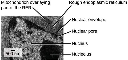
Figure 4.19 This transmission electron micrograph shows the rough endoplasmic reticulum and other organelles in a pancreatic cell. (credit: modification of work by Louisa Howard)
modification of work by Louisa Howard; Mary Ann Clark, Matthew Douglas, and Jung Choi, Biology 2e, OpenStax. · CC BY-NC-SA 4.0 · Original image ↗ · Access for free ↗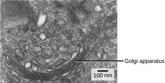
Figure 4.20 The Golgi apparatus in this white blood cell is visible as a stack of semicircular, flattened rings in the lower portion of the image. You can see several vesicles near the Golgi apparatus. (credit: modification of work by Louisa Howard)
modification of work by Louisa Howard; Mary Ann Clark, Matthew Douglas, and Jung Choi, Biology 2e, OpenStax. · CC BY-NC-SA 4.0 · Original image ↗ · Access for free ↗Source: Biology 2e §15.5 — Translation, folding, and targeting · Biology 2e §4.4 — The protein secretory pathway
Practice: charging and elongation
Question. A tRNA with an anticodon matching a phenylalanine codon is experimentally charged with alanine. Which amino acid can it insert? What would blocking EF-G change during bacterial elongation?
Show translation reasoning
The mischarged tRNA can insert alanine if it passes ribosomal selection: the anticodon directs codon recognition, but the attached amino acid is what enters the chain. Blocking EF-G impairs translocation, preventing the normal advance by one codon; it does not directly alter the mRNA sequence or tRNA-charging reaction.
Source: Biology 2e §15.5 — Translation, folding, and targeting
Practice: change the address
Question. Delete the only ER signal sequence from a normally secreted protein. Assume transcription, translation initiation, and cell viability remain intact. Predict localization and how to test the explanation.
Show targeting reasoning
The protein should lose efficient entry into the secretory pathway and is likely to accumulate in the cytosol or be degraded, rather than being secreted normally. Compare a normal construct, the signal-deletion construct, and a construct with the signal restored. Measure both cellular and medium-associated protein; also check RNA abundance so reduced expression is not mistaken for a targeting defect.
Source: Biology 2e §15.5 — Translation, folding, and targeting · Biology 2e §4.4 — The protein secretory pathway