Recognize each side chain and use chemical interactions to explain recognition and catalysis.
Side-chain recognition and stereochemistry
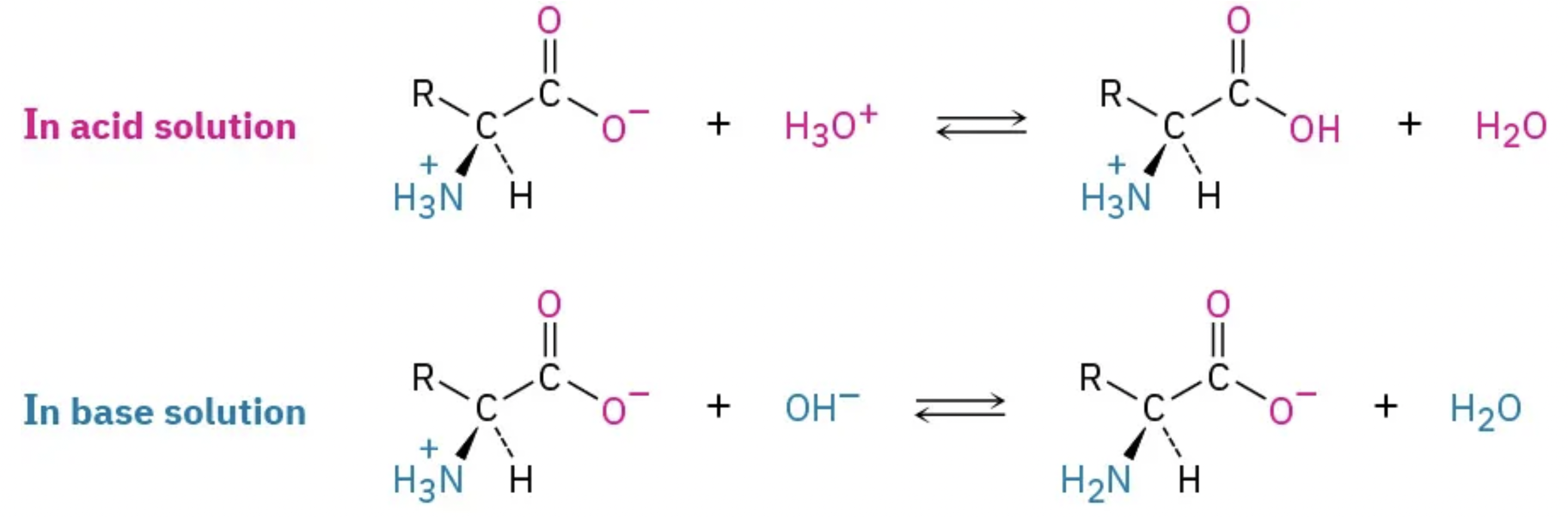
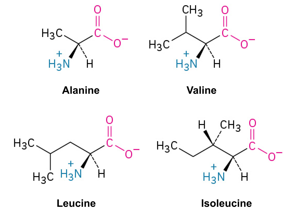
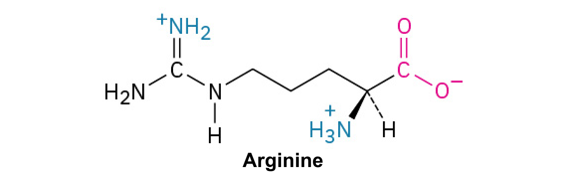
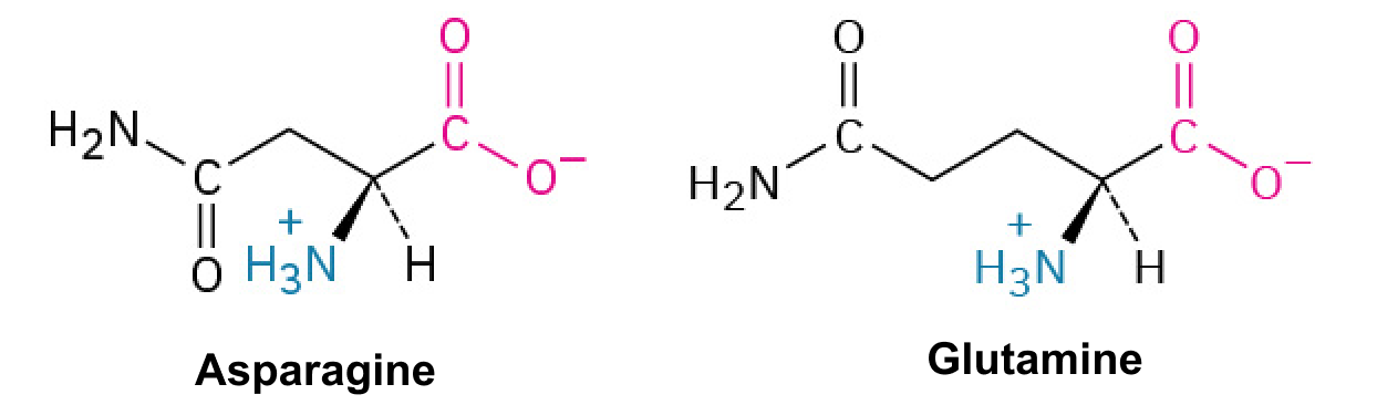
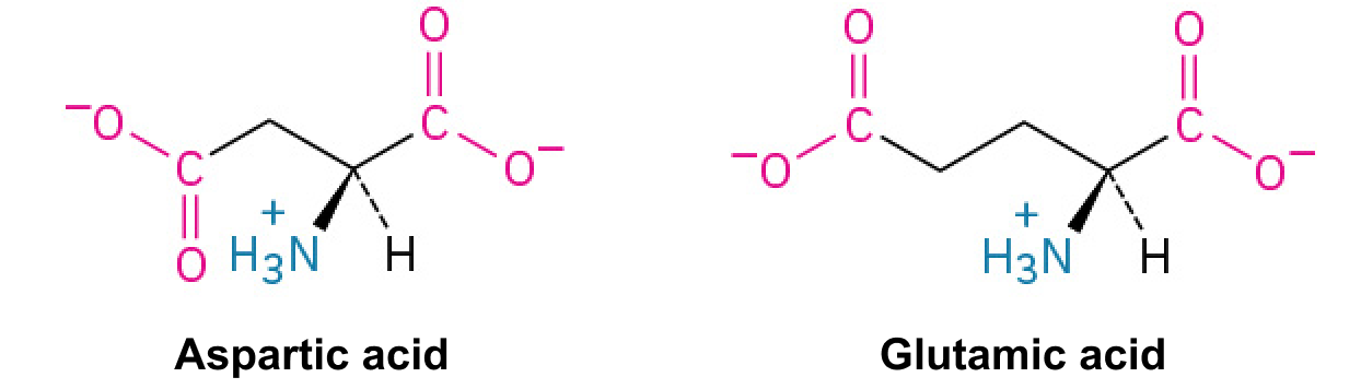
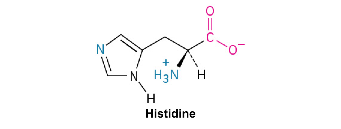
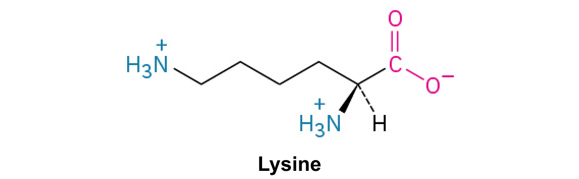
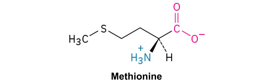
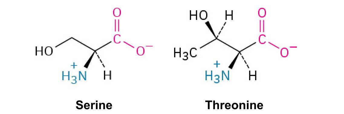
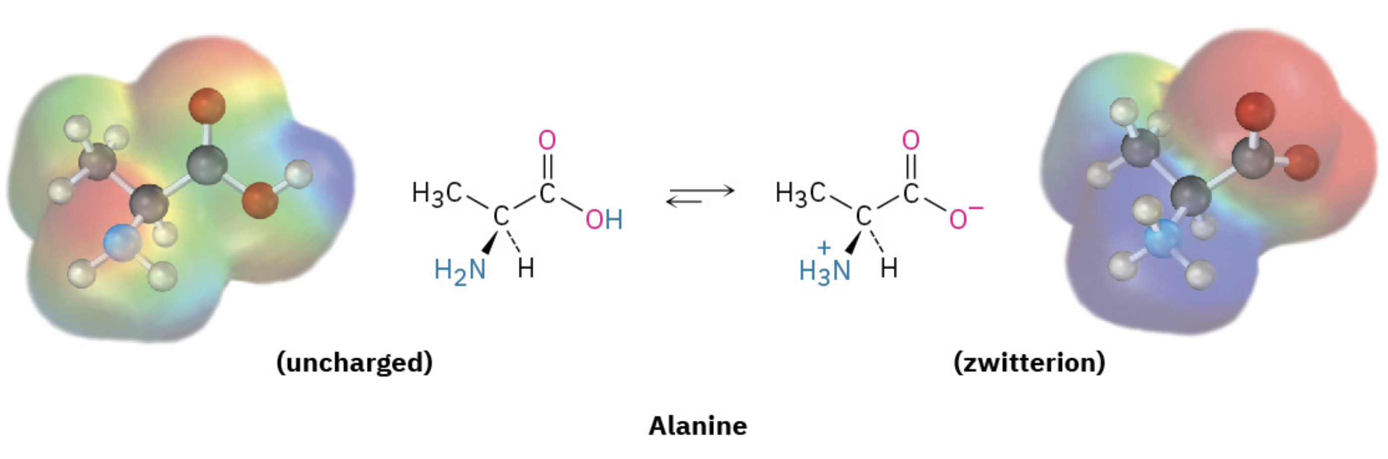
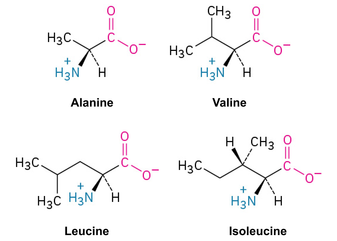
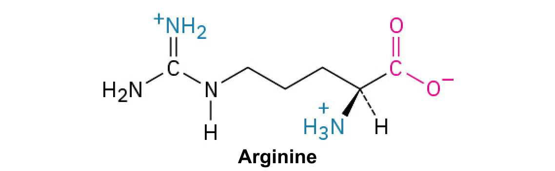
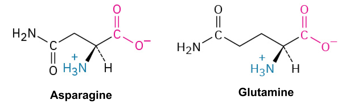
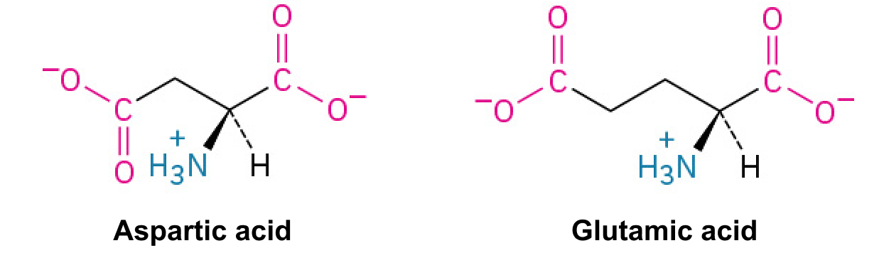
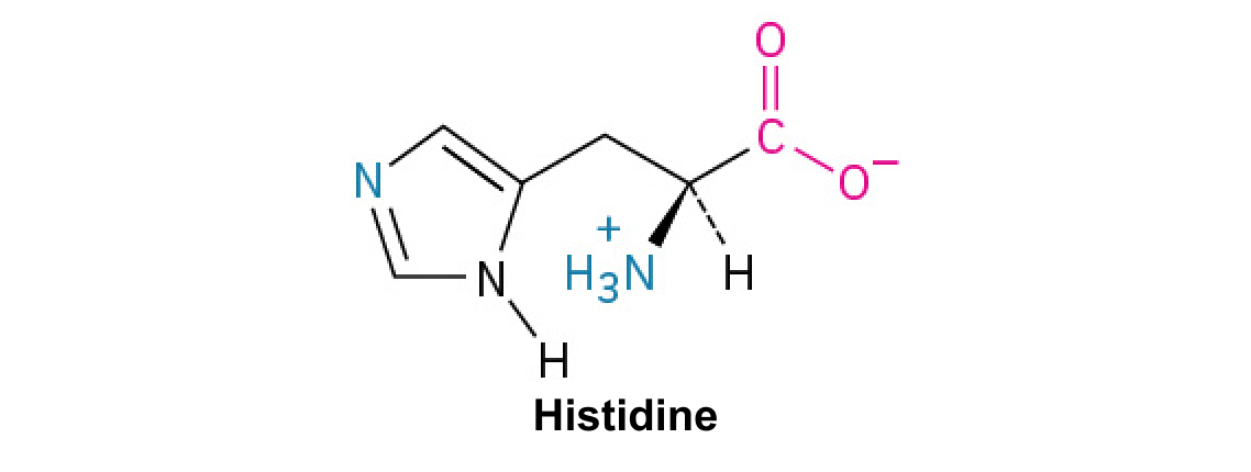
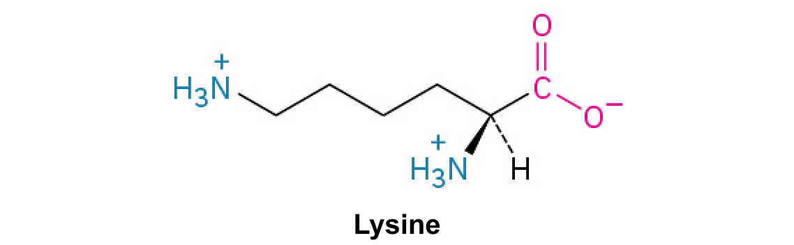
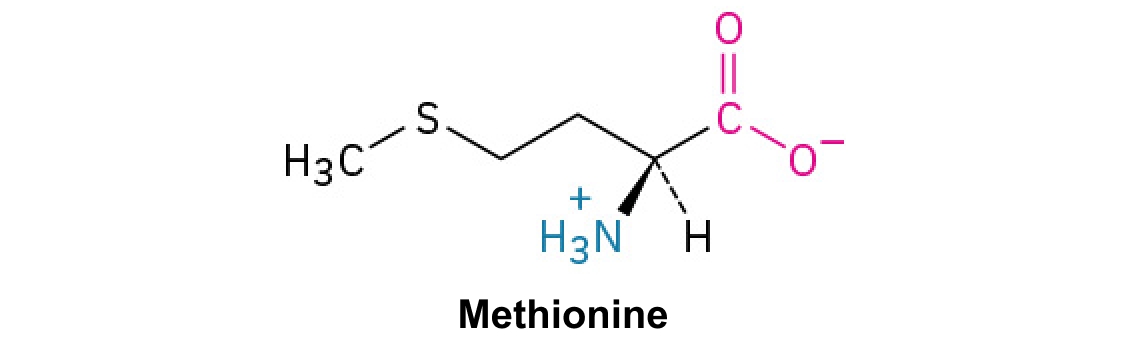
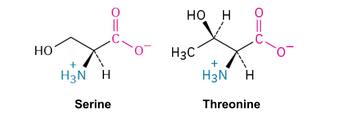
The common backbone is an α carbon bonded to amino, carboxyl, hydrogen, and side-chain groups. Near neutral pH, free amino acids are commonly zwitterions. Gly/G has H; Ala/A has methyl; Val/V has isopropyl; Leu/L has isobutyl; Ile/I has sec-butyl; Pro/P closes its side chain onto backbone nitrogen. Met/M contains a thioether; Cys/C has a thiol. Ser/S has hydroxymethyl and Thr/T has a hydroxyl-bearing branched chain. Asn/N and Gln/Q end in amides; Asp/D and Glu/E end in carboxylates, with glutamate one methylene longer. Lys/K ends in an amino group; Arg/R in guanidinium; His/H contains imidazole. Phe/F has benzyl, Tyr/Y hydroxybenzyl, and Trp/W an indole ring.
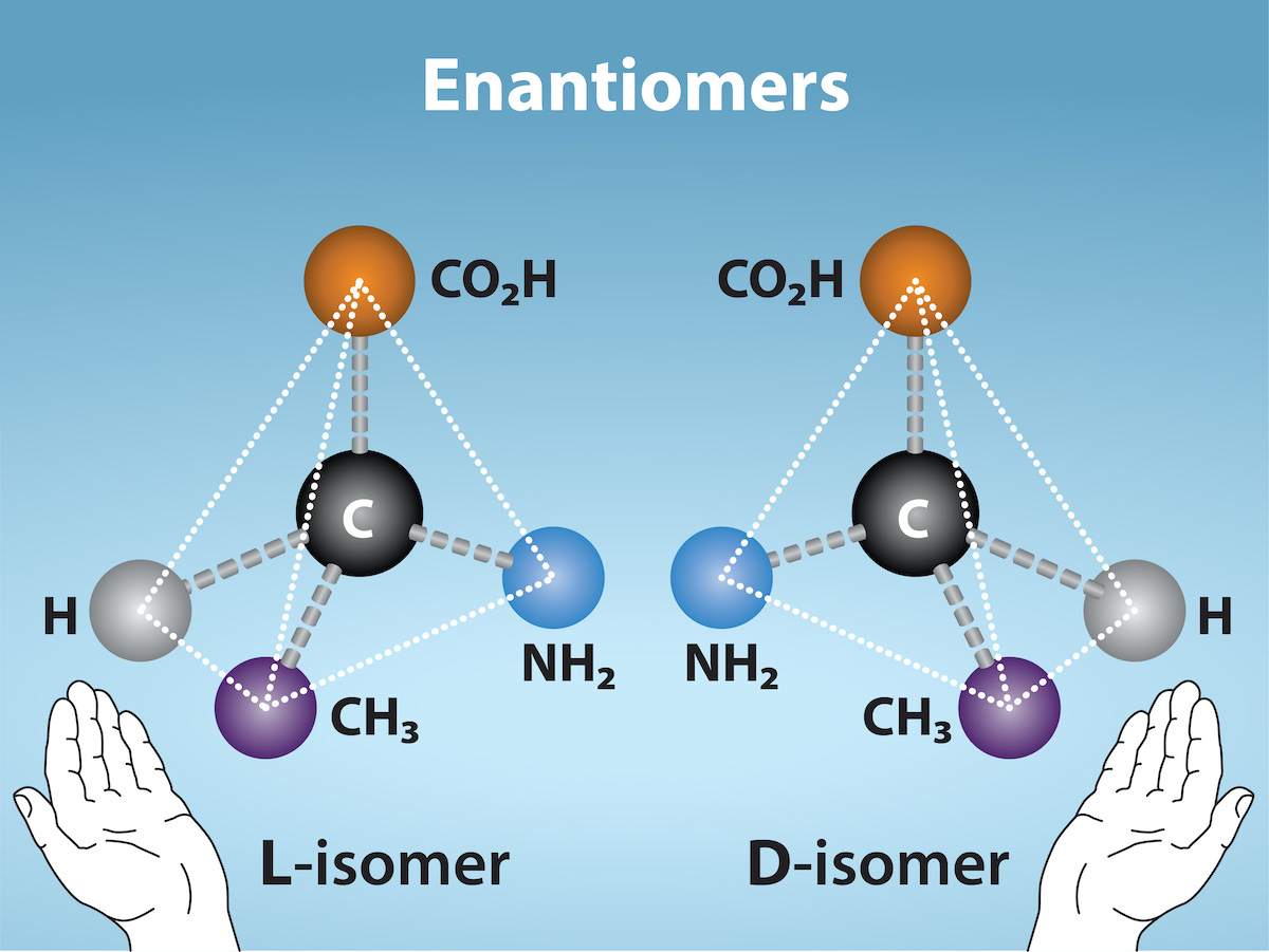
Glycine is achiral because two α-carbon substituents are H. Protein amino acids ordinarily use the L configuration; L/D specifies a reference configuration, not the direction of optical rotation. Most L amino acids have S absolute configuration at the α carbon. L-cysteine is R because sulfur changes the Cahn–Ingold–Prelog priority ordering. Assign priorities from actual atoms rather than memorizing L = S; isoleucine and threonine also contain a second stereocenter.
Source: 3.4 Proteins · 6.5 Enzymes
Complementarity is dynamic
Active-site geometry positions substrates while hydrogen bonding, ionic interactions, dispersion forces, and hydrophobic effects influence affinity. A chiral active site can distinguish enantiomers that share most bulk physical properties. Induced fit describes conformational adjustment upon binding; conformational selection emphasizes binding to pre-existing states, and both can contribute. Catalytic recognition must stabilize the transition state relative to the reactant state; extremely tight ground-state binding alone can impede reaction. Specificity is selective recognition, not necessarily absolute exclusivity.
Source: 3.4 Proteins · 6.5 Enzymes
Chemical routes lower activation barriers
General acid–base catalysis transfers protons through suitably placed groups, often side chains or water; the local environment can shift their pKa values. Covalent catalysis forms a temporary enzyme–substrate bond that creates an alternative reaction pathway and must later be broken to regenerate enzyme. Metal-assisted catalysis can stabilize negative charge, orient substrate, activate water, or transfer electrons. Proximity and orientation reduce the cost of productive encounters. These strategies can cooperate in one active site. They change activation free energy and rate, not overall reaction ΔG or equilibrium position.
Source: 3.4 Proteins · 6.5 Enzymes
Practice and reasoning
Question. Identify the amino acid with a thiol side chain and explain its usual L/R labels. A catalytic histidine accepts a proton while serine forms a transient substrate bond: name both catalytic strategies.
Show reasoning
Cysteine (Cys, C) has the thiol. Its usual protein form is L but has R α-carbon configuration because sulfur changes priority ordering. Histidine acts as a general base; transient serine–substrate bonding is covalent catalysis. Neither step alone establishes the reaction’s equilibrium constant.
Source: 3.4 Proteins · 6.5 Enzymes
3.4 Proteins — figures and tables
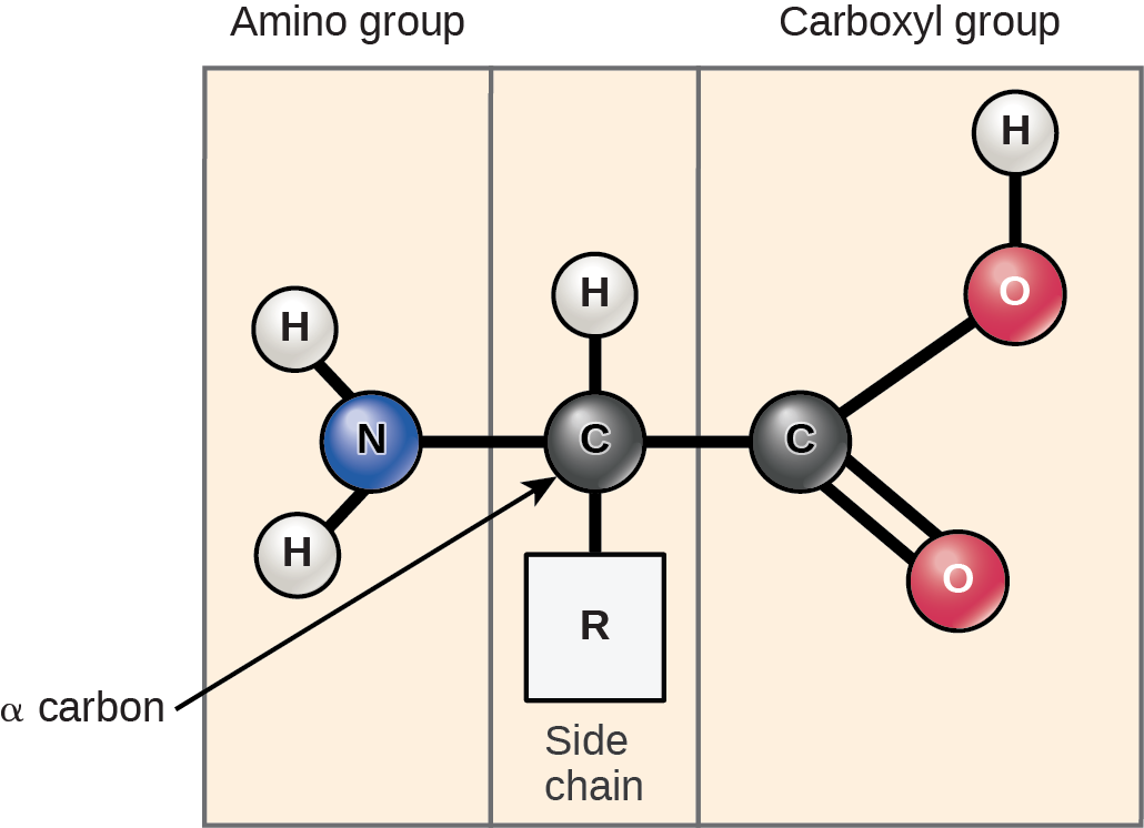
Figure 3.22 Amino acids have a central asymmetric carbon to which an amino group, a carboxyl group, a hydrogen atom, and a side chain (R group) are attached.
Mary Ann Clark, Matthew Douglas, and Jung Choi, Biology 2E, OpenStax. · CC BY-NC-SA 4.0 · Original image ↗ · Access for free ↗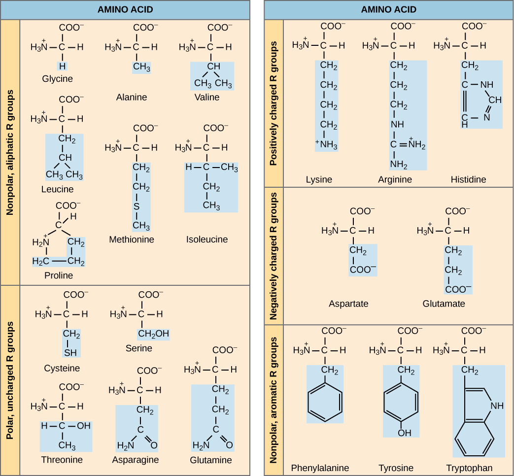
Figure 3.23 There are 20 common amino acids commonly found in proteins, each with a different R group (variant group) that determines its chemical nature.
Mary Ann Clark, Matthew Douglas, and Jung Choi, Biology 2E, OpenStax. · CC BY-NC-SA 4.0 · Original image ↗ · Access for free ↗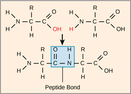
Figure 3.24 Peptide bond formation is a dehydration synthesis reaction. The carboxyl group of one amino acid is linked to the incoming amino acid's amino group. In the process, it releases a water molecule.
Mary Ann Clark, Matthew Douglas, and Jung Choi, Biology 2E, OpenStax. · CC BY-NC-SA 4.0 · Original image ↗ · Access for free ↗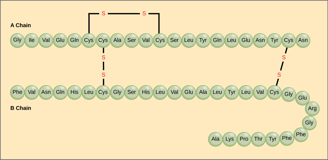
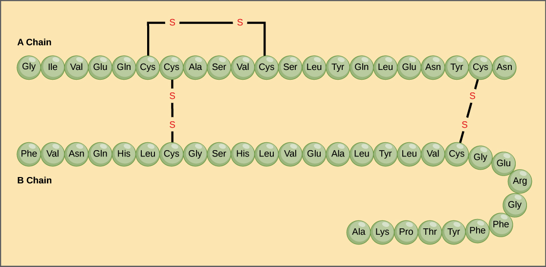
Figure 3.25 Bovine serum insulin is a protein hormone comprised of two peptide chains, A (21 amino acids long) and B (30 amino acids long). In each chain, three-letter abbreviations that represent the amino acids' names in the order they are present indicate primary structure. The amino acid cysteine (cys) has a sulfhydryl (SH) group as a side chain. Two sulfhydryl groups can react in the presence of oxygen to form a disulfide (S-S) bond. Two disulfide bonds connect the A and B chains together, and a third helps the A chain fold into the correct shape. Note that all disulfide bonds are the same length, but we have drawn them different sizes for clarity.
Mary Ann Clark, Matthew Douglas, and Jung Choi, Biology 2E, OpenStax. · CC BY-NC-SA 4.0 · Original image ↗ · Access for free ↗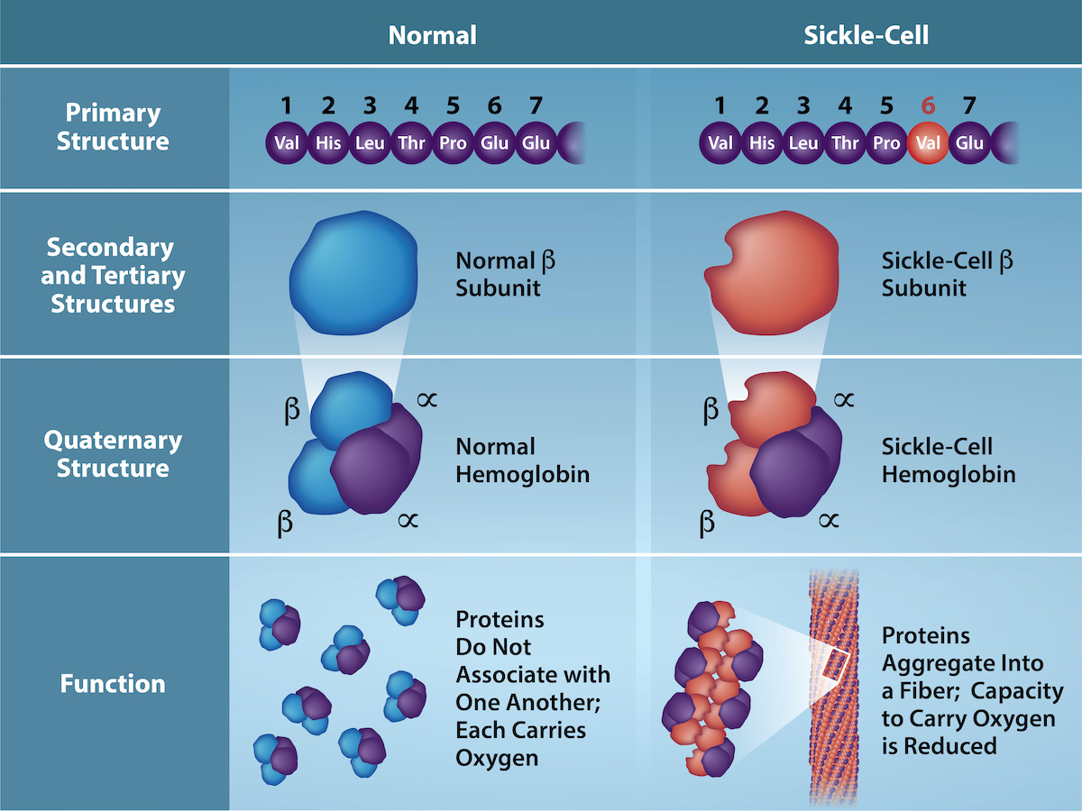
Figure 3.26 Because of this change of one amino acid in the chain, hemoglobin molecules form long fibers that distort the biconcave, or disc-shaped, red blood cells and causes them to assume a crescent or “sickle” shape, which clogs blood vessels (Figure 3.27). The beta (β)- chain of hemoglobin is 147 amino acids in length, yet a single amino acid substitution in the primary sequence leads to changes in secondary, tertiary, and quaternary structures and sickle cell anemia. In normal hemoglobin, the amino acid at position six is glutamate. In sickle cell hemoglobin glutamate is replaced by valine. (credit: Rao, A., Tag, A. Ryan, K. and Fletcher, S. Department of Biology, Texas A&M University)
Mary Ann Clark, Matthew Douglas, and Jung Choi, Biology 2E, OpenStax. (credit: Rao, A., Tag, A. Ryan, K. and Fletcher, S. Department of Biology, Texas A&M University) · CC BY-NC-SA 4.0 · Original image ↗ · Access for free ↗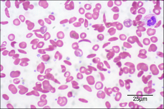
Figure 3.27 In this blood smear, visualized at 535x magnification using bright field microscopy, sickle cells are crescent shaped, while normal cells are disc-shaped. (credit: modification of work by Ed Uthman; scale-bar data from Matt Russell)
Mary Ann Clark, Matthew Douglas, and Jung Choi, Biology 2E, OpenStax. (credit: modification of work by Ed Uthman; scale-bar data from Matt Russell) · CC BY-NC-SA 4.0 · Original image ↗ · Access for free ↗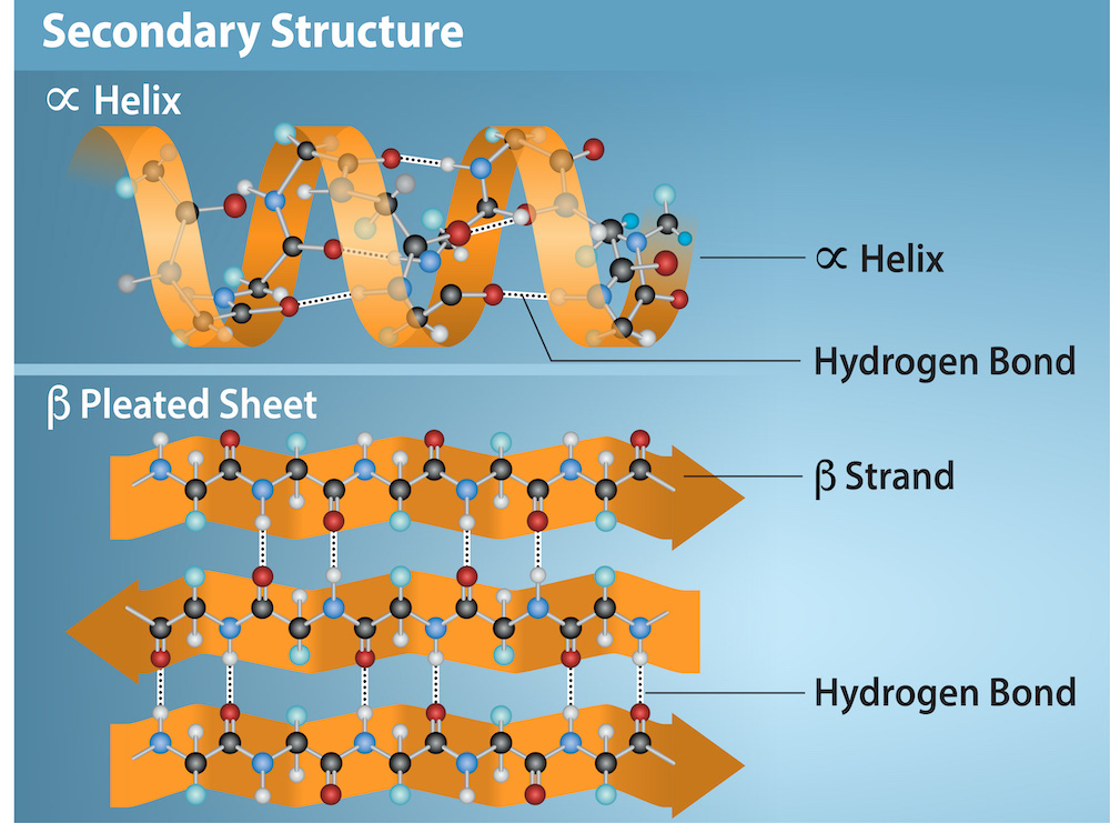
Figure 3.28 The α -helix and β -pleated sheet are secondary protein structures formed when hydrogen bonds form between the carbonyl oxygen and the amino hydrogen in the peptide backbone. Certain amino acids have a propensity to form an α-helix while others favor β-pleated sheet formation. Black = carbon, White = hydrogen, Blue = nitrogen, and Red = oxygen. Credit: Rao, A., Ryan, K. Fletcher, S. and Tag, A. Department of Biology, Texas A&M University.
Mary Ann Clark, Matthew Douglas, and Jung Choi, Biology 2E, OpenStax. · CC BY-NC-SA 4.0 · Original image ↗ · Access for free ↗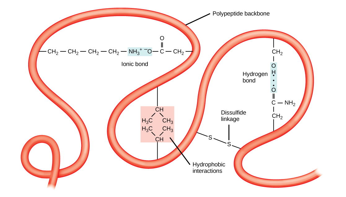
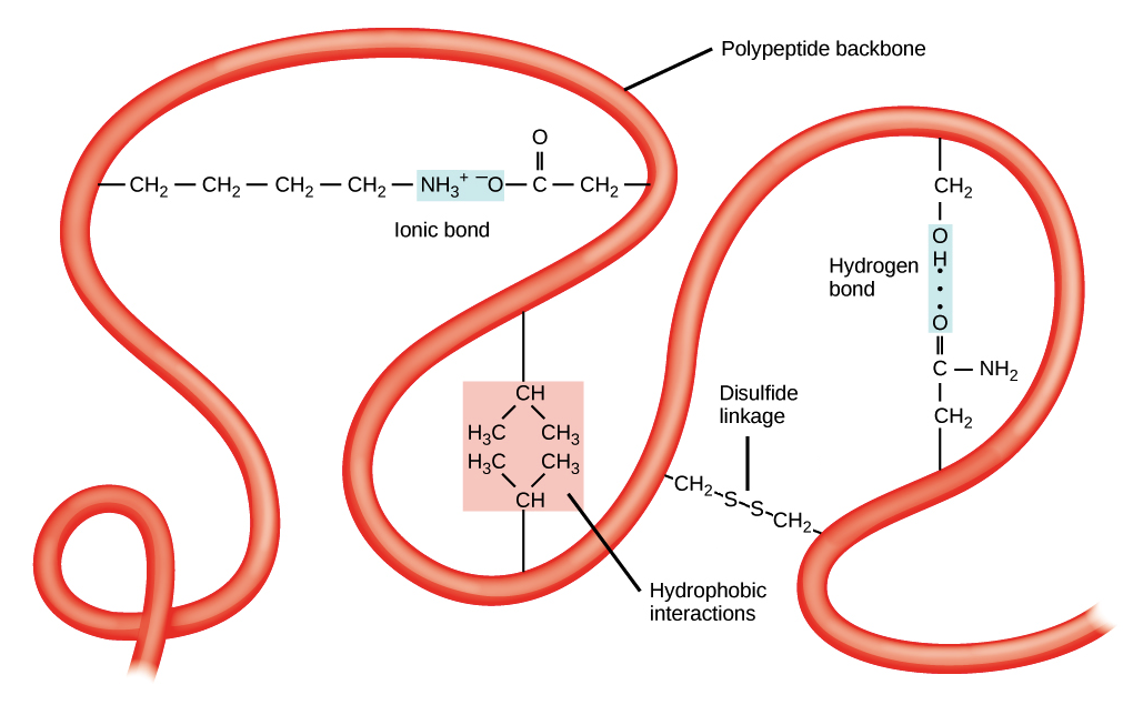
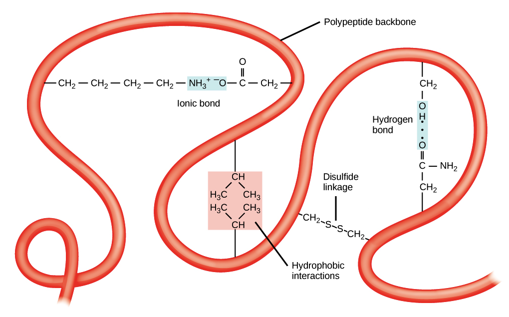
Figure 3.29 A variety of chemical interactions determine the proteins' tertiary structure. These include hydrophobic interactions, ionic bonding, hydrogen bonding, and disulfide linkages.
Mary Ann Clark, Matthew Douglas, and Jung Choi, Biology 2E, OpenStax. · CC BY-NC-SA 4.0 · Original image ↗ · Access for free ↗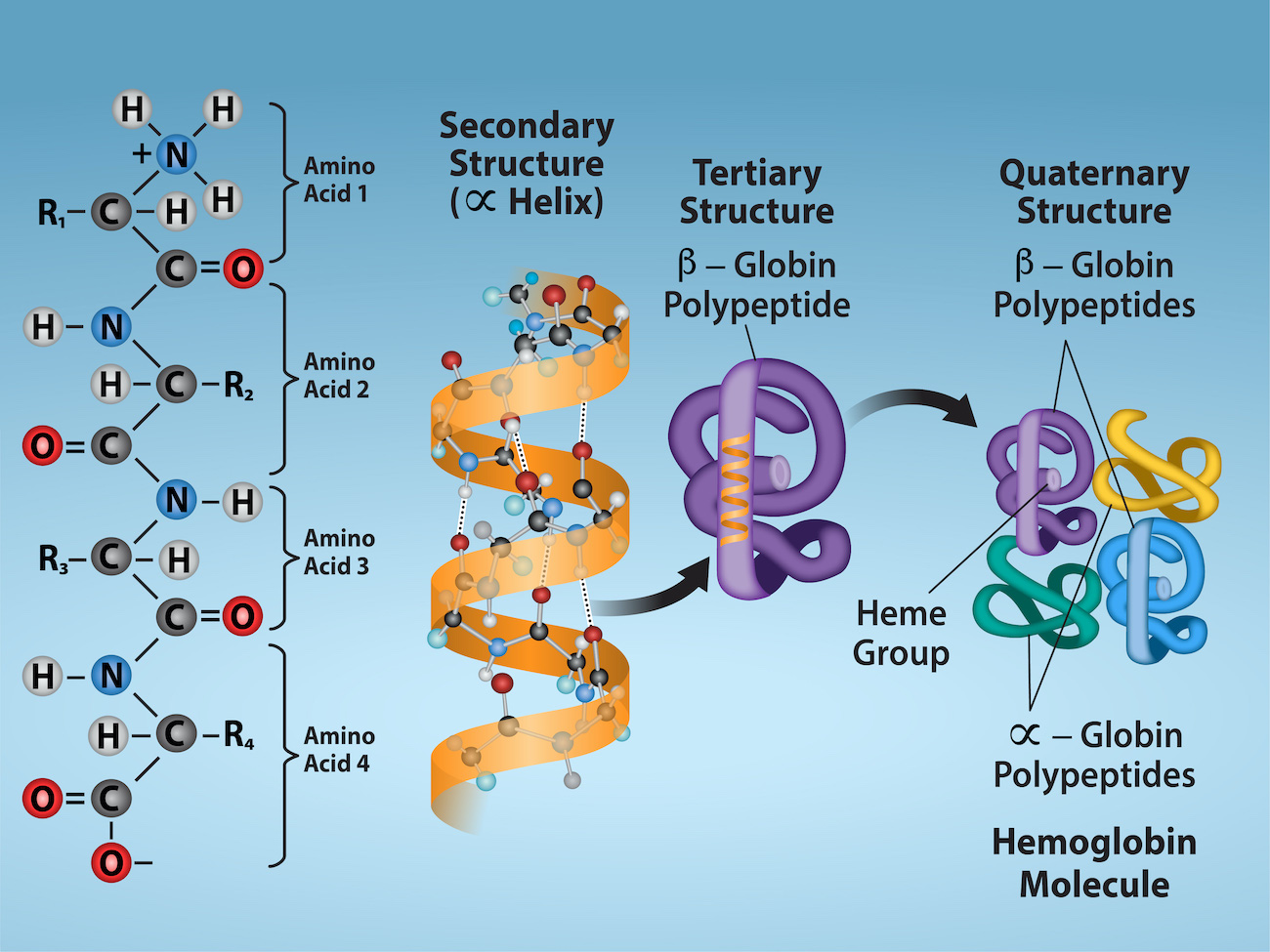
Figure 3.30 Observe the four levels of protein structure in these illustrations. Credit: Rao, A. Ryan, K. and Tag, A. Department of Biology, Texas A&M University.
Mary Ann Clark, Matthew Douglas, and Jung Choi, Biology 2E, OpenStax. · CC BY-NC-SA 4.0 · Original image ↗ · Access for free ↗| Type | Examples | Functions |
|---|---|---|
| Digestive Enzymes | Amylase, lipase, pepsin, trypsin | Help in food by catabolizing nutrients into monomeric units |
| Transport | Hemoglobin, albumin | Carry substances in the blood or lymph throughout the body |
| Structural | Actin, tubulin, keratin | Construct different structures, like the cytoskeleton |
| Hormones | Insulin, thyroxine | Coordinate different body systems' activity |
| Defense | Immunoglobulins | Protect the body from foreign pathogens |
| Contractile | Actin, myosin | Effect muscle contraction |
| Storage | Legume storage proteins, egg white (albumin) | Provide nourishment in early embryo development and the seedling |
Table 3.1. Mary Ann Clark, Matthew Douglas, and Jung Choi, Biology 2E, OpenStax.
Source: 3.4 Proteins
6.5 Enzymes — figures and tables
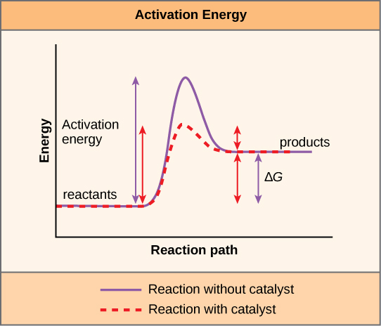
Figure 6.15 Enzymes lower the reaction's activation energy but do not change the reaction's free energy.
Mary Ann Clark, Matthew Douglas, and Jung Choi, Biology 2E, OpenStax. · CC BY-NC-SA 4.0 · Original image ↗ · Access for free ↗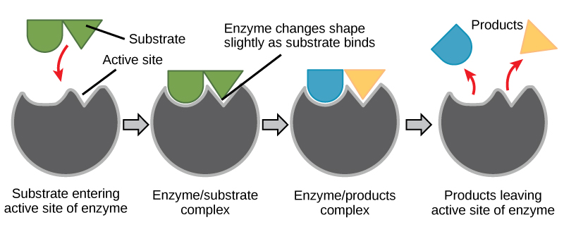
Figure 6.16 According to the induced-fit model, both enzyme and substrate undergo dynamic conformational changes upon binding. The enzyme contorts the substrate into its transition state, thereby increasing the reaction's rate.
Mary Ann Clark, Matthew Douglas, and Jung Choi, Biology 2E, OpenStax. · CC BY-NC-SA 4.0 · Original image ↗ · Access for free ↗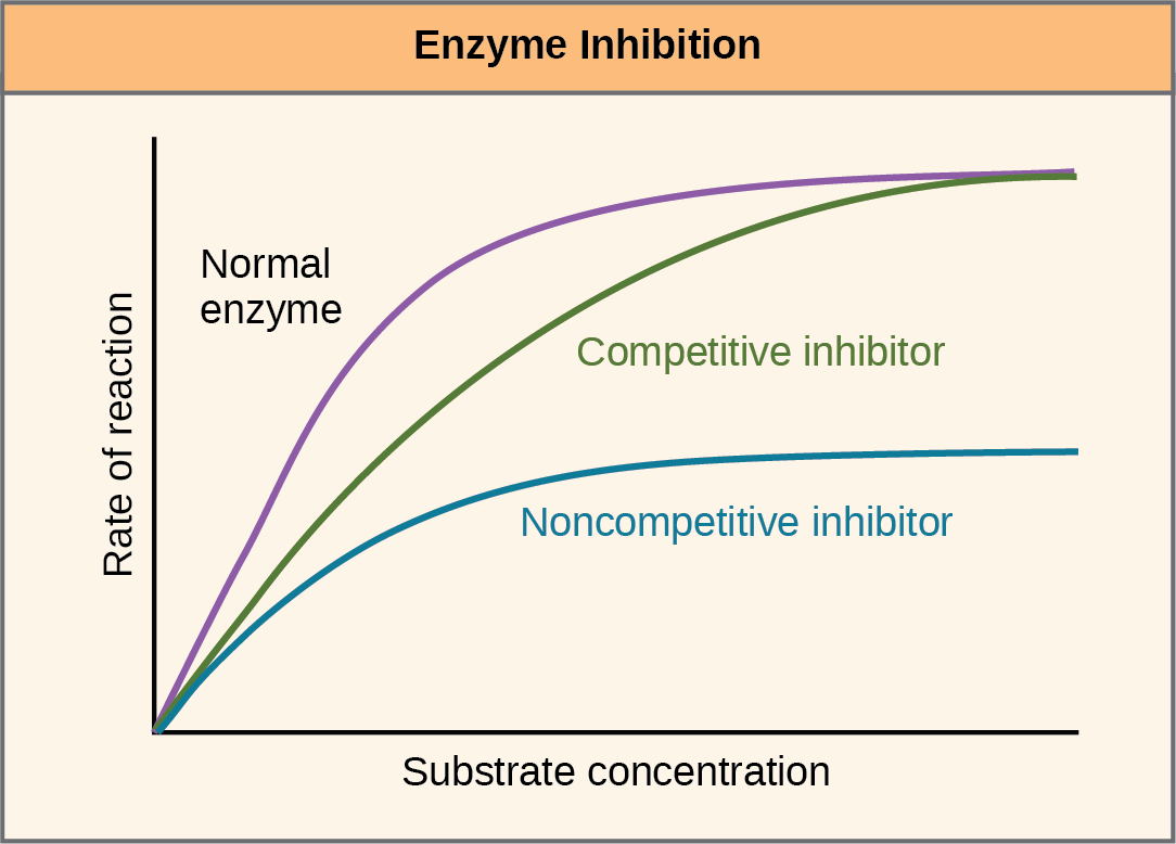
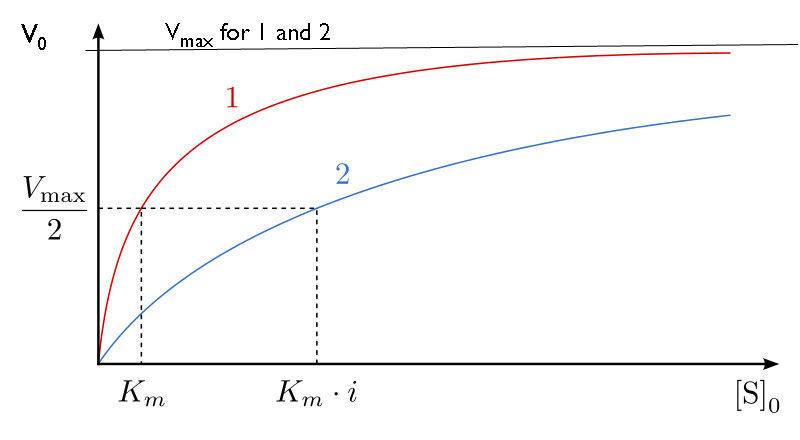
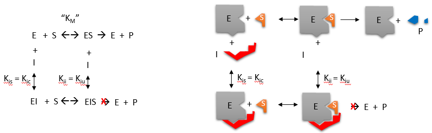
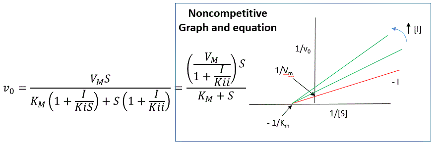
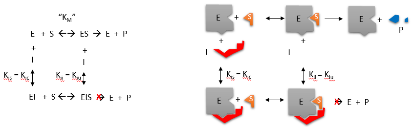
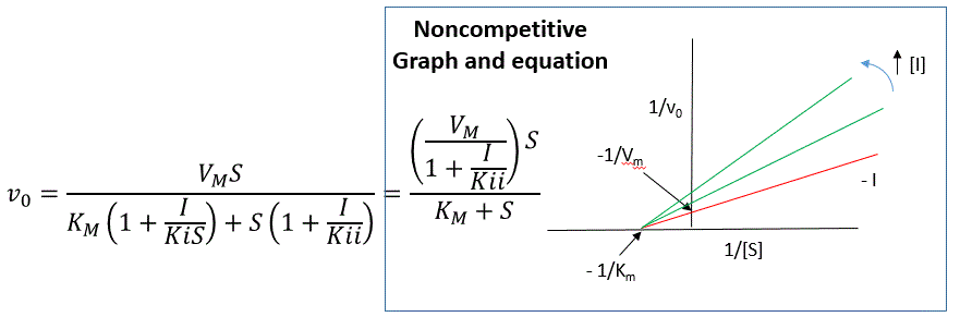
Figure 6.17 Competitive and noncompetitive inhibition affect the reaction's rate differently. Competitive inhibitors affect the initial rate but do not affect the maximal rate; whereas, noncompetitive inhibitors affect the maximal rate.
Mary Ann Clark, Matthew Douglas, and Jung Choi, Biology 2E, OpenStax. · CC BY-NC-SA 4.0 · Original image ↗ · Access for free ↗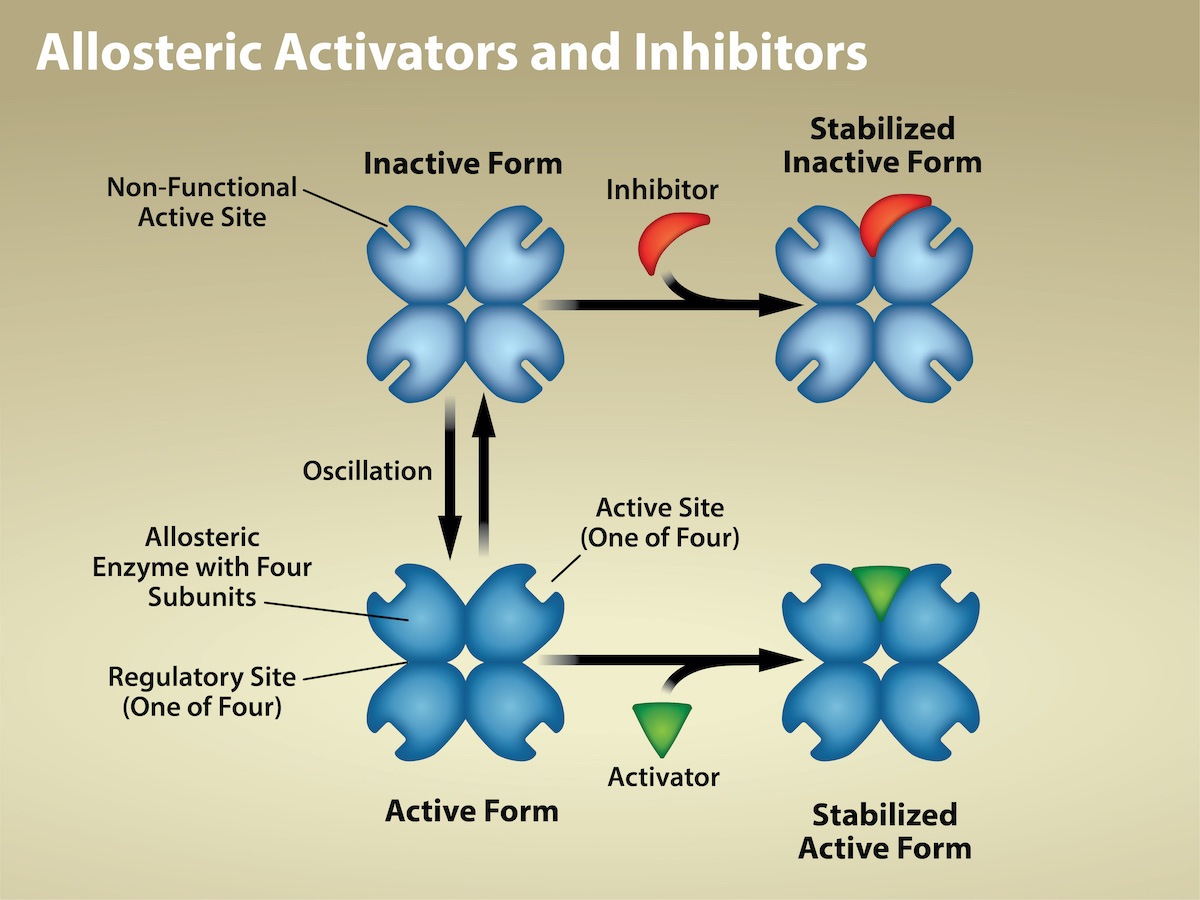
Figure 6.18 Allosteric inhibitors modify the enzyme's active site so that substrate binding is reduced or prevented. In contrast, allosteric activators modify the enzyme's active site so that the affinity for the substrate increases. Credit: Rao, A., Hawkins, A., Fletcher, S. and Tag, A. Department of Biology, Texas A&M University.
Mary Ann Clark, Matthew Douglas, and Jung Choi, Biology 2E, OpenStax. · CC BY-NC-SA 4.0 · Original image ↗ · Access for free ↗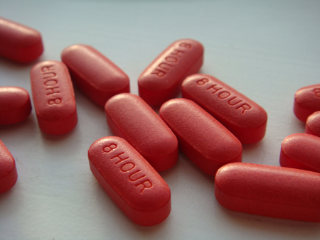
Figure 6.19 Have you ever wondered how pharmaceutical drugs are developed? (credit: Deborah Austin)
Mary Ann Clark, Matthew Douglas, and Jung Choi, Biology 2E, OpenStax. (credit: Deborah Austin) · CC BY-NC-SA 4.0 · Original image ↗ · Access for free ↗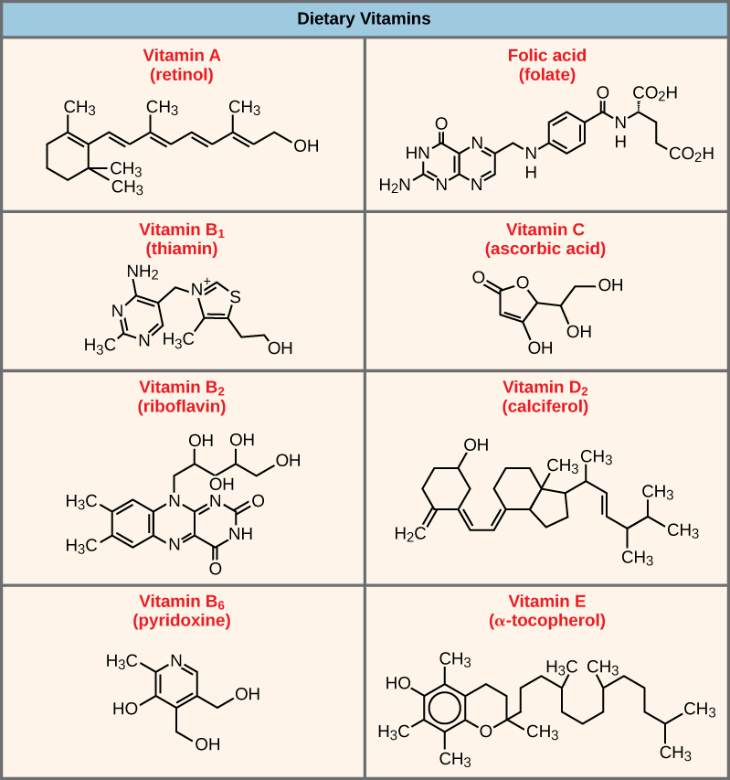
Figure 6.20 Vitamins are important coenzymes or precursors of coenzymes, and are required for enzymes to function properly. Multivitamin capsules usually contain mixtures of all the vitamins at different percentages.
Mary Ann Clark, Matthew Douglas, and Jung Choi, Biology 2E, OpenStax. · CC BY-NC-SA 4.0 · Original image ↗ · Access for free ↗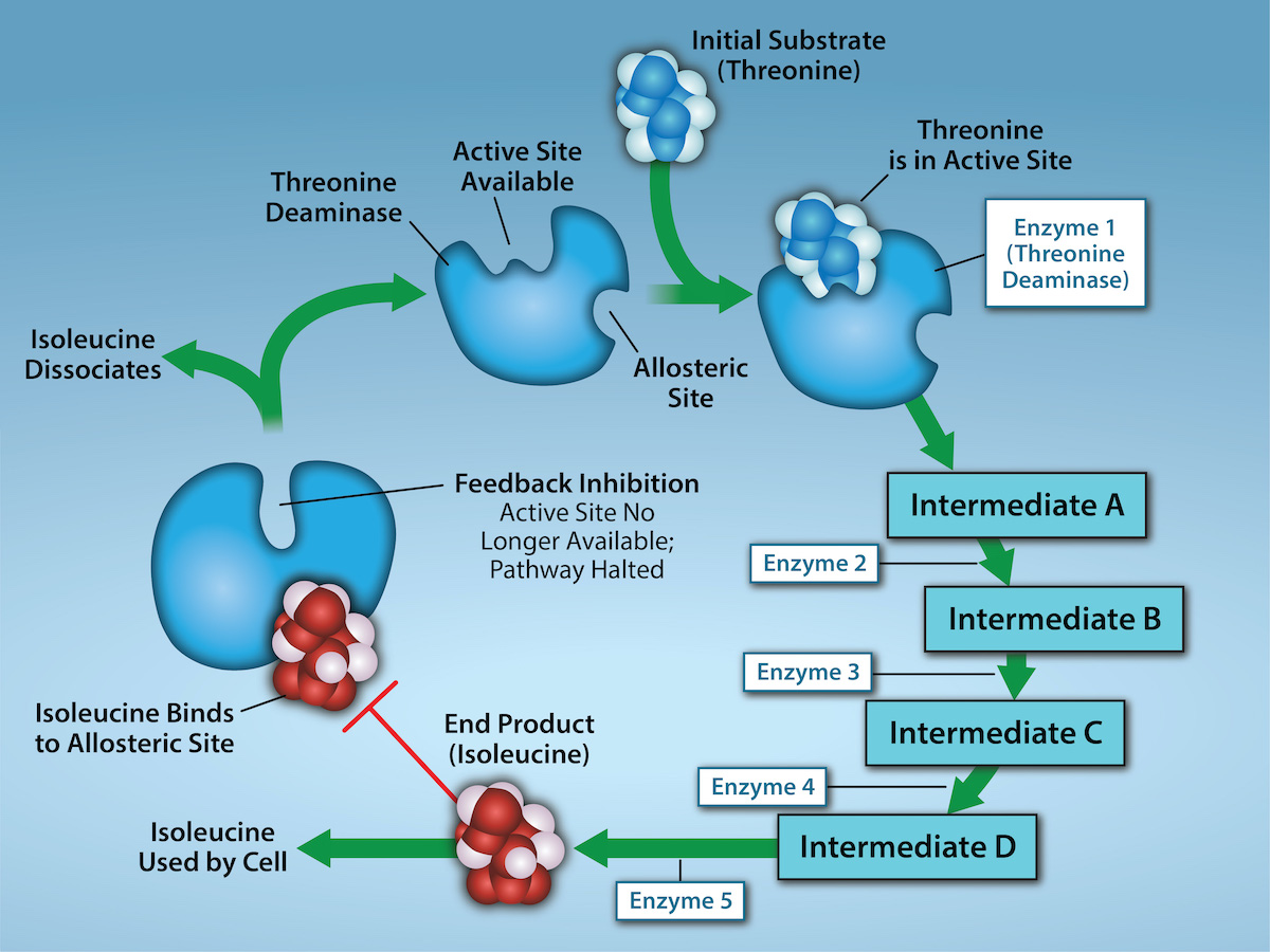
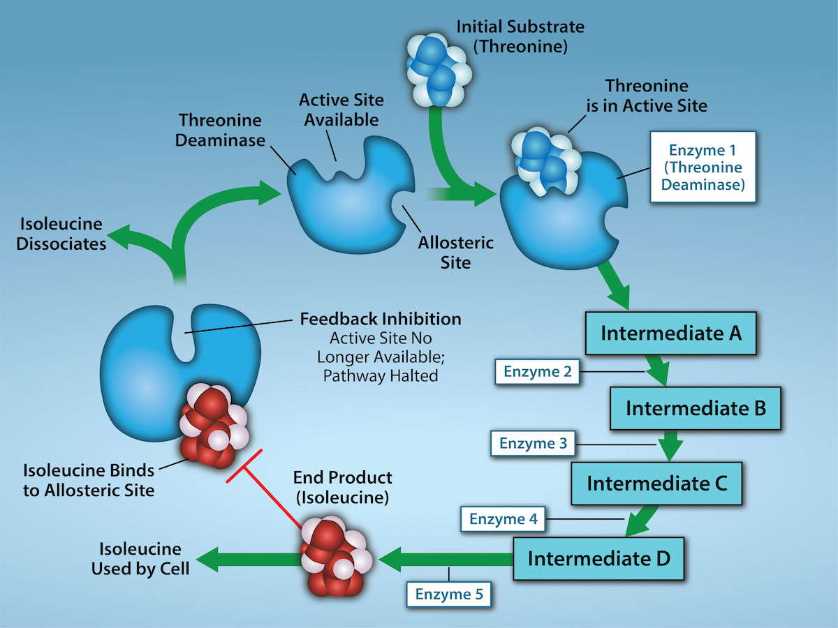
Figure 6.21 Metabolic pathways are a series of reactions that multiple enzymes catalyze. Feedback inhibition, where the pathway's end product inhibits an upstream step, is an important regulatory mechanism in cells. Metabolic pathways are a series of reactions catalyzed by multiple enzymes (Intermediates A – D, Enzymes 1 – 5). Feedback inhibition occurs when the pathway's end product (here isoleucine) inhibits an upstream enzyme (indicated by red bar). In this example, isoleucine will bind to Threonine Deaminase (at the allosteric site) and prevent threonine from binding to this enzyme’s active site, effectively blocking this metabolic pathway. When isoleucine levels decrease, Threonine will then bind to Threonine Deaminase’s active site and the metabolic pathway will resume. This is an important regulatory mechanism in cells to inhibit the overproduction of a product. Credit: Rao, A., Ryan, K., Tag, A., Hawkins, A. and Fletcher S. Department of Biology, Texas A&M University.
Mary Ann Clark, Matthew Douglas, and Jung Choi, Biology 2E, OpenStax. · CC BY-NC-SA 4.0 · Original image ↗ · Access for free ↗Source: 6.5 Enzymes