Try before revealing the answer. Why is the lone pair on pyridine more available for protonation than the lone pair on pyrrole?
Worked answer
Pyridine’s lone pair is not required for the six-electronaromatic π system, so protonation can preserve aromaticity. Pyrrole’s lone pair supplies two of its six aromaticelectrons; using it to form an additional bond disrupts that stabilizing arrangement, making the ring much less basic.
Figure 14.5 The purines have a double ring structure with a six-membered ring fused to a five-membered ring. Pyrimidines are smaller in size; they have a single six-membered ring structure.
A nitrogen-containing functional group. A free amino acid’s amino group is typically protonated (NH₃⁺) near physiological pH; proline’s ring nitrogen is an exception to this formula.
Figure 15.14 This is a space-filling model of a tRNA molecule that adds the amino acid phenylalanine to a growing polypeptide chain. The anticodon AAG binds the Codon UUC on the mRNA. The amino acid phenylalanine is attached to the other end of the tRNA.
Figure 15.4 This figure shows the genetic code for translating each nucleotide triplet in mRNA into an amino acid or a termination signal in a protein. (credit: modification of work by NIH)
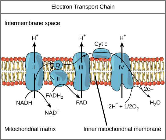
Figure 7.12 The electron transport chain is a series of electron transporters embedded in the inner mitochondrial membrane that shuttles electrons from NADH and FADH 2 to molecular oxygen. In the process, protons are pumped from the mitochondrial matrix to the intermembrane space, and oxygen is reduced to form water.
Figure 4.44 - Kinetic profile of an allosteric enzyme whose activity is controlled by a homotropic effector. Image by Aleia Kim
Kevin Ahern, Indira Rajagopal, and Taralyn Tan, Biochemistry Free For All, Oregon State University, via Biology LibreTexts. Image by Aleia Kim · CC BY-NC-SA 4.0
· Original image ↗ · Access for free ↗
Figure 14.5 The purines have a double ring structure with a six-membered ring fused to a five-membered ring. Pyrimidines are smaller in size; they have a single six-membered ring structure.
An electrical property that can be positive or negative. Opposite charges attract and like charges repel; net charge is the sum of all charges on a particle.
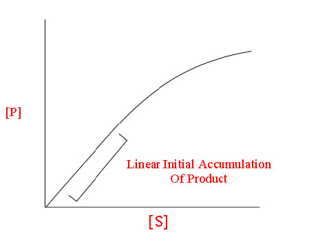
Figure 4.18 - Kinetics of an enzyme obeying Michaelis-Menten kinetics. Image by Aleia Kim
Kevin Ahern, Indira Rajagopal, and Taralyn Tan, Biochemistry Free For All, Oregon State University, via Biology LibreTexts. Image by Aleia Kim · CC BY-NC-SA 4.0
· Original image ↗ · Access for free ↗
Figure 15.11 Eukaryotic pre-mRNA processing. In addition to 5’ Cap and 3’ Poly-A Tail addition, introns must be precisely removed and exons joined to generate a functional mRNA. Nucleotides upstream (towards the 5’cap) of the translation START codon are part of the 5’ untranslated region (5’ UTR). Nucleotides downstream (towards 3’end) of the STOP codon form the 3’ UTR. Both 5’ and 3’ UTRs are important for regulating translation initiation and mRNA stability. Credit: Rao, A., Ryan, K. Fletcher, S. and Tag, A. Department of Biology, Texas A&M University.
Rao, A., Ryan, K. Fletcher, S. and Tag, A. Department of Biology, Texas A&M University.; Mary Ann Clark, Matthew Douglas, and Jung Choi, Biology 2e, OpenStax. · CC BY-NC-SA 4.0
· Original image ↗ · Access for free ↗
Figure 15.10 A generalized promoter of a gene transcribed by RNA polymerase II is shown. Transcription factors recognize the promoter. RNA polymerase II then binds and forms the transcription initiation complex.
Figure 14.5 The purines have a double ring structure with a six-membered ring fused to a five-membered ring. Pyrimidines are smaller in size; they have a single six-membered ring structure.
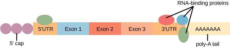
Figure 16.13 RNA-binding proteins. The protein-coding region of this processed mRNA is flanked by 5' and 3' untranslated regions (UTRs). The presence of RNA-binding proteins at the 5' or 3' UTR influences the stability of the RNA molecule.
Figure 4.18 - Kinetics of an enzyme obeying Michaelis-Menten kinetics. Image by Aleia Kim
Kevin Ahern, Indira Rajagopal, and Taralyn Tan, Biochemistry Free For All, Oregon State University, via Biology LibreTexts. Image by Aleia Kim · CC BY-NC-SA 4.0
· Original image ↗ · Access for free ↗
The early rate of product formation, measured before substantial substrate depletion or product accumulation, usually after the brief binding transient.
Figure 4.18 - Kinetics of an enzyme obeying Michaelis-Menten kinetics. Image by Aleia Kim
Kevin Ahern, Indira Rajagopal, and Taralyn Tan, Biochemistry Free For All, Oregon State University, via Biology LibreTexts. Image by Aleia Kim · CC BY-NC-SA 4.0
· Original image ↗ · Access for free ↗
Figure 15.11 Eukaryotic pre-mRNA processing. In addition to 5’ Cap and 3’ Poly-A Tail addition, introns must be precisely removed and exons joined to generate a functional mRNA. Nucleotides upstream (towards the 5’cap) of the translation START codon are part of the 5’ untranslated region (5’ UTR). Nucleotides downstream (towards 3’end) of the STOP codon form the 3’ UTR. Both 5’ and 3’ UTRs are important for regulating translation initiation and mRNA stability. Credit: Rao, A., Ryan, K. Fletcher, S. and Tag, A. Department of Biology, Texas A&M University.
Rao, A., Ryan, K. Fletcher, S. and Tag, A. Department of Biology, Texas A&M University.; Mary Ann Clark, Matthew Douglas, and Jung Choi, Biology 2e, OpenStax. · CC BY-NC-SA 4.0
· Original image ↗ · Access for free ↗
The exponent needed to obtain a number from a chosen fixed number. For decimal logarithms, log₁₀(100) = 2 because 10² = 100; pH uses the negative decimal logarithm of hydrogen ion activity.
Figure 4.18 Membrane and secretory proteins are synthesized in the rough endoplasmic reticulum (RER). The RER also sometimes modifies proteins. In this illustration, a (green) integral membrane protein is modified by attachment of a (purple) carbohydrate in the ER. Vesicles with the integral protein bud from the ER and fuse with the Golgi apparatus' cis face. As the protein passes along the Golgi’s cisternae, the addition of more carbohydrates further modifies it. After its synthesis is complete, it exits as an integral membrane protein of the vesicle that buds from the Golgi’s trans face. When the vesicle fuses with the cell membrane, the protein becomes an integral portion of that cell membrane. (credit: modification of work by Magnus Manske)
A supporting material, such as a porous gel or the solid support in a chromatography column. Its properties influence movement and binding of sample molecules.
A thin boundary or sheet. Cell membranes are lipid bilayers containing proteins; blotting membranes are synthetic supports that hold proteins or nucleic acids for detection.
Figure 15.11 Eukaryotic pre-mRNA processing. In addition to 5’ Cap and 3’ Poly-A Tail addition, introns must be precisely removed and exons joined to generate a functional mRNA. Nucleotides upstream (towards the 5’cap) of the translation START codon are part of the 5’ untranslated region (5’ UTR). Nucleotides downstream (towards 3’end) of the STOP codon form the 3’ UTR. Both 5’ and 3’ UTRs are important for regulating translation initiation and mRNA stability. Credit: Rao, A., Ryan, K. Fletcher, S. and Tag, A. Department of Biology, Texas A&M University.
Rao, A., Ryan, K. Fletcher, S. and Tag, A. Department of Biology, Texas A&M University.; Mary Ann Clark, Matthew Douglas, and Jung Choi, Biology 2e, OpenStax. · CC BY-NC-SA 4.0
· Original image ↗ · Access for free ↗
Figure 4.18 - Kinetics of an enzyme obeying Michaelis-Menten kinetics. Image by Aleia Kim
Kevin Ahern, Indira Rajagopal, and Taralyn Tan, Biochemistry Free For All, Oregon State University, via Biology LibreTexts. Image by Aleia Kim · CC BY-NC-SA 4.0
· Original image ↗ · Access for free ↗
Figure 4.18 Membrane and secretory proteins are synthesized in the rough endoplasmic reticulum (RER). The RER also sometimes modifies proteins. In this illustration, a (green) integral membrane protein is modified by attachment of a (purple) carbohydrate in the ER. Vesicles with the integral protein bud from the ER and fuse with the Golgi apparatus' cis face. As the protein passes along the Golgi’s cisternae, the addition of more carbohydrates further modifies it. After its synthesis is complete, it exits as an integral membrane protein of the vesicle that buds from the Golgi’s trans face. When the vesicle fuses with the cell membrane, the protein becomes an integral portion of that cell membrane. (credit: modification of work by Magnus Manske)
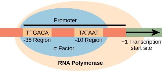
Figure 15.8 The σ subunit of prokaryotic RNA polymerase recognizes consensus sequences found in the promoter region upstream of the transcription start site. The σ subunit dissociates from the polymerase after transcription has been initiated.
Figure 14.5 The purines have a double ring structure with a six-membered ring fused to a five-membered ring. Pyrimidines are smaller in size; they have a single six-membered ring structure.
Figure 14.5 The purines have a double ring structure with a six-membered ring fused to a five-membered ring. Pyrimidines are smaller in size; they have a single six-membered ring structure.
Figure 12.19 Reaction diagrams for an endothermic process in the absence (red curve) and presence (blue curve) of a catalyst. The catalyzed pathway involves a two-step mechanism (note the presence of two transition states) and an intermediate species (represented by the valley between the two transitions states).
Figure 15.19 Translation Termination is Active. Translation is terminated when a STOP codon is in the A-site of the ribosome. Since there are no tRNAs corresponding to the STOP codons, the Release Factor protein enters and catalyzes the hydrolysis between the last amino acid and its tRNA. This hydrolysis releases the free carboxyl terminus (C-term) of the protein. Additional factors use the energy in GTP hydrolysis to disassemble the large and small ribosomal subunits and mRNA. Credit: Rao, A. and Ryan, K. Department of Biology, Texas A&M University.
Rao, A. and Ryan, K. Department of Biology, Texas A&M University.; Mary Ann Clark, Matthew Douglas, and Jung Choi, Biology 2e, OpenStax. · CC BY-NC-SA 4.0
· Original image ↗ · Access for free ↗
resonance
Large driven response near a system’s natural frequency.
Figure 15.13 Pre-mRNA splicing involves the precise removal of introns from the primary RNA transcript. The splicing process is catalyzed by protein complexes called spliceosomes that are composed of proteins and RNA molecules called small nuclear RNAs (snRNAs). Spliceosomes recognize sequences at the 5' and 3' end of the intron. Rao, A. and Ryan, K. Department of Biology, Texas A&M University.
Rao, A. and Ryan, K. Department of Biology, Texas A&M University.; Mary Ann Clark, Matthew Douglas, and Jung Choi, Biology 2e, OpenStax. · CC BY-NC-SA 4.0
· Original image ↗ · Access for free ↗
RNA stability
Persistence of an RNA before degradation; greater stability generally means a longer lifetime or half-life under specified conditions.
Figure 16.13 RNA-binding proteins. The protein-coding region of this processed mRNA is flanked by 5' and 3' untranslated regions (UTRs). The presence of RNA-binding proteins at the 5' or 3' UTR influences the stability of the RNA molecule.
Figure 15.8 The σ subunit of prokaryotic RNA polymerase recognizes consensus sequences found in the promoter region upstream of the transcription start site. The σ subunit dissociates from the polymerase after transcription has been initiated.
Figure 4.44 - Kinetic profile of an allosteric enzyme whose activity is controlled by a homotropic effector. Image by Aleia Kim
Kevin Ahern, Indira Rajagopal, and Taralyn Tan, Biochemistry Free For All, Oregon State University, via Biology LibreTexts. Image by Aleia Kim · CC BY-NC-SA 4.0
· Original image ↗ · Access for free ↗
Figure 7.45 - Non-homologous end joining (left) versus homologous recombination (right) - Wikipedia
Wikipedia, as credited by Kevin Ahern, Indira Rajagopal, and Taralyn Tan, Biochemistry Free For All, Oregon State University, via Biology LibreTexts. · Source attribution
· Original image ↗ · Access for free ↗
The amount of a substance that can dissolve in a solvent under specified conditions. Soluble means able to dissolve; insoluble means only very slightly soluble in that solvent.
Figure 15.4 This figure shows the genetic code for translating each nucleotide triplet in mRNA into an amino acid or a termination signal in a protein. (credit: modification of work by NIH)
Figure 14.5 The purines have a double ring structure with a six-membered ring fused to a five-membered ring. Pyrimidines are smaller in size; they have a single six-membered ring structure.
Figure 15.7 Messenger RNA is a copy of protein-coding information in the coding strand of DNA, with the substitution of U in the RNA for T in the coding sequence. However, new RNA nucleotides base pair with the nucleotides of the template strand. RNA is synthesized in its 5'-3' direction, using the enzyme RNA polymerase. As the template is read, the DNA unwinds ahead of the polymerase and then rewinds behind it.
Figure 15.14 This is a space-filling model of a tRNA molecule that adds the amino acid phenylalanine to a growing polypeptide chain. The anticodon AAG binds the Codon UUC on the mRNA. The amino acid phenylalanine is attached to the other end of the tRNA.
A fleeting, high-energy arrangement at a reaction barrier. It is an energy maximum along the reaction path, rather than a stable reaction intermediate.
Figure 12.19 Reaction diagrams for an endothermic process in the absence (red curve) and presence (blue curve) of a catalyst. The catalyzed pathway involves a two-step mechanism (note the presence of two transition states) and an intermediate species (represented by the valley between the two transitions states).
Figure 15.18 Translation begins when an initiator tRNA anticodon recognizes a start codon on mRNA bound to a small ribosomal subunit. The large ribosomal subunit joins the small subunit, and a second tRNA is recruited. As the mRNA moves relative to the ribosome, successive tRNAs move through the ribosome and the polypeptide chain is formed. Entry of a release factor into the A site terminates translation and the components dissociate.
Figure 4.21 - Kcat (turnover number) values for several enzymes. Image by Aleia Kim
Kevin Ahern, Indira Rajagopal, and Taralyn Tan, Biochemistry Free For All, Oregon State University, via Biology LibreTexts. Image by Aleia Kim · CC BY-NC-SA 4.0
· Original image ↗ · Access for free ↗
Figure 14.5 The purines have a double ring structure with a six-membered ring fused to a five-membered ring. Pyrimidines are smaller in size; they have a single six-membered ring structure.
No matching terms. Try a different name or abbreviation.
17.10 Reactions of Phenols
OpenStax · 17.10 Reactions of Phenols ↗ Instructional page including learning objectives, examples, figures, source tables and questions. Site navigation and embedded media players are omitted; source media links remain available.
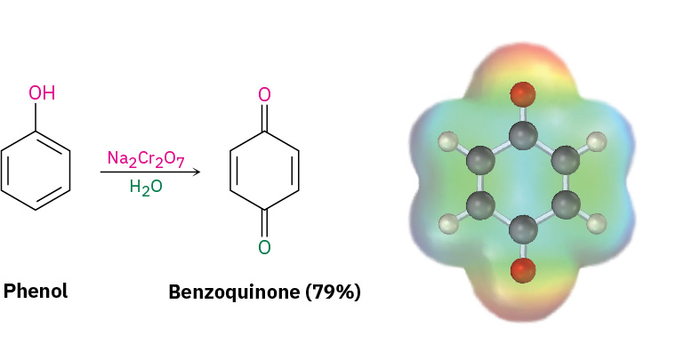
Phenols don’t undergo oxidation in the same way as alcohols because they don’t have a hydrogen atom on the hydroxyl-bearing carbon. Instead, oxidation of a phenol yields a 2,5-cyclohexadiene-1,4-dione, or quinone. Many different oxidizing agents will accomplish the transformation, with Na2Cr2O7 as a common choice for simple phenols and potassium nitrosodisulfonate [(KSO3)2NO], called Fremy’s salt, is often used.
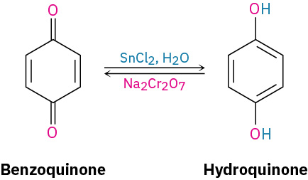
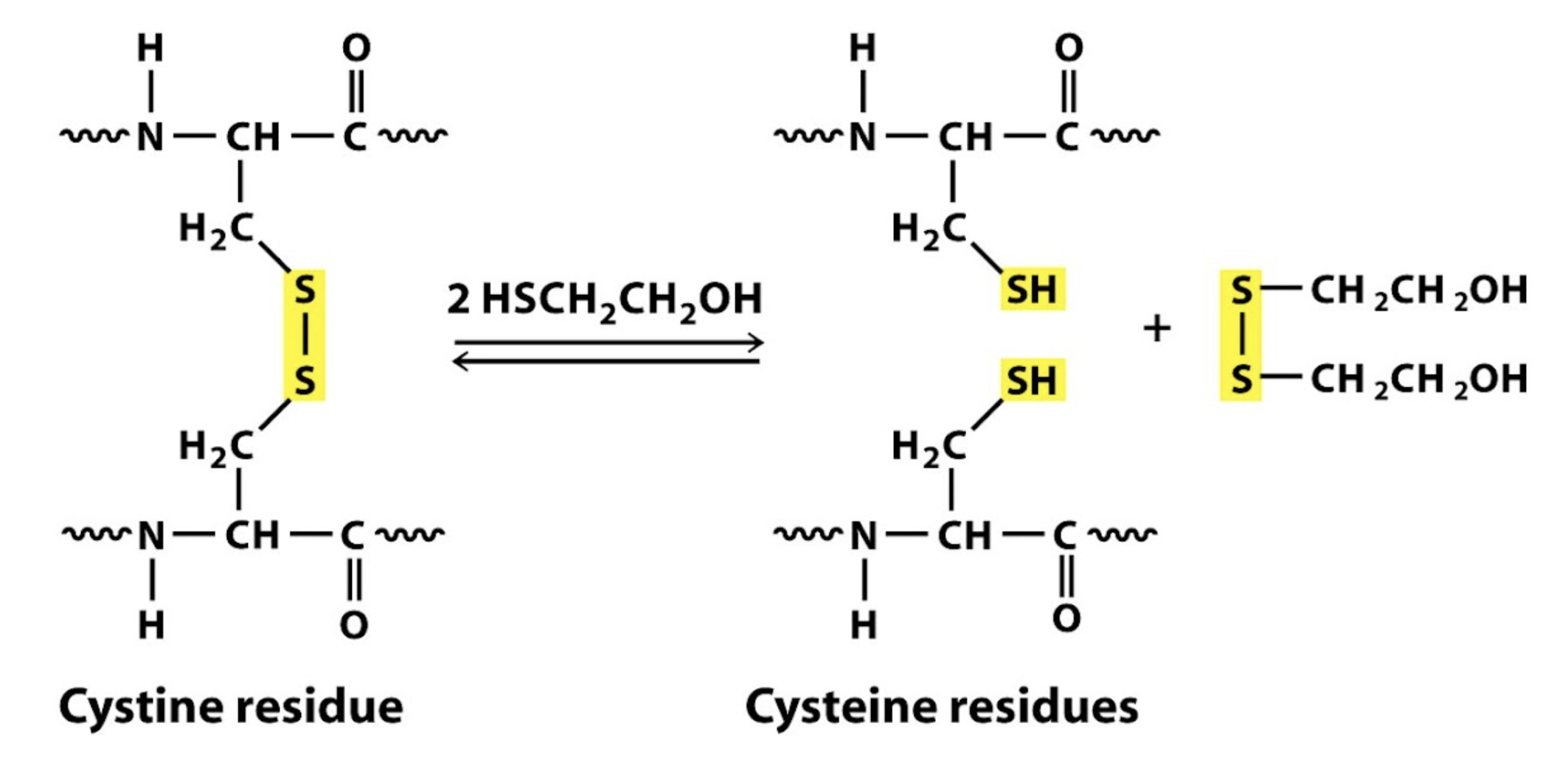
Quinones are a valuable class of compounds because of their oxidation–reduction, or redox, properties. They can be easily reduced to hydroquinones (p-dihydroxybenzenes) by reagents such as NaBH4 and SnCl2, and hydroquinones can be easily reoxidized back to quinones by Na2Cr2O7.
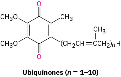
The redox properties of quinones are crucial to the functioning of living cells, where compounds called ubiquinones act as biochemical oxidizing agents to mediate the electron-transfer processes involved in energy production. Ubiquinones, also called coenzymesQ, are components of the cells in all aerobic organisms, from the simplest bacterium to humans. They are so named because of their ubiquitous occurrence throughout nature.
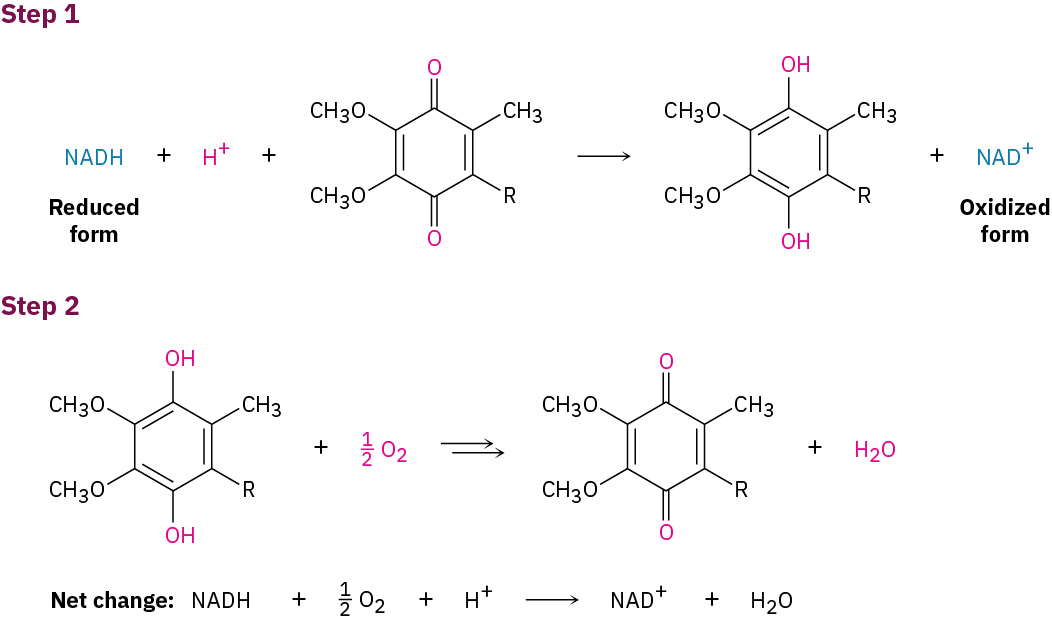
Ubiquinones function within the mitochondria of cells to mediate the respiration process in which electrons are transported from the biological reducing agentNADH to molecular oxygen. Through a complex series of steps, the ultimate result is a cycle whereby NADH is oxidized to NAD+, O2 is reduced to water, and energy is produced. Ubiquinone acts only as an intermediary and is itself unchanged.
OpenStax · 24.9 Heterocyclic Amines ↗ Instructional page including learning objectives, examples, figures, source tables and questions. Site navigation and embedded media players are omitted; source media links remain available.
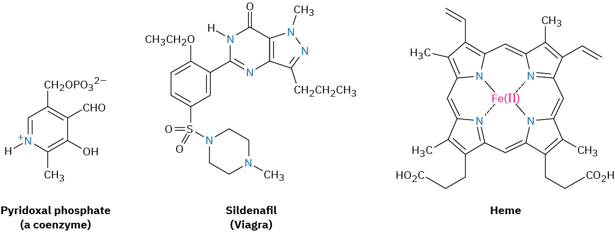
As noted in Section 15.5 in connection with a discussion of aromaticity, a cyclic organic compound that contains atoms of two or more elements in its ring is a called a heterocycle. Heterocyclic amines are particularly common, and many have important biological properties. Pyridoxal phosphate, a coenzyme; sildenafil (Viagra), a well-known pharmaceutical; and heme, the oxygen carrier in blood, are a few examples.
Most heterocycles have the same chemistry as their open-chain counterparts. Lactones and acyclic esters behave similarly, lactams and acyclic amides behave similarly, and cyclic and acyclic ethers behave similarly. In certain cases, however, particularly when the ring is unsaturated, heterocycles have unique and interesting properties.
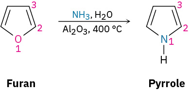
Pyrrole, the simplest five-membered unsaturated heterocyclic amine, is obtained commercially by treatment of furan with ammonia over an aluminacatalyst at 400 °C. Furan, the oxygen-containing analog of pyrrole, is obtained by acid-catalyzed dehydration of the five-carbon sugars found in oat hulls and corncobs.
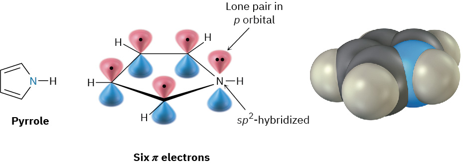
Although pyrrole appears to be both an amine and a conjugated diene, its chemical properties are not consistent with either of these structural features. Unlike most other amines, pyrrole is not basic—the pKa of the pyrrolinium ion is 0.4; unlike most other conjugated dienes, pyrrole undergoes electrophilic substitutionreactions rather than additions. The reason for both of these properties, as noted in Section 15.5, is that pyrrole has six πelectrons and is aromatic. Each of the four carbons contributes one πelectron, and the sp2-hybridized nitrogen contributes two more from its lone pair.
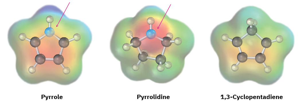
Because the nitrogen lone pair is a part of the aromatic sextet, protonation on nitrogen would destroy the aromaticity of the ring. The nitrogen atom in pyrrole is therefore less electron-rich, less basic, and less nucleophilic than the nitrogen in an aliphatic amine. By the same token, the carbon atoms of pyrrole are more electron-rich and more nucleophilic than typical double-bond carbons. The pyrrole ring is therefore reactive toward electrophiles in the same way as enamines (Section 23.11). Electrostatic potential maps show how the pyrrole nitrogen is electron-poor (less red) compared with the nitrogen in its saturated counterpart pyrrolidine, while the pyrrole carbon atoms are electron-rich (more red) compared with the carbons in 1,3-cyclopentadiene.
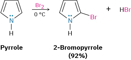
The chemistry of pyrrole is similar to that of activated benzene rings. In general, however, the heterocycles are more reactive toward electrophiles than benzene rings, and low temperatures are often necessary to control the reactions. Halogenation, nitration, sulfonation, and Friedel–Crafts acylation can all be accomplished. For example:
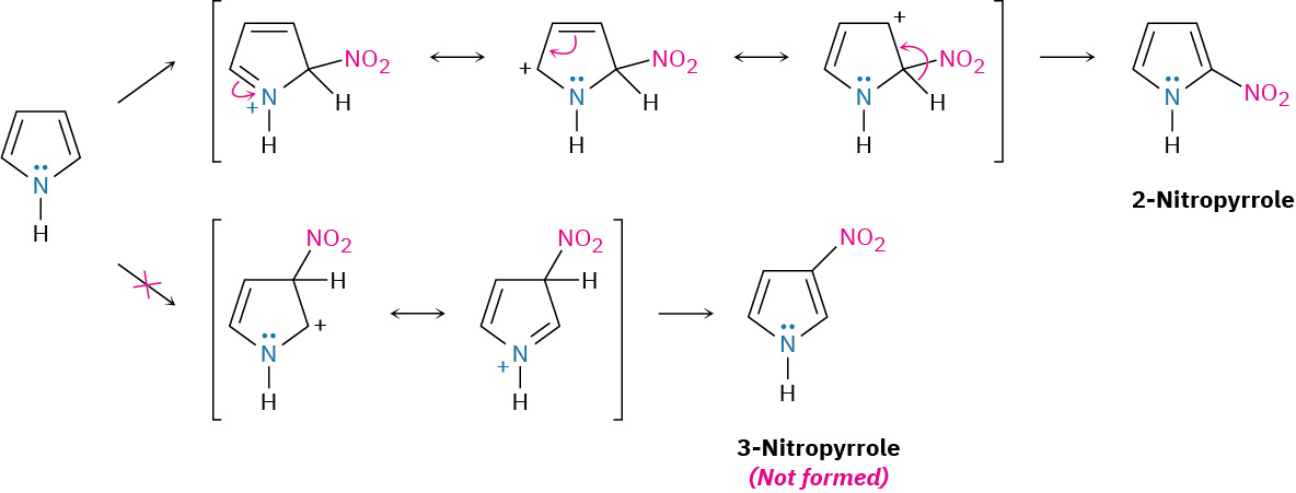
Electrophilic substitutions normally occur at C2, the position next to the nitrogen, because reaction at this position leads to a more stable intermediate cation having three resonance forms, whereas reaction at C3 gives a less stable cation with only two resonance forms (Figure 24.8).
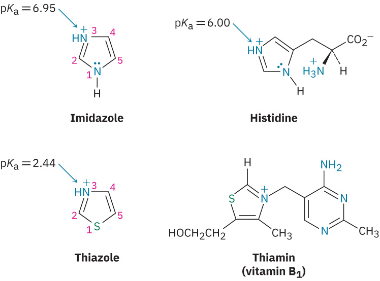
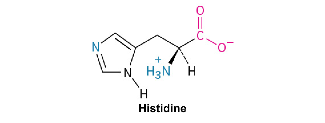
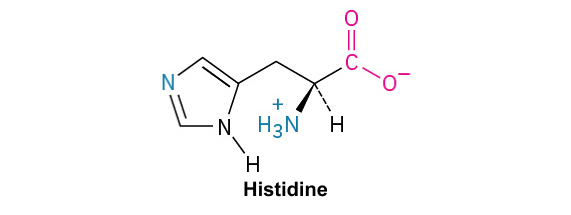
Other common five-membered heterocyclic amines include imidazole and thiazole. Imidazole, a constituent of the amino acidhistidine, has two nitrogens, only one of which is basic. Thiazole, the five-membered ring system on which the structure of thiamin (vitamin B1) is based, also contains a basic nitrogen that is alkylated in thiamin to form a quaternary ammonium ion.
Draw an orbital picture of thiazole. Assume that both the nitrogen and sulfur atoms are sp2-hybridized, and show the orbitals that the lone pairs occupy.
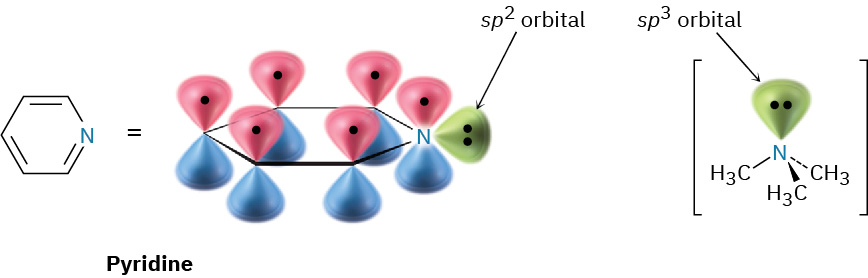
Pyridine is the nitrogen-containing heterocyclic analog of benzene. Like benzene, pyridine is a flat, aromaticmolecule, with bond angles of 120° and C–C bond lengths of 139 pm, intermediate between typical single and double bonds. The five carbon atoms and the sp2-hybridized nitrogen atom each contribute one πelectron to the aromatic sextet, and the lone-pair electrons occupy an sp2orbital in the plane of the ring (Section 15.5).
As shown previously in Table 24.1, pyridine (pKa = 5.25) is a stronger base than pyrrole but a weaker base than the alkylamines. The diminished basicity of pyridine compared with that of alkylamines is due to the fact that the lone-pair electrons on the pyridine nitrogen are in an sp2orbital, while those on an alkylamine nitrogen are in an sp3orbital. Because s orbitals have their maximum electron density at the nucleus but p orbitals have a node at the nucleus, electrons in an orbital with more s character are held more closely to the positively chargednucleus and are less available for bonding. As a result, the sp2-hybridized nitrogen atom (33% s character) in pyridine is less basic than the sp3-hybridized nitrogen in an alkylamine (25% s character).
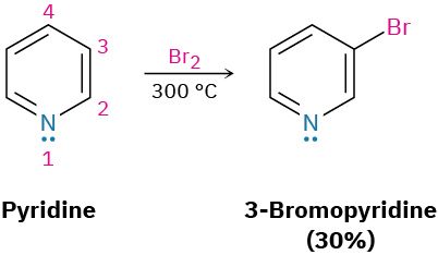
Unlike benzene, pyridine undergoes electrophilic aromaticsubstitutionreactions with difficulty. Halogenation can be carried out under drastic conditions, but nitration occurs in very low yield, and Friedel–Crafts reactions are not successful. Reactions usually give the 3-substituted product.
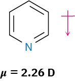
The low reactivity of pyridine toward electrophilic aromaticsubstitution is caused by a combination of factors. One is that acid–base complexation between the basic ring’s nitrogen atom and the incoming electrophile places a positive charge on the ring, thereby deactivating it. Equally important is that the electron density of the ring is decreased by the electron-withdrawing inductive effect of the electronegative nitrogen atom. Thus, pyridine has a substantial dipole moment (μ = 2.26 D), with the ring carbons acting as the positive end of the dipole. Reaction of an electrophile with the positively polarized carbon atoms is therefore difficult.
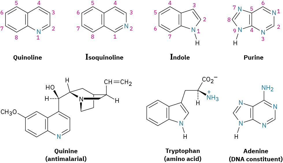
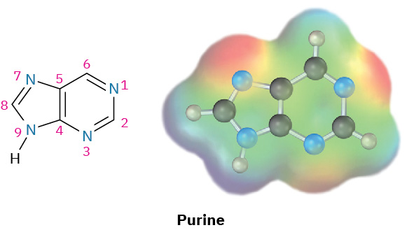
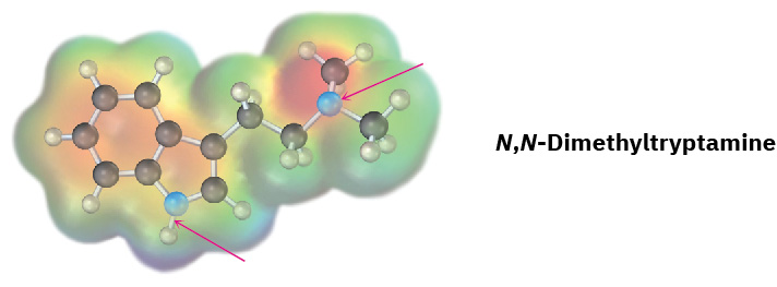
As we saw in Section 15.6, quinoline, isoquinoline, indole, and purine are common polycyclic heterocycles. The first three contain both a benzene ring and a heterocyclic aromatic ring, while purine contains two heterocyclic rings joined together. All four ring systems occur commonly in nature, and many compounds with these rings have pronounced physiological activity. The quinoline alkaloid quinine, for instance, is widely used as an antimalarial drug; tryptophan is a common amino acid; and the purineadenine is a constituent of nucleic acids.
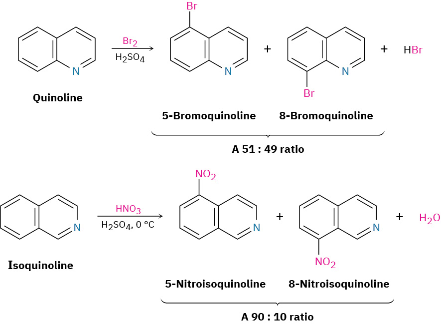
The chemistry of these polycyclic heterocycles is just what you might expect from a knowledge of the simpler heterocycles pyridine and pyrrole. Quinoline and isoquinoline both have basic, pyridine-like nitrogen atoms, and both undergo electrophilic substitutions. As with pyridine, both quinoline and isoquinoline are less reactive toward electrophilic substitution than benzene because of the electronegative nitrogen atom that withdraws electrons from the ring. Reaction occurs on the benzene ring rather than on the nitrogen-containing pyridine ring, and a mixture of substitutionproducts is obtained.
Purine has three basic, pyridine-like nitrogens with lone-pair electrons in sp2 orbitals in the plane of the ring. The remaining purine nitrogen is nonbasic and pyrrole-like, with its lone-pair electrons as part of the aromaticπelectron system.
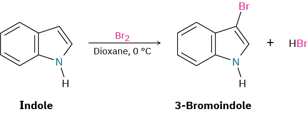
Indole reacts with electrophiles at C3 rather than at C2. Draw resonance forms of the intermediate cations resulting from reaction at C2 and C3, and explain the observed results.