Orders of protein structure

Source ↗

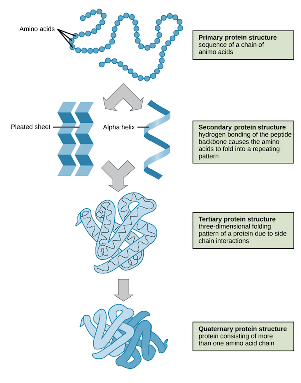
Primary → sequence; secondary → local backbone folds; tertiary → one chain; quaternary → multiple chains.

Primary: amino acid sequence

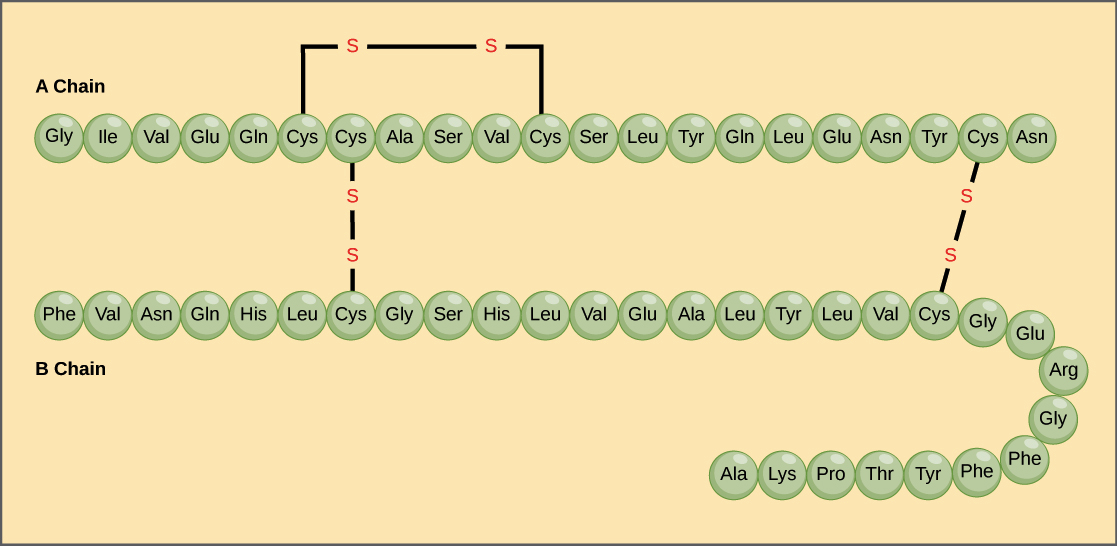
Peptide bonds connect amino acids into a polypeptide. Read its sequence from the N-terminus to the C-terminus. DNA determines the sequence; changing one residue can change the resulting protein’s behavior. Insulin has two mature chains connected by disulfide bonds.

Insulin A and B chains with amino acid sequences and disulfide links between cysteines.
Insulin’s amino acid sequence OpenStax Biology, via Khan Academy · CC BY 3.0 · Original image ↗

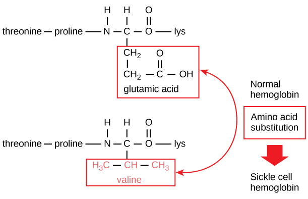
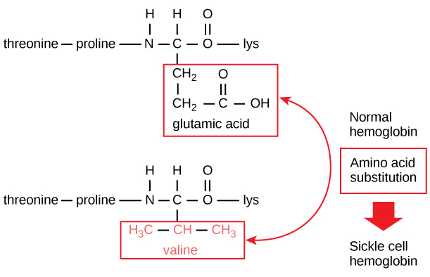
In sickle-cell disease, a mutation replaces glutamate with valine at position 6 of the hemoglobin beta chain. The new hydrophobic patch promotes aggregation of deoxygenated hemoglobin, distorting red blood cells and obstructing blood flow.

Normal and sickle hemoglobin sequences differ by glutamate versus valine; normal and sickled red blood cells are compared.
One residue changes hemoglobin Khan Academy, modified from OpenStax Biology · CC BY-NC-SA 4.0 · Original image ↗
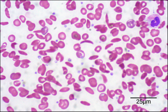
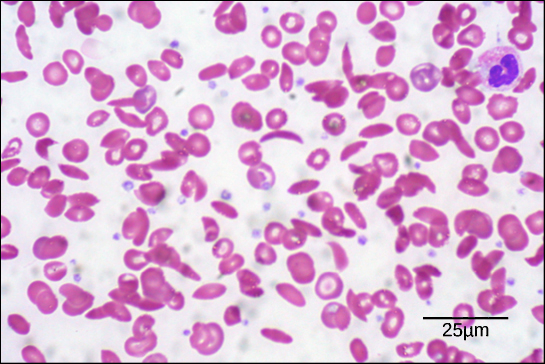
Blood micrograph showing crescent-shaped sickled red blood cells among other blood cells.
Sickled red blood cells OpenStax Biology; modification of work by Ed Uthman; scale bar by Matt Russell · CC BY 3.0 · Original image ↗

Secondary: local backbone folding

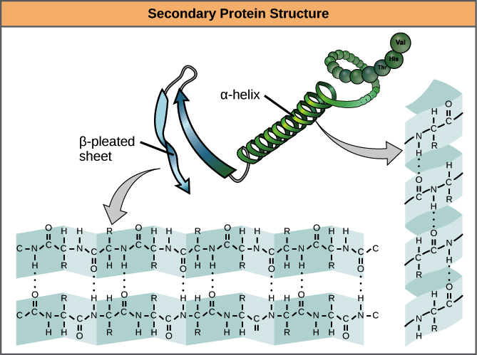
  • An alpha helix coils around its axis. Backbone carbonyl and amino groups four residues apart form hydrogen bonds; each turn contains about 3.6 residues. R groups point outward.
  • A beta sheet aligns extended strands. Hydrogen bonds connect their backbones; strands can run parallel or antiparallel. R groups alternate above and below the sheet.

These hydrogen bonds involve the backbone. Side-chain interactions are more important when describing tertiary structure.

Backbone hydrogen bonds stabilize an alpha helix and an antiparallel beta sheet; side chains extend away from the backbone.
Alpha helices and beta sheets OpenStax Biology, via Khan Academy · CC BY 3.0 · Original image ↗

Tertiary: one chain’s complete shape

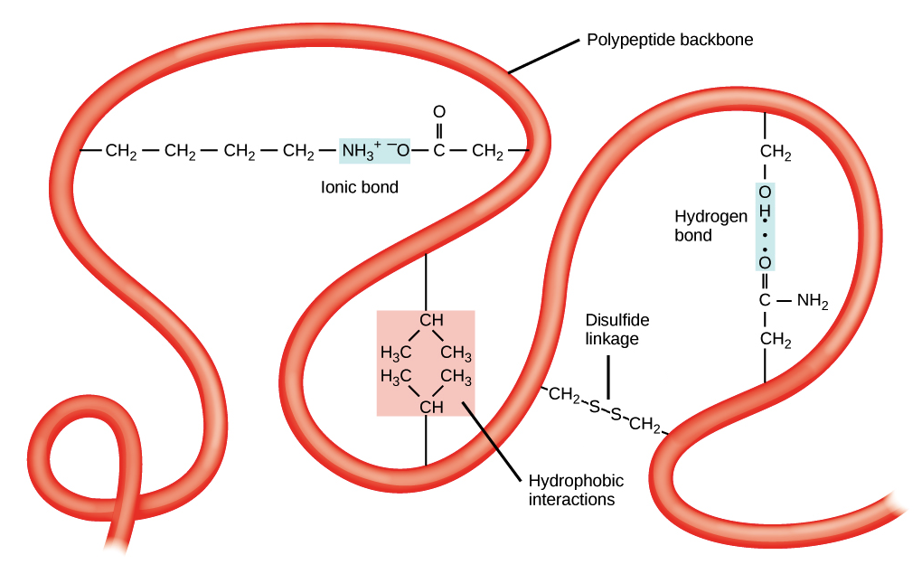
A single polypeptide folds into a three-dimensional structure. The hydrophobic effect often buries nonpolar R groups in soluble proteins, while polar groups face water. Hydrogen bonds, salt bridges, Van der Waals forces, and covalent disulfide bonds stabilize the fold. Disulfide bonds form between cysteine residues.

A folded polypeptide showing hydrogen bonding, ionic attraction, hydrophobic interactions, and a disulfide bond.
Interactions that stabilize a fold Khan Academy, modified from OpenStax Biology · CC BY-NC-SA 4.0 · Original image ↗

Quaternary: multiple chains together

Quaternary structure describes the arrangement of two or more subunits. Hemoglobin contains two alpha and two beta chains. The same types of interactions that stabilize tertiary structure can hold subunits together; disulfide bonds connect some complexes.

Primary amino acid sequence, secondary helix and sheet, tertiary fold, and quaternary assembly shown together.
The four levels together Khan Academy, modified from OpenStax Biology and NHGRI · CC BY-NC-SA 4.0 · Original image ↗

Shape, function, and denaturation

Heat or extreme pH can cause denaturation: loss of the native fold and often function. Peptide bonds usually remain intact, preserving primary structure. Some proteins refold when conditions recover; others aggregate. Chaperones help proteins fold and limit aggregation.

30 terms, alphabetically ordered

Alpha helixα helix · alpha helices · α helices
A helical secondary structure stabilized by hydrogen bonds between a backbone carbonyl oxygen and an amide hydrogen four residues later.
Amino acidamino acids
A protein building block containing amino and carboxyl groups and a characteristic side chain.
Beta sheetβ sheet · beta sheets · β sheets · β pleated sheet · beta pleated sheet
A secondary structure made of aligned polypeptide segments connected by backbone hydrogen bonds; strands may be parallel or antiparallel.
C-terminus
Carboxyl terminus: the end of a polypeptide with a free carboxyl group. Protein chains grow at this end.
CysteineCys · C
An amino acid with a thiol side chain. Two cysteines can form a disulfide bond.
Denaturation
Loss of a protein’s native shape, often with loss of function. This usually preserves the chain’s peptide bonds.
Deoxyribonucleic acidDNA
The nucleic acid that stores genetic information used to specify RNA and protein sequences.
Disulfide bond
A covalent sulfur–sulfur bond formed by oxidation of two cysteine thiol groups, which can stabilize protein structure.
GlutamateGlu · glutamic acid · E
An acidic amino acid whose side chain is usually negatively charged near physiological pH.
Hemoglobin
The oxygen-carrying protein in red blood cells, composed of four polypeptide subunits with heme groups.
Hydrogen bondhydrogen bonds · hydrogen bonding
An attraction between a hydrogen bonded to an electronegative atom and another electronegative atom. Backbone hydrogen bonds stabilize protein secondary structure.
Hydrophobic
Interacting poorly with water. Nonpolar side chains often gather in a soluble protein’s interior.
Hydrophobic effect
The tendency of nonpolar surfaces to cluster in water, reducing their exposure to water and helping stabilize folded proteins.
Insulin
A peptide hormone from pancreatic beta cells that lowers blood glucose by promoting uptake and storage and reducing glucose production.
Molecular chaperonemolecular chaperones · chaperone · chaperones
A protein that assists other proteins in folding or prevents aggregation, without specifying their amino acid sequence.
Mutation
A change in a DNA sequence; some mutations alter a protein’s amino acid sequence.
N-terminus
Amino terminus: the end of a polypeptide with a free amino group. Written first when displaying a protein sequence.
Peptide bondpeptide bonds
A covalent amide linkage between the carboxyl carbon of one amino acid and the amino nitrogen of the next.
pH
A measure of acidity, defined as the negative base-10 logarithm of hydrogen ion activity. Lower pH means greater acidity; pH influences amino acid charge.
Polypeptidepolypeptides
A chain of amino acid residues connected by peptide bonds. A functional protein can contain one or several such chains.
Primary structure
The amino acid sequence of a polypeptide, joined by covalent peptide bonds.
Quaternary structure
The arrangement and interactions of two or more polypeptide subunits in a protein complex.
R groupR groups · side chain · side chains
The variable side chain attached to an amino acid’s alpha carbon. It determines identity, polarity, charge, and chemical behavior.
Ribonucleic acidRNA
A nucleic acid with roles in gene expression, regulation, and catalysis; messenger RNA carries protein-coding information.
Salt bridgesalt bridges · ionic bond · ionic bonds
An electrostatic attraction between oppositely charged groups, such as acidic and basic amino acid side chains.
Secondary structure
Local backbone folding, such as alpha helices and beta sheets, stabilized mainly by backbone hydrogen bonds.
Subunitsubunits
An individual polypeptide chain within a protein containing multiple chains.
Tertiary structure
The overall three-dimensional shape of one polypeptide chain.
ValineVal · V
A nonpolar, hydrophobic amino acid with a branched side chain.
Van der Waals forcesLondon dispersion forces
Short-range attractions between nearby atoms, including London dispersion interactions, that help stabilize close packing.

View in terminology ↗