A protein contains one or more polypeptide chains. Each chain is a sequence of amino acids; its sequence helps determine the protein’s shape and function.
A nitrogen-containing functional group. A free amino acid’s amino group is typically protonated (NH₃⁺) near physiological pH; proline’s ring nitrogen is an exception to this formula.
Figure 15.4 This figure shows the genetic code for translating each nucleotide triplet in mRNA into an amino acid or a termination signal in a protein. (credit: modification of work by NIH)
An electrical property that can be positive or negative. Opposite charges attract and like charges repel; net charge is the sum of all charges on a particle.
Figure 4.18 Membrane and secretory proteins are synthesized in the rough endoplasmic reticulum (RER). The RER also sometimes modifies proteins. In this illustration, a (green) integral membrane protein is modified by attachment of a (purple) carbohydrate in the ER. Vesicles with the integral protein bud from the ER and fuse with the Golgi apparatus' cis face. As the protein passes along the Golgi’s cisternae, the addition of more carbohydrates further modifies it. After its synthesis is complete, it exits as an integral membrane protein of the vesicle that buds from the Golgi’s trans face. When the vesicle fuses with the cell membrane, the protein becomes an integral portion of that cell membrane. (credit: modification of work by Magnus Manske)
Figure 4.18 Membrane and secretory proteins are synthesized in the rough endoplasmic reticulum (RER). The RER also sometimes modifies proteins. In this illustration, a (green) integral membrane protein is modified by attachment of a (purple) carbohydrate in the ER. Vesicles with the integral protein bud from the ER and fuse with the Golgi apparatus' cis face. As the protein passes along the Golgi’s cisternae, the addition of more carbohydrates further modifies it. After its synthesis is complete, it exits as an integral membrane protein of the vesicle that buds from the Golgi’s trans face. When the vesicle fuses with the cell membrane, the protein becomes an integral portion of that cell membrane. (credit: modification of work by Magnus Manske)
Figure 15.4 This figure shows the genetic code for translating each nucleotide triplet in mRNA into an amino acid or a termination signal in a protein. (credit: modification of work by NIH)
Figure 4.18 Membrane and secretory proteins are synthesized in the rough endoplasmic reticulum (RER). The RER also sometimes modifies proteins. In this illustration, a (green) integral membrane protein is modified by attachment of a (purple) carbohydrate in the ER. Vesicles with the integral protein bud from the ER and fuse with the Golgi apparatus' cis face. As the protein passes along the Golgi’s cisternae, the addition of more carbohydrates further modifies it. After its synthesis is complete, it exits as an integral membrane protein of the vesicle that buds from the Golgi’s trans face. When the vesicle fuses with the cell membrane, the protein becomes an integral portion of that cell membrane. (credit: modification of work by Magnus Manske)
The exponent needed to obtain a number from a chosen fixed number. For decimal logarithms, log₁₀(100) = 2 because 10² = 100; pH uses the negative decimal logarithm of hydrogen ion activity.
Figure 10.16 The role of normal p53 is to monitor DNA and the supply of oxygen (hypoxia is a condition of reduced oxygen supply). If damage is detected, p53 triggers repair mechanisms. If repairs are unsuccessful, p53 signals apoptosis. A cell with an abnormal p53 protein cannot repair damaged DNA and thus cannot signal apoptosis. Cells with abnormal p53 can become cancerous. (credit: modification of work by Thierry Soussi)
A supporting material, such as a porous gel or the solid support in a chromatography column. Its properties influence movement and binding of sample molecules.
A thin boundary or sheet. Cell membranes are lipid bilayers containing proteins; blotting membranes are synthetic supports that hold proteins or nucleic acids for detection.
Figure 15.11 Eukaryotic pre-mRNA processing. In addition to 5’ Cap and 3’ Poly-A Tail addition, introns must be precisely removed and exons joined to generate a functional mRNA. Nucleotides upstream (towards the 5’cap) of the translation START codon are part of the 5’ untranslated region (5’ UTR). Nucleotides downstream (towards 3’end) of the STOP codon form the 3’ UTR. Both 5’ and 3’ UTRs are important for regulating translation initiation and mRNA stability. Credit: Rao, A., Ryan, K. Fletcher, S. and Tag, A. Department of Biology, Texas A&M University.
Rao, A., Ryan, K. Fletcher, S. and Tag, A. Department of Biology, Texas A&M University.; Mary Ann Clark, Matthew Douglas, and Jung Choi, Biology 2e, OpenStax. · CC BY-NC-SA 4.0
· Original image ↗ · Access for free ↗
Figure 4.18 Membrane and secretory proteins are synthesized in the rough endoplasmic reticulum (RER). The RER also sometimes modifies proteins. In this illustration, a (green) integral membrane protein is modified by attachment of a (purple) carbohydrate in the ER. Vesicles with the integral protein bud from the ER and fuse with the Golgi apparatus' cis face. As the protein passes along the Golgi’s cisternae, the addition of more carbohydrates further modifies it. After its synthesis is complete, it exits as an integral membrane protein of the vesicle that buds from the Golgi’s trans face. When the vesicle fuses with the cell membrane, the protein becomes an integral portion of that cell membrane. (credit: modification of work by Magnus Manske)
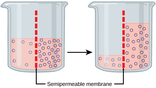
Figure 5.11 In osmosis, water always moves from an area of higher water concentration to one of lower concentration. In the diagram, the solute cannot pass through the selectively permeable membrane, but the water can.
Figure 12.19 Reaction diagrams for an endothermic process in the absence (red curve) and presence (blue curve) of a catalyst. The catalyzed pathway involves a two-step mechanism (note the presence of two transition states) and an intermediate species (represented by the valley between the two transitions states).
Figure 4.18 Membrane and secretory proteins are synthesized in the rough endoplasmic reticulum (RER). The RER also sometimes modifies proteins. In this illustration, a (green) integral membrane protein is modified by attachment of a (purple) carbohydrate in the ER. Vesicles with the integral protein bud from the ER and fuse with the Golgi apparatus' cis face. As the protein passes along the Golgi’s cisternae, the addition of more carbohydrates further modifies it. After its synthesis is complete, it exits as an integral membrane protein of the vesicle that buds from the Golgi’s trans face. When the vesicle fuses with the cell membrane, the protein becomes an integral portion of that cell membrane. (credit: modification of work by Magnus Manske)
Figure 7.45 - Non-homologous end joining (left) versus homologous recombination (right) - Wikipedia
Wikipedia, as credited by Kevin Ahern, Indira Rajagopal, and Taralyn Tan, Biochemistry Free For All, Oregon State University, via Biology LibreTexts. · Source attribution
· Original image ↗ · Access for free ↗
The amount of a substance that can dissolve in a solvent under specified conditions. Soluble means able to dissolve; insoluble means only very slightly soluble in that solvent.
Figure 15.7 Messenger RNA is a copy of protein-coding information in the coding strand of DNA, with the substitution of U in the RNA for T in the coding sequence. However, new RNA nucleotides base pair with the nucleotides of the template strand. RNA is synthesized in its 5'-3' direction, using the enzyme RNA polymerase. As the template is read, the DNA unwinds ahead of the polymerase and then rewinds behind it.
A fleeting, high-energy arrangement at a reaction barrier. It is an energy maximum along the reaction path, rather than a stable reaction intermediate.
Figure 12.19 Reaction diagrams for an endothermic process in the absence (red curve) and presence (blue curve) of a catalyst. The catalyzed pathway involves a two-step mechanism (note the presence of two transition states) and an intermediate species (represented by the valley between the two transitions states).
Figure 15.18 Translation begins when an initiator tRNA anticodon recognizes a start codon on mRNA bound to a small ribosomal subunit. The large ribosomal subunit joins the small subunit, and a second tRNA is recruited. As the mRNA moves relative to the ribosome, successive tRNAs move through the ribosome and the polypeptide chain is formed. Entry of a release factor into the A site terminates translation and the components dissociate.
Figure 4.18 Membrane and secretory proteins are synthesized in the rough endoplasmic reticulum (RER). The RER also sometimes modifies proteins. In this illustration, a (green) integral membrane protein is modified by attachment of a (purple) carbohydrate in the ER. Vesicles with the integral protein bud from the ER and fuse with the Golgi apparatus' cis face. As the protein passes along the Golgi’s cisternae, the addition of more carbohydrates further modifies it. After its synthesis is complete, it exits as an integral membrane protein of the vesicle that buds from the Golgi’s trans face. When the vesicle fuses with the cell membrane, the protein becomes an integral portion of that cell membrane. (credit: modification of work by Magnus Manske)
We tend to think of protein as a mass noun: a homogeneous substance, something that your diet should contain in a certain proportion. But if you ever work in a molecular biology lab (say, for a summer internship), protein may start to look very different to you.
How so? Well, you may see firsthand that protein isn’t just a single substance. Instead, there are lots and lots of different proteins in an organism, or even in a single cell. They come in every size, shape, and type you can imagine, and each one has a unique and specific job. Some are structural parts, giving cells shape or helping them move. Others act as signals, drifting between cells like messages in a bottle. Still others are metabolicenzymes, putting together or snapping apart biomolecules needed by the cell. And, odds are, one of these unique molecular players will become yours for the duration of your research!
Proteins are among the most abundant organic molecules in living systems and are way more diverse in structure and function than other classes of macromolecules. A single cell can contain thousands of proteins, each with a unique function. Although their structures, like their functions, vary greatly, all proteins are made up of one or more chains of amino acids. In this article, we will look in more detail at the building blocks, structures, and roles of proteins.
Proteins can play a wide array of roles in a cell or organism. Here, we’ll touch on a few examples of common protein types that may be familiar to you, and that are important in the biology of many organisms (including us).
One example of an enzyme found in your body is salivary amylase, which breaks amylose (a kind of starch) down into smaller sugars. The amylose doesn’t taste very sweet, but the smaller sugars do. This is why starchy foods often taste sweeter if you chew them for longer: you’re giving salivary amylase time to get to work.
Hormones are long-distance chemical signals released by endocrine cells (like the cells of your pituitary gland). They control specific physiological processes, such as growth, development, metabolism, and reproduction. While some hormones are steroid-based (see the article on lipids), others are proteins. These protein-based hormones are commonly called peptide hormones.
For example, insulin is an important peptide hormone that helps regulate blood glucose levels. When blood glucose rises (for instance, after you eat a meal), specialized cells in the pancreas release insulin. The insulin binds to cells in the liver and other parts of the body, causing them to take up the glucose. This process helps return blood sugar to its normal, resting level.
Some additional types of proteins and their functions are listed in the table below:
Provide food for the early development of the embryo or the seedling
Table modified from OpenStax College, Biology.
Proteins come in many different shapes and sizes. Some are globular (roughly spherical) in shape, whereas others form long, thin fibers. For example, the hemoglobinprotein that carries oxygen in the blood is a globular protein, while collagen, found in our skin, is a fibrous protein.
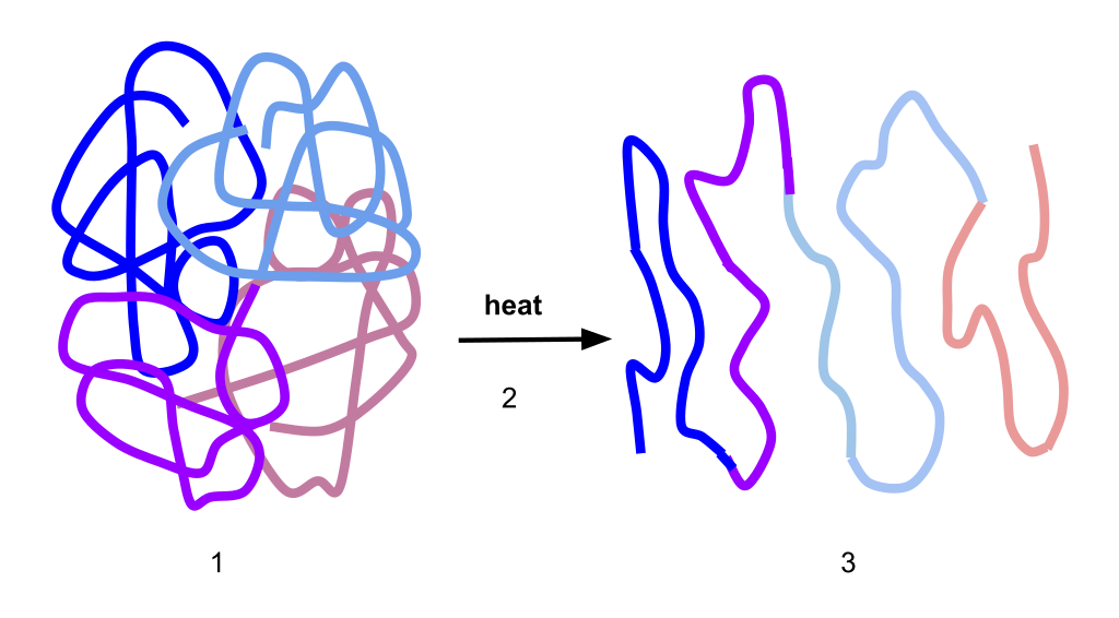
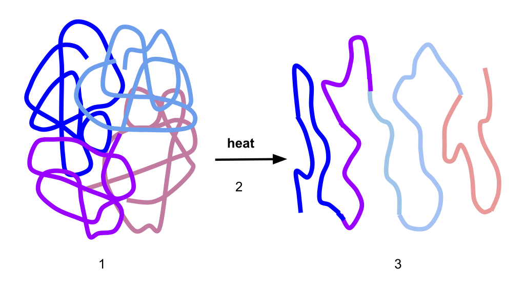
A protein’s shape is critical to its function, and, as we’ll see in the next article, many different types of chemical bonds may be important in maintaining this shape. Changes in temperature and pH, as well as the presence of certain chemicals, may disrupt a protein’s shape and cause it to lose functionality, a process known as denaturation.
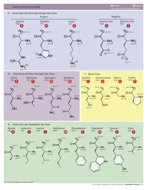
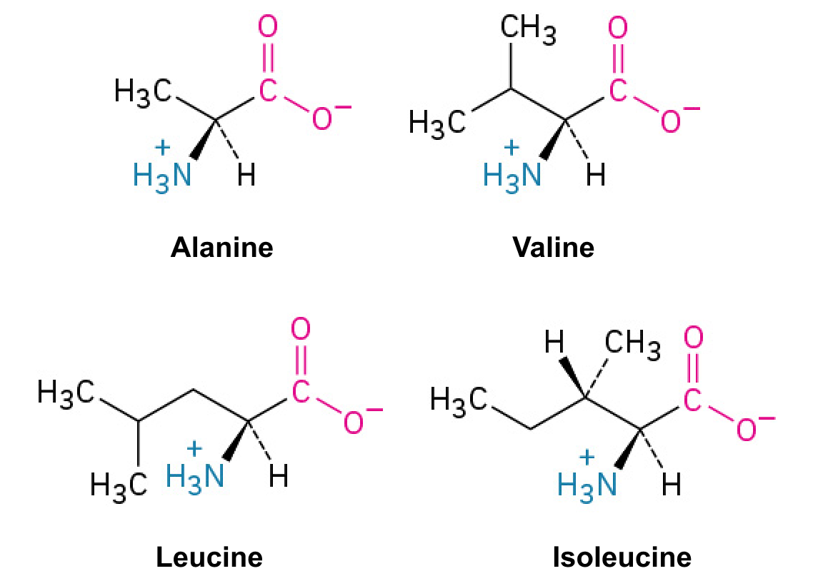
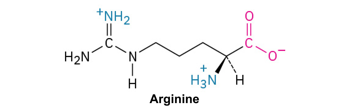
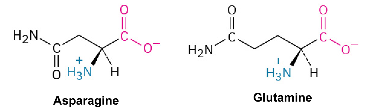
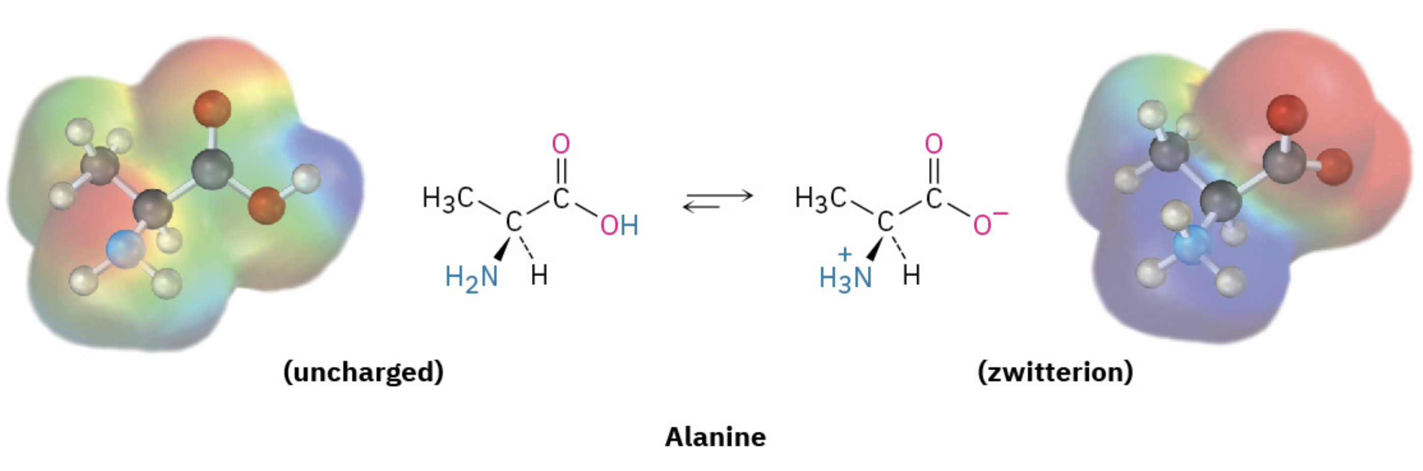
Amino acids share a basic structure, which consists of a central carbon atom, also known as the alpha (α) carbon, bonded to an amino group (), a carboxyl group (), and a hydrogen atom.
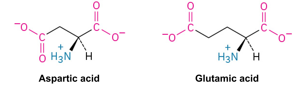
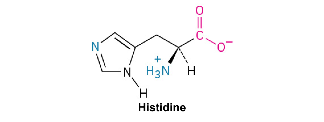
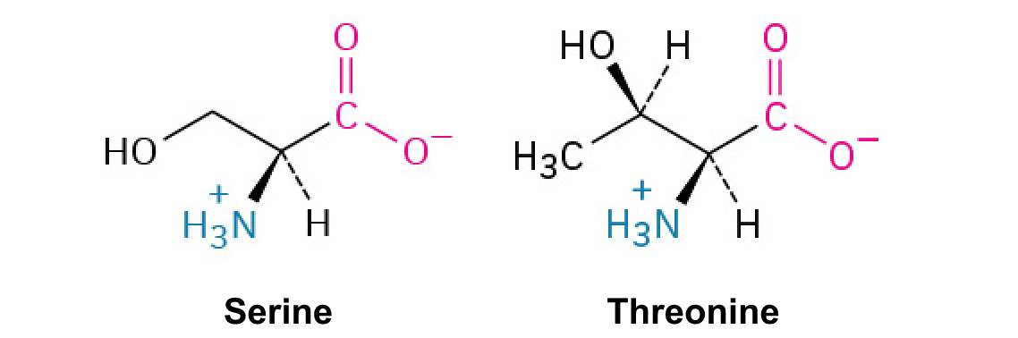
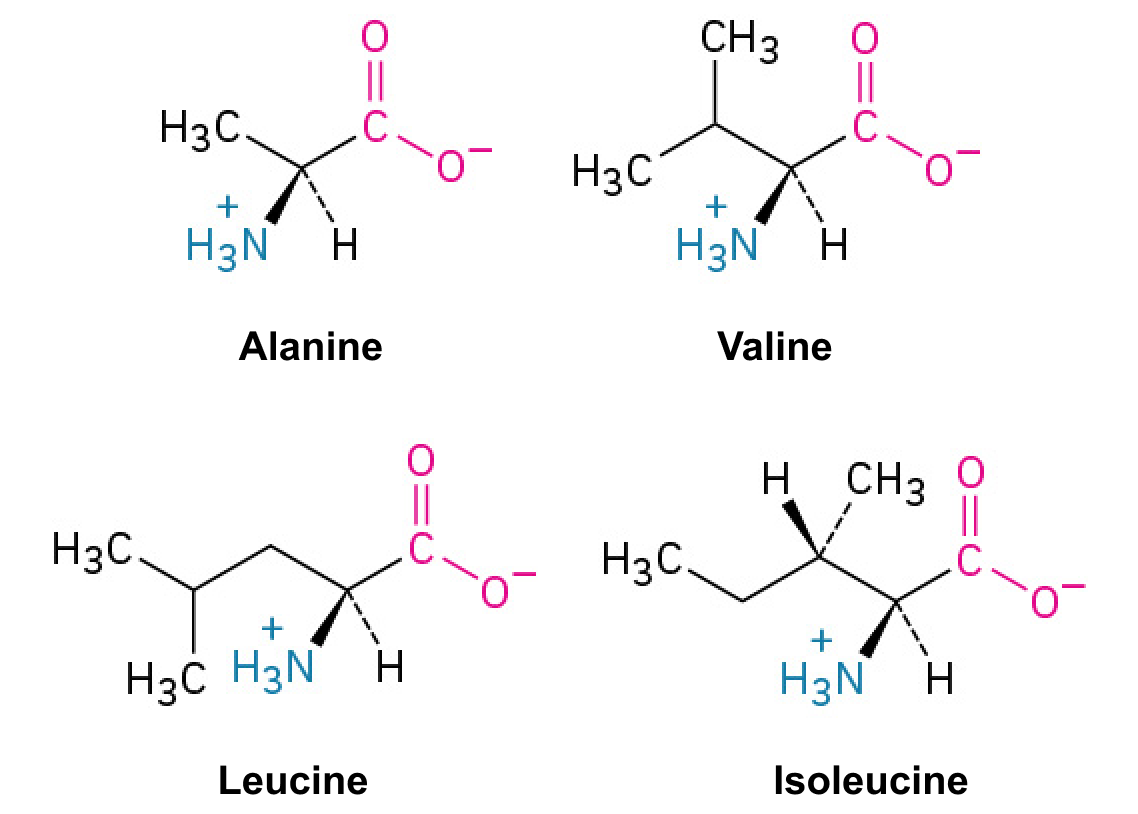
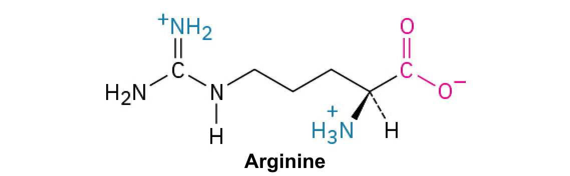
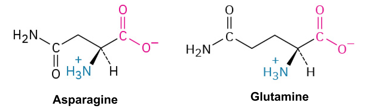
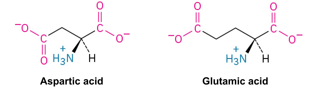
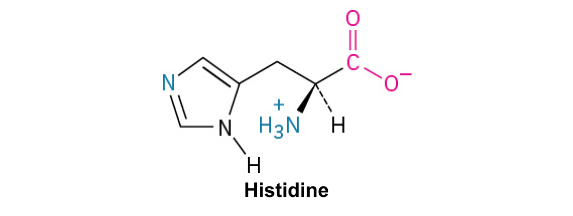
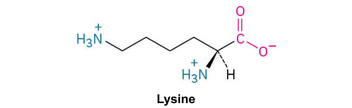
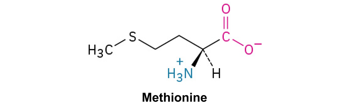
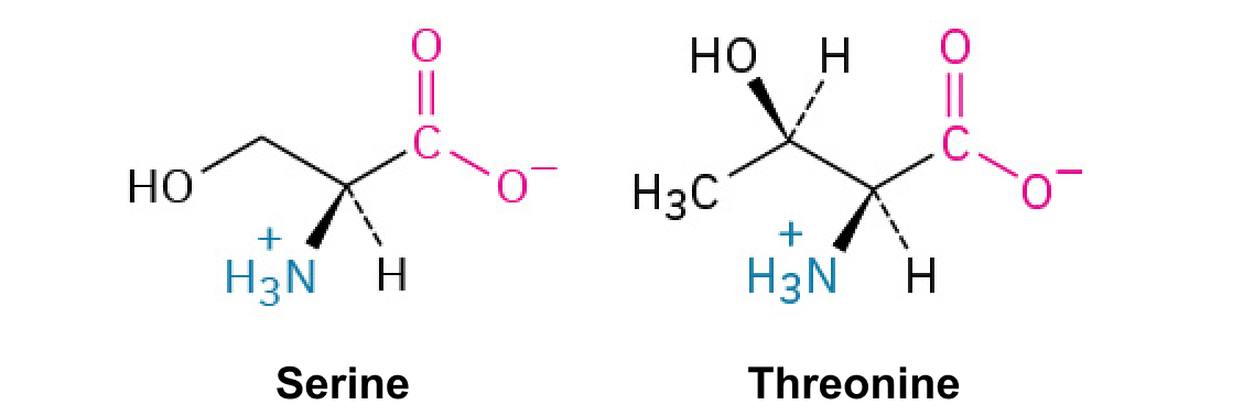
A few other amino acids have R groups with special properties, and these will prove to be important when we look at protein structure:
Proline has an R group that’s linked back to its own amino group, forming a ring structure. This makes it an exception to the typical structure of an amino acid, since it no longer has the standard NH amino group. If you think that ring structure looks a little awkward, you’re right: proline often causes bends or kinks in amino acid chains.
Finally, there are a few other “non-canonical” amino acids that are found in proteins only under certain conditions. Click here for cool details!
You may not need to know about these for your biology class, but they’re pretty darn interesting!
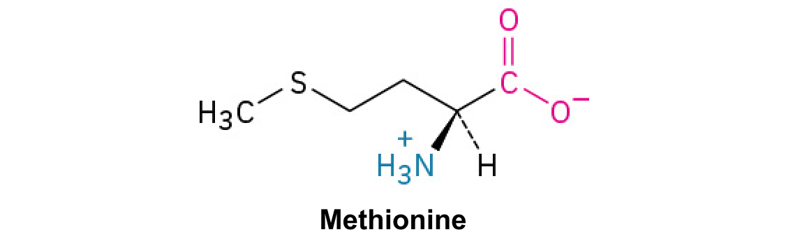
One is N-formylmethionine (abbreviated fMet), which is often the first amino acid of protein chains produced by bacteria. It’s similar in structure to methionine, but with an oxygen-containing group added to the amino group.
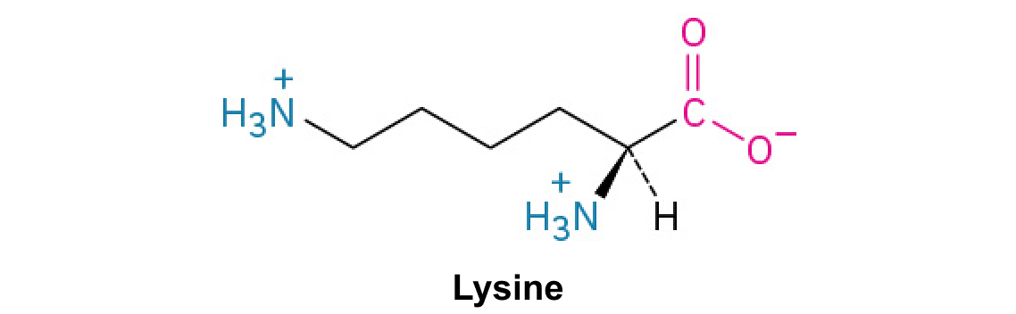
Another is pyrrolysine, which looks similar to lysine but has an extra ring-containing group attached to the end of the R group. Pyrrolysine is found mostly in the proteins of microorganisms called methanogenic (methane-producing) archaea.
Finally, selenocysteine is found in many different types of organisms, including humans, and is used to build selenium-containing proteins. Selenium is highly reactive, so this amino acid must be handled with care: it is specified in the genetic code, and added to protein chains, in a different way than a normal amino acid. Structurally, selenocysteine looks just like cysteine, but with a –SeH group in place of the –SH group.
Each protein in your cells consists of one or more polypeptide chains. Each of these polypeptide chains is made up of amino acids, linked together in a specific order. A polypeptide is kind of like a long word that is "spelled out" in amino acid letters. The chemical properties and order of the amino acids are key in determining the structure and function of the polypeptide, and the protein it's part of. But how are amino acids actually linked together in chains?
Because of the structure of the amino acids, a polypeptide chain has directionality, meaning that it has two ends that are chemically distinct from one another. At one end, the polypeptide has a free amino group, and this end is called the amino terminus (or N-terminus). The other end, which has a free carboxyl group, is known as the carboxyl terminus (or C-terminus). The N-terminus is on the left and the C-terminus is on the right for the very short polypeptide shown above.
Reece, J. B., Urry, L. A., Cain, M. L., Wasserman, S. A., Minorsky, P. V., and Jackson, R. B. (2011). Figure 5.18. Levels of protein structure. In Campbell Biology (10th ed., p. 80). San Francisco, CA: Pearson.
Raven, P. H., Johnson, G. B., Mason, K. A., Losos, J. B., and Singer, S. R. (2014). Proteins: Molecules with diverse structures and functions. In Biology (10th ed., AP ed., pp. 44-53. New York, NY: McGraw-Hill.
Reece, J. B., Urry, L. A., Cain, M. L., Wasserman, S. A., Minorsky, P. V., and Jackson, R. B. (2011). Proteins include a diversity of structures, resulting in a wide range of functions. In Campbell biology (10th ed., pp. 75-84). San Francisco, CA: Pearson.