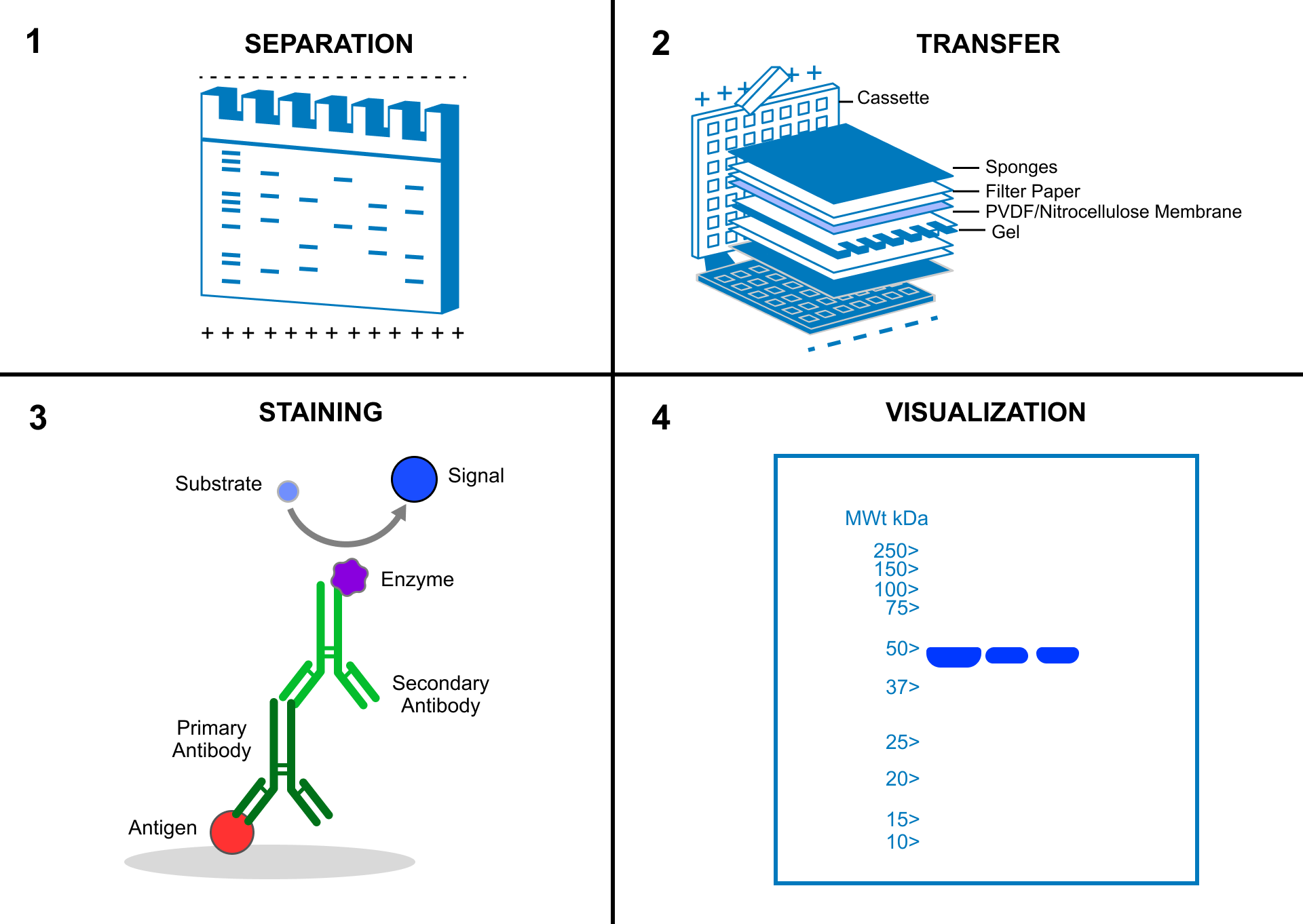
Detect the label by light emission, fluorescence, or color. A signal’s position estimates apparent size; its intensity can reflect abundance when loading, exposure, and signal range are controlled.
Specificity matters. An unexpected band can reflect cross-reactivity, degradation, or a modified protein. Appropriate controls and a molecular mass ladder help interpret the result. Some methods directly label the primary antibody.
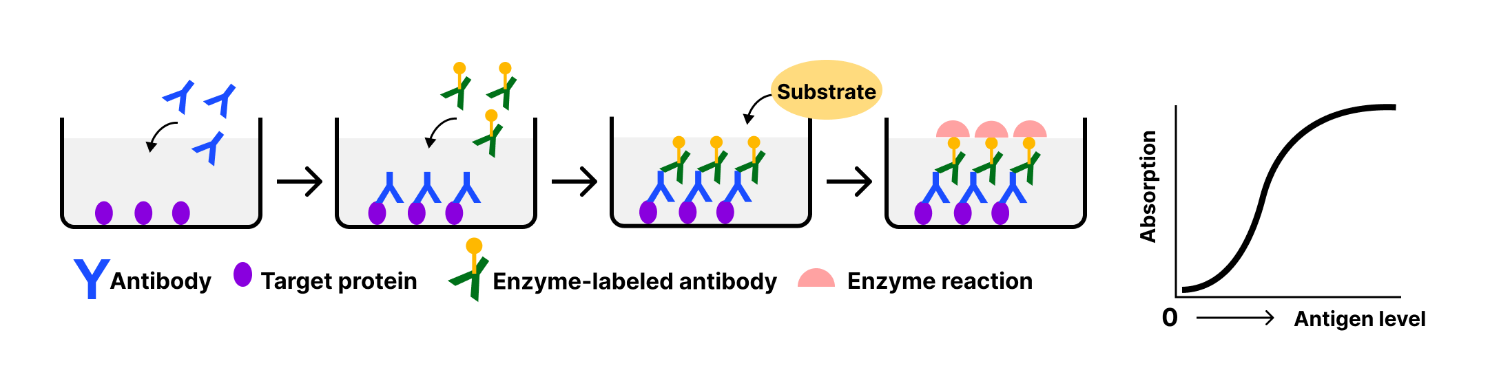
ELISA uses antibody binding on a solid surface, commonly a microplate, followed by an enzyme-generated signal. In a sandwich assay, a capture antibody binds the antigen and a detection antibody recognizes another site. Compare signal with standards to estimate concentration. ELISA does not provide the gel-based size information of a Western blot.
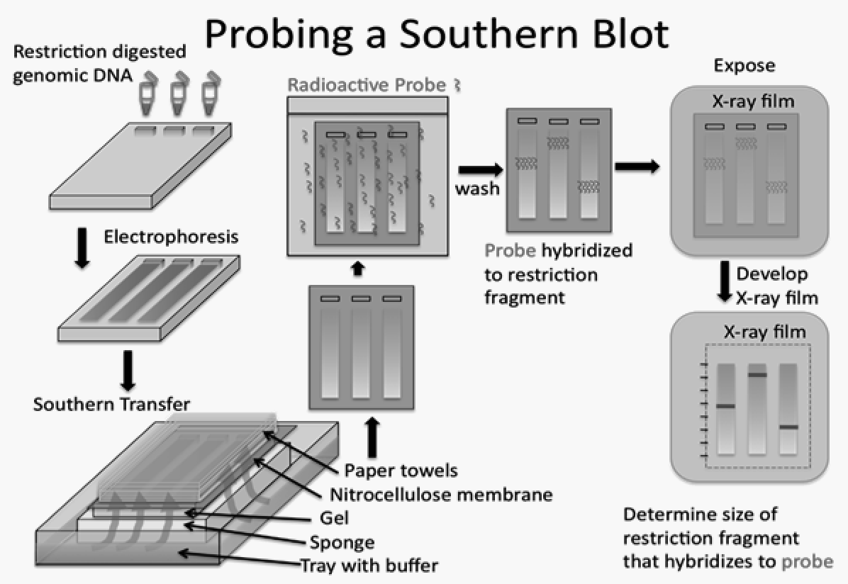
For a Southern blot, digest DNA into fragments, separate them by electrophoresis, denature the strands, and transfer them to a membrane. A labeled complementary probe hybridizes to the target sequence. Washing removes unbound probe before detection.
A Northern blot follows the same general separation–transfer–hybridization logic for RNA, usually without the restriction digestion used for DNA. It can reveal transcript size and relative abundance. Probes may use radioactive labels such as P-32 or nonradioactive labels.
Remember the targets. Southern → DNA; Northern → RNA; Western → protein. Nucleic acid blots use complementary base pairing; Western blots use antigen–antibody recognition.
An antibody-based assay performed on a solid surface, commonly a microplate, to detect or quantify an antigen. It does not provide gel-based size information.
Hybridization
Pairing of complementary nucleic acid strands, used for sequence-specific detection.