Protein function links selective binding to a change in conformation, activity, or movement.
Binding changes populations of conformations
For simple binding P + L ⇌ PL, Kd = [P][L]/[PL]; lower Kd means greater affinity at equilibrium. With independent identical sites, fractional occupancy is [L]/(Kd + [L]). Binding can shift a protein toward an active or inactive conformation. Cooperative sites influence each other, producing curves that depart from the independent-site equation. Affinity concerns one binding interaction, while avidity includes the combined strength of multiple contacts. Distinguish binding strength from specificity, reaction rate, and the number of available sites.
Source: 3.4 Proteins · 21.4 The Adaptive Immune Response: B-lymphocytes and Antibodies · 10.3 Muscle Fiber Contraction and Relaxation
Antibody structure supports recognition and effector action
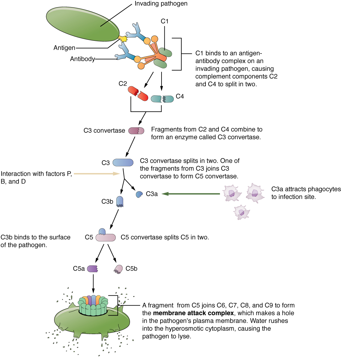
An antibody contains two heavy and two light chains linked partly by disulfides. Variable domains form antigen-binding sites; constant heavy-chain regions determine isotype and recruit effector functions. Fab binds antigen; Fc interacts with immune receptors or complement. Noncovalent shape and chemical complementarity permit specificity but can allow cross-reactivity. B-cell receptors recognize intact antigen, whereas conventional T-cell receptors recognize peptide–MHC complexes. Class switching changes the constant region while retaining antigen specificity; affinity maturation selects improved binding variants.
Source: 3.4 Proteins · 21.4 The Adaptive Immune Response: B-lymphocytes and Antibodies · 10.3 Muscle Fiber Contraction and Relaxation
ATP drives directed movement
Myosin moves along actin; kinesin and dynein move along microtubules. Most transport kinesins move toward the plus end and cytoplasmic dynein toward the minus end. ATP-dependent cycles alter track affinity and conformation, biasing movement rather than simply converting every hydrolysis into a fixed step under all conditions. In muscle, ATP binding detaches myosin, hydrolysis primes the head, and phosphate release is associated with the power stroke. Without ATP, attached crossbridges cannot detach normally, explaining rigor.
Source: 3.4 Proteins · 21.4 The Adaptive Immune Response: B-lymphocytes and Antibodies · 10.3 Muscle Fiber Contraction and Relaxation
Practice and reasoning
Question. An independent binding site has Kd = 2 µM. What occupancy is expected at free ligand 6 µM? Would increasing affinity necessarily increase a motor’s speed?
Show reasoning
Occupancy = 6/(2+6) = 0.75, or 75%. This assumes free ligand is known and sites behave independently. Increased track affinity need not increase speed: overly persistent attachment can slow detachment and cycling. Binding, ATP turnover, load, and movement should be measured separately.
Source: 3.4 Proteins · 21.4 The Adaptive Immune Response: B-lymphocytes and Antibodies · 10.3 Muscle Fiber Contraction and Relaxation
3.4 Proteins — figures and tables
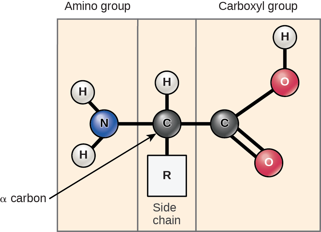
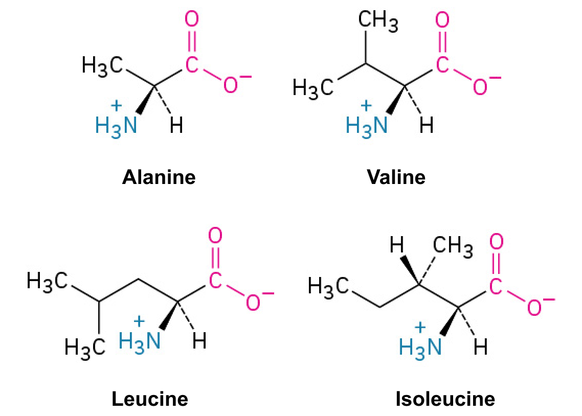
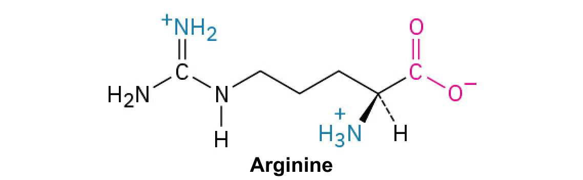
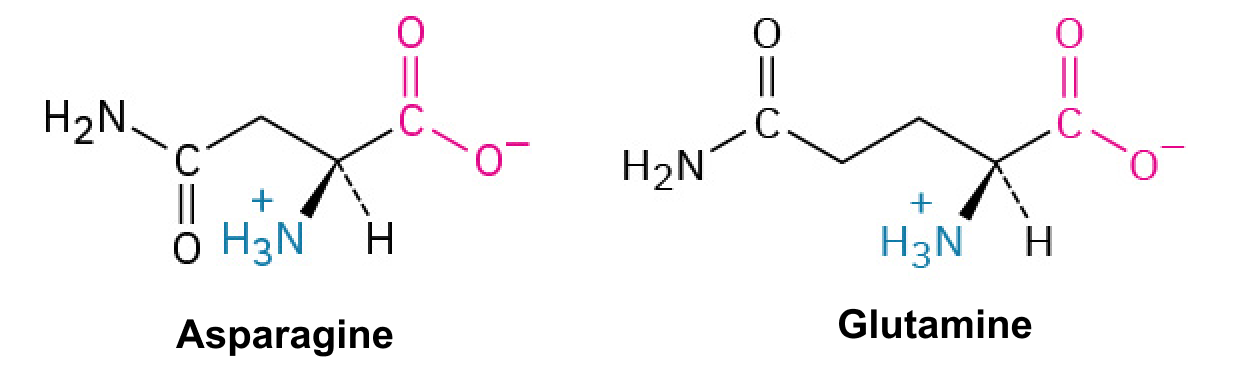
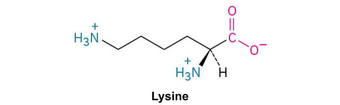
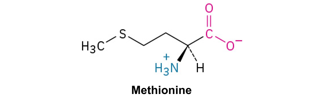
Figure 3.22 Amino acids have a central asymmetric carbon to which an amino group, a carboxyl group, a hydrogen atom, and a side chain (R group) are attached.
Mary Ann Clark, Matthew Douglas, and Jung Choi, Biology 2E, OpenStax. · CC BY-NC-SA 4.0 · Original image ↗ · Access for free ↗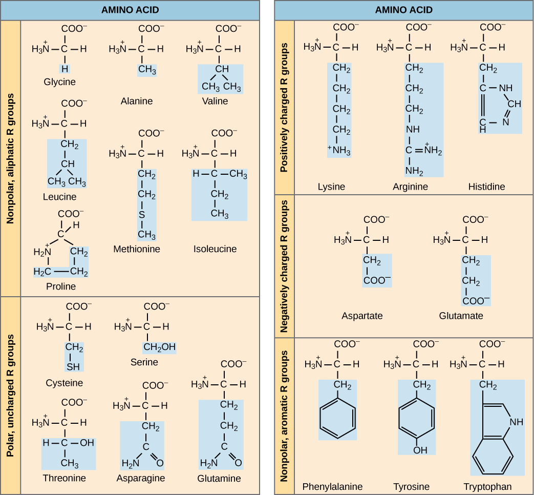
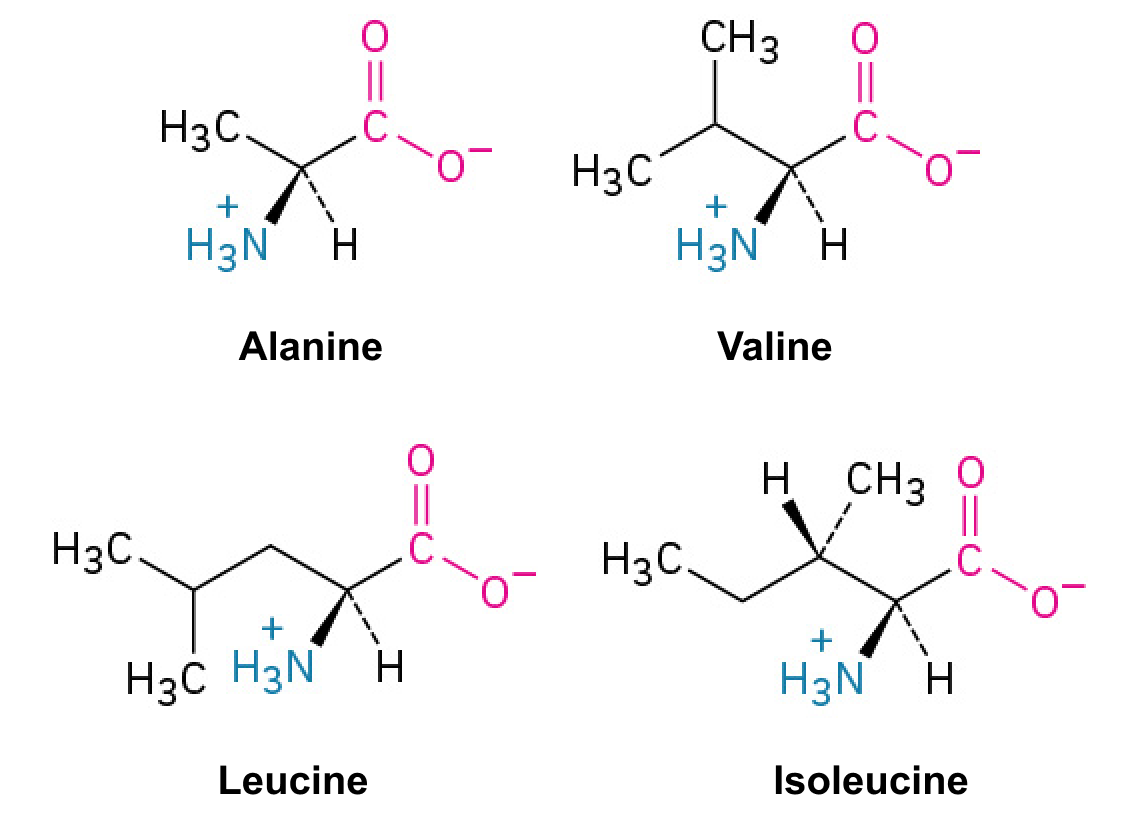
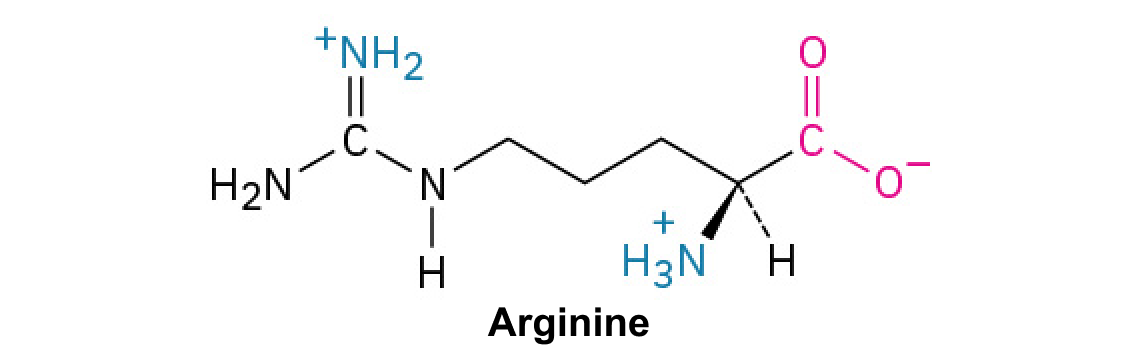
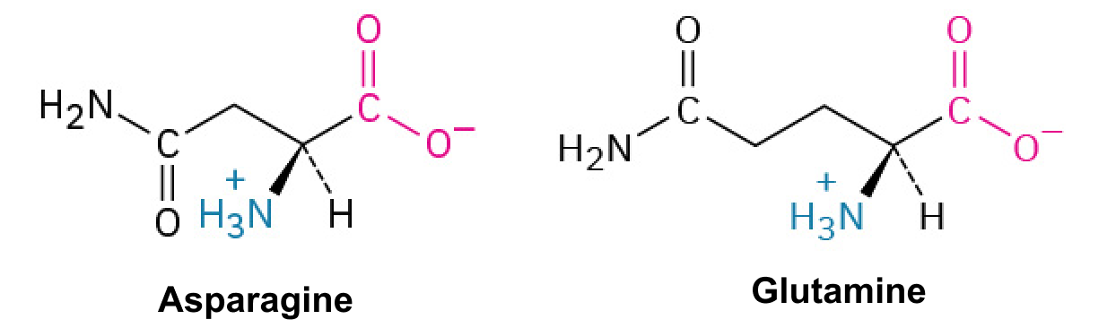
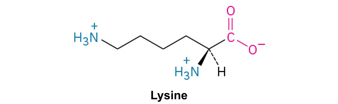
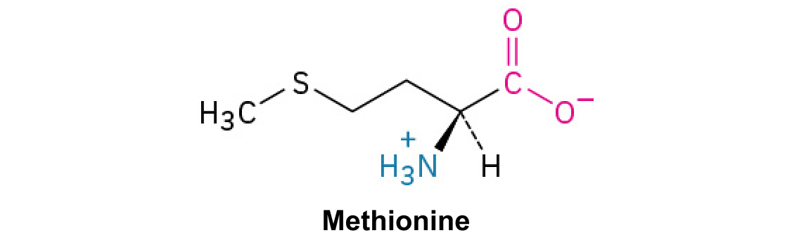
Figure 3.23 There are 20 common amino acids commonly found in proteins, each with a different R group (variant group) that determines its chemical nature.
Mary Ann Clark, Matthew Douglas, and Jung Choi, Biology 2E, OpenStax. · CC BY-NC-SA 4.0 · Original image ↗ · Access for free ↗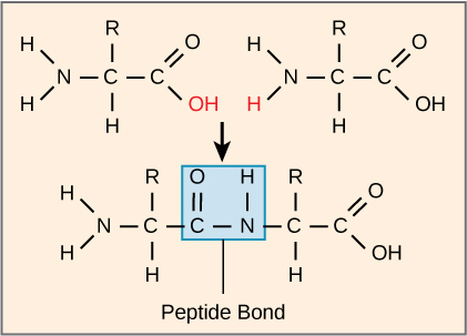
Figure 3.24 Peptide bond formation is a dehydration synthesis reaction. The carboxyl group of one amino acid is linked to the incoming amino acid's amino group. In the process, it releases a water molecule.
Mary Ann Clark, Matthew Douglas, and Jung Choi, Biology 2E, OpenStax. · CC BY-NC-SA 4.0 · Original image ↗ · Access for free ↗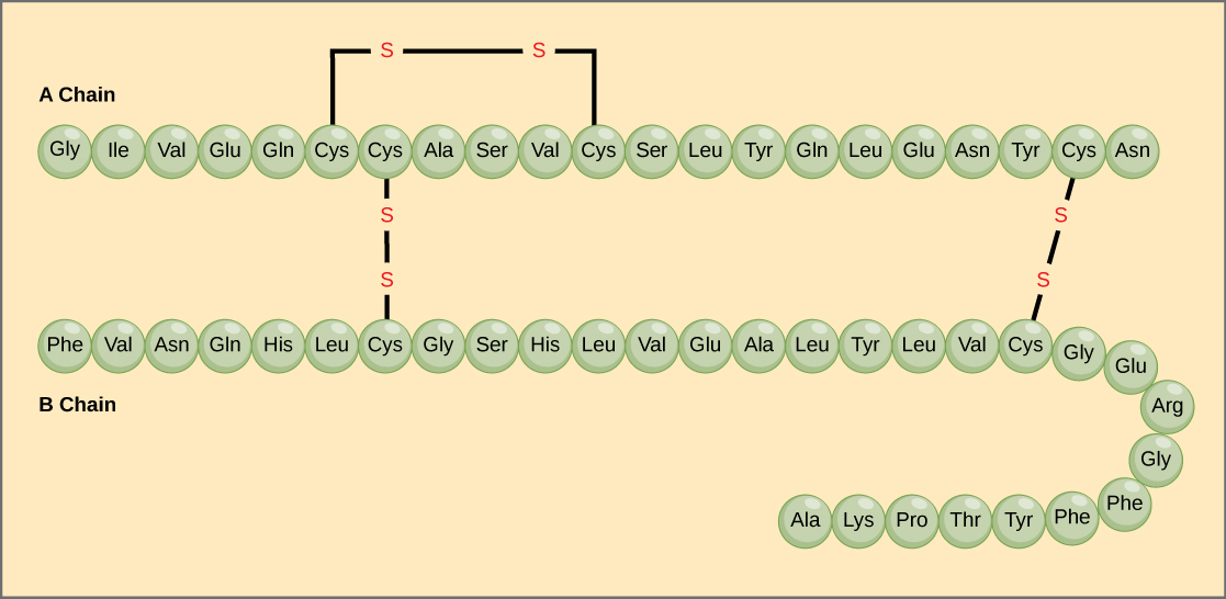
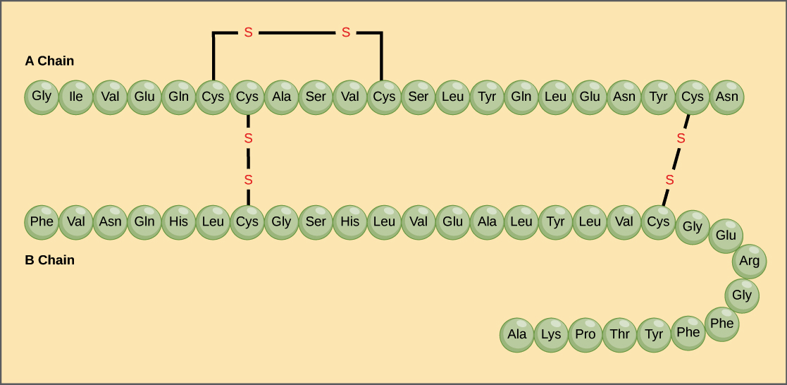
Figure 3.25 Bovine serum insulin is a protein hormone comprised of two peptide chains, A (21 amino acids long) and B (30 amino acids long). In each chain, three-letter abbreviations that represent the amino acids' names in the order they are present indicate primary structure. The amino acid cysteine (cys) has a sulfhydryl (SH) group as a side chain. Two sulfhydryl groups can react in the presence of oxygen to form a disulfide (S-S) bond. Two disulfide bonds connect the A and B chains together, and a third helps the A chain fold into the correct shape. Note that all disulfide bonds are the same length, but we have drawn them different sizes for clarity.
Mary Ann Clark, Matthew Douglas, and Jung Choi, Biology 2E, OpenStax. · CC BY-NC-SA 4.0 · Original image ↗ · Access for free ↗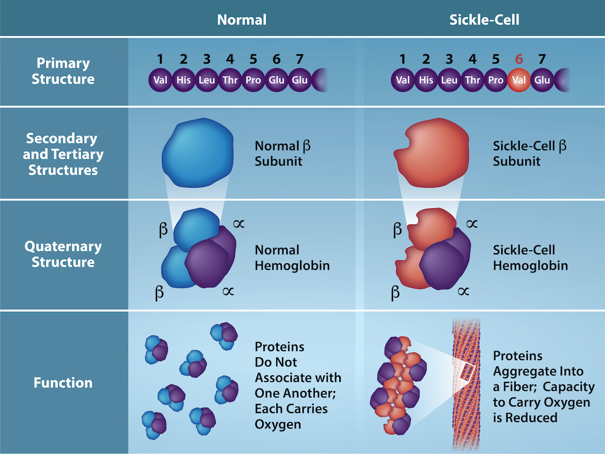
Figure 3.26 Because of this change of one amino acid in the chain, hemoglobin molecules form long fibers that distort the biconcave, or disc-shaped, red blood cells and causes them to assume a crescent or “sickle” shape, which clogs blood vessels (Figure 3.27). The beta (β)- chain of hemoglobin is 147 amino acids in length, yet a single amino acid substitution in the primary sequence leads to changes in secondary, tertiary, and quaternary structures and sickle cell anemia. In normal hemoglobin, the amino acid at position six is glutamate. In sickle cell hemoglobin glutamate is replaced by valine. (credit: Rao, A., Tag, A. Ryan, K. and Fletcher, S. Department of Biology, Texas A&M University)
Mary Ann Clark, Matthew Douglas, and Jung Choi, Biology 2E, OpenStax. (credit: Rao, A., Tag, A. Ryan, K. and Fletcher, S. Department of Biology, Texas A&M University) · CC BY-NC-SA 4.0 · Original image ↗ · Access for free ↗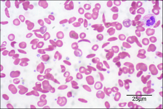
Figure 3.27 In this blood smear, visualized at 535x magnification using bright field microscopy, sickle cells are crescent shaped, while normal cells are disc-shaped. (credit: modification of work by Ed Uthman; scale-bar data from Matt Russell)
Mary Ann Clark, Matthew Douglas, and Jung Choi, Biology 2E, OpenStax. (credit: modification of work by Ed Uthman; scale-bar data from Matt Russell) · CC BY-NC-SA 4.0 · Original image ↗ · Access for free ↗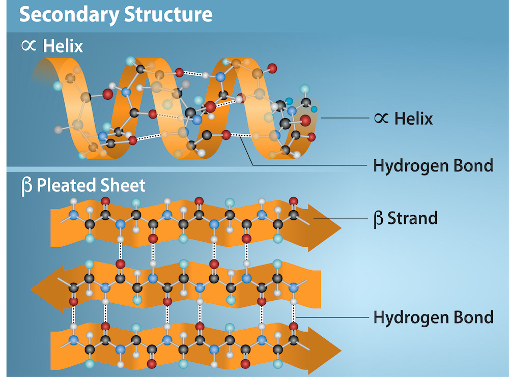
Figure 3.28 The α -helix and β -pleated sheet are secondary protein structures formed when hydrogen bonds form between the carbonyl oxygen and the amino hydrogen in the peptide backbone. Certain amino acids have a propensity to form an α-helix while others favor β-pleated sheet formation. Black = carbon, White = hydrogen, Blue = nitrogen, and Red = oxygen. Credit: Rao, A., Ryan, K. Fletcher, S. and Tag, A. Department of Biology, Texas A&M University.
Mary Ann Clark, Matthew Douglas, and Jung Choi, Biology 2E, OpenStax. · CC BY-NC-SA 4.0 · Original image ↗ · Access for free ↗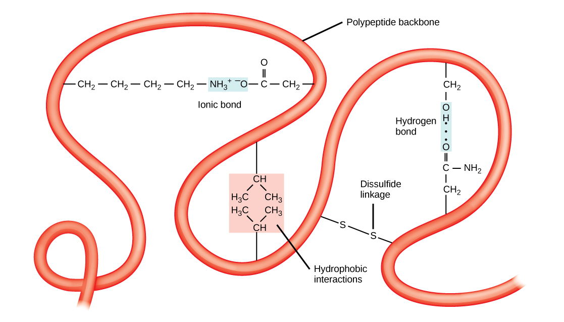
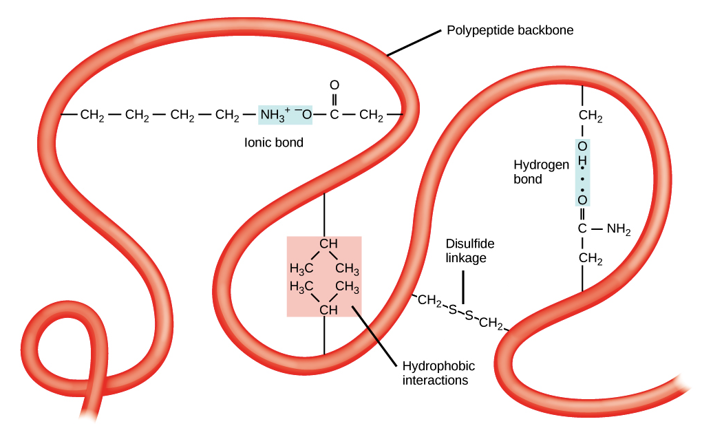
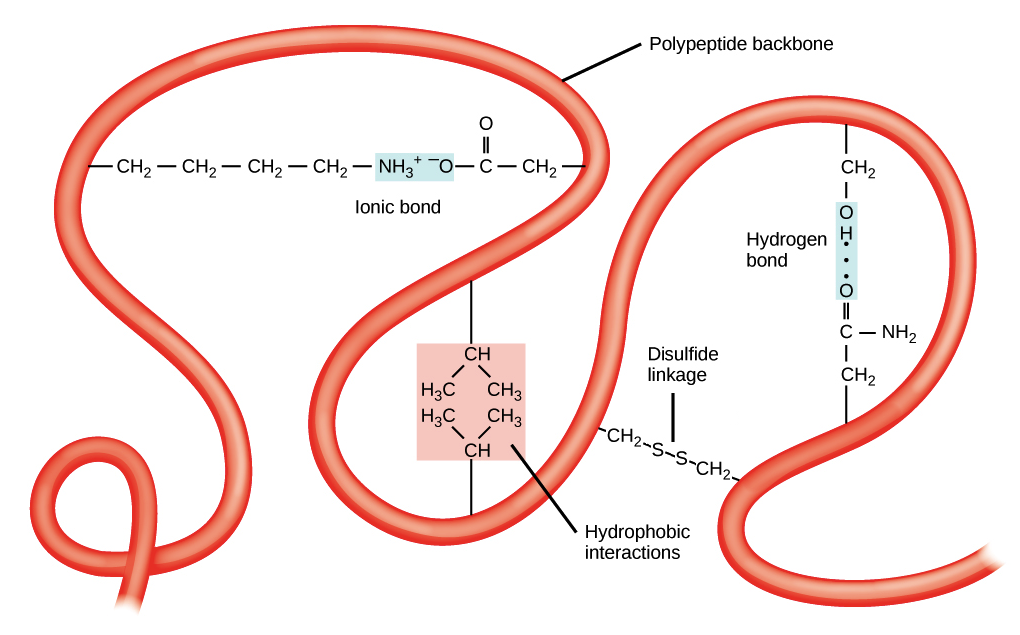
Figure 3.29 A variety of chemical interactions determine the proteins' tertiary structure. These include hydrophobic interactions, ionic bonding, hydrogen bonding, and disulfide linkages.
Mary Ann Clark, Matthew Douglas, and Jung Choi, Biology 2E, OpenStax. · CC BY-NC-SA 4.0 · Original image ↗ · Access for free ↗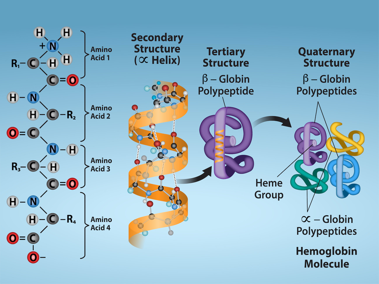
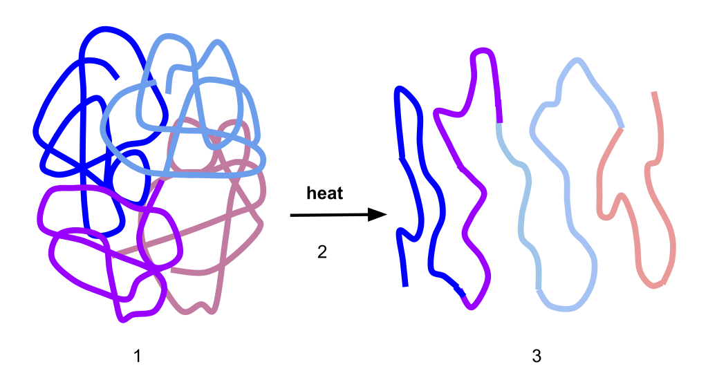
Figure 3.30 Observe the four levels of protein structure in these illustrations. Credit: Rao, A. Ryan, K. and Tag, A. Department of Biology, Texas A&M University.
Mary Ann Clark, Matthew Douglas, and Jung Choi, Biology 2E, OpenStax. · CC BY-NC-SA 4.0 · Original image ↗ · Access for free ↗| Type | Examples | Functions |
|---|---|---|
| Digestive Enzymes | Amylase, lipase, pepsin, trypsin | Help in food by catabolizing nutrients into monomeric units |
| Transport | Hemoglobin, albumin | Carry substances in the blood or lymph throughout the body |
| Structural | Actin, tubulin, keratin | Construct different structures, like the cytoskeleton |
| Hormones | Insulin, thyroxine | Coordinate different body systems' activity |
| Defense | Immunoglobulins | Protect the body from foreign pathogens |
| Contractile | Actin, myosin | Effect muscle contraction |
| Storage | Legume storage proteins, egg white (albumin) | Provide nourishment in early embryo development and the seedling |
Table 3.1. Mary Ann Clark, Matthew Douglas, and Jung Choi, Biology 2E, OpenStax.
Source: 3.4 Proteins
21.4 The Adaptive Immune Response: B-lymphocytes and Antibodies — figures and tables
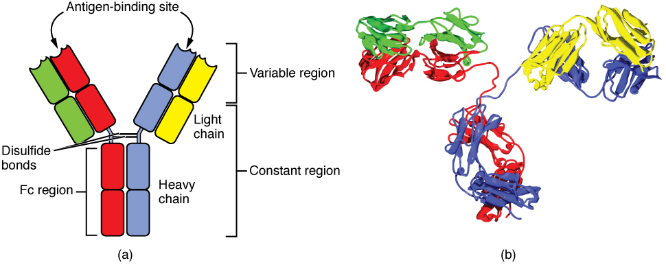
Figure 21.21 Antibody and IgG2 Structures The typical four chain structure of a generic antibody (a) and the corresponding three-dimensional structure of the antibody IgG2 (b). (credit b: modification of work by Tim Vickers)
J. Gordon Betts and colleagues, Anatomy And Physiology 2E, OpenStax. (credit b: modification of work by Tim Vickers) · CC BY-NC-SA 4.0 · Original image ↗ · Access for free ↗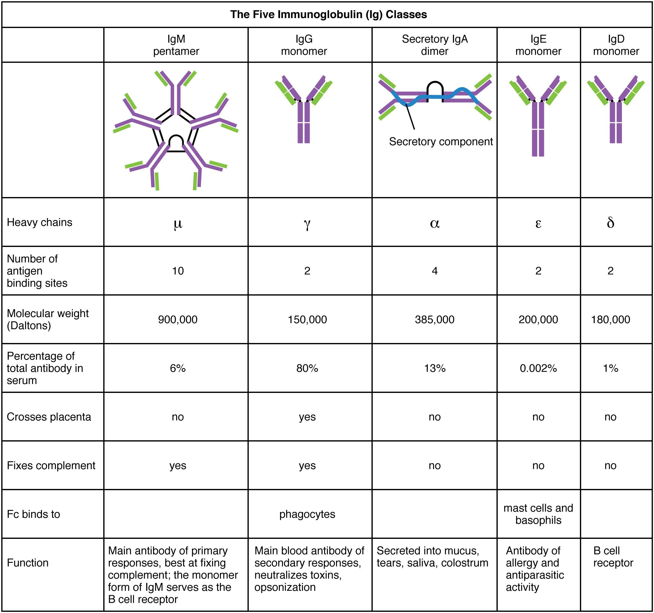
Figure 21.22 Five Classes of Antibodies
J. Gordon Betts and colleagues, Anatomy And Physiology 2E, OpenStax. · CC BY-NC-SA 4.0 · Original image ↗ · Access for free ↗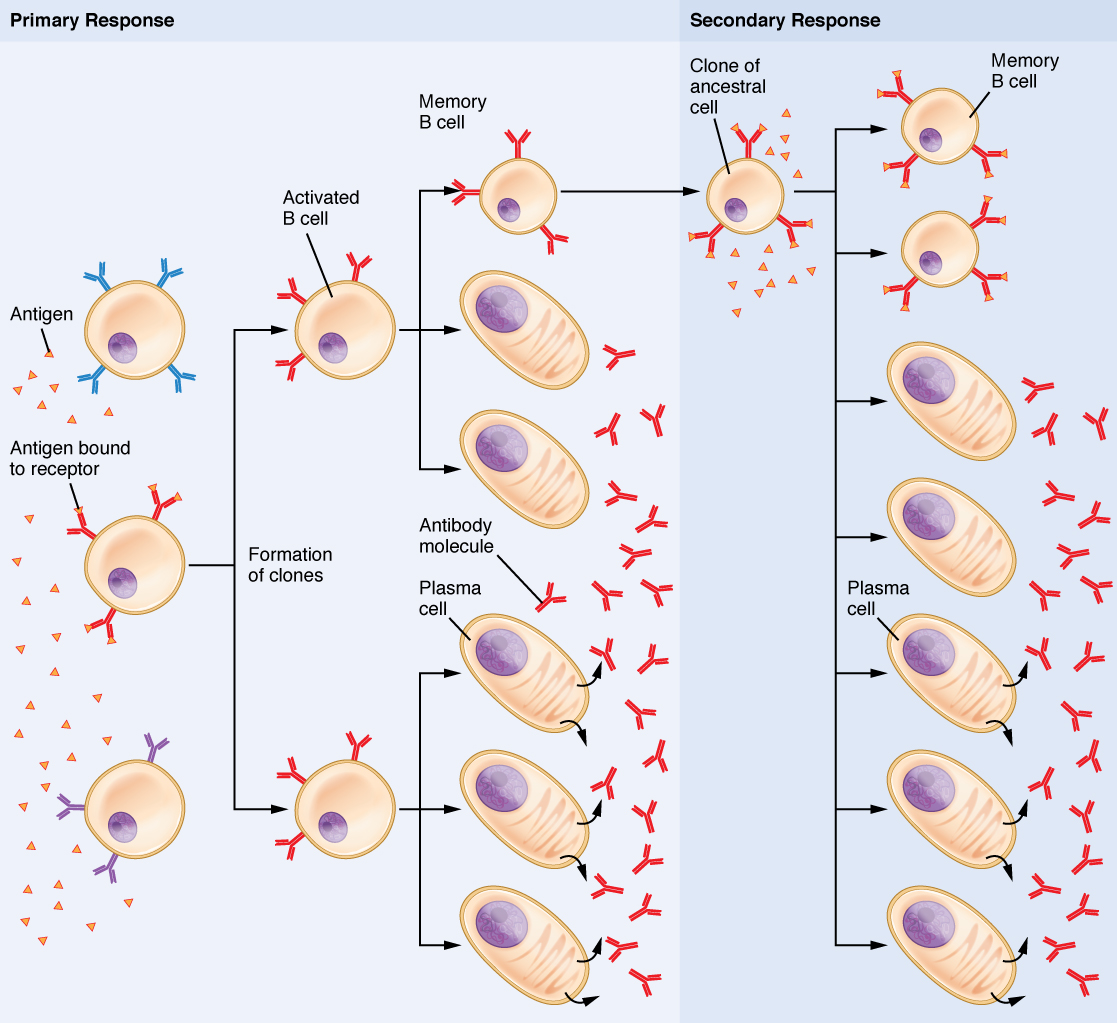
Figure 21.23 Clonal Selection of B Cells During a primary B cell immune response, both antibody-secreting plasma cells and memory B cells are produced. These memory cells lead to the differentiation of more plasma cells and memory B cells during secondary responses.
J. Gordon Betts and colleagues, Anatomy And Physiology 2E, OpenStax. · CC BY-NC-SA 4.0 · Original image ↗ · Access for free ↗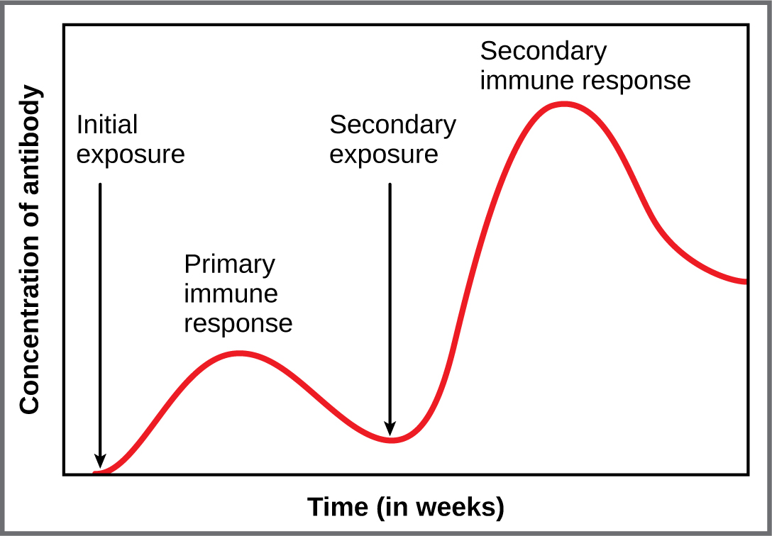
Figure 21.24 Primary and Secondary Antibody Responses Antigen A is given once to generate a primary response and later to generate a secondary response. When a different antigen is given for the first time, a new primary response is made.
J. Gordon Betts and colleagues, Anatomy And Physiology 2E, OpenStax. · CC BY-NC-SA 4.0 · Original image ↗ · Access for free ↗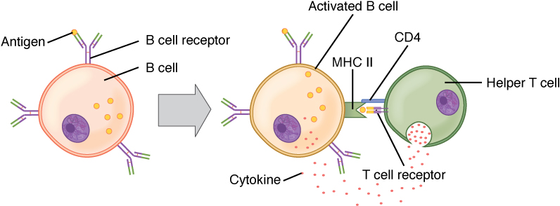
Figure 21.25 T and B Cell Binding To elicit a response to a T cell-dependent antigen, the B and T cells must come close together. To become fully activated, the B cell must receive two signals from the native antigen and the T cell’s cytokines.
J. Gordon Betts and colleagues, Anatomy And Physiology 2E, OpenStax. · CC BY-NC-SA 4.0 · Original image ↗ · Access for free ↗| Natural | Artificial | |
|---|---|---|
| Active | Adaptive immune response | Vaccine response |
| Passive | Trans-placental antibodies/breastfeeding | Immune globulin injections |
Table 21.6. J. Gordon Betts and colleagues, Anatomy And Physiology 2E, OpenStax.
Source: 21.4 The Adaptive Immune Response: B-lymphocytes and Antibodies
10.3 Muscle Fiber Contraction and Relaxation — figures and tables
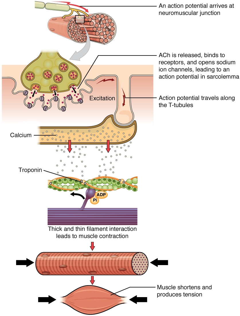
Figure 10.8 Contraction of a Muscle Fiber A cross-bridge forms between actin and the myosin heads triggering contraction. As long as Ca ++ ions remain in the sarcoplasm to bind to troponin, and as long as ATP is available, the muscle fiber will continue to shorten.
J. Gordon Betts and colleagues, Anatomy And Physiology 2E, OpenStax. · CC BY-NC-SA 4.0 · Original image ↗ · Access for free ↗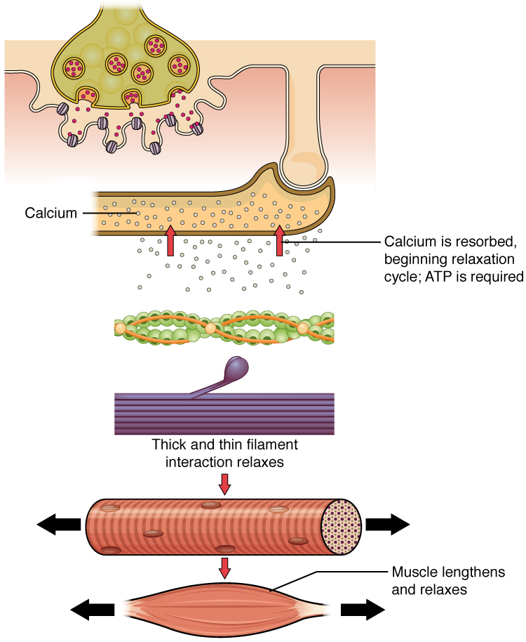
Figure 10.9 Relaxation of a Muscle Fiber Ca ++ ions are pumped back into the SR, which causes the tropomyosin to reshield the binding sites on the actin strands. A muscle may also stop contracting when it runs out of ATP and becomes fatigued.
J. Gordon Betts and colleagues, Anatomy And Physiology 2E, OpenStax. · CC BY-NC-SA 4.0 · Original image ↗ · Access for free ↗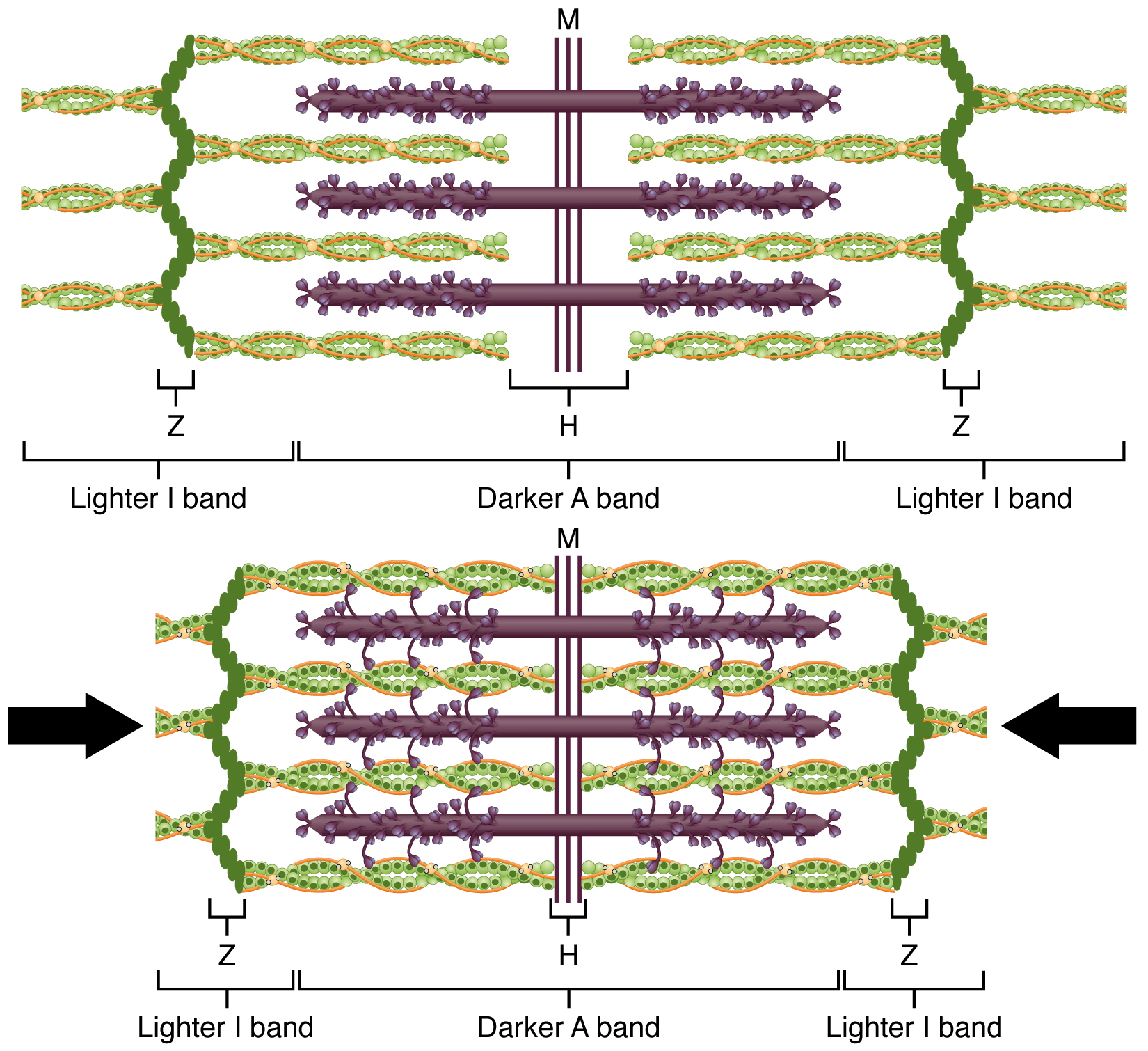
Figure 10.10 The Sliding Filament Model of Muscle Contraction When a sarcomere contracts, the Z lines move closer together, and the I band becomes smaller. The A band stays the same width. At full contraction, the thin and thick filaments overlap completely.
J. Gordon Betts and colleagues, Anatomy And Physiology 2E, OpenStax. · CC BY-NC-SA 4.0 · Original image ↗ · Access for free ↗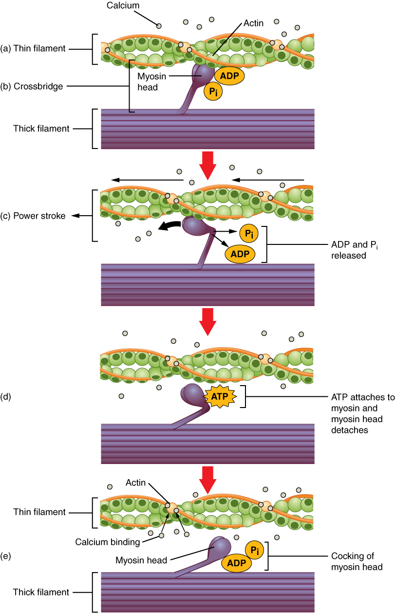
Figure 10.11 Skeletal Muscle Contraction (a) The active site on actin is exposed as calcium binds to troponin. (b) The myosin head is attracted to actin, and myosin binds actin at its actin-binding site, forming the cross-bridge. (c) During the power stroke, the phosphate generated in the previous contraction cycle is released. This results in the myosin head pivoting toward the center of the sarcomere, after which the attached ADP and phosphate group are released. (d) A new molecule of ATP attaches to the myosin head, causing the cross-bridge to detach. (e) The myosin head hydrolyzes ATP to ADP and phosphate, which returns the myosin to the cocked position.
J. Gordon Betts and colleagues, Anatomy And Physiology 2E, OpenStax. · CC BY-NC-SA 4.0 · Original image ↗ · Access for free ↗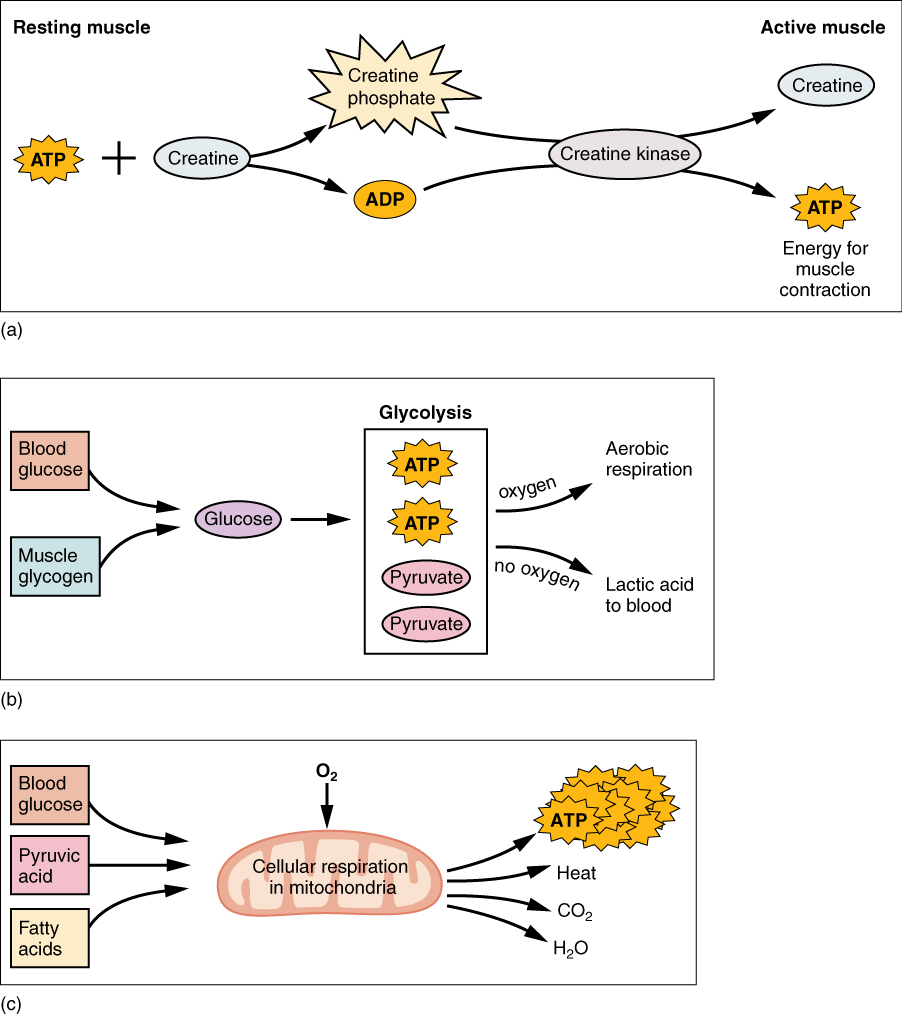
Figure 10.12 Muscle Metabolism (a) Some ATP is stored in a resting muscle. As contraction starts, it is used up in seconds. More ATP is generated from creatine phosphate for about 15 seconds. (b) Each glucose molecule produces two ATP and two molecules of pyruvic acid, which can be used in aerobic respiration or converted to lactic acid. If oxygen is not available, pyruvic acid is converted to lactic acid, which may contribute to muscle fatigue. This occurs during strenuous exercise when high amounts of energy are needed but oxygen cannot be sufficiently delivered to muscle. (c) Aerobic respiration is the breakdown of glucose in the presence of oxygen (O 2 ) to produce carbon dioxide, water, and ATP. Approximately 95 percent of the ATP required for resting or moderately active muscles is provided by aerobic respiration, which takes place in mitochondria.
J. Gordon Betts and colleagues, Anatomy And Physiology 2E, OpenStax. · CC BY-NC-SA 4.0 · Original image ↗ · Access for free ↗